submitted to the
Combined Faculties for the Natural Sciences and for Mathematics of the Ruperto-Carola University of Heidelberg, Germany
for the degree of Doctor of Natural Sciences
presented by
Mila Mahilum, Master of Science in Biology Cebu City, Philippines
Oral examination: February, 2003
Biochemical, molecular and microscopic studies on mosquitoes with special emphasis on Wolbachia infections, diagnostic
assessment of Dengue viruses and biological control of Dengue vectors
Referees: Prof. Dr. Volker Storch Prof. Dr. Elisabeth Pollerberg
Heartfelt thanks and gratitude to: Prof. Dr. Volker Storch, Head of the Zoological Institute, University of Heidelberg, for acting as adviser and first referee of the Ph. D. work, for sharing his expertise concerning the techniques on electron microscopic study and most especially for the guidance and help in making this work possible.
Dr. Norbert Becker, Scientific Director of the German Mosquito Control Association (KABS)/GFS, for acting also as adviser of the Ph. D. work, for the guidance and support in the course of the studies and most especially for sharing his valuable knowledge especially in regards to the biology and control of the mosquitoes.
Prof. Dr. Elisabeth Pollerberg, Zoology Institute, University of Heidelberg, for the help and being the second referee of the Ph. D. work.
Dr. Susanne Lankenau and Dr. Dirk Lankenau, Zoological Institute, University of Heidelberg, for the support of the Wolbachia study. Mrs. Renate Mummert, Mrs. Claudia Kempendorf and Mrs. Gisela Adam, Zoology Institute, University of Heidelberg, for the assistance on electron microscopic study and Mrs. Gabriele van der Linden for the technical assistance.
Mr. Thomas Weitzel and Mr. Artur Jöst for teaching the techinque on enzyme electrophoresis as well as for the continuous support of the biochemical study. Mr. Heiko Kotter and Mr.
Mattias Beck for helping in the collection of mosquito larvae. Ms. Silvia Guerreri for assisting the Elisa study, Ms. Regina Teo for reviewing some parts of the manuscript, Dr. Beate Heeger and Dr. Thomas Heeger, Dr. Filipina Sotto, Ms. Agnes Tomayao, Ms. Abigail Genelsa, Ms.
Pia Ternes, and Mr. Markus Ulrich for the help of the study. Dr. Paul Schädler, President of the German Mosquito Control Association (KABS) and Gesellschaft zur Förderung der Stechmückenbekämpfung (GFS) for the support of the study.
To all the people who spared assistance whom I may not have mentioned above, to all my beloved friends, my family for giving me the inspiration. Above all, the author is most grateful and thankful to the German Academic Exchange Program (DAAD) for making this work possible and most especially for the financial support during the whole period of the Ph.
D. work.
1. General Introduction
1.1. General background of Culicidae 1
1.2. Biology of Culicidae 2
1.3. Medical importance of mosquitoes 6
1.4. Significance of the studies 9
2. Protein electrophoretic studies on mosquitoes 2.1. Introduction 11
2.2. Materials and Methods 15
2.3. Results 26 2.4. Discussion 57
3. Studies on ovaries of mosquitoes using light, transmission and scanning microscopy 3.1. Introduction 61
3.2. Materials and Methods 63
3.2.1. Transmission electron microscopy 63
3.2.2. Scanning electron microscopy 66
3.2.3. Light microscopy 67
3.3. Results 67 3.3.1. Transmission electron microscopy 67
3.3.2. Scanning electron microscopy 78
3.3.3. Light microscopy 79
3.4. Discussion 80
4. Molecular and electron miscroscopic identification of Wolbachia sp. in populations of the Culex Pipiens Complex from the Upper Rhine Valley, Germany, and Cebu City, Philippines 4.1. Introduction 83
4.2. Materials and Methods 86
4.3. Results 90 4.4. Discussion 98
5. IgG-Capture Enzyme-Linked Immunosorbent Assay (ELISA) of serum samples from Filipino patients, Cebu City, Philippines 5.1. Introduction 100
5.2. Materials and Methods 101
5.3. Results 103
5.4. Discussion 104
6. Control of dengue vectors in Cebu City, Philippines 6.1. Introduction 107
6.2. Materials and Methods 111
6.3. Results 113
6.4. Discussion 121
7. Summary 123
8. Literature cited 126
9. Appendix 138
1. GENERAL INTRODUCTION
1.1. General background of Culicidae
Insects are the largest and most successful group of arthropods (Scott, 1980). Reasons for their success is attributed to their high rate of reproduction, their ability to adapt to almost every environmental condition and lastly, their accessibility to a variety of food sources.
Many studies on insects are focused on such different research fields such as genetics, evolutionary biology, systematics, ecology and applied zoology because of their great medical and agricultural importance. Due to their advantageous characteristics in the laboratory, some species are good experimental animals (e.g. Drosophila melanogaster Meigen) and also serve as model organisms in various researches.
Insects and humans affect each other in many ways. Since both are very successful species, they sometimes affect each other in a negative way. Insects are considered by most people to be annoying. They bite or sting and can infect humans and other tetrapods with diseases. The most important vectors of diseases are the mosquitoes. For this reason, they are certainly among the best known family of insects that have of great importance in research. Their systematics have been studied not only at the traditional morphological level but also utilizing, at least in some groups, crossing experiments, cytological and molecular genetic analysis (Bullini & Coluzzi,1978).
Mosquitoes are placed in the Family Culicidae, suborder Nematocera of the order Diptera (the two-winged flies or true flies). The Culicidae contain over 3,200 species and are divided into 3 subfamilies: Anophelinae, Culicinae, and Toxorhynchitinae (Scott, 1980). They are one of the more primitive families of Diptera being more closely related to midges, gnats and crane flies. They occur throughout the world except in places that are permanently frozen (Clements, 1992). Mosquitoes are widely investigated by the researchers for the reason that they are the hosts to a variety of pathogens and parasites including viruses, bacteria, protozoans and nematodes. Thus, by their blood-sucking habit, they serve as agents of human and animal diseases.
1.2. The biology of Culicidae
Like all other Dipterans, mosquitoes exhibit complete metamorphosis. They need aquatic habitats for their development. After hatching they pass through four larval instars and a pupal stage where the transformation to the adult stage takes place. Most species are unautogenous, that means after copulation the females have to take a blood meal to complete egg development. Only a few species of mosquitoes are autogenous, they develop first egg batches without a blood-meal (e.g. Cx. p. pipiens biotype molestus) (Becker et al., 2003).
1.2.1 Oviposition
Female mosquitoes are capable of laying between 50 to almost 500 eggs, two to four days or even more in cool temperate climates after having a blood meal. Mosquitoes can be divided into two groups depending on their egg laying behaviour and whether or not the embryos enter into a period of dormancy (externally triggered resting period) or diapause “genetically fixed resting period” (Barr, 1958).
In the first group, females deposit the eggs onto the surface of the water either singly (e.g.
Anopheles) or in egg batches (e.g. Culex). To cite an example, females of Culex lay their eggs in rafts which are comprised of several hundred eggs locked together in a boat-shaped structure. Anophelines lay eggs singly while standing on the water or hovering over it. The eggs of this mosquito group are adapted for floating and can easily be destroyed by dessication. The embryos do not enter a dormancy or diapause and hatch when the embryonic development is completed. Species having these non-dormant eggs usually produce several generations each year. Their developing stages are found for the most part in more permanent waters where one generation succeeds another during the breeding season. The number of generations depends on the length of the breeding season as well as abiotic and biotic conditions, most especially the temperature which influences the speed of the development.
The factors or conditions that lead to the choice of a breeding site by the females in laying their eggs onto the water surface are still unknown for many species. Factors such as water quality, light intensity, availability of food, and local vegetation, are some of the conclusive factors for the choice of a breeding site.
The second group of female mosquitoes lays eggs which do not hatch immediately after oviposition. The most interesting is the egg-laying behaviour of the floodwater mosquitoes ( Aedes vexans). The eggs are laid into small depressions or between particles of moss with a high degree of soil moisture in order to protect the sensitive eggs from drying out during embryogenesis (Barr & Azawi, 1958; Horsfall et al., 1973).
1.2.2. Embryonic development
The embryonic development of mosquitoes starts almost immediately after the eggs have been laid. It takes about two days to a week or more depending on the temperature until the embryos are fully developed .
The course of embryonic development reflects also a special adaptation to various abiotic conditions in the larval habitat (Becker, 1989a). The non-dormant eggs of Culex hatch shortly after the embryonic development is completed. The length of time required is dependent almost entirely on the temperature. The embryonic development of Aedes species usually takes longer. For instance, larvae of Ae. vexans are ready to hatch four or eight days after oviposition, when the eggs are kept at 25°C and 20°C, respectively (Horsfall et al., 1973; Becker, 1989a). This means that the embryonic development of Culex pipiens usually takes only half as long as that of Ae. vexans which assures a quicker generation renewal of the former.
1.2.3. Hatching
Aedes mosquitoes have developed a high sophisticated mechanism which regulates the hatching process as a direct adaptation to the greatly fluctuating abiotic conditions existing in the temporary waters where these mosquitoes breed (Gillett, 1955; Telford, 1963;
Horsfall et al., 1973; Beach, 1978; Becker, 1989b; Becker et al., 2003).
Temperature fluctuations, varying degrees of moisture in the air and soil as well as changes in the day-length are the factors that are most likely to have an influence on diapause or on hatching inhibition and readiness (Brust & Costello, 1969).
The hatching process has been extensively investigated in Ae. vexans eggs (Becker, 1989a;
Horsfall et al., 1973). After the content of oxygen decreases in the water of the breeding site, the larvae initiates the shell rupture by pressing the so-called “egg tooth”, an egg burster located posterio-dorsally on the head capsules of the larva, onto the egg shell. As a result the shell splits along a particular line at the anterior end of the egg. A cap (anterior part of the egg shell) comes away and the larvae escape by swallowing water into the gut which forces the body from the shell (Clements, 1992). The whole process of hatching takes only a few minutes.
1.2.4. Larvae
The larva is divided into three distinct parts: a) the head with mouthparts, eyes and antennae; b) the broader thorax and c) the abdomen which is composed of seven almost identical segments and three modified posterior segments. The posterior segments bear 4 anal papillae which function in regulating electrolyte levels. At the VIII abdominal segment, a siphon in culicines, or only spiracular lobes in anophelines, are developed where the tracheal trunks open at spiracles for the intake of oxygen. Usually the culicine larvae hang their head downwards from the water surface whereas, anopheline larvae lie horizontally at the water surface.
Food of the larvae consist of microorganisms, algae, protozoans, invertebrates and detritus.
On the basis of their feeding behaviour they may be classified into filter or suspension feeders, browsers or predators. The filter feeders collect food particles which are suspended in the water column (Culex) or the microbial film at the air-water interface (Anopheles). The larvae of the filter feeders (Culex) typically hang on the water surface, filtering the water column beneath the surface by beating their head brushes (lateral palatal brushes) towards the preoral cavity. This generates water currents which carry food particles towards the mouth. Mosquito larvae are usually not selective with regards to the food they ingest. However, the size of the particles should be less than 50 µm. Larvae can move also slowly in the water column while filtering the food. The browsers (mostly Aedes) collect food by re-suspension, scraping or shredding particles, microorganisms, algae, and protozoa from the surface of submerged substrates or the microbial film at the air-water interface (Anopheles). Even small parts of dead invertebrates and plants can be bitten off with the mouth parts (Becker et al., 2003).
In contrary, the anopheline larva hangs horizontally under the water surface with its dorsal side uppermost and the mouthparts directed downwards. Upon feeding, the larva rotates its head through 180° and creates a water current by beating its head brushes to collect the food organisms in the surface film.
Disturbances of the water surface cause the larvae to dive for a short period of time. They dive by flexing the abdomen and moving backwards. When the larvae return to the water surface, they swim backwards until the abdomen comes into contact with the surface (Becker et al., 2003). Larvae moult four times at intervals before reaching the pupal stage.
At each moult the head capsule is increased to full size which is a characteristic of the next instar, whereas the body grows continuously. Each moult is coordinated by the relative concentrations and interactions of juvenile hormone and ecdysone ( a moulting hormone).
1.2.5. Pupae
Just like the larvae, pupae are also aquatic. The pupal stage usually lasts about two days.
However, this period may be reduced or extended, at higher or lower temperatures, repectively. During the pupal stage the process of metamorphosis takes place. Some larval organs are histolized, while the body of the adult is formed through the development of imaginal discs (cells or groups of cells that stayed quiescent in the larval body until the pupal stage). In particular, the fat body of the larvae is transferred to the adult stage and used as a source of vitellogenines for autogenous egg formation or as a reserve for hibernation. Characteristically, the head and thorax of the pupa are fused into a prominent cephalothorax which carries anterio-laterally two respiratory trumpets. The abdomen which terminates with two paddles and is kept flexed under the cephalothorax. The trumpets are connected with the mesothoracic spiracles of the developing adult to provide the organism with oxygen. When at rest the pupae float motionless in the water surface.
When the pupa is disturbed, it dives by straightening the abdomen and spreading the paddles, then it rapidly flexes the abdomen which has retained the larval musculature. In contrast to the larvae which have to swim actively to the water surface, the pupa floats passively back to the surface after diving. Mosquito pupae, unlike the pupae of most other insects, are quite mobile (Becker et al., 2003).
1.2.6. Emergence of adults
When the final stage of metamorphosis is completed, gas is forced between the pupa and the adult cuticle as well as into its midgut. The pupa straightens the abdomen into a horizontal position, and by swallowing air, it further increases the internal pressure. The cephalothoracic cuticle of the pupa splits along the ecdysial line and the adult slowly emerges from the pupal skin. The emerging adult moves carefully to avoid falling onto the water surface, while its appendages are still partly in the exuvia. In this phase the emerging individual is highly susceptible to strong winds.
After emergence the adult increases the haemolymph pressure which causes the legs and wings to stretch. It immediately ejects droplets of fluid to empty the gut, while air disappears from the gut for some hours later. Within a few minutes when the soft cuticle has sclerotized, it is able to fly (Gillet, 1983; Becker et al., 2003).
There is also a difference between male and female sexual maturity at the time of emergence. Male mosquitoes are not sexually mature at emergence. They have to rotate their hypopygium at 180° before they are ready to mate, which takes about one day. The males emerge 1-2 days before the females, in order to achieve sexual maturity at the same time as the females.
1.3. Medical importance of mosquitoes
Human population especially in developing countries is increasing continously. As this happens, breeding sites of mosquitoes like garbages and stagnant canals are also increasing.
This situation has greatly affected the population of mosquitoes that carry diseases (WHO, 1997a).
Mosquitoes are responsible for the transmission of many medically important pathogens and parasites such as viruses, protozoans and nematodes that cause serious diseases like malaria, dengue, yellow fever, encephalitis or filariasis (Kettle, 1995; Beaty & Marquardt, 1996;
Lehane, 1991). Transmission can be mechanical (e.g. myxoma virus causing myxomatosis in rabbits) or biological. The latter is more complex because it involves an obligatory period of replication and/or development of the pathogen or parasite in the vector insect. Due to their
blood-sucking behaviour, mosquitoes are able to acquire the pathogens or parasites from one vertebrate host and pass them to another, if the mosquito's ecology and physiology is appropriate for the transmission. Highly efficient vectors have to be closely associated with the hosts, and their longevity has to be sufficient to enable the pathogens/parasites to proliferate and/or to develop to the infective stages in the vector. For successful transmission, usually multiple blood-meals are necessary (Becker et al., 2003).
In terms of morbidity and mortality caused by vector-borne diseases, mosquitoes are the most dangerous insects confronting mankind. They threaten more than two billion people in tropical and subtropical regions, and have substantially influenced the development of mankind, not only socio-economically but also politically (Bruce-Chwatt & de Zulueta, 1980).
1.3.1. Malaria
Malaria is caused by the protozoans Plasmodium spp. and continues to be the most important vector-borne disease. It affects more than 100 tropical countries, with more than 40% of the world population are at risk. Some 300 million people are believed to be infected with malaria parasites, with 90% of them living in tropical Africa (WHO, 1993; WHO 1997a,b). In Africa, the disease is probably responsible for 500,000 to 1.2 million deaths annually, mainly among children below the age of five. The enormous total of lives and days of labour lost, the costs of treatment of patients, emphasize the negative socio-economic impact of the disease. The annual costs of malaria in Africa alone were estimated to be almost 2 billion US$ in 1995 (WHO, 1997a,b).
Four species of the genus Plasmodium (P. falciparum, P. vivax, P. ovale and P. malariae) cause human malaria and are transmitted solely by anopheline mosquitoes. The Plasmodium species have a complex replication and transmission cycle with the sexual replication in mosquitoes and the asexual replication in vertebrates.
In humans, malignant malaria caused by P. falciparum is the most severe form resulting in a life-threatening complications such as anaemia and cerebral malaria. This is a frequent cause of mortality among children and can kill up to 25% of non-immune adults within two weeks.
This form of malaria is called malaria tropica and occurs mostly in tropical and subtropical
areas, being limited by a summer isotherm of 20°C which is necessary to complete sporogony of the parasite in the mosquito. In contrast, P. vivax can complete sporogony in mosquitoes in areas with a summer isotherm of 16°C (Wernsdorfer, 1980).
1.3.2. Virosis
Arboviruses (Arthropod-Borne-Viruses) are defined as viruses that replicate in arthropods and are transmitted by arthropods to vertebrates. The arthropod becomes infected by feeding on blood from an infected vertebrate during viremia (virus circulation in the peripheral blood vessels), and after proliferation in the vector, the virus can be transmitted to another vertebrate-host (horizontal transmission). Arboviruses can also be passed from one arthropod generation to another by transovarian transmission (vertical transmission). Thus, some of these viruses are known to be capable of overwintering in the egg stage of the vector (e.g.
some Aedes species).
More than 300 arboviruses are listed by Francki et al. (1991) and more than 500 by Karabatsos (1985). Approximately 100 viruses can infect humans and 40 are able to infect livestock (Monath, 1998). The most important viruses transmitted by mosquitoes to humans or other vertebrates are found in three families: the Togaviridae with the genus Alphavirus (e.g. Sindbis virus, equine encephalitis viruses), Flaviviridae with the genus Flavivirus (e.g.
yellow fever virus, dengue 1-4 viruses, West Nile virus, Japanese and St. Louis encephalitis/SLE-viruses) and the Bunyaviridae with the genera Bunyavirus (e.g. California Group), and Phlebovirus (Rift Valley fever) (Murphy et al., 1995). Human arboviral diseases are classified by the major clinical symptoms they cause such as encephalitis, febrile illness accompanied by rash and arthritis as well as haemorrhagic fever. Infections can cause a wide range of mild or severe symptoms with significant morbidity and mortality especially in tropical countries (Becker et al., 2003).
Dengue and dengue haemorrhagic fever which are primarily transmitted by the mosquito Ae.
aegypti, constitute increasingly an important burden to mankind in terms of morbidity and mortality. About 1.5 billion people in the tropics, mainly in Asia, the Western Pacific region, the Caribbean, as well as Central and South America, live under the risk of dengue virus infection (Halstead, 1980; 1982; 1992; Becker et al., 1991; Gratz, 1999).
Besides the clinical symptoms, records of arboviruses are based on virus isolation from mosquitoes and vertebrates including the sentinel-method (serologicaly negative vertebrates, e.g. rabbits or chickens, are exposed to mosquito bites in the field; after a while the blood is tested for virus antibodies) as well as serological investigations to detect virus antibody in vertebrates including humans. More recently molecular techniques e.g. polymerase chain reaction (PCR) have been applied (Puri et al., 1994).
1.3.3. Filariasis
Vertebrates are the primary hosts for all filarial nematodes. They are the hosts for adult worms, and sexual reproduction occurs within these hosts. The tissues in which adult worms occur characterize the disease caused by the worms. Adult Wuchereria bancrofti remain in the lymphatic vessels and lymph nodes, and the disease caused by these and similar parasites is called lymphatic filarisis (Eldridge et al., 2000).
In the tropics, lymphatic filarial diseases caused by the nematodes Wuchereria bancrofti, Brugia malayi and Brugia timori affect an estimated 120 million people in Asia, Africa and South America; 90% of the infections are caused by W. bancrofti. An estimated 905 million people are directly exposed to the infection by transmission of various genera of mosquitoes, the most important are Cx. quinquefasciatus and Mansonia spp. (Becker et al., 2003).
Mosquitoes can become infected by feeding on the peripheral blood of microfilaremic hosts.
In all cases, larval development takes place in the indirect flight muscles. As with all filariae, the duration of vector phase development in the lymphatic dwelling parasites is a function of ambient temperature and usually ranges from 8-16 days. Third stage larvae (L3) are transferred to the host from the mouthparts of the mosquito during a subsequent bloodfeeding (Eldridge et al., 2000).
1.4. Significance of the study
The general objective of the Ph. D. work is to increase the knowledge on the bionomics of nuisance and vector mosquitoes. Specifically, it aims: 1) to identify by protein electrophoresis the genetic variation between sibling species or subspecies as vector or nuisance mosquitoes;
2) to investigate the ultrastructure of mosquito ovaries as a basis for the identification of
Wolbachia sp. infections; 3) to investigate the presence of Wolbachia sp. infections in mosquito populations from Germany and the Philippines in order to have a better understanding on the reproduction of mosquitoes infected with Wolbachia sp.; 4) to assess the prevalence of dengue viruses among Filipino patients in order to emphasize the biomedical role of Aedes aegypti as vector of dengue as well as to support the implementation of vector control programmes and 5) to support the implementation of new biological control tool in order to combat mosquito borne diseases.
2. Protein electrophoretic study of mosquitoes (Diptera: Culicidae)
2.1. INTRODUCTION 2.1.1. Molecular and biochemical methods in taxonomy
Some mosquito groups can hardly be differentiated and are more oftenly indistinguishable by morphological traits especially members of species complexes as the Culex Pipiens Complex.
In order to obtain more information on their taxonomic differentiation, biochemical and molecular methods are applied since the 1960‘s. Both DNA and gene products are used for this purpose. Gene products (proteins) are heritable traits and differences in protein structure reflect the genetic differentiation on DNA-level due to the colinearity of encoding gene sequences and the resulting amino acid sequences of the gene products.
Proteins are charged particles with different sizes and shapes. For this reason they can be distinguished in an electrical field by gel electrophoresis and mainly blood proteins and enzymes are being used. Enzymes are excellent diagnostic characters, particularly valuable for the identification of individuals or morphologically nearly indistinguishable species. Many enzymes are genetically polymorphic hence, individuals of the same population may differ from each other in their enzyme pattern. The differences between populations by considering the variation in allele frequencies are taxonomically significant.
Elucidation of the genetics of enzyme variability in culicine mosquitoes continues to contribute immensely to the understanding of the molecular biology and the vector competence of various species and populations (Laven, 1967; Wagner & Selander, 1970;
Tibayrene, 1979; Gubler et al., 1982). The detectable biochemical differences between species make electrophoretic techniques of great value in describing and identifying members of different species. The application of electrophoretic techniques to identify variable (polymorphic) gene loci in mosquitoes is a suitable tool in mosquito taxonomy.
Electrophoretic markers in mosquitoes are useful for formal genetics (genetic maps), release experiments and the study of reproductive behaviour (multiple insemination, mating propensity, effective population size, etc.). Such investigations are considered especially useful in view of the present attempt of genetic control (Bullini & Coluzzi, 1972a). These
techniques have been available since 1961 and are now widely used in the study of the genetics and evolutionary biology of a wide range of organisms. Electromorphic and genetic variability within several enzyme systems has been reported in the Culex Pipiens Complex by several authors (Simon, 1969; Garnett & French, 1971; Narang et al., 1977; Pasteur et al., 1981; Pryor et al., 1979; Pryor & Ferrell, 1981; Igbokwe, 1988; Igbokwe & Braden, 1989).
Electrophoretic techniques are able to distinguish multiple fractions of proteins migrating through a gel matrix under the influence of an electric current. The electrophoretic study of gene-enzyme systems represents a powerful tool in research fields of genetics, evolutionary biology, systematics, ecology and applied zoology. Various recent studies on gene-enzyme systems deal with diptera, particularly the genus Drosophila. Due to their ecological and medical importance, a number of contributions are also available for mosquitoes.
There are actually two forms of protein data that can be obtained simultaneously by using electrophoretic methods. One is derived from isozymes, which are functionally similar forms of enzymes, including all polymeres of sub-units produced by different gene loci (mostly paralogous traits) or by different alleles at the same locus (homologous traits) (Markert &
Moller, 1959). The latter data consists of allozymes, a subset of isozymes, which are variants of polypeptides representing different allelic alternatives of a distinct gene locus (Murphy et al., 1995).
Proteins have been studied by a number of techniques: SDS electrophoresis, two dimensional chromatography and electrophoresis, immunology, "finger-printing" of peptides and amino acid sequencing. However, most of the work has been done by using the standard methods of gel electrophoresis because this technique can be easily performed and is among the most cost-effective methods of investigating genetic phenomena at the molecular level (Ferguson, 1980).
Protein electrophoresis is defined as the migration of proteins under the influence of an electrical field (Pasteur et al., 1981). Proteins are composed of amino acids joined by covalent peptide bonds to form polypeptides. These sequences, or primary structures, are genetically determined. Each of the 20 eukaryotic amino acids has a unique side chain, characterized by its shape, size, and charge. The side chains on five of these amino acids are either basic (potentially positively charged), or acidic (negatively charged) depending on their
environment. The amino acid sequences of proteins may be altered by mutations in the encoding DNA locus. Resulting amino acid exchanges may alter the shape and net charge, as well as the catalytic efficiency and stability of an enzyme.
Polypeptides migrate through an electric field at different rates, according to their net charge, size and shape. Proteins which migrate different distances usually differ by at least one amino acid. The colinearity of amino acid sequence and nucleotide sequence in the DNA imply that these proteins were encoded by segments of DNA differing in at least one base pair. Thus, electrophoretic mobility of proteins provides indirect information on the DNA.
2.1.2. Morphology and ecology of the Culex Pipiens Complex
Moquitoes of the Culex Pipiens Complex include a number of closely related taxa, showing extensive variation in their morphological and ecological characteristics (Urbanelli et al.,1995). The Culex Pipiens Complex includes the following subspecies, namely; Culex pipiens quinquefasciatus Say, Cx. p. pipiens Linnaeus, Cx. p. pipiens biotype molestus Forskal, Cx. p. phytophaga Ficalbi, Cx. p. comitatus Dryar and Knab, Cx. p. dipseticus Dryar and Knab, Cx. p. pallen Coquillett, Cx. p. australicus Dobrotworsky and Drummond, Cx. p.
globocoxitus Dobrotworsky, Cx. p. berbericus Roubaud and Cx. p. torridus Iglisch.
Culex p. pipiens and Cx. p. quinquefasciatus are the most widespread members of the Culex Pipiens Complex, the former being present in temperate regions and the latter in tropical regions. Zones of overlap and/or intergradation in the subtropics have been extensively studied in North America and Japan and have also been reported in areas of the Middle and Far East , South America, Australia and Africa (Urbanelli & Bullini, 1985).
There are different features or characteristics that have been used to distinguish between the typical Cx. p. pipiens and Cx. p. quinquefasciatus including such morphological features as colour, scale patterns, wing venation, aedeagus morphology, shape of the pupal trumpets, larval siphon indices, branching of the siphonal tufts and the lateral and transutural setae, as well as the biological characteristics of mating behaviour, host preferences, occurrence of adult diapause and larval habitat (Barr, 1982). However, as Belkin (1962) states, that the only reliable diagnostic character up to the present has been the phallosome morphology of the male genitalia. The differences of this character involve (a) the shape of the tips of the pair of dorsal arms of the phallosome, which are thin and pointed in Cx. p. quinquefasciatus and
thick and blunt in Cx. p. pipiens; (b) the allignment of the dorsal arms which are more or less parallel in Cx. p. quinquefasciatus and divergent in Cx. p. pipiens; and (c) the shape of the ventral arms of the phallosome which are widely expanded in Cx. p. quinquefasciatus and narrow in Cx. p. pipiens. The latter two features are generally combined in a single measure, expressed as the DV/D ratio, where DV is the distance between the tips of the dorsal and ventral arm of the male genitalia, and D is the distance between the tips of the dorsal arms (Sundararaman, 1949). This ratio has been proven to be reliable by several authors (Barr, 1957; Bullini & Coluzzi, 1973; Tabachnick & Powell, 1983; Humeres et al., 1990).
The name Culex p. pipiens dates from the 10th edition of Linnaeus' Systema Naturae in 1758 in which the distribution was given in Europe and America. This polytypic species of the subgenus Culex is one of the most widely distributed of all mosquitoes. It occurs around the world between latitudes of 60°C North and 40° South and it has been extensively studied because of its parasitological interest and its complex taxonomic status (Bullini & Coluzzi, 1978). Among the populations of Cx. p. pipiens, the biological form known as 'autogenicus Roubaud' or 'molestus Forskal' is characterized by autogeny, stenogamy and other features, but these forms have been synonymized with Cx. p. pipiens (Barr, 1981).
Culex p. quinquefasciatus was described by Thomas Say in 1823 from western United States which at that time extended only to the Mississippi River. The type of locality was later restricted to the vicinity of New Orleans (Belkin et al., 1966) .
2.1.3. Genetics in taxonomy of mosquitoes
Populations belonging to different species almost invariably oftenly show considerably more genetic differences than con-specific populations. Morphological characters are mainly formed by selection pressure while protein characteristics are more plastic and their variability is influenced by chance besides selection. Consequently, divergence of protein traits is possible even when no morphological differentiation between taxa is detectable. In this case not only polymorphic gene loci with shared alleles, but also monomorphic loci with fixed alleles in each population, contribute to observed genetic distances, since species are frequently monomorphic for different alleles. Biochemical differences between species make electrophoretic studies of great value in describing and identifying members of the closely related taxa of the Culex Pipiens Complex.
The Culex peus population is not a member of the Culex Pipiens Complex. All developmental stages are well differentiated from the Culex pipiens species by morphological characters. It is included in the study because of it's clear taxonomic status and it serves as an outgroup taxon for the evaluation of the genetic differentiation between the Culex pipiens populations.
The study aims to identify the two sibling species or subspecies, namely: Cx. p. pipiens and Cx. p. quinquefasciatus by using electrophoretic analysis of gene-enzyme systems in order to emphasize the medical as well as the epidemiological importance of these mosquitoes complex as vectors of several parasites.
2.2. MATERIALS AND METHODS
2.2.1. Mosquito strains
A total of 668 mosquito individuals were used in the study. The individuals were composed of both sexes because no differences in allele frequencies were observed between males and females (Urbanelli et al., 1995). These were composed of Culex. p. pipiens, Culex p.
quinquefasciatus and Culex peus. The mosquitoes were sampled during larval stage from three different countries. Populations of Cx. p. quinquefasciatus were collected from Cebu City, Philippines as well as from Los Angeles, USA. The population of Cx. peus was sampled from the same breeding site as Cx. p. quinquefasciatus in Los Angeles, USA and the population of Cx. p. pipiens were collected from Lahr, Germany.
2.2.2. Collection of mosquito larvae
Larvae were sampled in the field and reared until adult stage in the laboratory. The collection was done by scooping the mosquito larvae in the water with a fine mesh net. Larvae and adult mosquitoes (48 h after emergence) were stored in liquid nitrogen until use for electrophoresis.
2.2.3. Mode of rearing the mosquito larvae under laboratory conditions
All larvae were raised and held in the laboratory at a temperature of 26 ±°C and relative humidity between 70% and 80%. The larvae were reared in a round enameled pans (30 cm diameter by 9 cm deep) with corresponding labels according to their localities. Each pan was filled up with 2,000 mL of pond water and the larvae were allowed to developed until pupal
stage. The principal food of these mosquito larvae was the detritus or particulate matters that were present in the water. A small amount of pond water (about 100 mL) was added daily in each enameled pan in order to sustain the principal food. Liver powder was also added in small amounts to avoid malnutrition of the larvae. The pans were covered with fine nets in order to avoid egg-laying of adult mosquitoes from different sources.
Cultures of mosquito larvae were monitored daily. Important was to avoid the growth of a bacterioneuston layer, a thin organic surface microlayer that covers the water surface and has a negative impact on oxygen intake leading to high mortality rate of the larvae (Norkrans, 1980). Therefore, the microlayers were regularly removed by scraping a tissue paper across the water surface.
Pupae were removed daily by using a small pipette and transferred into small cups with 20 mL of pond water. The cups were transferred to their corresponding cages and the adults were allowed to emerge.
Slices of apples or sucrose cubes were placed at the top of the cages in order to provide food for the adults. Adult mosquitoes, approximately two days old, were collected from the cages with a suction tube and were transferred to a 1.5 mL reaction tubes or Eppendorf vials in batches of 25 and stored in dry ice or liquid nitrogen until use for electrophoresis analysis.
Some adult mosquitoes were retained in the cages in order to continue the cycle of rearing process to ensure sufficient samples for the study.
2.2.4. Enzymes
Eight enzymes were used for the protein electrophoresis in populations of Culex (Table 2-1).
Table 2-1. List of enzymes used in the electrophoretic study.
Enzyme Abbreviation E.C. No.
Adenylate Kinase AK 2.7.4.3
Glucose-6-phosphate-dehydrogenase GPD 1.1.1.8
Hexokinase HK 2.7.1.1
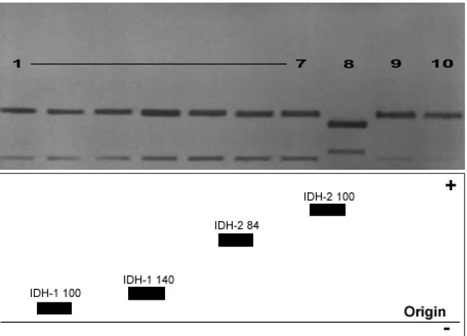
Isocitrate-dehydrogenase IDH 1.1.1.42
Malate-dehydrogenase (NAD-dependent) MDHNAD 1.1.1.37 Malate-dehydrogenase (NADP-dependent) MDHNADP (ME) 1.1.1.40
Mannose-phosphate-isomerase MPI 5.3.1.8
Phosphoglucomutase PGM 5.4.2.2
2.2.5. Preparation of samples
One mL buffer solution of 10 mM Tris/citric acid, pH 7.5 including 10 mM 2- mercaptoethanol solution was prepared. Fifteen up to 22 plastic reaction tubes (1.5 mL) were prepared with corresponding labels. Each of the vial was filled with 40 µl of buffer solution.
The mosquito samples were removed from the liquid nitrogen and each one specimen was transferred into each of the 22 vials. The samples in the vials were homogenized for 15 sec.
by using an ultrasound sonicator instrument (Sonopuls). The homogenate was centrifuged for a period of 1 min. at 14,000 rpm in a table Eppendorf centrifuge and the supernatants including the soluble proteins were used for electrophoresis.
2.2.6. Performance of electrophoresis
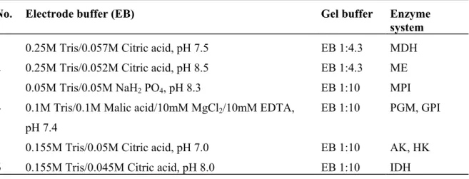
Five hundred mL of electrophoresis buffer were prepared based on the standard procedures from Harris and Hopkinson (1976) and Shaw and Prasad (1970). Table 2-2 shows the different buffer systems adapted to the enzyme specific requirements used in the the study.
Table 2-2. Buffer systems that were used for enzyme electrophoresis and gel preparation following Harris and Hopkinson (1976) and Shaw and Prasad (1970).
No. Electrode buffer (EB) Gel buffer Enzyme system 1 0.25M Tris/0.057M Citric acid, pH 7.5 EB 1:4.3 MDH 2 0.25M Tris/0.052M Citric acid, pH 8.5 EB 1:4.3 ME 3 0.05M Tris/0.05M NaH2 PO4, pH 8.3 EB 1:10 MPI 4 0.1M Tris/0.1M Malic acid/10mM MgCl2/10mM EDTA,
pH 7.4
EB 1:10 PGM, GPI
5 0.155M Tris/0.05M Citric acid, pH 7.0 EB 1:10 AK, HK 6 0.155M Tris/0.045M Citric acid, pH 8.0 EB 1:10 IDH
2.2.7. Gel preparation
Gels were prepared using the appropriate gel buffer for the protein systems in a ratio of 1:10 (buffer : agarose + H2O). Nine mL of the prepared electrode buffer (Table 2-2) and 0.9 g agarose (Applichem, LEEO) were filled up to 90 mL with distilled water in an Erlenmeyer
flask. The mixture was heated on a magnetic heating stirrir with continuous swirling until a vigorously boiling, clear, viscous solution was obtained (4-5 min.).
A glass plate (19x25 cm) serving as a carrier for the gel was prepared. The glass was labelled with the name of the buffer system, the date and the names of the mosquito samples. A single line was drawn (4cm) from the edge of the glass plate in order to serve as the point of origin for sample application.
The gel solution was poured onto the glass plate. It was cooled at room temperature for 10 min. until the agarose substance solidified. It was transferred into a moist box and placed immediately in the refrigerator (4°C) for a minimum of 30 min. Before putting the homogenate samples onto the gel, a longitudinal paper strip was applied (2 min.), 2 inches from 1 end of the cooled gel in order to remove the excess amount of moisture as well as it enabled the samples to penetrate the gel. A slot guide of plexiglass with corresponding numbers from 1 to 15 or to 22 was placed at the line of origin prior to electrophoresis. Three to 7 µl of the homogenate samples were pippeted from each of the fifteen vial and placed into the sample slots of the prepared gel.
2.2.8. Equipment preparation
The gel with the samples was set on the cooling plate (+4°C) and connected to the buffer tanks by electrode wicks and thus, the gel served as a bridge between both tanks. About 250 mL of electrolyte or electrode buffer were poured into each tank for an electrophoretic run.
The chamber started to operate by switching on the power supply at 200 Volts for 10 min.
(pre-run) and was turned to 500 Volts for 1 h (main run).
A stain solution for a specific enzyme was prepared following Harris and Hopkinson (1976) and Murphy et al. (1996). Each enzyme used in the experiments had a corresponding stain (Table 2-3).
Table 2-3. The different enzyme systems with their corresponding stains.
Enzyme Abbreviation E.C. No. Stain Composition Quantity Malate-dehydrogenase (NAD) MDH(NAD) 1.1.1.37 0.1 M Tris/HCl, pH 8.0 25 ml
L-Malic Acid 350 mg, pH 8.0
NAD 10 mg
MTT 7.5 mg
PMS 5 mg
Agar, 2 % 25 ml Enzyme Abbreviation E.C. No. Stain Composition Quantity Malate-dehydrogenase (NADP) MDH(NADP),
(ME) 1.1.1.40 0.1 M Tris/HCl, pH 7.0 25 ml
L-Malic acid 100 mg, pH 7.0 MgCl2, 1M 0.5 ml
NADP 5 mg
MTT 5 mg
PMS 0.5 mg
Agar, 2% 25 ml Isocitrate-dehydrogenase IDH 1.1.1.42 0.5 M Tris/HCl, pH 8.0 25 ml Isocitric acid 20 mg MgCl2, 1M 3 ml
NADP, MTT, PMS 5 mg each Agar, 2% 25 ml Hexokinase HK 2.7.1.1 0.05 M Tris/HCl, pH 7.1 25 ml
MgCl2*6H2O 9 mg
NADP 5 mg
Glucose 40 mg
ATP 10 mg
G6PDH 35 µl (80u)
MTT 8 mg
PMS 2 mg Agar, 2% 25 ml
Enzyme Abbreviation E.C. No. Stain Composition Quantity Adenylate Kinase AK 2.7.4.3 0.05 M Tris/HCl, pH 7.1 20 ml
MgCl2*6H2O 9 mg
NADP 5 mg
Glucose 40 mg
ADP 8 mg
Hexokinase 0.08 µl (160u) G6PD 35 µl (80u)
MTT 8 mg
PMS 2 mg
Agar, 2% 30 mL Mannose-phosphate-isomerase MPI 5.3.1.8 0.2 M Tris/HCl, pH 7.5 25 ml
Mannose-6 phosphate
(Ba-Salz) 10 mg
1 M MgCl2 0.2 ml Glucose-phosphate-
isomerase
2 µl
Glucose-6-phosphate-
dehydrogenase 3 µl NADP, MTT, PMS 5 mg Agar, 2% 25 mL Glucose-6-phosphate-isomerase GPI 5.3.1.9 0.2 M Tris/HCl, pH 8.0 25 ml
1 M MgCl2 0.4 ml
Fructose-6-phosphate
(Ba-Salz)
20 mg
Glucose-6-phosphate-
dehydrogenase 2.5 µl NADP, MTT, PMS 5 mg each Agar, 2% 25 ml
Enzyme Abbreviation E.C. No. Stain Composition Quantity Phosphoglucomutase PGM 2.7.5.1 0.05 M Tris/HCl, pH 8.0 25 ml
Glucose-1-phosphate 40 mg Glucose-1,6-diphosphate 0.1 mg MgCl2, 1 M 0.5 ml
Glucose-6-phosphate- dehydrogenase
2.5 µl
NADP, MTT, PMS 5 mg Agar, 2% 25 ml
The individual proteins had to be selectively stained and thus, a specific substrate for the enzyme was desired to allow the catalyzation of the particular reaction. The development of a dye, visualized the formation of bands of enzyme activity in the gel when electrophoresis was successful. The stain solution including substrate, co-factors and dye was poured into the gel and was immediately incubated in the oven (37°C). The results were observed (the earliest was 20 min.) by the appearance of the different bands caused by enzyme activity in the gel.
Photoes were taken from these results and the migration of different isozymes were also recorded and drawn for the interpretation of data.
2.2.9. Data nomenclature and analyses
The appearance of enzyme bands in the gels with different electrophoretic mobilities from different samples implies that these electrophoretic variants of enzymes are encoded either by different alleles (allozymes) or by different gene loci (isozymes), respectively. The appearance of one band or single band implies a gene locus whose gene products are electrophoretically indistinguishable and that no allelic variation is detectable. These gene loci are considered as monomorphic and homozygous. In contrary, the appearance of more than one band implies heterozygosity depending on the quarternary structure of the protein, or homozygote at more than one locus. After the representation of the alleles, a hyphenated number (if the enzyme is encoded by more than one locus) is assigned according to their electrophoretic mobility (no. 1 is closer to the origin, i.e. less mobile, than no. 2). Allelic variants of enzymes are assigned by a subscript number depending on their relative electrophoretic mobility while the mobility of the most common allozyme of the reference population (Cx. p. pipiens, Germany) is expanded to the value of 100 mm. The relative
mobility of each other allozyme is expanded by the same factor (e.g. IDH-2*70, IDH-2*100 and IDH-2*120, where IDH-2*100 is the most common allozyme with intermediate electrophoretic mobility).
The zymograms or the allozyme patterns on gels obtained after electrophoresis depend on the structure of the enzymes (Evans, 1987). A monomeric enzyme consists of one polypeptide chain, thus, an electrophoretic detectable heterozygous individual possesses two bands. First band corresponds to the position of one homozygote and the second band corresponds to the other homozygote. For enzymes which consist of two polypeptides (dimeric structure such as IDH), heterozygotes are represented by three bands with the middle band usually being the strongest due to the combination of both variants in one protein molecule. Nevertheless, monomorphic enzymes or homozygous gene loci are always represented by one band except of post-translational (non-genetic) modifications.
Furthermore, the band patterns that appeared in the gels represent the phenotypes with a clear genetic background. Allozymic variation reflects a part of the genetic variation on DNA-level.
Diploid organisms such as mosquitoes consist of two copies of each gene locus (except of the few genes of the sex-chromosomes in males). The products of both gene copies can be identical (monomorphic) or can differ in amino acid sequence and the underlying variants of the same gene locus are called alleles. The genotype describes the combination of identical or different alleles in one specimen. Genotypic frequencies in a certain population depend on the allele frequencies of that population.
The degree of heterozygosity reflects the combination frequencies of different alleles and provides a measure of the genetic variability within the population. This is calculated by adding up the numbers of each heterozygous genotype at a given locus and dividing each by the total number of all genotypes combined (total sample).
Beside the genotypic frequencies, the frequencies of each allele at each locus were calculated.
This was done by counting the number of times each variant appeared and dividing it by the total number of distinguishable alleles in the sample. The frequency of an allele calculated in this way is the frequency of individual homozygous for that allele, plus half the frequency of heterozygotes for that allele. Each allele frequency was calculated separately and when totalled the sum should be equal to 1.0. This was done by using the formula below (Evans, 1987).
where A, B are allele frequencies; N is the sample size.
Allele frequencies were obtaind from the randomly choosen samples out of the whole population. They accurately reflect the frequencies in the population as a whole, particularly if the samples are large enough in number. In this case, it is of great importance to calculate the variance of each allele frequency as a measure of accuracy for the estimated real population's status. Formula for doing this together with an algebraic representation of the above method for estimating allele frequencies is given below (Evans, 1987).
Var (pA) = pA (1-pA)/2 Ntotal
where Var (pA) is the variance of allele frequency pA of allele A and Ntotal is the size of the mosquito sample.
The calculation of the Confidence Interval was taken from Weir (1990) for the probability of 95%.
CI(pA) = pA±2√Var (pA)
By 5% probability, a randomly choosen sample posseses allele frequencies beyond the calculated interval.
2.2.10. The Hardy-Weinberg Equilibrium
The Hardy-Weinberg Equilibrium was founded by Hardy and Weinberg in the year 1908. It states that in a randomly mating population, the expected distribution of genotypes is determined by the random combination of alleles and this results in an equilibrium being set up among the genotypic frequencies at any given locus that remains constant from one generation to the other.
For the proof of the individual genotypic frequencies, the expected frequency values of each genotype have to be determined. The Hardy-Weinberg formula states for homozygote genotypes Npi2 and 2Npipj for the heterozygote genotypes. In case of a limited number of individuals (N<100 individuals, accdg. to Spiess, 1989) the number of homozygotes is over
total AB AA
N N pA N +1/2
= ;
total BB AB
N N
pB N +
=1/2
( ) ( )
∑
∑
< =− + −
−
= − n
i ii
ii n ii
j
i j
j j
E c E O Ei
c Ei Oi
1 2 2
χ2
represented and has to be corrected (Levene, 1949; Haldene, 1954; Smith, 1970). The genotype frequencies are corrected according to the following formula (Lessios, 1992):
1 2 4 2
= − N
p p
Eij N i j for heterozygote
1 2
) 1 2
(
−
= −
N Np
Eii Npi i for homozygote
In order to prove whether the observed genotype distribution is corresponding to the expected genotype distribution of a Hardy-Weinberg population or a significant difference is given, a χ2 test was done according to the following formula (Lessios, 1992):
c refers to the correction factor which amounts to 0.25 if two alleles or is zero if three and more alleles are considered (Emigh, 1980).
As for the estimated allele frequency the degree of freedom is reduced to one. The degree of freedom is calculated in this test not according to the genotypes –1, but acording to the following formula (Dobzhansky & Levene, 1948; Crisp, 1978).
2 ) 1 ( −
= n n FG
n = number of alleles at the given locus.
In order to have a valid data, it was assumed according to Cochran (1954) that the expected frequency is greater than 1 and at the same time 60% of the frequencies are higher than 5. In the χ2 test, all rare alleles were combined and the new frequencies were computed. In case one locus had 2 alleles icluding one rare allele, a table after Vithayasai (1973) was used to determine the heterozygosity in relation to the sample size and the frequency of the rare alleles.
2.2.11. Genetic identity and distances
The genetic distances between two populations for one gene locus can be determined according to Nei (1972) after the following formula:
∑ ∑
=
∑
2 2
i i
i i
L x y
y I x
For the average of all determined loci, the following formula has been applied:
I Ø
y x xy
J J
= J , where Jxy=ΣΣxiyi, Jx =ΣΣx2i, Jy =ΣΣy2i
The genetic identity is expressed in numbers between zero (populations don’t have any common allele) and one (the populations have identical allele frequencies).
The genetic distance D is calculated by
D∅ = -ln I∅
according to Nei (1972). D varies between zero for populations having the same frequencies until indefinite (theoretically) for populations with no common alleles. The genetic distances between the Culex populations were calculated using Gendist from the Phylip programme package (Phylgeny inference package, windows version 3.6C) by Felsenstein (2002).
The UPGMA method
For a better documentation of the calculated paired distances between the populations, dendrograms were constructed based on the UPGMA-cluster analyses (Unweighted Pair Group Method with Arithmetic Means) with the Neighbor program (Phylip programme package, Felsenstein, 2002).
2.3. RESULTS
2.3.1. Results of enzyme electrophoresis in Culex populations
The data presented below provide indications of protein electrophoretic variability in the populations of Culex. The genetic variability within as well as the differentiation among the populations of Culex were analyzed.
Each enzyme was investigated in several electrophoretic runs in populations of Culex.
Specific banding patterns in the gels were observed in each of the enzyme system. Original photoes were taken from these bands for documentation processes. The electrophoretic conditions were adjusted individually so that the proteins usually migrated from the origin to the top (anode). Only few variants migrated to the cathode. Allele frequencies, genotype frequencies as well as the variances in comparison between the different populations were calculated.
Oxidoreductases
Buffer system no. 1, Running time: 66 min., Voltage: 450 V, Ampere: 105 mA, Incubation time for staining: 30 min.
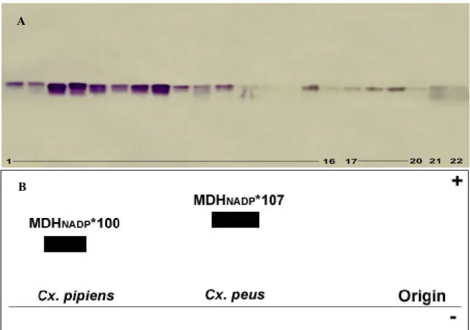
The results obtained from MDHNAD in populations of Culex showed the presence of two MDHNAD- loci. These were clearly seen by the formation of bands in the gel with different electrophoretic mobilities, thus, it imply that these enzyme variants are encoded by different gene loci (Fig. 2-1A & B).
Malate-dehydrogenase(NADdependent)(MDHNAD) E.C. 1.1.1.37
Figure 2-1A shows that only one common electrophoretic variant in populations of Culex was observed in MDHNAD-1* (MDHNAD-1*100). All of the investigated Culex populations had the same allele frequency which is equal to 1.0000.
In regards to the locus MDH-2*, three allelic variants were detected, MDHNAD-2*76, MDHNAD-2*100 and MDHNAD-2*124. Cx. p. pipiens, Cx. p. quinquefasciatus (Phils.) and Culex p. quinquefasciatus (USA), showed a common allozyme at +25 mm (MDHNAD-2*100) and a rare one at +31 mm (MDHNAD-2*124). Culex peus was differentiated by its own fixed allele (MDHNAD-2*76), the corresponding allozyme had a lower electrophoretic mobility (19 mm) than the Cx. p. pipiens reference allozyme (MDHNAD-2*100).
The absolute (Abs.) and relative (Rel.) allele frequencies as well as variances (Var.) and the confidence intervals (CI) of locus MDH-1* and MDH-2* in populations of Culex were calculated (Table 2-1, 2-2, 2-3 & Graph 2-1).
Figure 2-1A. Electrophoretic mobilities of MDHNAD enzyme in populations of Culex. Lanes: 1 to 5:
Cx .p. pipiens, 6 to 12: Cx. p. quinquefasciatus (USA), 13 to 14: Cx. peus. B. Electrophoretic mobility model of Malate-dehydrogenase (MDHNAD) enzyme.
A
B
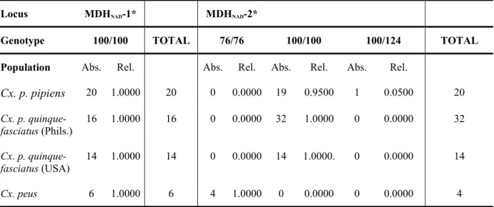
Table 2-1. Absolute (Abs.) and relative (Rel.) genotype frequencies of MDHNAD-1* and MDHNAD-2* in populations of Culex.
Locus MDHNAD-1* MDHNAD-2*
Genotype 100/100 TOTAL 76/76 100/100 100/124 TOTAL Population Abs. Rel. Abs. Rel. Abs. Rel. Abs. Rel.
Cx. p. pipiens 20 1.0000 20 0 0.0000 19 0.9500 1 0.0500 20 Cx. p. quinque-
fasciatus (Phils.)
16 1.0000 16 0 0.0000 32 1.0000 0 0.0000 32
Cx. p. quinque-
fasciatus (USA) 14 1.0000 14 0 0.0000 14 1.0000. 0 0.0000 14
Cx. peus 6 1.0000 6 4 1.0000 0 0.0000 0 0.0000 4
Table 2-2. Absolute (Abs.) and relative (Rel.) allele frequencies as well as variances (Var.) and confidence intervals (CI) of MDHNAD-1*. The confidence interval results from the relative allele frequency ± the V-value (see 2.2.9).
Allele MDHNAD-1* 100
CI
Population Abs. Rel. V(±) Var.
Cx. p. pipiens 40 1.0000 0.0000 0.0000
Cx. p. quinquefasciatus (Phils.) 32 1.0000 0.0000 0.0000
Cx. p. quinquefasciatus (USA) 28 1.0000 0.0000 0.0000
Cx. peus 12 1.0000 0.0000 0.0000
Table 2-3. Absolute (Abs.) and relative (Rel.) allele frequencies as well as variances (Var.) and confidence intervals (CI) of locus MDHNAD-2* in populations of Culex. The confidence interval results from the relative allele frequency ± the V-value (see 2.2.9).
The results presented in Table 2-3 show that there were three different alleles represented by the bands MDH-2*76, MDH-2*100 and MDH-2*124. In MDH-2*124, Culex p. pipiens has an additional rare allele, whereas MDH-2* in Culex p. quinquefasciatus from the Philippines and USA was monomorphic showing a fixed common allele. On the other hand, Culex peus was well differentiated by one exclusive allozyme representing the allele MDH-2*76.
Allele MDHNAD-2*76 MDHNAD-2*100 MDHNAD-2*124
CI CI CI
Population Abs. Rel. V(±) Var. Abs. Rel. V(±) Var. Abs. Rel. V(±) Var.
Cx. p. pipiens 0 0.0000 0.0000 0.0000 38 0.9500 0.0346 0.0012 2 0.0500 0.0346 0.0012 Cx. p. quinque-
fasciatus (Phils.)
0 0.0000 0.0000 0.0000 64 1.0000 0.0000 0.0000 0 0.0000 0.0000 0.0000
Cx. p. quinque- fasciatus (USA)
0 0.0000 0.0000 0.0000 28 1.0000 0.0000 0.0000 0 0.0000 0.0000 0.0000
Cx. peus 8 1.0000 0.0000 0.0000 0 0.0000 0.0000 0.0000 0 0.0000 0.0000 0.0000
Graph 2-1. The distribution of allele frequencies of MDH-2* in populations of Culex. Cx.. p. p.=
Cx.p.p.
Cx.p.qu. (Phils.)
Cx.p.qu. (USA)
Cx. peus
Frequency
0.0 0.2 0.4 0.6 0.8 1.0
M D H -2*76 M D H -2*100 M D H -2*124
Population
Allele Frequencies MDH-2*