Technische Universität Dortmund Fakultät für Chemie und Chemische Biologie
Dissertation zur
Erlangung des Grades Dr. rer. nat.
Systems Analysis of the spatial Regulation of oncogenic Ras
signalling
vorgelegt von
Kaatje Friederike Heinelt Matrikel-Nr. 173007 Peterstr. 1 | 26316 Varel
angefertigt am
Max-Planck-Institut für Molekulare Physiologie Arbeitsgruppe Bastiaens
1. Gutachter: Prof. Dr. Philippe Bastiaens 2. Gutachter: PD Dr. Leif Dehmelt
Eidesstattliche Versicherung
Ich versichere hiermit an Eides statt, dass ich die vorliegende Dissertation mit dem Titel
“Systems Analysis of the spatial Regulation of oncogenic Ras Signalling“ selbstständig und ohne unzulässige fremde Hilfe angefertigt habe. Ich habe keine anderen als die angegebenen Quellen und Hilfsmittel benutzt sowie wörtliche und sinngemäße Zitate kenntlich gemacht.
Die Arbeit hat in gegenwärtiger oder in einer anderen Fassung weder der TU Dortmund noch einer anderen Hochschule im Zusammenhang mit einer staatlichen oder akademischen Prüfung vorgelegen.
__________________________ __________________________
Ort, Datum Unterschrift
Table of contents
1 Abstract... 1
2 Zusammenfassung ... 3
3 Introduction ... 5
3.1 Ras family... 5
3.1.1 Ras GTPases – an overview ...5
3.1.2 The GTPase cycle of Ras proteins...6
3.1.2.1Ras and its interaction with GEFs...8
3.1.2.2Ras and its interaction with GAPs...9
3.1.3 The clinically most notable members of the Ras subfamily...11
3.1.4 Downstream effector signalling of Ras ...16
3.1.5 Kras ...21
3.1.6 Kras mutations and their impact in cancer... 22
3.1.7 Inhibition of oncogenic Ras – The “undruggable” protein ...27
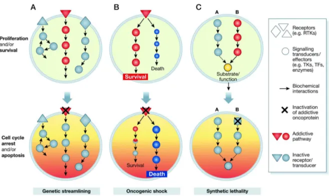
3.1.8 The principle of oncogene addiction ...28
3.1.9 Another way to target Ras – small molecule PDEδ inhibitors...31
3.2 Organoids as a new model system... 32
4 Objectives ... 34
5 Material... 35
5.1 Cell lines used ... 35
5.2 Antibodies ... 35
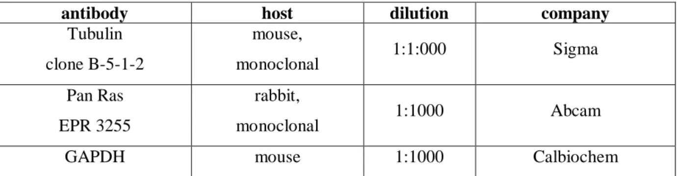
5.2.1 Primary antibodies for Western Blots ...35
5.2.2 Secondary antibodies for Western Blots ... 35
5.2.3 Primary antibodies for Immunofluorescence... 36
5.2.4 Secondary antibodies for Immunofluorescence... 36
5.3 Materials and Media for cell culture ... 36
5.4 Transfection ... 37
5.5 Oligonucleotides ... 37
5.5.1 Primer for LIC...37
5.5.2 Primer for sequencing ... 38
5.5.3 Primer for genotyping ... 40
5.6 Animal resources, organoid preparation and organoid culture... 41
5.6.1 Mice ... 41
5.6.2 Material and Media for organoid culture... 42
5.7 Bacteria ... 43
5.8 Plasmids ... 43
5.8.1 Eukaryotic Vectors ... 43
5.8.2 Viral Vectors...43
5.9 Media for Bacteria... 43
5.10 PFA preparation ... 44
5.10.1 4% PFA in PBS ...44
5.10.2 4% PFA in PME...44
5.11 Commercial Material and Kits... 44
5.12 Inhibitors... 45
5.13 Standards ... 45
5.14 General chemicals... 45
5.15 Buffer... 47
5.16 Devices and expendables ... 47
5.17 Microscopes... 49
6 Methods ... 50
6.1 Culture of cell lines ... 50
6.2 Freezing and thawing cell lines ... 51
6.3 Transfection of cell lines... 52
6.3.1 Transfection with Fugene ... 52
6.3.2 Transfection with Effectene ... 52
6.3.3 Transfection with Lipofectamine 2000...53
6.4 Preparation of small intestine organoids... 54
6.5 Culture of small intestine organoids ... 55
6.6 Freezing and thawing organoids ... 57
6.7 Retroviral infection of organoids... 58
6.8 Whole cell lysates ... 61
6.8.1 Mammalian cells ... 61
6.8.2 Organoids ... 62
6.9 SDS-PAGE ... 63
6.10 Western Blot – Tank Blot method ... 64
6.11 Immunodetection ... 65
6.12 Antibody stripping of Western Blot membranes... 66
6.13 Bradford assay ... 66
6.14 DNA preparation using Quick-DNATM Universal Kit... 67
6.15 Determination of DNA concentration with NanoDrop Spectral
photometer... 67
6.16 Bacterial culture... 67
6.17 Transformation of bacteria... 68
6.18 Polymerase Chain Reaction (PCR) methods ... 69
6.18.1 Ligation independent cloning ... 70
6.18.2 Genotyping PCR (Organoids) ... 70
6.19 DNA preparation M&N Midi kit ... 71
6.20 DNA preparation using Roti®-Prep Plasmid Mini Kit ... 72
6.21 Agarose Gelelectrophoresis ... 72
6.22 Purification of DNA from Agarose gels... 73
6.23 Restriction digest... 73
6.24 Dephosphorylation of 5’-phosphorylated DNA ... 74
6.25 PCR product purification... 74
6.26 Ligation... 75
6.27 Sequencing using BigDye® Terminator kit... 75
6.28 Preparation of Cryoslices using a Cryostat ... 77
6.29 Immunofluorescence ... 78
6.29.1 Cells ... 78
6.29.2 Organoids on slides... 78
6.29.3 Organoids in SPIM ... 79
6.30 Life cell imaging experiments... 81
6.30.1 In cells... 81
7 Results ... 84
7.1 Comparison of the localization of different over expressed Ras isoforms upon Deltarasin and Deltazinone 1 treatment in MDCK cells ... 84
7.2 Comparison of the effect of Deltarasin and Deltazinone 1 treatment on localization of endogenous Ras in A431 cells ... 87 7.3 Deltarasin relocalizes Ras in colorectal cancer cells ... 88
7.4 Comparison of the effect of Deltarasin and Deltazinone 1 treatment on Localization of mCherry KrasWT in PancTuI cells ... 90 7.5 Characterization of small intestine organoids ... 95
7.6 Infection with Cre recombinase leads to DNA recombination and
expression of mTFP-KrasG12D protein in small intestine organoids... 96
7.6.1 Cre-mediated DNA recombination in organoids to switch mTFP-KrasG12D expression on ... 97 7.6.2 mTFP Kras expression in organoids ... 101
7.7 Deltarasin causes mTFP-KrasG12D relocalization in small intestine
organoids... 106
8 Discussion ... 109
8.1 mCherry KrasWT and mCitrine HrasG12V relocalize in parallel in MDCK cells upon PDEδ inhibition by Deltarasin but not by Deltazinone 1 ... 109
8.2 Endogenous Ras relocalizes in Kras-independent cells upon PDEδ inhibition by Deltarasin in a dose dependent manner ... 112
8.3 mCherry KrasWT relocalizes in Kras-dependent cells upon PDEδ inhibition by Deltarasin and Deltazinone 1 ... 113 8.4 Why use small intestinal organoids?... 115 8.5 The mTFP-KrasG12D organoid system... 116
8.7 Endogenous levels of mTFP-KrasG12D relocalize small intestinal organoids upon PDEδ inhibition by Deltarasin... 120
9 Outlook ... 122
1 Abstract
Ras (Rat sarcoma) isoforms are small GTP-binding proteins that play a major role in the signalling networks controlling cell growth and survival. The Kras isoform is of particular interest as in many severe kinds of cancer the presence of oncogenic Kras mutations is associated with a poor prognosis.
Kras is associated with the plasma membrane due to its farnesyl moiety and a polybasic motif and functions as a signalling hub. If Kras gets lost from the plasma membrane due to spontaneous dissociation or endocytosis, it will equilibrate over the extensive endomembrane system inside the cell. With Kras no longer present at the plasma membrane, its activation and the following activation of subsequent pathways can no longer take place. However, to remain on the plasma membrane, Kras has to be constantly enriched there. This enrichment must be actively maintained in the cell by an energy-driven mechanism involving the solubilising factor PDEδ. Consequently, inhibition or down-modulation of PDEδ results in mislocalisation of Kras, making PDEδ an interesting target for anti-cancer drug development.
In 2013 a small molecule, Deltarasin, was identified as a potent inhibitor of PDEδ causing a redistribution of Kras from the plasma membrane towards endomembranes when applied to cells.
This work investigates whether small molecule PDEδ inhibitors such as Deltarasin affect (K) Ras localization in space and time. It demonstrates that PDEδ inhibition causes Ras relocalisation from the plasma membrane towards the endomembranes in different human cancer cell lines and in murine small intestine organoids, which express endogenous levels of oncogenic Kras.
Nonetheless, it has been shown that Deltarasin has certain side effects, e.g. it becomes cytotoxic at higher concentrations. Hence, a new PDEδ inhibitor, Deltazinone 1, which is supposed to be less cytotoxic in comparison to Deltarasin, was synthesized. In order to determine whether it represents a viable alternative to Deltarasin, its ability to relocalise Kras in a panel of cancer cell lines was tested and it was indeed possible to mislocalise Kras with this inhibitor in Kras-dependent PancTuI cells. Deltazinone 1 and Deltarasin both had a demonstrable effect on cell growth/survival, respectively. In this way it appears that PDEδ constitutes a valid target for the pharmacological therapy of Kras-dependent tumours.
This work demonstrates that two specific PDEδ inhibitors with completely different lead structures are capable of mislocalising Kras to endomembranes. In sum, it demonstrates that
the availability of PDEδ is essential to ensure (K) Ras localization at the plasma membrane in Kras-dependent cancer cells and thus that the survival of those cells are ultimately dependent on PDEδ.
2 Zusammenfassung
Bei Ras (Rat sarcoma) Isoformen handelt es sich um kleine GTP-bindende Proteine, die eine wichtige Rolle in Signalwegen zur Kontrolle des Wachstums und Überlebens von Zellen spielen. Dabei ist besonders die Kras Isoform von Bedeutung, da in vielen Krebsarten das Vorhandensein onkogener Mutationen von Kras mit einer schlechten Prognose verbunden ist.
Kras kann mittels seines Farnesylschwanzes und einer Abfolge basischer Aminosäuren an die Plasmamembran binden und wirkt dort als Knotenpunkt im Verlauf der Signalübertragung.
Wenn Kras durch spontane Dissoziation oder Endocytose die Plasma Membran verlässt, equilibriert es über das im Inneren der Zelle vorhandene ausladende System von Endomembranen. In Folge dessen können die Aktivierung von Kras und auch die der nachfolgenden Signalwege nicht länger stattfinden. Kras muss kontinuierlich an der Plasmamembran angereichert werden, um dort verbleiben zu können. Diese Anreicherung ist ein aktiver Prozess und wird durch einen Energie-abhängigen Mechanismus aufrecht erhalten, an welchem der Lösungsfaktor PDEδ beteiligt ist. Folglich führen Inhibition oder Herabregulierung von PDEδ zu einer falschen Lokalisation von Kras in der Zelle, was PDEδ zu einem überaus interessanten Zielprotein für die Entwicklung neuer Medikamente zur Behandlung von Krebs macht. Im Jahr 2013 wurde ein kleines Molekül mit dem Namen Deltarasin identifiziert, von welchem gezeigt werden konnte, dass es ein potenter Inhibitor für PDEδ ist. Es führt zu einer Umlagerung von Kras von der Plasmamembran in Richtung der Endomembranen, wenn es zu Zellen gegeben wird.
In dieser Arbeit wird untersucht, inwieweit PDEδ Inhibitoren auf Basis kleiner Moleküle, wie z.B. Deltarasin, die Lokalisation von (K)Ras in Raum und Zeit beeinflussen. Es kann in verschiedenen humanen Krebszelllinien und in Dünndarmorganoiden aus der Maus, welche endogene Level von oncogenem Kras exprimieren, gezeigt werden, dass die Inhibition von PDEδ zu einer Relokalisation von Ras von der Plasmamembran in Richtung der Endomembranen führt.
Dennoch wurde gezeigt, dass Deltarasin unerwünschte Nebeneffekte hat, beispielsweise wirkt es in höheren Konzentrationen cytotoxisch. Daher wurde ein neuer Inhibitor für PDEδ, Deltazinone 1, synthetisiert, welcher weniger cytotoxisch sein sollte. Um herauszufinden, ob er eine gute Alternative zu Deltarasin darstellt, wurde seine Fähigkeit zur Relokalisation von Kras in einem Panel von Krebszelllinien überprüft. Es war tatsächlich möglich Kras mittels dieses Inhibitors in Kras-abhängigen PancTuI Zellen von der Plasmamembran in Richtung
der Endomembranen zu verlagern. Da Deltazinone 1 und Deltarasin beide nachweislich Effekte auf Wachstum und Überleben von Zellen haben, scheint PDEδ ein lohnendes Ziel für die pharmakologische Therapie Kras-abhängiger Tumore zu sein.
Diese Arbeit zeigt, dass zwei spezifische PDEδ Inhibitoren mit vollkommen verschiedenen Leitstrukturen Kras in Richtung der Endomembranen mislokalisieren können.
Zusammengefasst zeigt sie, dass das Vorhandensein von PDEδ essentiell ist, um in Kras- abhängigen Krebszellen die Lokalisation von (K)Ras an der Plasmamembran zu gewährleisten. Somit hängt das Überleben dieser Zellen komplett von PDEδ ab.
3 Introduction
Ras proteins are the biological background of this work. Accordingly, their role in signal transduction under both normal and pathological conditions and the manner in which they are regulated to be at the right place at the right time are outlined in this chapter.
3.1 Ras family
3.1.1 Ras GTPases – an overview
The Ras (Rat sarcoma) protein superfamily comprises more than 150 members in humans and is divided into five major branches based on sequence and functional similarities: Ras, Rho, Rab, Ran and Arf (Wennerberg et al., 2005). All members of the Ras superfamily are small GTPases that act as molecular switches that cycle between inactive GDP-bound and active GTP-bound states and share a common biochemical mechanism (Vetter and Wittinghofer, 2001). Members of the Ras branch play a major role in the signalling networks controlling cell growth and survival (Nandan and Yang 2011; Courtney et al., 2010) and were found to be some of the first oncogenes (Der et al., 1982; Shimizu et al., 1983).
Ras proteins share a similar topological structure, which is characterized by a G-domain with 6 β-strands flanked by 5 α-helices and 10 connecting loops (Figure 1, Milburn et al., 1990).
The proteins have a molecular weight of 21 kDa and are 188/189 amino acids in length.
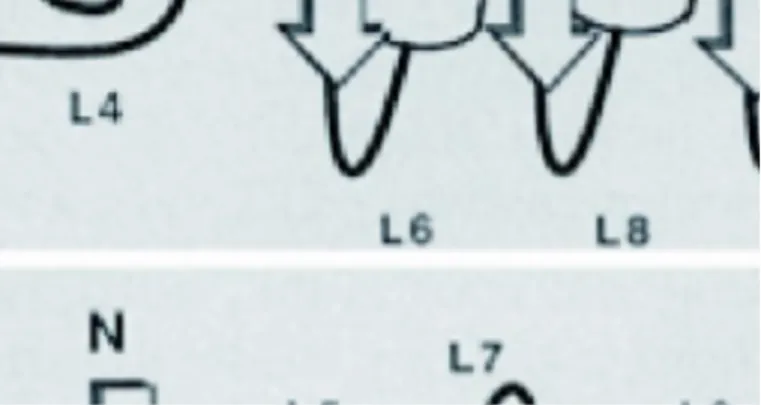
Figure 1: Topological structure of Ras proteins
The topological structure of Ras proteins: α -helices are represented by cylinders and β –strands by arrows. The current assignments of the beginning and the ending residue numbers for each secondary structural element are; β 1 (1-9), β 2 (38-46), β 3 (50-58), β 4 (77-84), β 5 (110-117), β 6 (140-144), α l (15-26), α 2 (67-75), α 3 (87- 104), α 4 (126-137), α 5 (151-171), L1 (10-14), L2 (27-37), L3 (47-49), L4 (59-66), L5 (76), L6 (85-86), L7 (105-109), L8 (118-125), L9 (138-139), and L1O (145-150). A shaded string represents the COOH-terminal 18 residues that are lacking or disordered in the crystal structures. (Milburn et al., 1990)
The catalytical G domains that form the guanine nucleotide-binding pocket of all Ras proteins are highly conserved. They share 100% identity in the N-terminal lobe 1 (residues 1–86) and have an identity of 90% in the C-terminal lobe 2 (residues 87–171) (Buhrman, et al., 2011).
The only region of significant difference between the Ras-isoforms is the C-terminal hypervariable region (HVR), which contains isoform-specific trafficking and membrane binding determinants. Each Ras-isoform therefore shares overlapping but distinct localizations at the plasma membrane and on intracellular organelles (Eisenberg et al., 2013).
Lobe 1, the effector lobe containing protein/protein interaction sites with effectors, includes the catalytic machinery with the active site for GTP hydrolysis (switch I, switch II, residues 10-17 of the P-loop) and a large part of the nucleotide binding pocket.
Lobe 2, on the other side of the molecule, contains the membrane-interacting portions of Ras and is called the “allosteric” lobe. It contains several affinity hot spots that interact directly with membrane components, the allosteric site and the allosteric switch components. The active site and allosteric site of Ras are connected together through helix 3 at one edge of the interlobal region and switch II at the other (Buhrman, et al., 2011; Buhrman, et al., 2010) Many residue differences between the Ras-isoforms outside the hypervariable C-terminal region can be found in the hot spots that interact with membranes of that lobe (Nussinov et al., 2013).
The third domain is the HVR, which is terminated by a CAAX box motif (Cox and Der, 2002). Here C is a cysteine, A must be an aliphatic amino acid (leucine, isoleucine, or valine) and X can be any amino acid. This motif, when coupled together with residues immediately upstream (e.g. cysteine residues modified by the fatty acid palmitate) comprises the targeting sequences for interactions with distinct membrane compartments inside the cell. It is also the recognition sequence for farnesyltransferase and geranylgeranyltransferase I, which lead to addition of farnesyl or geranylgeranyl isoprenoid to the cysteine residue of the CAAX motif (Wennerberg et al., 2005).
3.1.2 The GTPase cycle of Ras proteins
In general all Ras proteins function as molecular switches that cycle between inactive guanosine diphosphate (GDP)-bound and active guanosine triphosphate (GTP)-bound states (Figure 2; King et al., 2013). They become activated by binding GTP in response to receptor protein tyrosine kinases or other growth factor–dependent stimuli and both interact with and
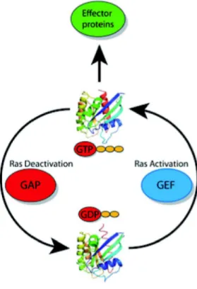
Figure 2: GTPase cycle
Ras cycles between a GTP-bound state (top) in which it interacts with downstream effector proteins, and a GDP-bound state (bottom) in which it is inactive. Different proteins regulate the activity of Ras.
The activity of the proteins that activate Ras (called GEFs) and deactivate Ras (called GAPs) is also strictly controlled by the cells. (Hein et al., 2013)
Guanine-nucleotide exchange factors (GEFs) such as Son of sevenless (SOS) stimulate the guanine-nucleotide exchange, allowing GTP, more prevalent in the cell than GDP, to bind and thus activate Ras. This is necessary as Ras has no preference in binding GTP or GDP, therefore stimulating the guanine-nucleotide exchange by GEFs allow GTP to bind and subsequently activate the protein (Boriack-Sjodin et al., 1998; Scheffzek et al., 1997; Bos et al., 2007). Ras has only weak intrinsic GTPase activity and therefore the conversion back to its inactive state is dependent upon Ras GTPase-activating proteins (Ras-GAPs), closing the GTPase cycle (Bos et al., 2007; Donovan et al., 2002). Ras-GAPs increase the intrinsic GTP hydrolysis by several orders of magnitude and thus help to make Ras inactive (Scheffzek et al., 1997). Both GEFs and GAPs are usually multidomain proteins. As many of these domains are protein or lipid interaction domains it is likely that they serve as localization signals and/or as scaffolds for the formation of protein complexes (Bos et al., 2007).
3.1.2.1 Ras and its interaction with GEFs
All GEFs for Ras have a common ~250 amino acid CDC25 homology catalytic domain (the RasGEF domain) and an adjacent ~50 amino acid N-terminal Ras exchange motif (REM; the RasGEFN domain). Three main classes of RasGEFs (Sos, Ras-GRF, and RasGRP) exist and can be distinguished by additional flanking domains and motifs that facilitate their activation or possess additional catalytic functions. CDC25 homology domains possess the ability to concurrently activate other Ras family proteins. Links between Ras activation and the function of Ras and Rho family proteins are provided by the presence of separate Rho- specific GEF catalytic domains in Sos and Ras-GRF proteins. The N-termini of Sos proteins contain the CDC25 domain and additionally Dbl homology (DH; also called RhoGEF) and pleckstrin homology (PH) domains (Mitin et al., 2005). The tandem DH–PH domain cluster is a signature motif of Dbl family proteins, comprising the majority of GEFs for the Rho family of GTPases (Rossman et al., 2006). The GDP/GTP exchange on Rho GTPases is mediated by the catalytic DH domain, while the PH domain is responsible for modulating the activity of the DH domain e.g. by promoting membrane association, facilitating GTPase substrate binding, or by controlling intramolecular interactions. Thus, Sos proteins are capable to perform GEF catalytic activity on Ras and Rac GTPases (Mitin et al., 2005).
After activation of receptor protein tyrosine kinases a GEF such as Sos is transported from the cytoplasm to the activated receptor by adapter proteins such as Grb2 in a phosphotyrosine- dependent manner. The SH3 domains of Grb2 are bound to a proline rich region in the C terminus of Sos and this Grb2–Sos complex interacts with phosphotyrosine residues on activated receptors via the SH2 domains of Grb2. Receptor activation results in an increase in the effective concentration of Sos in the vicinity of the plasma membrane localized Ras, facilitating the exchange of bound guanine nucleotide for free cellular guanine nucleotides (Boriack-Sjodin et al., 1998). The affinity of most small G proteins such as Ras for GDP/GTP is in the lower nanomolar to picomolar range. A slow dissociation rate of nucleotides with a half-life on the order of one or more hours is the direct consequence of this high affinity. In biological processes exchange of GDP for GTP and subsequent activation Ras occurs within minutes or even less. Therefore, the activity of GEFs is required for the exchange of GDP for GTP as GEFs accelerate the exchange reaction by several orders of magnitude (Vetter and Wittinghofer, 2001; Bos et al., 2007). GEFs modify the nucleotide-binding site in such a way that the nucleotide affinity is decreased which catalyzes the dissociation of the nucleotide from Ras. Thus, the nucleotide is released and subsequently replaced. The affinity of Ras for
GTP and GDP is similar and the GEF does not favour rebinding of GDP or GTP. Thus, the resulting increase in GTP-bound over GDP-bound is due to the approximately ten times higher cellular concentration of GTP compared to GDP (Bos et al. 2007). Distinct RasGEFs are capable to determine the membrane compartments in which Ras becomes activated as well as the effectors or downstream pathways that are utilized (Mitin et al., 2005).
The nucleotide binding site of Ras opens as a result of the displacement of Ras Switch 1, which is caused by the insertion of a helical hairpin formed by aH and aI of Sos when Sos binds to Ras. The insertion of helix aH into the Ras active site leads to the introduction of a hydrophobic side chain (Leu 938) which blocks magnesium binding, and an acidic side chain (Glu 942) which overlaps the side where the α-phosphate of the nucleotide would otherwise be bound (Boriack-Sjodin et al., 1998).
Ras Glu 62 and 63 are crucial for its interaction with Sos: Although mutation of both negative charged glutamic acids to positive charged histidines had no effect on the stability of Ras- GDP complexes, the ability of mutants to be activated by the CDC25 domain of the GEF was severely compromised (Boriack-Sjodin et al., 1998).
3.1.2.2 Ras and its interaction with GAPs
In about 30% of human tumours, oncogenic mutations of Ras that lead to impaired intrinsic GTPase activity and insensitivity to GAPs are found. These changes result in an inability to switch off the transmitted signal. In unstimulated cells, WT Ras is found in the inactive (GDP) state due to the presence of the negative regulator GAP, while oncogenic Ras remains in the active (GTP) state (Scheffzek et al., 1997). Ras GAPs have a common ~250 amino acid catalytic domain, but otherwise exhibit no crucial sequence similarity or domain architecture in the sequences that flank this catalytic domain (Mitin et al., 2005).
Many parts of the Ras molecule play a role in its interaction with its GAP. Participants in the interaction include residues 10 to 16 of the phosphate binding (P-) loop, residues 30 to 37 of switch region I, 60 to 76 of switch region II and possibly residues 87 to 98 of helix a3 (Scheffzek et al., 1997).
Polar interactions (involving several water molecules) and van der Waals interactions build the partly hydrophobic interface of the complex between Ras and RasGAP and may explain why the oncogenic mutation Gln61Leu in Ras, which cannot hydrolyze GTP efficiently, has a higher affinity than the wild type for GAP-334 (Scheffzek et al., 1997).
While Ras is localized in the complex with its GAP, loop L4 and helix a2 in the switch II region are well defined. In contrast, in isolated Ras these structures are highly mobile. In the complex, residues 61 to 63 of L4 are arranged in a short 310 helix which precedes a2.
Tyrosine-64 plays a major role in forming the polar contact between Ras and its GAP by contributing to the formation of the hydrophobic interface, consistent with the observation that it can be mutated to Phe but not to Glu without affecting the interaction. Switch II may be further stabilized by interactions between Glu62 or Glu63 of Ras and arginines of the GAP.
Ras-mediated GTP hydrolysis is blocked if glutamine-61 is mutated to any amino acid other than Glu leading to tumour formation. Gln61 points toward the phosphate chain of the nucleotide in the complex with GAP-334 and is stabilized in its orientation by a hydrogen bond with the main chain carbonyl group of the invariant Arg789 (Scheffzek et al., 1997). In this stabilized transition state the nucleophilic attack of a water molecule on the γ-phosphate of GTP is possible, and thus this state is a requirement for the Mg2+-dependent hydrolysis reaction (Vetter and Wittinghofer, 2001). Another important position in Ras is Gly12, which contacts the loop L1c region of GAP-334 at Arg789 in the complex via van der Waals interactions. This explains the block in GAP-accelerated GTP hydrolysis for Gly12 mutants.
Although they bind to GAP with an affinity similar to wild type Ras, it appears from the structure that even replacement with alanine would be highly unfavourable, leading to steric clashes with the main chain of Arg789 and with the side chain NH2 of Gln61, thus preventing proper hydrolysis of GTP (Scheffzek et al., 1997).
Therefore, the most frequently occurring oncogenic mutations in Ras proteins are missense gain-of-function mutations at one of three mutational hotspots: G12, G13 and Q61. They are considered to be defective in GAP-mediated GTP hydrolysis, resulting in an accumulation of constitutively GTP-bound Ras in cells. Ras can further be activated in cancer by loss of its GAPs (e.g. neurofibromin 1) or persistent receptor mediated activation of GEFs (e.g. Sos) (Hobbs et al., 2016).
The “molecular switch” model is based on the changes in conformation that occur upon hydrolysis of GTP to GDP in switch I (residues 30-40) and switch II (residues 60-76) (Milburn et al., 1990; Buhrman et al., 2011). In addition to this model, other structural features also need to be considered. In GTP-bound Ras there is an equilibrium between two distinct conformational states modulated by an allosteric switch mechanism (Buhrman et al., 2010). The Ras form that is predominately found under solution conditions used for in vitro
catalytic residue Q61. Its allosteric site is “empty”, which could explain the slow GTP hydrolysis rates measured for Ras in vitro (Buhrman et al., 2011). The other conformational state displays an ordered active site, with Q61 positioned near the catalytic centre and a shift in helix 3/loop 7 toward helix 4. It has been proposed that this is the active state in which intrinsic hydrolysis is somehow promoted by an allosteric modulator (Buhrman et al., 2010).
In the crystals investigated, bound calcium acetate was found in the allosteric site, which mimicked the modulator during the experiment (Buhrman et al., 2010). In the complex of Ras with GAPs, which promotes hydrolysis of GTP, an ordered switch II region combined with a shift of helix 3 toward helix 4 is also found. The difference is the insertion of the arginine finger from RasGAP into the active site (Scheffzek et al., 1997; Buhrman et al., 2011).
3.1.3 The clinically most notable members of the Ras subfamily
The clinically most notable members of the Ras subfamily are Harvey-Ras (Hras), neuroblastoma-Ras (Nras), and 2 variants of Kirsten-Ras (Kras), Kras4a and Kras4b. The two variants of Kras are produced by alternative splicing from the same gene (McCubrey et al., 2006). The N-terminal catalytic domains (amino acids 1–165) of Hras, Nras and Kras are highly conserved (90–100% identical). Therefore, the Ras isoforms differ primarily with respect to the sequence of the hypervariable region (HVR), which contains the protein sequences necessary for Ras to associate with the inner leaflet of the plasma membrane, and in the types of posttranslational modifications that characterize each Ras isoform. The HVR comprises the minimal Ras anchor which comprises the carboxy-terminal CAAX motif in addition to a second signal, which can be a single palmitoylation site (C181) in Nras, two palmitoylation sites in Hras (C181 and C184) and a polybasic domain of six contiguous lysine residues in Kras (K175–K180) (Figure 3; Hancock, 2003).
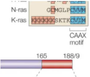
Figure 3: Domain structure of the Ras proteins
The N-terminal catalytic domains of all Ras isoforms are highly conserved. The C-terminal hypervariable regions (HVR) diverge significantly. The HVR comprises anchor sequences that also operate as Ras trafficking signals. The minimal Ras anchor comprises the carboxy-terminal CAAX motif in addition to a second signal. The second signal (in orange) can be a single palmitoylation site (C181) in Nras, two palmitoylation sites in Hras (C181 and C184) and a polybasic domain (six contiguous lysine residues) in Kras (K175–K180). In this figure Kras refers to the ubiquitously expressed Kras4B, and not to the alternatively spliced isoform Kras4A. The sequence between the anchor and the conserved domain constitutes the linker domain. (Hancock, 2003).
In order to be able to associate with cell membranes, Ras proteins undergo posttranslational modifications as they are synthesized as cytosolic precursors. In a first step, the protein farnesyl transferase, a cytosolic enzyme, attaches a farnesyl group to the cysteine residue of the CAAX motif. The farnesylated CAAX sequence is a localization motif for the ER and consequently Ras is targeted to the ER’s cytosolic surface. Here, the AAX tripeptide is removed by an endopeptidase, Rce1 (Ras and a-factor converting enzyme). The α-carboxyl group on the c-terminal farnesylated cysteine is then methylated by isoprenylcysteine carboxyl methyltransferase (Icmt). After methylation, Ras proteins move to the plasma membrane by one of two routes, depending on a second targeting signal located amino- terminal to the farnesylated cysteine. In the case of Hras and Nras, palmitoylation on cysteines in the HVRs occurs and they enter the exocytic pathway, trafficking to the plasma membrane via the Golgi. As Kras has a polybasic stretch rather than cysteine residues, it bypasses the Golgi and reaches the plasma membrane through direct vesicular transport from the ER (Hancock, 2003). Enrichment at the plasma membrane is subsequently maintained by an energy-driven mechanism that involves PDEδ (Schmick et al., 2014).
PDEδ, a 17 kDa protein, was discovered as a subunit of phosphodiesterase 6 (PDE6) in
subunits. A hydrophobic cavity in the immunoglobulin-like β sandwich fold of PDEδ binds the farnesyl anchor at the C terminus of Ras and other cargo (Ismail et al., 2011) independent of the nucleotide state. PDEδ is allosterically induced to relinquish its cargo by binding of Arl2-GTP or Arl3-GTP (Hanzal-Bayer et al., 2002; Ismail et al., 2011). In complex with Arl2-GTP, residues of PDEδs hydrophobic pocket are shifted toward the inside of the pocket and clash with the farnesyl group (closed conformation). When a substrate such as Ras is bound rather than Arl2-GTP, the hydrophobic pocket is open (open conformation) (Ismail et al., 2011). Reduced expression of PDEδ has been shown to mislocalise Kras and Hras (Chandra et al, 2012).
The inner leaflet of the plasma membrane is exposing negatively charged phospholipids to the cytoplasm which offer binding sites for lysines, e.g. those in the polybasic stretch of the HVR of Kras. Such an electrostatic interaction strengthens the weak, lipophilic association to the plasma membrane conferred by prenylation (Schmick et al., 2015). The pericentriolar recycling endosome (RE) (Chen et al., 2010), which recycles cargo-containing vesicles from the plasma membrane, features negatively charged phospholipids on its outer (cytoplasm- facing) membrane leaflet. Kras that is displaced from PDEδ by Arl2 activity on perinuclear membranes has a high probability of encountering RE membranes. Due to a much lower dissociation rate from negatively charged surfaces compared with other perinuclear membranes, Kras rapidly dissociates from uncharged membrane surfaces until it becomes trapped on the RE (Schmick et al., 2015).
Kras is constantly depleted from the plasma membrane by spontaneous dissociation and vesicular internalization of the plasma membrane through endocytosis, phagocytosis, and pinocytosis. The rate of plasma membrane vesiculation is five times higher than the level of spontaneous dissociation of Kras from membranes (Schmick et al., 2014).
Until it reaches membranes to associate with, e.g. the endomembranes, Kras diffuses through the cytosol with no membrane association due to the loss of charge that occurs when vesicles are formed (Yeung et al., 2006). It equilibrates to all endomembranes in a highly dynamic way, constantly moving between the membranes via its soluble fraction. Equilibration occurs relatively slowly due to the large surface area of the endomembranes in comparison to the plasma membrane (Schmick et al., 2015).
Kras can escape from the endomembranes as a result of a competition between its low affinity endomembrane binding and the solubilising activity of PDEδ (Schmick et al., 2014; Chandra
mediated localized release of Kras from PDEδ and trapping of Kras at the recycling endosome takes place. Arl2 releases cargo from PDEδ in the perinuclear area (Ismail et al., 2011). The interaction between the G-protein Arl2/3 and PDEδ (Ismail et al., 2011) leads to a GTP- dependent release of its cargo, which can then either associate with (endo-) membranes or be rebound by free PDEδ. The presence of Arl2-GTP increases the dissociation rate of Kras 10- fold (Ismail et al., 2011). The recycling endosome is a compartment with negatively charged membrane surfaces (Chen et al., 2010), which in comparison to endomembranes are bound preferentially by Kras due to its polybasic stretch (Schmick et al., 2014). From the RE Kras is shuttled back to the plasma membrane via directed vesicular transport (Figure 4; Schmick et al., 2014).
Hras and Nras localize to the plasma membrane and the Golgi apparatus, respectively due to being irreversibly farnesylated and reversibly palmitoylated, resulting in distinct cellular responses for each isoform (Lorentzen, 2010). Leakage from the plasma membrane is countered via an acylation cycle that helps to maintain the distribution of palmitoylated and depalmitoylated Hras and Nras (Figure 4; Rocks et al., 2005).
In both cases, the strong hydrophobic interaction of the palmitoylated Ras with the membranes is weakened by cytosolic acyl protein thioesterases (APTs). These enzymes remove the S-palmitoylation from Ras proteins but leave the farnesyl moiety (Dekker et al., 2010). Absence of electrostatic interactions and depalmitoylation destabilize the membrane association and thereby increase the cytosolic fraction of Hras and Nras to speed-up equilibration to all membranes. This is further enhanced by the passive sequestration of soluble farnesylated Ras by the solubilisation factor PDEδ, which facilitates Ras diffusion in the cytoplasm (Chandra et al., 2012; Hanzal-Bayer et al., 2002; Ismail et al., 2011). The mechanism of PDEδ-mediated solubilisation followed by Arl-2-mediated localized release works in both the Kras-cycle and the Hras/Nras-cycle. Palmitoylation by palmitoyl transferases (PATs), which are localized to the cytoplasmic face of the Golgi apparatus increases the membrane affinity of H- and Nras by more than 100-fold as compared to only farnesylated Ras. Post-translational palmitoylation of depalmitoylated H- and Nras thus provides the means of trapping these molecules at the Golgi (Figure 4).
Due to depalmitoylating activity of APT 1/2, Nras and Hras dissociate from the plasma membrane, mono-palmitoylated Nras being faster than Hras with its two palmitoylation sites.
The number of palmitoylation sites also means that Nras net palmitoylation is less stable
thioesterases is an additional factor diminishing palmitate stability and thus the membrane affinity of the protein. After losing its palmitate Nras can be solubilised by PDEδ and be trapped at the Golgi apparatus again, resulting in clearer steady-state Golgi localization in comparison to Hras (Rocks et al., 2010).
From the recycling endosome and the Golgi apparatus directed vesicular transport facilitates trapping and enrichment of Ras at the plasma membrane, thereby closing the Ras spatial cycles (Rocks et al., 2005; Rocks et al., 2010; Vartak and Bastiaens, 2010; Schmick et al., 2014; Figure 4).
Ras homolog enriched in brain (Rheb) is another member of the Ras family which is farnesylated and interacts with PDEδ (Hanzal-Bayer et al., 2002; Chandra et al., 2012;
Schmick et al., 2014). It is part of the PI3K-mTORC1 pathway regulated by amino acid availability, energy status, and oxygen levels. One of the mechanisms by which amino acids positively regulate mTORC1 involves activation of the Rag GTPases, which recruit mTORC1 to lysosomes where it co-localizes with Rheb. In the case of glucose deprivation or hypoxia 5’
AMP-activated protein kinase (AMPK) activation due to an increase in AMP: ATP ratios negatively regulates mTORC1. AMPK then phosphorylates TSC2, priming it for additional activating phosphorylations by glycogen synthase kinase 3 (GSK3) α and β. This results in inhibition of Rheb-mTORC1 signalling as the GAP function of TSC is activated. AMPK is also a direct inhibitor of mTORC1 signalling as it phosphorylates Raptor, which is an adapter protein forming a stoichiometric complex with mTOR to regulate cell growth in response to nutrient and insulin levels (Mendoza et al., 2011).
Rheb has neither a polybasic sequence nor an additional palmitoylation site which would trap it in a specific membrane compartment. Therefore Rheb equilibrates relatively quickly over all endomembranes from the perinuclear area once it is released from PDEδ (Figure 4;
Schmick et al., 2015).
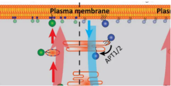
Figure 4: One delivery system to rule them all
Displacing farnesylated cargo from phosphodiesterase 6δ (PDEδ) by Arl2 activity in the perinuclear area is responsible for enrichment of Kras4B (left) and Hras (middle) at the plasma membrane, as well as for perinuclear enrichment of Rheb (right).
Ras is displaced from membranes by vesiculation and spontaneous dissociation. Its dissociation into the cytosol is enhanced by a changed surface or lack of depalmitoylation by acyl protein thioesterase (APT) activity, where the solubilisation factor PDEδ enhances diffusional intracellular exploration.
Farnesylated cargo can explore the perinuclear membranes for a trapping compartment as active disruption of the farnesyl-binding capacity of PDEδ by Arl2-GTP in the perinuclear area (green gradient) takes place. Palmitoylation by palmitoyl transferase (PAT) activity at the Golgi traps Hras by increasing its residence time on these membranes, as interaction with the negatively charged recycling endosome (RE) for Kras does. By directed vesicular export both Ras isoforms are displaced to the plasma membrane. Rheb is enriched in all perinuclear membranes by the PDEd–Arl2 delivery system and due to the lack of trapping on a vesicular transport compartment. The blue arrows denote the magnitude of entropic leakage from target membranes; red arrows represent the magnitude of the countering, energy-driven relocalisation processes. (Schmick et al., 2015)
3.1.4 Downstream effector signalling of Ras
During the 1980s a number of research groups reported several changes in cellular biochemistry that were caused by Ras proteins, but the precise mechanism underlying Ras’
function in cellular signalling remained to be determined. Over the years many studies have focused on finding the direct downstream signalling effectors that interact directly with Ras and mediate its various functions (Karnoub and Weinberg, 2008; Figure 5).
In 1993 the first interaction partner of Ras was identified, the Raf1 Ser/Thr kinase (Karnoub and Weinberg, 2008; Moodie et al., 1993; Warne et al., 1993; Zhang et al., 1993; Vojtek et
involves the mitogen-activated protein kinase (MAPK)/extracellular signal-regulated kinase (ERK) kinase (MEK), ERK1/2, and the E26-transcription factor proteins (ETS) (Wood et al., 1992; Leevers and Marshall, 1992). In Ras biology MAPK signalling has been shown to be both sufficient and necessary for Ras-induced transformation of mammalian cell lines (Leevers et al., 1994; Stokoe et al., 1994; Khosravi-Far et al., 1995; White at al., 1995;
Karnoub and Weinberg, 2008). This signalling cascade became the prototype for a number of other pathways that signal from the plasma membrane towards the nucleus (Karnoub and Weinberg, 2008). The importance for aberrant Raf–MEK–ERK signalling in oncogenesis was established by the identification of B-Raf mutations in cancers such as melanoma or colon cancer that occur in non-overlapping frequencies with Ras mutations (Rajagopalan et al., 2002; Karnoub and Weinberg, 2008).
The p110 catalytic subunit of the class I phosphoinositide 3‑kinases (PI3Ks) (Rodriguez- Viciana et al., 1994) and the guanine nucleotide-exchange factors for the Ras-like (RalA and RalB) small GTPases (Ral guanine nucleotide-dissociation stimulator (RalGDS) and RalGDS-like protein (RGL)) (Hofer et al., 1994; Kikuchi et al., 1994; Spaargaren and Bischoff., 1994) were identified as Ras effectors in 1994 (Karnoub and Weinberg, 2008). It was demonstrated that PI3K activity was required for Ras dependent transformation of NIH 3T3 cells (Rodriguez-Viciana et al., 1997). The signalling pathway of activated PI3K involves the Ser/Thr kinase AKT/protein kinase B and the transcription factor nuclear factor-κB (NF- κB), both of which have crucial roles in preventing anoikis and provide an explanation for the anti-apoptotic effects of Ras activation (Marte and Downward, 1997; Khwaja et al., 1997;
Mayo et al., 1997; Karnoub and Weinberg, 2008). PI3K or Raf pathways alone were not sufficient to promote Ras transformation in human kidney epithelial cells, whereas activation of the RalGEF–Ral pathway did prove sufficient (Hamad et al., 2002). Another study showed prominent but distinct roles for the highly related RalA and RalB GTPases in countering apoptosis and regulating cellular proliferation (Chien and White, 2003; Karnoub and Weinberg, 2008).
Ras interacts with effectors like Raf or PI3K through the effector lobe, which is oriented toward the cytoplasm. The allosteric lobe on the other side interacts with the membrane and contains the Ras-GTP surface. GEFs like Sos or GAPs interact with Ras at the effector lobe and at the interface between the two lobes.
Other Ras effectors with diverse roles in cell physiology include phospholipase C-ε (PLCε), T‑cell lymphoma invasion and metastasis-1 (TIAM1), Ras interaction/interference protein-1
(RIN1), ALL (acute lymphoblastic leukaemia) 1 fused gene on chromosome 6 (AF‑6) protein, and the Ras association domain-containing family (RASSF) proteins (Karnoub and Weinberg, 2008).
PLCε is a direct effector of Ras (Kelley et al., 2001; Song et al., 2001) and its activation enables it to cleave PtdIns(4,5)P2 into inositol-1,4,5- trisphosphate (Ins(1,4,5)P3) and diacylglycerol (DAG), promoting the release of Ca2+ and the activation of PKC (Karnoub and Weinberg, 2008). PLCε is also an upstream activator of the Ras–MAPK pathway as it contains a Cdc25 domain (Lopez et al., 2001).
The activity of the Ras effector TIAM1 has been shown to be required for Ras transformation (Malliri et al., 2002). AF-6 may participate in cytoskeletal activities downstream of Ras as it contains both microtubule- and actin-binding motifs (Ponting and Benjamin, 1996) and was shown to localize to adherens junctions and to associate with proteins that are involved in regulating cell polarity (such as ponsin and profilin) (Mandai et al., 1997). RIN1 and the RASSF proteins have reported tumour-suppressor activities and RIN1 was the first downstream effector of Ras that was shown to block Ras transformation (Han and Colicelli, 1995). RASSF proteins act as tumour suppressors although the detailed mechanism of action for RASSF proteins has not been fully discovered. RASSF kinases participate in a pathway that suppresses the activity of cyclin E and leads to cell cycle arrest and apoptosis as they associate with the mammalian sterile-20-like protein kinase-1 (MST1) and MST2 in D.
melanogaster. Pro-apoptotic functions have been shown for RASSF1, RASSF2 and RASSF5 (Khokhlatchev et al., 2002; Vos et al., 2003; Vos et al., 2003a; Karnoub and Weinberg, 2008).
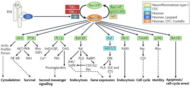
Figure 5: Ras signalling networks
Ras cycles between a GTP-bound state in which it interacts with downstream effector proteins, and a GDP-bound state in which it is inactive. A slow intrinsic GTPase activity cleaves off the γ‑phosphate of active GTP bound Ras, leading to Ras functional inactivation and thus the termination of signalling. The activity of the proteins that activate Ras (GEFs) and deactivate Ras (GAPs) is strictly controlled by the cells. GAPs, such as p120GAP or neurofibromin (NF1), enhance the intrinsic GTPase activity and hence negatively regulate Ras protein function. Conversely, GEFs (also known as GTP-releasing proteins/factors, termed GRPs or GRFs), such as son of sevenless (Sos), catalyse nucleotide ejection and therefore facilitate GTP binding and protein activation. After stimulation of receptor Tyr kinase (RTK) by growth factors (GF) Ras is activated and recruited to the plasma membrane. Several signal- transduction cascades can be initiated by effector molecules that are engaged by activated Ras. Outputs shown represent the main thrusts of the indicated pathways. Ras activation can also occur in endomembrane compartments (endoplasmic reticulum and the Golgi). Activating mutations in the different components of the Ras–Raf–mitogen-activated protein kinase (MAPK) pathway are associated with the indicated developmental disorders.
AF-6, acute lymphoblastic leukaemia‑1 fused gene on chromosome 6; CD1, cadherin domain-1;
CDC42, cell division cycle-42; ELK, ETS-like protein; ERK, extracellular signal regulated kinase;
ETS, E26-transcription factor proteins; Ins(1,4,5)P3, inositol-1,4,5-trisphosphate; JNK, Jun N-terminal kinase; MEK, mitogen-activated protein kinase/ERK kinase; NF-κB, nuclear factor-κB; PI3K, phosphoinositide 3‑kinase; PKB/C, protein kinase B/C; PLA/Cε/D, phospholipase A/Cε/D; RalBP1, Ral-binding protein-1; RASSF, Ras association domain-containing family; Rin1, Ras interaction/interference protein-1; SAPK, stress-activated protein kinase; SHP2, Src-homology‑2 domain-containing protein Tyr phosphatase-2; TIAM1, T‑cell lymphoma invasion and metastasis-1.
(Karnoub and Weinberg, 2008)
In primary cells oncogenic Ras has been shown to induce senescence through activation of the p53–p21WAF and/or p16INK4A– Retinoblastoma (Rb) tumour-suppressor pathways (Serrano et al., 1997; Wei et al., 2003; Voorhoeve and Agami, 2003; Karnoub and Weinberg, 2008; Figure 6). ‘Normal’ cells undergo cell-cycle arrest or senescence in response to hyperactive Ras signalling rather than unlimited proliferation or transformation. Moreover, the susceptibility of certain normal cells to Ras-mediated transformation seems to rely on prior inactivation of these tumour-suppressor pathways (Karnoub and Weinberg, 2008) Therefore, Ras activation in normal cells does not cause an initiation of tumourigenesis, but instead a halt in cell proliferation. This Ras induced senescence in normal cells can be explained by the oncogenic cooperation model proposed in 1983 (Land et al., 1983; Ruley, 1983), as Ras-collaborating oncoproteins, like E1A, SV40 and E6/E7 are all well-known inactivators of the Rb and the p53 pathways (Karnoub and Weinberg, 2008).
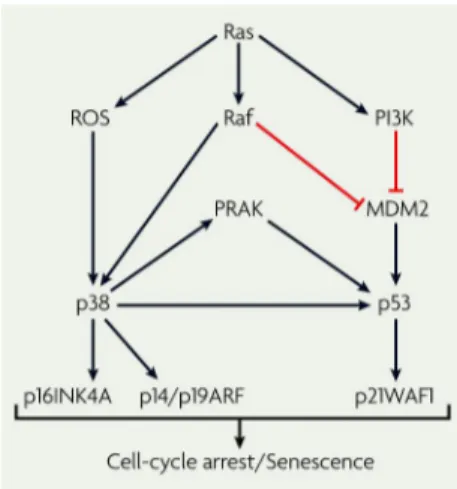
Figure 6: The tumour suppressor effect of Ras
Ras activation of p16 and p53 depend, in part, on the activation of the p38 mitogen-activated protein kinase (MAPK) pathway through unidentified mechanisms. Ras activation has been shown to cause elevated reactive oxygen species (ROS) levels, and ROS promote p38 activation. The Ras-induced Raf–MAPK pathway might feed into the p53 pathway by activating the p38-regulated/activated protein kinase (PRAK), which in turn phosphorylates and activates p53.
MDM2, murine double minute-2; PI3K, phosphoinositide 3-kinase; p14/19ARF, p14ARF in humans and p19ARF in mice. (Karnoub and Weinberg, 2008)
Whether cells become transformed or senescent in response to Ras activation is determined by p16, which appears to be the key factor in ‘making’ this decision. High levels of Ras expression result in cell-cycle arrest due to an acute elevation of p16. However, moderate Ras activation allows Ras-induced transformation of cells as it does not induce an acute p16 response (Karnoub and Weinberg, 2008). It has been demonstrated in mice that in vivo
expression of oncogenic Kras at endogenous levels could cause cellular transformation (Tuveson et al., 2004; Johnson et al., 2001).
3.1.5 Kras
Kras is a protein consisting of 188/189 amino acids with a molecular mass of 21.6 kDa. It is the only isoform of the Ras family that is essential in mouse embryogenesis.
Knockout of Kras is embryonic lethal at day 12-14, which is not the case for either Hras or Nras (Johnson et al., 1997).
Kras was first identified in the rat genome in 1981 (DeFeo at al., 1981) as a cellular homolog of the Kirsten transforming Ras sequence and was subsequently also found in the human and mouse genomes (Chang et al., 1982; Ellis et al., 1982). These studies revealed that Kras behaved similarly to the Src oncogene of Rous sarcoma virus (RSV), the origins of which were first reported in 1976 (Stehelin et al., 1976). Soon mutant alleles of Kras were discovered in human cancer cell lines including e.g. those of bladder and lung carcinoma (Der at al., 1982). These mutations did not arise from culturing the cancer cells in vitro - in 1984 mutant Kras alleles were found for the first time in lung cancer specimens (Santos et al., 1984; Nakano et al., 1984). Associations between particular types of human cancer and specific Ras oncogenes are known to exist (Karnoub and Weinberg, 2008).
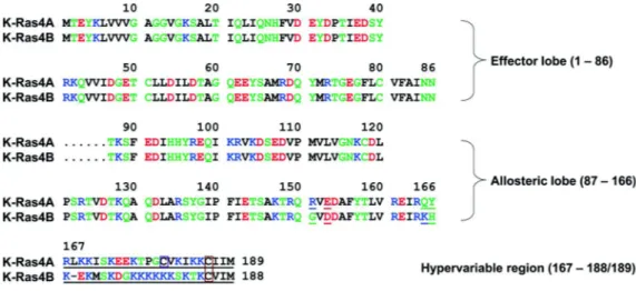
Two copies of the Kras gene can be found in the human genome. One of them, Kras1, has been assigned to chromosome 6 (O’Brien et al., 1983) and is reported to be a pseudogene (McGrath et al., 1983); and the second, Kras2, resides on chromosome 12 (Sakaguchi et al., 1983). Kras also has two splice variants, Kras4A and Kras4B, arising from alternative mRNA splicing of exon 5 (fourth coding exon) of the Kras mRNA transcript (Carta et al., 2006; Pells et al., 1997). Exon 6 encodes the HVR at the C-terminus of Kras4B and remains untranslated in Kras4A. The two splice variants of Kras therefore differ only in four catalytic domain residues (151, 153, 165, and 166) and in their HVRs (Chakrabarti et al., 2016; Figure 7).
Figure 7: Multiple sequence alignment of the amino acids in the Kras4A and Kras4B proteins
In the sequence, hydrophobic, polar/glycine, positively charged, and negatively charged residues are coloured black, green, blue, and red, respectively. Underlined text indicates the non-identity of residues in the alignment. In the HVRs a purple box denotes the palmitoylated cysteine in Kras4A, and an orange-box indicates the farnesylated cysteines in Kras4A and Kras4B. (Chakrabarti et al., 2016)
Kras4A undergoes additional palmitoylation upstream of the CAAX motif whereas Kras4B shows no detectable palmitoylation (Hancock et al., 1989). In older studies it was hypothesized that Kras4A is a minor species in comparison to Kras4B (Jackson et al., 1994;
Plowman et al., 2006; Normanno et al., 2009; Stone, 2011). However, a more recent study using splice variant-specific primers and antibodies could show that Kras4A and Kras4B mRNA transcript and protein are expressed at equal levels - or even higher levels of Kras4A can be found - in colorectal and bladder-derived tumour cell lines and in primary human colorectal adenocarcinoma tissues (Tsai et al., 2015; Chakrabarti et al., 2016). In colorectal cancers the ratio of Kras4A to 4B expression is commonly altered in favour of Kras4B, which may be due to a tumour-suppressive effect of the Kras4A isoform in colon adenomas that favours Kras4B expression (Luo et al., 2010). In cancer cells both splice variants are expressed (Plowman et al., 2006; Butz et al., 2004).
As the Kras4B gene product was the only Kras variant used in the present work it will henceforth be termed Kras.
3.1.6 Kras mutations and their impact in cancer
The Ras genes account for approximately 30% of all cancers in humans (Roberts and Der, 2007; Thumar et al., 2014). Kras is of particular interest, as its mutations account for nearly 86% of all Ras mediated cancers (van Hattum and Waldmann, 2014; Cox et al., 2014). In
mutations leading to a permanently active Kras molecule can be found (Blum and Kloog, 2005, Blum et al., 2008; Vogelstein et al., 1988; Mitin et al., 2005).
Kras protein translation and expression is poor in comparison to e.g. Hras due to a high frequency of rare codons in the Kras DNA coding sequence (Lampson et al., 2013). As described earlier, high expression levels of activated Ras induce senescence rather than uncontrolled proliferation. But a cell with mutated Kras does not harbour the combination of an activated Ras protein with high expression of the mutated Ras protein due to Kras’ low expression levels. Consequently, this cell will survive and other genetic events can take place in order to promote tumour progression (Hobbs et al., 2016).
In the case of Kras, the three mutation hotspots (G12, G13, Q61) show the following mutation frequency: Most mutations occur at G12 (83%), followed by G13 mutations (14%), while Q61 mutations are rare (2%). Between different cancer types the mutation frequency within Kras also exhibits significant differences. In colorectal adenocarcinoma (CRC) a relatively high frequency of G13 mutations can be found while in pancreatic ductal adenocarcinoma (PDAC) the dominating mutation is located at G12 and mutations of G13 and Q61 are exceptional. Therefore, it is likely that different Ras mutations may have different functional consequences and depending on the tissue of origin the properties that are crucial for their oncogenic functions vary (Hobbs et al., 2016). It has been shown that the Q61L mutation leads to reduced intrinsic hydrolysis and GAP sensitivity, as well as increased intrinsic nucleotide exchange while the G12Vmutation leads to a loss of GAP sensitivity (Smith et al., 2013; Hobbs et al., 2016). In comparison to WT Ras the G13D mutant displays decreased GAP-mediated hydrolysis and an increased rate of intrinsic nucleotide exchange (Smith et al., 2013; Hobbs et al., 2016).
The mutations at different hotspots could be of great consequence for the clinical outcome and treatment of cancer patients. Initially, patients with Kras G12 or G13 mutations were excluded from anti-epidermal growth factor receptor (EGFR) therapy to treat CRC because EGFR is a receptor tyrosine kinase that is positioned upstream of Ras. The US Food and Drug Administration (FDA) reviewed this advice (Allegra et al., 2009) and a subsequent analysis has recommended that CRC patients with Kras G13 mutations would benefit from anti-EGFR therapy (Tejpar et al., 2012; Hobbs et al., 2016). However, another study concludes that CRC patients with any Kras mutation, including Q61 and A146, will not benefit from anti-EGFR therapy (Tran et al., 2015). There is thus still much work to be done to elucidate the
underlying mechanisms and thereby to determine in which cases anti-EGFR therapy is beneficial for patients and in which cases the relevant mutation status precludes any benefit.
The different amino acid substitutions at the mutation hotspots also appear to be of importance for cancer patient survival. For PDAC, different clinical outcomes for different Kras mutations have been described. It was demonstrated that Kras G12D and G12R mutations are a negative prognostic factor for patient survival (Ogura et al., 2013). Overall survival rates are increased when only Kras G12R mutations are present, whereas poor survival rates are associated with a mutation of Kras G12D (Hobbs et al., 2016). Improved survival could also be detected for patients with Kras Q61 mutations (Witkiewicz et al., 2015). Therefore, to determine the prognostic value of the mutation, it is necessary to consider cancer type, amino acid substitution and mutation site of each patient (Hobbs et al., 2016).
In PDAC the substitution found most frequently at codon G12 of Kras is G12D, followed by G12V. In contrast, in lung adenocarcinoma (LAC) the main substitution is G12C, which is rare in PDAC (3%) (Cox et al., 2014). The tissue-specific exposure to certain carcinogens could play a role in establishing these distinct frequencies and the different substitutions at one position may have different biological consequences, also in different Ras isoforms (Hobbs et al., 2016).
A model of progression has been established for the development of PDAC based on genomic analyses and molecular pathology studies. Kras mutations appear to contribute to its inception as activating mutations of Kras are found in most tumours of PDAC patients (>90%) (Kanda et al., 2012; Hwang et al., 2016). To accelerate the progression of PDAC other genetic alterations affecting CDKN2A, TP53 and SMAD4 cooperate with oncogenic Kras (Morris et al., 2010). In premalignant lesions associated with the pancreatic ducts, named ‘pancreatic intraepithelial neoplasia’ (PanIN), the signature mutations of PDAC have been identified.
PanIN exhibit alterations in their cellular architecture, such as mucinous cytoplasm, nuclear crowding and nuclear atypia (Hruban et al., 2001; Hwang et al., 2016). In mice, activation of oncogenic Kras in pancreatic epithelial cells is sufficient to initiate PDAC and PDAC progression can be accelerated by additional mutation of Trp53, Cdkn2a or Smad4, sharing many features of the human disease (Hingorani et al., 2005; Bardeesy et al., 2006).
Many human PDAC cell lines have been characterised in terms of molecular aberrations and cellular phenotype (Deer et al., 2010) and several pancreatic cell lines have been shown to be
seen that acute inactivation of transforming oncogenes leads to cell-cycle arrest, differentiation or apoptosis. Therefore, it is sufficient to induce tumour regression in such cases by abrogating the function of individual oncogenic products. For cell lines gene expression signatures were established that can distinguish two groups of cells (Kras- dependent/ Kras-independent) after genes were identified that are specifically up regulated in Kras-dependent cells and are required for their viability (Singh et al., 2009). It has been possible to develop prognostic classifiers for patients and predictive biomarkers for drug response because the cell lines harbour a distinct set of mutations with corresponding transcription profiles (Hwang et al., 2016).
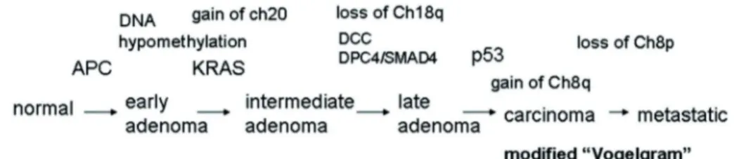
In the 1980s, it had already been found that most, if not all, malignant colorectal carcinomas arise from pre-existing benign tumours (adenomas) and that both hereditary and environmental factors contribute to the development of colorectal neoplasia by leading to genetic alterations (Fearon and Vogelstein, 1990). Even a small number of cells within a pocket of epithelial stem cells are capable of initiating the process of neoplasia by clonal expansion as it was found that adenomas arise from a single pocket in this way (Ponder and Wilkinson, 1986).
Thus, colon cancer develops in stages. After normal tissues have acquired certain mutations, they develop into hyperplastic epithelia and then into early adenomas. Early adenomas develop into intermediate and late adenomas, then into carcinomas with additional key gene mutations, activation of oncogenes, loss and gain of chromosomes, and/or chromosome amplifications (Fearon and Vogelstein, 1990). This development takes a long time, usually decades. The transition from colorectal carcinoma to a metastatic CRC can take another 2-3 years (Rao and Yamada, 2013).
Approximately 50% of colorectal carcinomas (Bos et al., 1987; Forrester et al., 1987) have been found to harbour (K) Ras gene mutations, which could also be found in adenomas > 1 cm in size but in fewer than 10% of adenomas less than 1 cm in size (Vogelstein et al., 1988;
Farr et al., 1988). In a subset of colorectal tumours and adenomas with Ras gene mutations they may also be the initiating event. However, in most tumours they occur in cells of a pre- existing adenoma and are then responsible for the conversion of a small adenoma into a larger and more dysplastic one, through clonal expansion of the cell with the mutation (Fearon and Vogelstein, 1990).
In colorectal neoplasia a loss of specific chromosomal regions frequently occurs, usually