Acid sphingomyelinase is required for efficient phago-lysosomal fusion
I n a u g u r a l – D i s s e r t a t i o n
zur
Erlangung des Doktorgrades
der Mathematisch-Naturwissenschaftlichen Fakultät der Universität zu Köln
vorgelegt von Michael Schramm
aus Köln
Köln 2008
Berichterstatter: Prof. Dr. Martin Krönke Prof. Dr. Jens Brüning
Table of contents
1 Introduction ... 1
1.1 Acid sphingomyelinase... 1
1.2 Antibacterial effector mechanisms of macrophages... 2
1.2.1 Oxidative effector mechanisms... 2
1.2.2 Non-oxidative effector mechanisms... 3
1.3 Phagosome maturation... 4
1.4 Listeria monocytogenes... 6
1.5 Aim of this study... 7
2 Material and Methods ... 8
2.1 Material... 8
2.1.1 Mice... 8
2.1.2 Bacteria... 8
2.1.3 Chemicals, buffers and solutions... 8
2.1.4 Antibodies... 12
2.1.5 Technical equipment... 14
2.1.6 Kits... 14
2.2 Methods... 15
2.2.1 Preparation of peritoneal macrophages... 15
2.2.2 Immunomagnetic enrichment of peritoneal macrophages... 15
2.2.3 Preparation of bone marrow-derived macrophages... 15
2.2.4 Cell culture... 16
2.2.5 Labelling of L. monocytogenes with fluorescent dyes... 16
2.2.6 Preparation of heat-killed L. monocytogenes... 16
2.2.7 Immunofluorescence microscopy (IFM)... 16
2.2.8 Pulse/chase labelling of lysosomes with fluid phase markers... 17
2.2.9 Western blotting (WB)... 17
2.2.10 Preparation of phagosomes containing L. monocytogenes... 18
2.2.11 Measurement of pH in phagosomes containing L. monocytogenes... 18
2.2.12 Measurement of proteolytic activity... 18
2.2.13 Quantification of reactive oxygen intermediates... 19
2.2.14 Quantification of reactive nitrogen intermediates... 19
3 Results ... 20
3.1 Impaired inactivation of Listeria monocytogenes despite normal generation of ROI and RNI in ASMase-/- macrophages... 20
3.2 Impaired maturation of phagosomes containing L. monocytogenes into phagolysosomes in ASMase-/- macrophages... 24
3.3 Impaired transfer of lysosomal cargo into phagosomes containing L. monocytogenes in ASMase-/- macrophages... 33
4 Discussion ... 42
5 References ... 49
6 Summary ... 65
7 Zusammenfassung... 66
8 Danksagung... 67
9 Erklärung... 68
10 Curriculum vitae... 69
Abbreviations
ASMase Acid sphingomyelinase
ASMase-/- Acid sphingomyelinase-deficient
BHI Brain Heart Infusion
BMDM Bone marrow-derived macrophages
BSA Bovine serum albumin
CFU Colony forming unit(s)
DMEM Dulbecco’s Modified Eagle Medium
DMSO Dimethyl sulfoxide
EEA1 Early endosomal antigen 1
Fig. Figure
FITC Fluorescein isothiocyanate
H+ Proton
HBSS Hanks Balanced Salt Solution
HKLM Heat-killed Listeria monocytogenes
i.p. intra peritoneal
IFM Immunofluorescence microscopy
IFN-γ Interferon gamma
IgG Immunoglobulin G
iNOS Inducible nitric oxide synthase
kDa kiloDalton
L.m. Listeria monocytogenes
Lamp1 Lysosome-associated membrane protein 1
LLO Listeriolysin O
M6PR Cation-independent mannose 6-phosphate receptor
MACS Magnetic cell sorting
MEF Mouse embryonic fibroblasts
MOI Multiplicity of infection
MW Molecular weight
NADPH Nicotinamide adenine dinucleotide phosphate
NMS Normal mouse serum
NO Nitric oxide
NPD Niemann-Pick disease
O2 molecular oxygen
O2- Superoxide
OD600 Optical density at 600 nm
PBS Phosphate buffered saline
PFA Paraformaldehyde
pH pH value; negative decadic logarithm of the H+
concentration
Phox Phagocyte oxidase
PMA Phorbol 12-myristate 13-acetate
RNS Reactive nitrogen species
ROS Reactive oxygen species
RT Room temparature
s.e. Standard error of the mean
SDS Sodium dodecyl sulfate
SMase Sphingomyelinase
SNARE soluble N-ethylmaleimide-sensitive factor attachment protein receptor
SV Simian virus
TNF Tumour necrosis factor
TRITC Tetramethylrhodamine isothiocyanate
vATPase vesicular proton pump
w/v Weight to volume
WB Western blot
wt Wild type
1
1 Introduction
1.1 Acid sphingomyelinase
Sphingomyelinases (SMases) catalyze the hydrolysis of the membrane lipid sphingomyelin into ceramide and phosphorylcholine. So far, seven sphingomyelinases have been identified and classified with regard to pH optimum, subcellular localization or dependency on bivalent metal ions. The most intensively studied SMase is the acid sphingomyelinase (ASMase) (Levade and Jaffrezou 1999).
ASMase is expressed ubiquitously in mammals but confined to a few subcellular locations, i.e. the extracellular leaflet of the plasma membrane, endosomes, phagosomes and lysosomes. Activity of ASMase is maximal at acidic pH of about 4.5 to 5 (Kanfer, Young et al. 1966; Schuchman, Suchi et al. 1991; Schissel, Schuchman et al. 1996; Schissel, Keesler et al. 1998; Kronke 1999; Grassme, Schwarz et al. 2001; Schneider-Brachert, Tchikov et al. 2004).
Deficiency in ASMase in human leads to autosomal recessive inherited Niemann- Pick disease (NPD) (Brady, Kanfer et al. 1966; Schneider and Kennedy 1967). NPD is a lipid storage disorder caused by abolished (type A NPD) or strongly reduced (type B NPD) activity of ASMase. Intracellular accumulation of sphingomyelin in neuronal cells of the cerebellum leads to severe neurodegeneration and death during early childhood (type A NPD) or adolescence (type B NPD). ASMase-deficient (ASMase-/-) mice show first clinical symptoms of NPD at an age of about three months and die at about nine months of age (Horinouchi, Erlich et al. 1995; Lozano, Morales et al. 2001).
By hydrolyzing sphingomyelin, SMases alter the lipid composition and thereby the biochemical and biophysical properties of biomembranes. Sphingomyelin is an important component of the extracellular leaflet of biomembranes (Futerman and Riezman 2005; Holthuis and Levine 2005) and interacts with cholesterol to form lipid rafts which are central for the spatial organization of integral membrane proteins.
Hydrolysis of sphingomyelin into ceramide alters lipid raft composition as ceramide spontaneously self-associates into ceramide-enriched membrane domains (Holopainen, Subramanian et al. 1998). Removal of the large polar head group phosphorylcholine from sphingomyelin also promotes transition from lamellar phase
2
into hexagonal II phase which facilitates membrane reorganization processes (Kronke 1999). Furthermore, ceramide is an important signalling molecule involved in programmed cell death, cell differentiation and proliferation (Futerman and Hannun 2004). By hydrolyzing sphingomyelin into ceramide SMases also directly influence the fusogenicity of biomembranes (Grassme, Gulbins et al. 1997; Zha, Pierini et al.
1998; Goni and Alonso 2000; Holopainen, Angelova et al. 2000; Goni and Alonso 2002) and promote liposome fusion in vitro (Ruiz-Arguello, Basanez et al. 1996;
Basanez, Ruiz-Arguello et al. 1997; Hinkovska-Galcheva, Boxer et al. 1998; Oorni, Hakala et al. 1998; Ruiz-Arguello, Goni et al. 1998).
ASMase is activated by a number of pro-inflammatory cytokines, e.g. tumour necrosis factor (TNF), interleukin (IL)-1ß or interferon-γ (IFN-γ) suggesting a role of ASMase in immune responses (Schutze, Potthoff et al. 1992; Cifone, Roncaioli et al.
1995; Pushkareva, Obeid et al. 1995; Kronke 1999; Schneider-Brachert, Tchikov et al. 2004). Furthermore, NPD patients are more susceptible to respiratory diseases indicating a role of ASMase during infection (Jonas 1966; Dechovitz and Moffet 1968). Importantly, our group has shown that ASMase-/- mice are highly susceptible to infection with the facultative intracellular bacteria Listeria monocytogenes (L. monocytogenes) and Salmonella typhimurium because ASMase-/- macrophages are unable to restrict intracellular growth of these bacteria (Utermohlen, Karow et al.
2003). The precise antibacterial mechanism of ASMase, however, remains elusive.
1.2 Antibacterial effector mechanisms of macrophages
Macrophages are of critical importance in innate immunity as they specialize in phagocytosis and subsequent killing of invading bacteria. To inactivate and degrade phagocytosed bacteria, macrophages employ both oxidative and non-oxidative effector mechanisms (Scott, Botelho et al. 2003; Haas 2007).
1.2.1 Oxidative effector mechanisms
Macrophages generate oxidative effector molecules by two distinct pathways (Vazquez-Torres, Jones-Carson et al. 2000). First, the nicotinamide adenine dinucleotide phosphate-oxidase complex (NADPH oxidase complex; phox) generates reactive oxygen intermediates (ROI). Second, the inducible nitric oxide synthase (iNOS) generates reactive nitrogen intermediates (RNI).
3
The NADPH oxidase complex is assembled from its subunits in the membrane of phagosomes containing bacteria (Nauseef 2004; Minakami and Sumimotoa 2006).
The catalytic subunit, flavocytochrome b558, is a heterodimer composed of gp91phox and p22phox. The flavocytochrome b558 is an integral membrane protein of the plasma membrane and of vesicles fusing with phagosomes very early after generation of phagosomes (Ginsel, Onderwater et al. 1990; Nauseef 2004). The regulatory subunits p40phox, p47phox, p67phox and Rac2 are recruited to flavocytochrome b558
from their location in the cytosol (DeLeo, Allen et al. 1999; van Bruggen, Anthony et al. 2004). After assembly, the NADPH oxidase complex catalyses the transfer of one electron from NADPH through the phagosomal membrane onto molecular oxygen (O2) which leads to generation of superoxide (O2-) inside phagosomes. The highly reactive radical superoxide reacts with intraphagosomal molecules which gives rise to diverse bactericidal ROI, e.g. hydrogen peroxide, hydroxyl radicals or hypochloride (Quinn and Gauss 2004; Robinson, Ohira et al. 2004).
Cytosolic iNOS is recruited to phagosomes containing bacteria where its homodimeric form catalyses the oxidation of L-arginine into L-citrulline and nitric oxide (NO) (Vodovotz, Russell et al. 1995; Miller, Fratti et al. 2004). The lipid and water permeable radical NO diffuses into the phagosome where it reacts with oxygen-containing molecules leading to generation of diverse bactericidal RNI, e.g. nitrogen dioxide, nitrite, nitrate or S-nitrothioles (MacMicking, Xie et al. 1997).
After activation of macrophages, expression of iNOS is strongly up-regulated while resting macrophages express almost no iNOS (Aktan 2004).
The simultaneous generation of ROI and RNI has a synergistic effect on killing of phagocytosed bacteria because O2- and NO form the highly bactericidal peroxynitrite (ONOO-) (Nathan and Shiloh 2000).
1.2.2 Non-oxidative effector mechanisms
For many years, ROI and RNI have been assumed to be the major antibacterial effector mechanisms (Klebanoff 1975; Nathan and Shiloh 2000). Only recently, it has been established that non-oxidative effector mechanisms, particularly lysosomal acid hydrolases, are at least as important for effective control of infectious pathogens within phagosomes (Segal 2005). Neutrophil elastase and cathepsin G are necessary for effective killing of Staphylococcus aureus and Candida albicans in neutrophils (Reeves, Lu et al. 2002). Furthermore, the lysosomal aspartic protease cathepsin D
4
has been identified as an important factor in the defence against Listeria monocytogenes in macrophages as well as in fibroblasts (del Cerro-Vadillo, Madrazo-Toca et al. 2006). The bactericidal acid hydrolases are transferred from lysosomes into phagosomes by phago-lysosomal fusion at the end of the phagosomal maturation process (Haas 2007).
1.3 Phagosome maturation
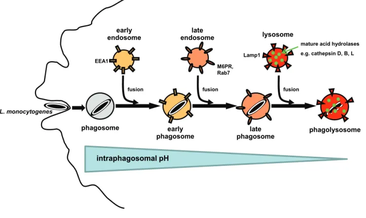
Phagosome maturation into phagolysosomes is achieved stepwise by sequential fusion of the phagosome with early endosomes, late endosomes and finally lysosomes (Mayorga, Bertini et al. 1991; Pitt, Mayorga et al. 1992; Desjardins, Celis et al. 1994; Desjardins, Huber et al. 1994; Desjardins, Nzala et al. 1997; Henry, Hoppe et al. 2004). Each maturation stage is characterized by acquisition or loss of specific marker molecules (Fig. 1). After separation from the plasma membrane, phagosomes fuse with early endosomes and by this acquire early endosomal markers like the ‘early endosomal antigen 1’ (EEA1). Via fusion with late endosomes, phagosomes acquire the ‘cation-independent mannose 6-phosphate receptor’
(M6PR) and the small GTPase Rab7. Only late phagosomes are capable of fusing with lysosomes into phagolysosomes. Phago-lysosomal fusion leads to transfer of mature acid hydrolases like the lysosomal cathepsins D, B and L from lysosomes into phagosomes. Furthermore, phagolysosomes are positive for the ‘lysosome- associated membrane protein 1’ (Lamp1) but lack M6PR and Rab7. With progressing phagosomal maturation, activity of the vesicular proton pump (vATPase) leads to a drop of the intraphagosomal pH from about 6.5 in early phagosomes to between 6.0 and 5.0 in late phagosomes and finally 5.0 to 4.0 in phagolysosomes.
Several molecules orchestrating the tethering, docking and final membrane fusion of endosomes or lysosomes with phagosomes have been identified in recent years (Luzio, Pryor et al. 2005). Key regulators of the phago-endosomal fusion machinery are Rab GTPases which recruit specific sets of effector proteins to vesicular membranes (Jordens, Marsman et al. 2005). After docking and tethering, membrane fusion itself is mediated by ‘soluble N-ethylmaleimide-sensitive factor attachment receptor proteins’ (SNAREs) (Jahn and Scheller 2006). In reconstituted systems in vitro, SNAREs have been shown to provide the minimal machinery for membrane
5
fusion
early
endosome late
endosome lysosome
M6PR, Rab7
Lamp1
early
phagosome late
phagosome phagolysosome
fusion fusion
phagosome L. monocytogenes
mature acid hydrolases e.g. cathepsin D, B, L
intraphagosomal pH
EEA1
Fig. 1: Schematic overview of phagosomal maturation into phagolysosomes by sequential fusion with early endosomes, late endosomes and finally lysosomes.
fusion (Weber, Zemelman et al. 1998; Hu, Ahmed et al. 2003; Bonifacino and Glick 2004). However, this minimal fusion machinery acts very slowly and inefficiently so that additional factors are supposed to be involved in the rapid and efficient membrane fusion processes observed in intact, viable cells (Weber, Zemelman et al.
1998; Bonifacino and Glick 2004). Specifically, it has been hypothesized that lipid modifying enzymes might contribute to efficient vesicle fusion via alteration of the biochemical and/or biophysical properties of cellular membranes (Chernomordik and Kozlov 2003; Bonifacino and Glick 2004).
The kinetics of the phagosomal maturation and subsequent phago-lysosomal fusion are particularly critical during phagocytosis of intracellular bacteria which subvert phagosome maturation for their own needs (Scott, Botelho et al. 2003; Haas 2007). Some bacteria like for example Mycobacterium tuberculosis or Salmonella typhimurium arrest phagosome maturation at pre-phagolysosomal stage and thus avoid exposure to lysosomal acid hydrolases. Other bacteria like Shigella flexneri or L. monocytogenes disrupt the membrane of maturing phagosomes before phago- lysosomal fusion and escape into the cytosol.
6 1.4 Listeria monocytogenes
L. monocytogenes are a gram-positive rods causing severe, often lethal food-borne infections in immunocompromised individuals, pregnant women and newborns. The facultative intracellular bacteria cross the intestinal barrier by invading intestinal epithelial cells and primarily infect hepatocytes. For elimination of L. monocytogenes during primary infection, concerted action of almost all regulatory and effector mechanisms of innate and adaptive immunity is necessary (Gellin and Broome 1989).
For both pathogenesis and elimination, phagocytosis of L. monocytogenes by macrophages plays a crucial role. In resident macrophages, L. monocytogenes escape from phagosomes into the cytosol at about 30 min after infection (Myers, Tsang et al. 2003) and then proliferate with a generation time of about 1 h (Portnoy, Jacks et al. 1988). By disrupting the membrane of maturing phagosomes prior to phago-lysosomal fusion, L. monocytogenes avoid exposure to lysosomal acid hydrolases and at the same time gain access to the nutrient-rich cytosol. Three virulence factors are crucial for membrane disruption: the pore forming hemolysin listeriolysin O (LLO) (Leimeister-Wachter, Haffner et al. 1990; Beauregard, Lee et al.
1997) and the phospholipases PlcA and PlcB (Portnoy, Smith et al. 1994; Portnoy, Auerbuch et al. 2002). In the cytosol, L. monocytogenes recruit actin via its virulence factor ActA. The cloud of actin molecules surrounding L. monocytogenes is then rearranged into a comet-tail like structure. Continuous polymerization of actin at one bacterial pole propels L. monocytogenes towards the plasma membrane. There, L. monocytogenes induce formation of a pseudopod-like structure which is phagocytosed by neighbouring cells. L. monocytogenes disrupt the double membrane of the phagosome and enter the cytosol of the new host cell (Portnoy, Smith et al. 1994).
In contrast to resident macrophages, activated macrophages efficiently kill phagocytosed L. monocytogenes. Generation of large quantities of ROI and RNI (Dinauer, Deck et al. 1997; Shiloh, MacMicking et al. 1999; Alvarez-Dominguez, Carrasco-Marin et al. 2000; Myers, Tsang et al. 2003) and exposure to lysosomal acid hydrolases (del Cerro-Vadillo, Madrazo-Toca et al. 2006) prevent escape into the cytosol and lead to degradation of L. monocytogenes in phagolysosomes.
7 1.5 Aim of this study
ASMase-/- macrophages cannot kill phagocytosed L. monocytogenes despite activation (Utermohlen, Karow et al. 2003) indicating a role of ASMase in listeriocidal activity of macrophages. The aim of this study is to elucidate the listeriocidal mechanism of ASMase in macrophages.
8
2 Material and Methods
2.1 Material
2.1.1 MiceBreeding pairs of mice heterozygously deficient for acid sphingomyelinase were kindly provided by R. Kolesnick (Memorial Sloan-Kettering Cancer Center, New York, NY; originally obtained from E. H. Schuchmann, Mount Sinai School of Medicine, New York, NY (Horinouchi, Erlich et al. 1995)). Mice were heterozygously backcrossed to the C57BL/6 strain to the 10th generation under specific pathogen- free conditions at the animal facilities of the Medical Centre of the University of Cologne (Cologne, Germany). Experiments were performed in accordance with the Animal Protection Law of Germany in compliance with the Ethics Committee at the University of Cologne with 6–10 weeks old ASMase-/- mice and wt littermates.
2.1.2 Bacteria
L. monocytogenes, strain EGD, serotype 1/2a, were kindly provided by C. Kocks (Harvard Medical School, Boston, USA). Following in vivo passage, single colonies of L. monocytogenes were expanded in brain-heart infusion (BHI) medium. Aliquots of log-phase growing cultures were stored at -80° C.
The isogenic ΔprfA- and Δhly-deletion mutants of L. monocytogenes (Peters, Domann et al. 2003) were kindly provided by Eugen Domann (University of Giessen, Germany).
2.1.3 Chemicals, buffers and solutions
Chemicals were of research grade and from Sigma-Aldrich (Steinhausen, Germany), AppliChem (Darmstadt, Germany) or Becton Dickinson GmbH (Heidelberg, Germany) unless stated otherwise. Buffers and solutions were prepared using bidestilled H2O from an EASYpure UV/UF H2O purification unit (Werner Reinstwassersysteme, Leverkusen), degassed and sterilised by autoclaving or filtration through a 0.2 µm filter membrane if necessary.
9
0.1% saponin in PBS 0.1% (w/v) saponin in PBS, stored at 4° C 3 kDa dextran conjugated to TexasRed Dextran, Texas Red, 3000 MW, lysine fixable
(Invitrogen, Karlsruhe, Germany), stored at -20° C
3% PFA in PBS 3% paraformaldehyde in PBS, stored
at -20° C
70 kDa dextran conjugated to TexasRed Dextran, Texas Red, 70000 MW, lysine fixable (Invitrogen, Karlsruhe, Germany), stored at -20° C
Alexa Fluor 594-succinimidyl ester 5 mg/ml in DMSO (Invitrogen, Karlsruhe, Germany), stored at -20° C
Amersham ECL hyperfilm GE Healthcare (München, Germany), stored at 4° C
BHI medium Brain Heart Infusion medium
Blocking buffer (for IFM) 3% BSA, 0.1% saponin in PBS, stored at 4° C
Blocking buffer (for WB) 5 % non-fat dried milk powder in TBS-T, stored at -20° C
BSA Albumin from bovine serum, fraction V,
stored at 4° C
Carboxylated 1 µm silica beads Silica particles carboxylated, 1,0 µm, 50 mg/ml (Kisker Biotech, Steinfurt, Germany), stored at 4° C
CD11b MicroBeads CD11b MicroBeads, human and mouse (Miltenyi Biotec, Bergisch Gladbach, Germany), stored at 4° C
Chicken ovalbumin conjugated to TexasRed
Ovalbumin, Texas Red conjugate (Invitrogen, Karlsruhe, Germany), stored at -20° C
DMEM 1x Dulbecco`s Modified Eagle Medium,
stored at 4° C
DMSO Dimethyl sulfoxide (Merck, Darmstadt,
Germany)
Ferricytochrome c 0,27 g cytochrome c from equine heart, type VI, in HBSS with Ca2+ and Mg2+, stored at -20° C
Fetal calf serum (FCS) heat-inactivated at 56° C for 30 min
10
(Biochrom AG, Berlin, Germany), stored at 4° C
FITC Fluorescein isothiocyanate isomer I,
20 mg/ml in DMSO, stored at -20° C
Greiss reagent 1% (w/v) sulfanilamide in 2,5% phosphoric acid and 1% (w/v) naphthyl ethylene diamine dihydrochloride in 2,5% phosphoric acid, stored separately at 4° C
HBSS “Hanks Balanced Salt Solution” with Ca2+ and Mg2+
HEPES 1 M HEPES solution (Invitrogen, Karlsruhe,
Germany), stored at 4° C
Human IgG IgG from human serum, stored at -20° C Immobilon-PSQ transfer membrane Millipore (Bedford, USA)
Interferon-γ (IFN-γ) 10 µg/ml interferon-γ from mouse in H2O (R&D Systems, Wiesbaden-Nordenstadt, Germany), stored at -20° C
Laemmli buffer (5x) 60 mM Tris-HCl (pH 6.8), 2% SDS solution, 25% Glycerol, 0.2% bromphenol blue in H2O, stored at -20° C; 10% ß-mercaptoethanol added before use
Lysis buffer 1% Triton X-100, 5 mM MgCl2, 20 mM Tris- HCl in H2O, pH 7.5
MES buffer 2 mM NaCl, 115 mM KCl, 1,2 mM MgSO4, 25 mM MES in H2O, pH 7.5; For calibration:
adjusted to pH 3.0-7.5, stored at 4° C
Monensin 0.1 M monensin sodium salt in ethanol,
stored at -20° C
MS columns MS MACS Separation Columns (Miltenyi
Biotec, Bergisch Gladbach, Germany), stored at 4° C
Nigericin Nigericin sodium salt from Streptomyces
hygroscorpicus, 50 mg/ml in chloroform, stored at -20° C
NuPAGE Novex 10% Bis-Tris Midi gels Invitrogen (Karlsruhe, Germany), stored at 4° C
Oregon Green 488 Oregon Green 488-X succinimidyl ester, 6-
11
isomer (Invitrogen, Karlsruhe, Germany), 25 mM in DMSO, stored at -20° C
PBS 1 x Dulbecco´s phosphate buffered salt
solution, pH 7.4 (Biochrom AG, Berlin), stored at 4° C
Penicillin/Streptomycin Penicillin (10000 U/ml) and streptomycin (10 ng/ml) in H2O (Biochrom AG, Berlin, Germany), stored at -20° C
Peptide substrate (Biotin-LC-Phe-Arg)2-rhodamine 110
(AnaSpec Inc., San Jose, USA), stored at -20° C
Phalloidin Texas Red-X phalloidin (Invitrogen,
Karlsruhe, Germany), stored at -20° C Phorbol 12-myristate 13-acetate (PMA) 2mg/ml in H2O, stored at -20° C
ProLong Gold antifade reagent (Invitrogen, Karlsruhe, Germany), stored at -20° C
Protein marker High-Range Rainbow Molecular Weight
Marker (GE Healthcare, München, Germany), stored at -20° C
Proteose peptone solution 10 % proteose peptone No.3 in H2O, stored at 4° C
Red blood cell lysis buffers 0.2% or 1.6% NaCl in H2O, stored at 4° C Red-fluorescent latex beads with a
diameter of 20 nm (20 nm LB)
FluoSpheres fluorescent microspheres, carboxylate-modified, red (580/605), 0.02 µm (Invitrogen, Karlsruhe, Germany), stored at 4° C
RPMI 1x VLE RPMI 1640 medium, stored at 4° C
Sheep blood agar plates Stored at 4° C (Heipha, Eppelheim, Germany)
ß-mercaptoethanol (ß-ME) ß-mercaptoethanol, min. 98%
Streptavidin Promega (Madison, USA), stored at -20° C
TBS 10 mM Tris-HCl and 150 mM NaCl in H2O,
pH 7.4
TBS-T 0.1% Tween-20 in TBS
TRITC Tetramethylrhodamine isothiocyanate
Isomer R, 8 mM in DMSO, stored at -20° C Trypanblue solution 1 x ready to use solution
12
Trypsin-EDTA solution 10 x Trypsin-EDTA solution (Biochrom AG, Berlin), made up to 1 x using H2O, stored at 4° C
2.1.4 Antibodies
Purified rabbit antibodies recognizing murine cation-independent M6PR, Rab7 or the E subunit of the vATPase were kindly provided by Gustav E. Lienhard (Dartmouth Medical School, Hanover, USA) (Tanner and Lienhard 1989), Marino Zerial (Max Planck-Institute of Molecular Cell Biology and Genetics, Dresden, Germany) (Rink, Ghigo et al. 2005) or Dennis Brown (Harvard University, Boston, USA) (Alexander, Brown et al. 1999), respectively. Rabbit serum against p22phox was kindly donated by Mary Dinauer (Indiana University School of Medicine, Indianapolis, USA).
Antigen Specification Provider Primary
Cathepsin B Purified polyclonal goat anti- mouse, WB 1:200
R&D Systems (Wiesbaden- Nordenstadt, Germany) Cathepsin D (C-20) Purified polyclonal goat anti-
human, WB 1:200
SantaCruz Biotechnology (Heidelberg, Germany) Cathepsin L (M-19) Purified polyclonal goat anti-
mouse, WB 1:200
SantaCruz Biotechnology (Heidelberg, Germany) cation-independent M6PR Purified rabbit anti-mouse, IFM
1:250, WB 1:1000
Gustav E. Lienhard
hen egg lysozyme Purified polyclonal rabbit anti- lysozyme, WB 1:5000
Abcam (Cambridge, UK)
horseradish peroxidase Purified polyclonal rabbit anti- HRP, WB 1:30000
Abcam (Cambridge, UK)
L. monocytogenes Purified polyclonal rabbit anti- L. monocytogenes, IFM 1:250
US Biologicals (Swampscott, USA)
Lamp1 Purified monoclonal rat anti- mouse, IFM and WB 1:1000
Becton Dickinson GmbH, Heidelberg, Germany p22phox Rabbit anti-mouse serum, IFM
1:100
Mary Dinauer
Rab7 Purified rabbit anti-mouse, IFM 1:250, WB 1:200
Marino Zerial
13
ß-actin Monoclonal chicken anti-
mouse (clone AC-15), WB 1:10000
Sigma-Aldrich (Steinhausen, Germany)
vATPase, E subunit Purified rabbit anti-mouse, WB 1:500
Dennis Brown
Secondary
Goat -HRP Rabbit anti-goat IgG conjugated with HRP, WB 1:10000
Dianova (Hamburg, Germany)
Mouse -HRP Goat anti-mouse IgG (γ chain) conjugated with HRP, WB 1:10000
Sigma-Aldrich (Steinhausen, Germany)
Rabbit -Alexa Fluor 405 Goat anti-rabbit IgG
conjugated with Alexa Fluor 405, IFM 1:1000
Invitrogen (Karlsruhe, Germany)
Rabbit -Alexa Fluor 594 Chicken anti-rabbit IgG conjugated with Alexa Fluor 594, IFM 1:1000
Invitrogen (Karlsruhe, Germany)
Rabbit -HRP Goat anti-rabbit IgG conjugated with HRP, WB 1:10000
Sigma-Aldrich (Steinhausen, Germany)
Rat -Cy3 Goat anti-rat IgG conjugated with Cy3, IFM 1:1000
Dianova (Hamburg, Germany)
Rat -HRP Goat anti-rat IgG (H+L) conjugated with HRP, WB 1:10000
Dianova (Hamburg, Germany)
Phalloidin
Phalloidin -Texas Red-X Phalloidin conjugated with Texas Red-X, IFM 1:1000
Invitrogen (Karlsruhe, Germany)
14 2.1.5 Technical equipment
Device Specification Provider Blot and gel chambers Criterion-blotter Bio-Rad Laboratories
(Hercules, USA)
Centrifuge 5417 R Eppendorf (Hamburg,
Germany)
Centrifuge Megafuge 1.0 R Heraeus Instruments GmbH (Düsseldorf, Germany) Confocal laser scanning
microscope
DMIRE2 with Leica Confocal Software TCS SP2 (Version 2.5.1347d)
Leica Mikrosysteme Vertrieb GmbH (Bensheim, Germany)
Developer Curix AGFA (Düsseldorf, Germany)
Flow cytometer FACSCalibur with CellQuest Pro software
Becton Dickinson GmbH (Heidelberg, Germany)
Fluorescence microscope IX81 with Analysis 3.2 software Olympus (Hamburg, Germany) Fluorometer Victor2 1420 Multilabel Counter Wallac Oy (Turku, Finnland) Incubator (bacteria) Kelvitron t Heraeus Instruments GmbH
(Düsseldorf, Germany) Incubator (cells) Hera Cell 240 Heraeus Instruments GmbH
(Düsseldorf, Germany) Magnetic separator OctoMACS Miltenyi Biotec (Bergisch
Gladbach, Germany)
Microscope Axiovert 25 Carl Zeiss MicroImaging
GmbH (Göttingen, Germany) Microtiter plate reader MRX with Revelation Software
(version 4.06)
Dynex (Denkendorf, Germany)
Photometer Genesys 20 Thermo Spectronic
(Rochester, USA) Power supply Power Pac 3000 Bio-Rad Laboratories
(Hercules, USA)
2.1.6 Kits
BCA Protein Assay kit Thermo Spectronic (Rochester, USA) ECL Western Blotting Detection kit GE Healthcare (München, Germany)
15
2.2 Methods
All incubations were performed in Dulbecco’s minimal essential medium (DMEM) supplemented with 10% heat-inactivated fetal calf serum (FCS) but without antibiotics at 37° C, 5% CO2 and water vapour saturated atmosphere unless stated otherwise.
2.2.1 Preparation of peritoneal macrophages
For in vivo generation of listeriocidal peritoneal macrophages (Alford, King et al.
1991), 1 ml proteose peptone solution was injected into the peritoneal cavity of mice two days before sacrifice by cervical dislocation. Proteose peptone-elicited peritoneal exudate cells were harvested by peritoneal lavage with ice cold PBS. After red blood cell lysis in 5 ml 0.2% NaCl in H2O for 30 seconds, isotonic conditions were reconstituted with 5 ml 1.6% NaCl in H2O. Viable peritoneal macrophages were counted using Trypanblue exclusion in a Neubauer chamber.
2.2.2 Immunomagnetic enrichment of peritoneal macrophages
Proteose peptone-elicited peritoneal macrophages were enriched from peritoneal exudate cells by magnetic cell sorting using CD11b-specific monoclonal antibodies conjugated to paramagnetic beads (CD11b MicroBeads) according to the instructions of the manufacturer (Miltenyi Biotec, Bergisch Gladbach, Germany). After elution from the MS column, peritoneal macrophages were counted using Trypanblue exclusion in a Neubauer chamber.
2.2.3 Preparation of bone marrow-derived macrophages
Bone marrow cells prepared from the femurs of mice were incubated in RPMI supplemented with 10% FCS, 1% penicillin/streptomycin, 1% HEPES and 15% L929- conditioned medium for 10 days to differentiate into bone marrow-derived macrophages (BMDM). Antibiotics were removed 24 h prior to infection of BMDM.
16 2.2.4 Cell culture
Simian virus (SV) 40-transformed C57BL/6 ASMase-/- and wt mouse embryonic fibroblasts (MEF) were grown and maintained in tissue culture flasks and passaged weekly after treatment with trypsin-EDTA.
2.2.5 Labelling of L. monocytogenes with fluorescent dyes
L. monocytogenes were grown overnight in BHI medium, resuspended in fresh BHI medium and harvested during mid-log phase. After washing once with PBS, density of L. monocytogenes was estimated by OD measurement at 600 nm (OD600 1 = 5 x 10⁸ CFU/ml). L. monocytogenes at a density of 1 x 10⁹ CFU/ml were incubated with either 2 mg/ml of FITC isomer I or 1 mM TRITC isomer R and 1 mM Oregon Green 488-X succinimidyl ester in 0,1 M NaHCO3 in H2O, pH 9 for 1 h at room temperature (RT). Unbound dye was removed by repeated washing with PBS. Stocks of labelled L. monocytogenes were stored in PBS supplemented with 10% DMSO at -80° C until further use. For each experiment, a fresh aliquot was thawed.
2.2.6 Preparation of heat-killed L. monocytogenes
Heat-killed L. monocytogenes (HKLM) were prepared by incubating L. monocytogenes at 60° C for 60 min. Inactivation of HKLM was proven by plating on sheep blood agar plates before use.
2.2.7 Immunofluorescence microscopy (IFM)
Proteose peptone-elicited peritoneal macrophages were allowed to adhere to sterile 15x15 mm cover slips at a density of 2 x 10⁵ cells/well in 12-well plates. Non- adherent cells were removed after 1 h by washing once with PBS. Afterwards, either viable or heat-killed L. monocytogenes were added at a MOI of 5 in ice cold DMEM with 10% FCS and 5% normal mouse serum (NMS). Adherence of L. monocytogenes to macrophages was synchronized by centrifugation at 850 g, 4° C for 5 min.
Subsequently, non-adherent L. monocytogenes were removed by triple washing with ice cold PBS. Infected macrophages were incubated in pre-warmed DMEM with 10% FCS. At distinct times after infection, samples were fixed in 3% PFA in PBS for 20 min at RT. Cells were permeabilized with 0.1% saponin in PBS for 20 min, blocked with blocking buffer (3% BSA and 0.1% saponin in PBS) for 15 min and then stained with the primary antibody diluted in blocking buffer for 30 min at RT. After
17
triple washing with 0.1% saponin in PBS, the secondary antibody diluted in blocking buffer was added for 30 min at RT. Samples were washed three times with 0.1% saponin in PBS and then mounted on glass microscopic slides in ProLong Gold antifade reagent. Sections of the preparations were analysed by confocal laser scanning microscopy.
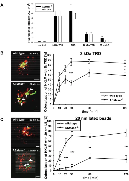
2.2.8 Pulse/chase labelling of lysosomes with fluid phase markers
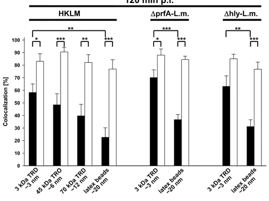
Peritoneal macrophages at a density of 2 x 10⁵ cells/ml were pulsed for 1 h with either 0.6 mg/ml 3 kDa dextran conjugated to TexasRed, 30 µg/ml chicken ovalbumin conjugated to TexasRed or 1 mg/ml 70 kDa dextran conjugated to TexasRed in DMEM with 10% FCS or with 5 x 10¹⁰ red-fluorescent latex beads with a diameter of 20 nm in DMEM without FCS. Non-endocytosed fluid phase markers were removed by triple washing with PBS. The fluorescent fluid phase markers were chased into lysosomes by incubation in fresh medium for 2 h. Afterwards, the cells were prepared for immunofluorescence microscopy as described above.
To quantify uptake of fluid phase markers, immunomagnetically enriched peritoneal macrophages were pulse/chase-labelled as described above and then lysed in lysis buffer for 30 min on ice. Fluorometric analysis of macrophage lysates was performed in a Wallac Victor2 1420 Multilabel Counter.
Stoke’s radii as an indicator for the diameter of dextrans and ovalbumin were calculated as described by Venturoli and Rippe (Venturoli and Rippe 2005).
2.2.9 Western blotting (WB)
Protein concentration in samples was determined by BCA Protein Assay according to the instructions of the supplier (Thermo Scientific, Rockford, USA). Equal amounts of protein in 1x Laemmli buffer were denatured for 5 min at 95° C. Samples were loaded onto NuPAGE Novex 10% Bis-Tris Midi gels and run under standard conditions. For immunoblotting, proteins were electrophoretically transferred to an Immobilon-PSQ transfer membrane at 250 mA for 1.5 h. Membranes were blocked with blocking buffer for 1h at RT before incubation with primary antibody in blocking buffer overnight at 4° C. After triple washing with TBS-T, membranes were incubated with horseradish peroxidase-conjugated secondary antibodies in blocking buffer for 1h at 4° C. Unbound antibodies were removed by washing three times with TBS-T
18
and twice with TBS. The immune complex was visualised by an enhanced chemiluminescence system (ECL Western Blotting reagent) and detected by Amersham ECL hyperfilm. A commercial protein marker was used for identification of protein size.
Specific bands on immunoblots were quantified by densidometry with imageJ (NIH, USA).
2.2.10 Preparation of phagosomes containing L. monocytogenes
1x10⁸ MEF or BMDM were infected with the ΔprfA-mutant of L. monocytogenes (ΔprfA-L. monocytogenes) at a MOI of 10 for 1 h or 20 min, respectively. After removal of extracellular L. monocytogenes by triple washing with PBS, cells were incubated in fresh medium for 2 h or 100 min, respectively. Phagosomes containing ΔprfA-L. monocytogenes were prepared as described by Lührmann et al. (Luhrmann and Haas 2000) and analysed via Western blotting.
2.2.11 Measurement of pH in phagosomes containing L. monocytogenes
Immunomagnetically enriched, proteose peptone-elicited macrophages were co- incubated at a MOI-equivalent of 50 with HKLM double-labelled with Oregon Green 488 and TRITC in DMEM with 10% FCS and 5% NMS for 20 min. Non-phagocytosed HKLM were removed by washing with ice cold PBS. At distinct times after co- incubation, the ratio of pH-dependent Oregon Green 488 fluorescence intensity to pH-independent TRITC fluorescence intensity in MES buffer was determined by flow cytometry. A calibration curve of samples treated with 50 µM monensin and 10 µM nigericin for 15 min in MES buffer of graded pH (3.0 to 7.5) was used to calculate the pH in phagosomes containing HKLM (Vergne, Constant et al. 1998;
Holopainen, Saarikoski et al. 2001).
2.2.12 Measurement of proteolytic activity
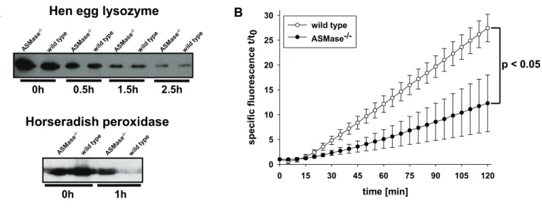
Proteose peptone-elicited peritoneal macrophages at a density of 1,5x10⁶ cells/ml were co-incubated with 0.5 mg/ml hen egg lysozyme or horseradish peroxidase in DMEM with 5% FCS. After 20 min, non-endocytosed protein was removed by triple washing with ice cold PBS. After distinct times of incubation in DMEM with 5% FCS,
19
macrophages were washed twice with PBS, lysed in Laemmli buffer for 5 min and subsequently analysed by Western blotting.
To determine intraphagosomal proteolytic activity, carboxylated 1 µm silica beads were coated with human IgG and streptavidin and labelled with Alexa Fluor 594- succinimidyl ester and the peptide substrate (Biotin-LC-Phe-Arg)2-rhodamine 110 as described by Yates et al. (Yates, Hermetter et al. 2005). Immunomagnetically enriched, proteose peptone-elicited macrophages at a density of 0,75x10⁸ cells/ml were co-incubated with labelled beads for 3 min at 37° C in a very small volume to facilitate bead phagocytosis. The concentration of beads was titrated to achieve an average of 1-2 beads per macrophage and quantitative bead uptake was assessed via fluorescence microscopy. Rhodamine 110 and Alexa Fluor 594 fluorescence intensity was determined in a Wallac Victor2 1420 Multilabel Counter immediately (t0) and at specific time points (t) of co-incubation with beads. Proteolysis of the substrate was expressed as ratio of specific rhodamine 110 fluorescence intensity t / t0 and Alexa Fluor 594 fluorescence intensity.
2.2.13 Quantification of reactive oxygen intermediates
Superoxide production of resident or IFN-γ pre-activated, immunomagnetically enriched peritoneal macrophages after stimulation with PMA was measured by the reduction of ferricytochrome c as described by Kruisbeek et al. (Kruisbeek and Vogel 1999).
2.2.14 Quantification of reactive nitrogen intermediates
Production of nitrite by resident or IFN-γ pre-activated, immunomagnetically enriched peritoneal macrophages after stimulation with HKLM at a MOI-equivalent of 10 was determined using the Greiss reagent as described by Kruisbeek et al.
(Kruisbeek and Vogel 1999).
2.2.15 Statistical analysis
For statistical analysis, the data were subjected to one-way analysis of variance (ANOVA) using SigmaStat 3.5 (Systat Software GmbH, Erkrath, Germany).
20
3 Results
3.1 Impaired inactivation of Listeria monocytogenes despite normal generation of ROI and RNI in ASMase-/- macrophages
To elucidate the role of ASMase in the listeriocidal activity of macrophages, we investigated both the oxidative and non-oxidative bactericidal effector mechanisms of proteose peptone-elicited peritoneal macrophages from ASMase-/- and wild type (wt) mice.
Deficiency in ASMase impairs mannose 6-phosphate receptor-mediated endocytosis in macrophages (Dhami and Schuchman 2004) and internalization of Neisseria gonorrhoeae and Pseudomonas aeruginosa into human epithelial cells (Grassme, Gulbins et al. 1997; Grassme, Jendrossek et al. 2003). Thus, we first examined phagocytic uptake of L. monocytogenes into macrophages from ASMase-/- and wt mice.
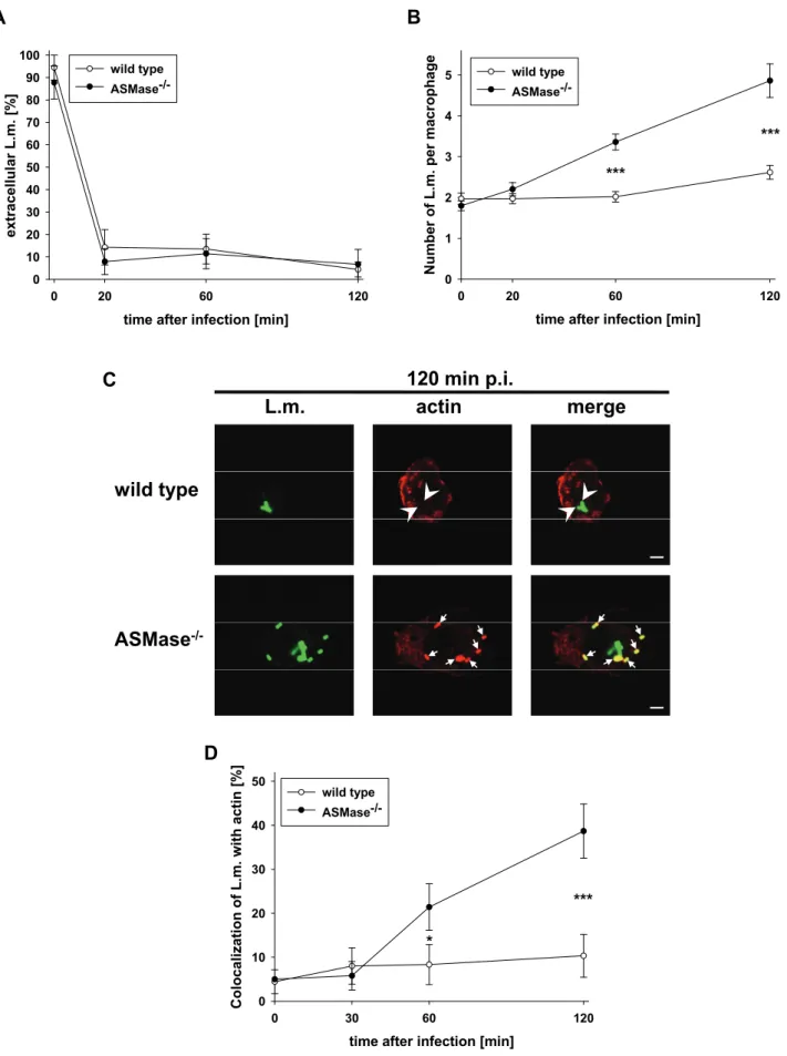
ASMase-/- and wt macrophages phagocytosed fluorescein isothiocyanate (FITC)- labelled L. monocytogenes with identical kinetics (Fig. 2A) and in similar numbers (Fig. 2B, t = 0 min). Importantly, while wt macrophages restricted intracellular proliferation, ASMase-/- macrophages allowed significant multiplication of L. monocytogenes (Fig. 2B). Correspondingly, L. monocytogenes colocalized with actin in ASMase-/- macrophages but not in wt macrophages which indicates escape of L. monocytogenes from phagosomes into the cytosol of ASMase-/- macrophages (Fig. 2C and D). Together, these data show that ASMase-/- macrophages efficiently phagocytose L. monocytogenes but subsequently fail to inactivate the bacteria.
21
time after infection [min]
0 20 60 120
extracellular L.m. [%]
0 10 20 30 40 50 60 70 80 90
100 wild type
ASMase-/-
A
L.m. actin merge
ASMase-/- wild type
D C
time after infection [min]
0 30 60 120
Colocalization of L.m. with actin [%]
0 10 20 30 40
50 wild type
ASMase-/-
***
*
120 min p.i.
time after infection [min]
0 20 60 120
Number of L.m. per macrophage
0 1 2 3 4
5 wild type
ASMase-/-
***
***
B
Fig. 2: Listeria monocytogenes escapes from phagosomes, recruits actin and rapidly proliferates in ASMase-/- but not wt macrophages.
(Legend continued on following page)
22
(Legend of Fig. 2 / continued)
Proteose peptone-elicited peritoneal macrophages of ASMase-/- or wt littermates were infected at a MOI of 5 with FITC-labelled L. monocytogenes (L.m.). At the indicated times after infection, samples were fixed and analysed by confocal laser scanning microscopy.
(A) Kinetics of the decrease in extracellular L. monocytogenes. Extracellular L. monocytogenes were stained with an antibody against L. monocytogenes without permeabilization. L. monocytogenes double-stained with antibody and FITC were considered extracellular, FITC-only L. monocytogenes intracellular. (B) Kinetics of the number of intracellular L. monocytogenes per macrophage. (C) Representative micrographs indicating colocalization (arrows) or non-colocalization (arrow heads) of L. monocytogenes with phalloidin-stained actin at 120 min after infection. Bars are 4 µm.
(D) Kinetics of the colocalization of L. monocytogenes with phalloidin-stained actin. At each time point, a minimum of 100 cells from three independent experiments was analysed. Data are shown as mean +/- s.e. Statistical significance of the difference between ASMase-/- and wild type was determined by one-way ANOVA. P-values are indicated by * (p < 0.05) or *** (p < 0.001).
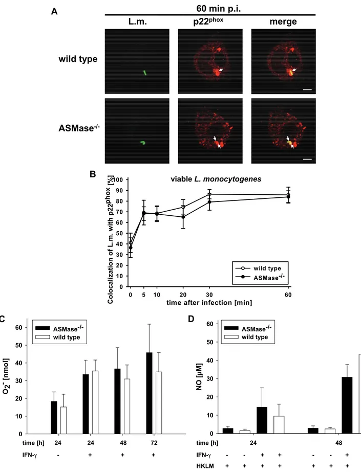
ROI are generated after assembly of the NADPH oxidase complex in the phagosomal membrane. L. monocytogenes colocalized with the catalytic p22phox subunit of the NADPH oxidase complex with similar kinetics in ASMase-/- and wt macrophages (Fig. 3A and B) indicating unimpaired recruitment of p22phox to the phagosomal membrane. Furthermore, both resident and IFN-γ pre-activated ASMase-/- macrophages generated similar amounts of superoxide (O2-) as wt macrophages after stimulation with PMA (Fig. 3C) showing unimpaired generation of ROI. Additionally, ASMase-/- and wt macrophages generated similar amounts of NO after pre-activation with IFN-γ and stimulation with HKLM (Fig. 3D) indicating unimpaired generation of RNI. Thus, ASMase-/- macrophages generate normal amounts of oxidative bactericidal effector molecules but cannot kill L. monocytogenes suggesting that non-oxidative bactericidal effector mechanisms might depend on ASMase activity.
23
time [h]
ohne IFN 24 48 72
O2
- [nm
ol]
0 10 20 30 40 50
60 ASMase-/-
wild type
time [h] 24 24 48 72 IFN-γ - + + +
C
B
NO [µM]
0 10 20 30 40 50
60 ASMase-/-
wild type
D
time [h] 24 48 IFN-γ - - + + - - + + HKLM + + + + + + + + time after infection [min]
0 5 10 20 30 60
Colocalization of L.m. with p22phox [%]
0 10 20 30 40 50 60 70 80 90 100
wild type ASMase-/-
viableL. monocytogenes ASMase-/-
wild type
L.m. p22phox merge
60 min p.i.
A
Fig. 3: Normal activity of oxidative effector mechanisms in ASMase-/- macrophages.
(A, B) Proteose peptone-elicited peritoneal macrophages from ASMase-/- or wt littermates were infected at a MOI of 5 with FITC-labelled L. monocytogenes. At the (Legend continued on following page)
24
(Legend of Fig. 3 / continued)
indicated times after infection, samples were fixed and analysed by confocal laser scanning microscopy. (A) Representative micrographs indicating colocalization (arrows) of L. monocytogenes with p22phox at 60 min after infection. Bars are 4 µm. (B) Kinetics of the colocalization of L. monocytogenes with p22phox. At each time point, a minimum of 100 phagosomes from three independent experiments was analysed. Percentage of L. monocytogenes per macrophage colocalizing with p22phox are shown as mean +/- s.e.
(C) Generation of superoxide by immunomagnetically enriched, resident or IFN-γ pre- activated ASMase-/- and wt macrophages after stimulus with PMA was measured after 24, 48 and 72 h by the reduction of ferricytochrome c. Results from four independent experiments are shown as mean +/- s.e. (D) Generation of NO by immunomagnetically enriched, resident or IFN-γ pre-activated ASMase-/- and wt macrophages stimulated with HKLM was measured after 24 and 48 h using the Greiss reagent. Results from three independent experiments are shown as mean +/- s.e.
3.2 Impaired maturation of phagosomes containing L. monocytogenes into phagolysosomes in ASMase-/- macrophages
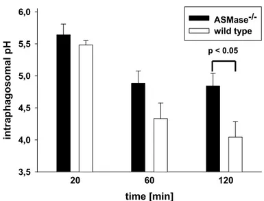
Non-oxidative bactericidal effector mechanisms exerted by acid hydrolases, particularly the lysosomal protease cathepsin D, are essential for listeriocity of macrophages (del Cerro-Vadillo, Madrazo-Toca et al. 2006). These acid hydrolases are transferred from lysosomes into phagosomes at the end of the phagosomal maturation process (Haas 2007).
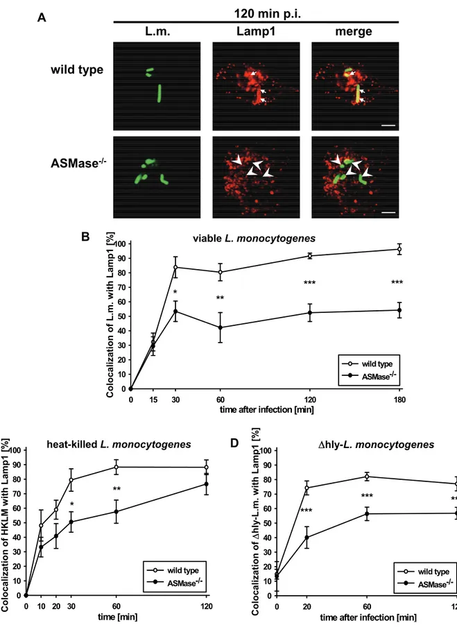
To investigate maturation of phagosomes into phagolysosomes, we analysed the kinetics of the colocalization of L. monocytogenes with the (phago)lysosomal marker
‘lysosome-associated membrane protein 1’ (Lamp1) in ASMase-/- and wt macrophages. In wt macrophages, more than 90 % of the phagocytosed L. monocytogenes colocalized with Lamp1 from 30 min post infection (p.i.) on (Fig.
4A and B). In contrast, only 50% of the phagocytosed L. monocytogenes colocalized with Lamp1 in ASMase-/- macrophages indicating impaired maturation of phagosomes into phagolysosomes.
The reduced colocalization of L. monocytogenes with Lamp1 in ASMase-/- macrophages could be due to (i) interference of L. monocytogenes with the phagosomal maturation (Alvarez-Dominguez, Barbieri et al. 1996; Alvarez- Dominguez, Roberts et al. 1997; Prada-Delgado, Carrasco-Marin et al. 2005;
Alvarez-Dominguez, Madrazo-Toca et al. 2008) (ii) escape of L. monocytogenes from
25
phagosomes (Tilney and Portnoy 1989) or (iii) a cellular defect in the maturation of phagosomes into phagolysosomes.
To discriminate between these alternatives, we performed two additional series of experiments. First, we analysed the colocalization of heat-killed L. monocytogenes (HKLM) with Lamp1 because HKLM can neither actively interfere with the phagosomal maturation nor escape from phagosomes (Alvarez-Dominguez, Roberts et al. 1997). As illustrated in Fig. 4C, HKLM colocalized significantly less frequently with Lamp1 in ASMase-/- than in wt macrophages at 30 min and 60 min after co- incubation of HKLM with macrophages. Afterwards, the percentage of HKLM colocalizing with Lamp1 in ASMase-/- macrophages slowly increased to reach almost the level observed in wt macrophages at 120 min after co-incubation. Second, we studied colocalization of the viable but apathogenic listeriolysin O-deficient Δhly-strain of L. monocytogenes (Δhly-L. monocytogenes) with Lamp1. Listeriolysin O (LLO) is the major virulence factor required for escape of L. monocytogenes from phagosomes (Leimeister-Wachter, Haffner et al. 1990; Beauregard, Lee et al. 1997).
Fig. 4D shows that Δhly-L. monocytogenes colocalized significantly less frequently with Lamp1 in ASMase-/- than in wt macrophages from 20 min p.i. on. These data show that reduced colocalization of L. monocytogenes with Lamp1 in ASMase-/- macrophages is independent of the viability or the virulence of L. monocytogenes.
26
time [min]
0 10 20 30 60 120
Colocalization of HKLM with Lamp1 [%]
0 10 20 30 40 50 60 70 80 90 100
wild type ASMase-/-
**
*
heat-killed L. monocytogenes C
A
ASMase-/- wild type
L.m. Lamp1 merge
D
time after infection [min]
0 20 60 120
Colocalization of Δhly-L.m. with Lamp1 [%]
0 10 20 30 40 50 60 70 80 90 100
wild type ASMase-/-
***
***
Δhly-L. monocytogenes
***
120 min p.i.
time after infection [min]
0 15 30 60 120 180
Colocalization of L.m. with Lamp1 [%]
0 10 20 30 40 50 60 70 80 90 100
wild type ASMase-/-
B
**
*** ***
*
viable L. monocytogenes
Fig. 4: Reduced colocalization of viable, heat-killed and Δhly-L. monocytogenes with Lamp1 in ASMase-/- macrophages.
Proteose peptone-elicited peritoneal macrophages from ASMase-/- or wt littermates were infected at a MOI of 5 with FITC-labelled (A, B) wild type or (Legend continued on following page)
27
(Legend of Fig. 4 / continued)
(D) Δhly-L. monocytogenes or (C) incubated with an equivalent number of heat-killed L. monocytogenes (HKLM). At the indicated times after infection, samples were fixed and analysed by confocal laser scanning microscopy. (A) Representative micrographs indicating colocalization (arrows) or non-colocalization (arrow heads) of viable wild type L. monocytogenes with Lamp1 at 120 min after infection. Bars are 4 µm. (B, C, D) Kinetics of the colocalization of (B) viable wt L. monocytogenes, (C) HKLM or (D) Δhly- L. monocytogenes with Lamp1. At each time point, a minimum of 100 phagosomes from three independent experiments was analysed. Percentage of Listeria per macrophage colocalizing with Lamp1 are shown as mean +/- s.e. The statistical significance of the difference between ASMase-/- and wild type was determined by one-way ANOVA.
P-values are indicated by * (p < 0.05), ** (p < 0.01) or *** (p < 0.001).
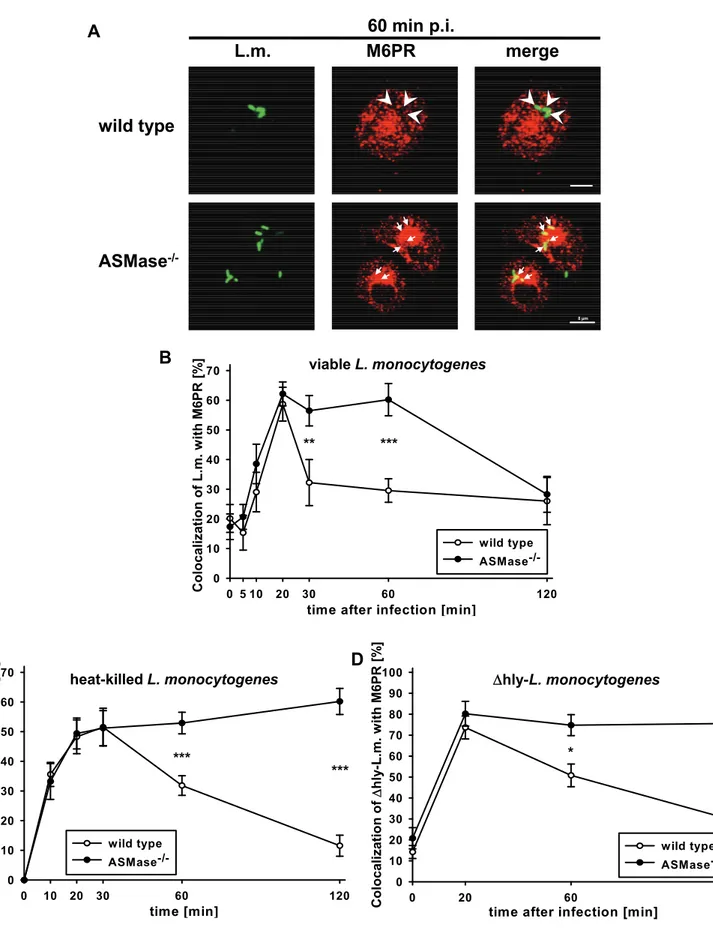
To further investigate whether a cellular defect of ASMase-/- macrophages impairs the maturation of phagosomes containing L. monocytogenes into phagolysosomes, we analysed acquisition of the ‘cation-independent mannose 6-phosphate receptor’
(M6PR) by phagosomes containing L. monocytogenes. M6PR is present on late phagosomes but not on phagolysosomes (Haas 2007).
In both ASMase-/- and wt macrophages, more than 60% of the phagocytosed L. monocytogenes colocalized with M6PR within 20 min p.i. (Fig. 5A and B).
Afterwards, most phagosomes containing L. monocytogenes in wt macrophages lost colocalization with M6PR within just 10 min (30% at 30 min p.i.). In sharp contrast, phagosomes containing L. monocytogenes in ASMase-/- macrophages remained positive for M6PR until at least 60 min p.i. and slowly lost colocalization with M6PR between 60 min and 120 min post infection.
To discriminate whether this late loss of colocalization of L. monocytogenes with M6PR in ASMase-/- macrophages was caused by escape of L. monocytogenes from phagosomes or by maturation of late phagosomes into phagolysosomes, we used HKLM and Δhly-L. monocytogenes. In both ASMase-/- and wt macrophages, more than 50% of HKLM colocalized with M6PR within 20 min after co-incubation (Fig. 5C).
Afterwards, the percentage of HKLM colocalizing with M6PR in wt macrophages dropped significantly to about 30% at 60 min after co-incubation and 15% at 120 min after co-incubation. In ASMase-/- macrophages, however, phagosomes containing HKLM remained positive for M6PR until at least 120 min after co-incubation.
Similarly, after infection with Δhly-L. monocytogenes, colocalization with M6PR was sustained at peak levels in ASMase-/- macrophages but rapidly declined in wt
28
macrophages (Fig. 5D). These data show that colocalization of viable L. monocytogenes with M6PR in ASMase-/- macrophages beyond 60 min p.i. dropped only because the bacteria escaped from phagosomes. Both HKLM and Δhly- L. monocytogenes remained within M6PR-positive phagosomes in ASMase-/- macrophages further indicating an impaired maturation of phagosomes into phagolysosomes.
29
time [min]
0 10 20 30 60 120
Colocalization of HKLM with M6PR [%]
0 10 20 30 40 50 60 70
wild type ASMase-/-
*** ***
heat-killedL. monocytogenes C
time after infection [min]
0 5 10 20 30 60 120
Colocalization of L.m. with M6PR [%]
0 10 20 30 40 50 60 70
wild type ASMase-/-
B
** ***
viable L. monocytogenes A
8 µm
L.m. M6PR merge
8 µm
ASMase-/- wild type
D
time after infection [min]
0 20 60 120
Colocalization of Δhly-L.m. with M6PR [%]
0 10 20 30 40 50 60 70 80 90 100
wild type ASMase-/-
Δhly-L. monocytogenes
* ***
60 min p.i.
Fig. 5: Prolonged colocalization of viable, heat-killed and Δhly-L. monocytogenes with M6PR in ASMase-/- macrophages.
Proteose peptone-elicited peritoneal macrophages from ASMase-/- or wt littermates (Legend continued on following page)
30
(Legend of Fig. 5 / continued)
were infected at a MOI of 5 with FITC-labelled (A, B) wild type or (D) Δhly- L. monocytogenes or (C) incubated with an equivalent number of heat-killed L. monocytogenes (HKLM). At the indicated times p.i., samples were fixed and analysed by confocal laser scanning microscopy. (A) Representative micrographs indicating colocalization (arrows) or non-colocalization (arrow heads) of viable wt L. monocytogenes with M6PR at 60 min after infection. Bars are 4 µm. (B, C, D) Kinetics of the colocalization of (B) viable wt L. monocytogenes, (C) HKLM or (D) Δhly-L. monocytogenes with M6PR.
At each time point, a minimum of 100 phagosomes from three independent experiments was analysed. Percentage of Listeria per macrophage colocalizing with M6PR are shown as mean +/- s.e. The statistical significance of the difference between ASMase-/- and wild type was determined by one-way ANOVA. P-values are indicated by * (p < 0.05),
** (p < 0.01) or *** (p < 0.001).
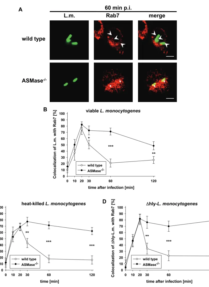
The small GTPase Rab7 mediates the maturation of late phagosomes into phagolysosomes (Mullock, Bright et al. 1998; Bucci, Thomsen et al. 2000; Ceresa and Bahr 2006). To investigate the role of Rab7 in the phagosomal maturation defect of ASMase-/- macrophages, we analysed the colocalization of L. monocytogenes with Rab7. In both ASMase-/- and wt macrophages, more than 70% of viable L. monocytogenes (Fig. 6A and B), HKLM (Fig. 6C) or Δhly-L. monocytogenes (Fig. 6D) colocalized with Rab7 at 20 min post infection. Afterwards, phagosomes containing L. monocytogenes lost Rab7 already at 30 min p.i. in wt macrophages but remained positive for Rab7 until 120 min p.i. in ASMase-/- macrophages. These data confirm a block in the maturation of phagosomes at the late phagosomal stage in ASMase-/- macrophages.