Research Collection
Doctoral Thesis
Compensating impaired movements: design principles for lower- limb exoskeletons
Author(s):
Schmidt, Kai Publication Date:
2021
Permanent Link:
https://doi.org/10.3929/ethz-b-000476794
Rights / License:
In Copyright - Non-Commercial Use Permitted
This page was generated automatically upon download from the ETH Zurich Research Collection. For more information please consult the Terms of use.
ETH Library
DISS. ETH NO. 27445
Compensating impaired movements: design principles for lower-limb exoskeletons
A thesis submitted to attain the degree of
DOCTOR OF SCIENCES OF ETH ZURICH (Dr. sc. ETH Zurich)
presented by KAI SCHMIDT
M.Sc., Technische Universität Berlin born on 10.01.1989
citizen of Germany
accepted on the recommendation of
Prof. Dr. Ing. Dr. med. h.c. Robert Riener - examiner Prof. Dr. Roger Gassert - co-examiner
2021
i
Abstract
Walking is one of the top priorities of patients that suffer from reduced locomotor function. Exoskeletons bear the promise of restoring that function by providing active support, and thus, enable economical movements again. However, there is a disparity of support strategies and designs with limited clinical validation. The fundamental re- quirements that drive these designs are manifold and partially lack validated approaches.
The purpose of the thesis is to propose and validate essential requirements for assistive exoskeletons independent of their technical implementation while also ensuring causal tracebility. The requirements have been derived from mechanisms and features of ef- ficient movements and prevalent design principles of devices that are able to improve movement economy. As characteristics of unimpaired movements do not necessarily apply to patients, sources of pathological movements and design principles of move- ment aids have been identified and compared. To determine the most beneficial support method, an assessment framework based on gait deviations and underlying causes has been proposed. The requirements have been implemented in a first prototype: the Myosuit. The Myosuit was used for initial validation tests on a single participant during sitting transfers. The requirements have been further validated with regard to their fea- sibility and usability in five patients with different pathologies.
The assessment framework based on gait deviations identified external support of the knee extensor muscles and flexor muscles of the hip, knee and ankle joint as the most promising interventions to improve impaired walking. When considering other move- ments besides walking, also supporting hip extension can provide a benefit. A set of essential requirements and design principles related to the exoskeleton interface, pro- vision of force and controls have been derived and implemented in the Myosuit. The Myosuit uses a bi-articular, cable driven design to assist knee and hip extension simul- taneously and a posture based control approach. Elastic elements support the flexor muscles antagonistic to the active cable drive. The Myosuit was able to reduce extensor muscle activity during sitting transfers and could be used without any serious adverse events in a patient population during a feasibility test. These initial tests validated the essential requirements and related design principles. This shows that the applied meth- ods of deriving the requirements can lead to an exoskeleton design that fulfill basic needs of various pathologies and may provide a functional benefit.
iii
Überblick
Das Gehen ist eine der obersten Prioritäten von Patienten, die unter eingeschränkter Bewegungsfunktion leiden. Exoskelette versprechen diese Funktion durch aktive Unter- stützung wiederherzustellen und ermöglichen so wieder effiziente Bewegungen. Aller- dings gibt es eine Vielzahl von Unterstützungsstrategien und Designs mit begrenzter klinischer Validierung. Die Anforderungen, die diesen Designs zugrunde liegen, sind vielfältig und es fehlen häufig substantielle Ansätze. Ziel der Arbeit ist es, wesentliche An- forderungen an unterstützende Exoskelette unabhängig von ihrer technischen Umsetzung abzuleiten und zu validieren, wobei auch die kausale Nachverfolgbarkeit sichergestellt wird. Die Ableitung der Anforderungen erfolgte durch die Identifizierung von Mechanis- men und Funktionen, die effiziente Bewegungen ermöglichen, und vorherrschende Kon- struktionsprinzipien von Geräten, die die Bewegungsökonomie verbessern können. Da diese Merkmale nicht unbedingt auf Patienten zutreffen, wurden die Ursachen pathol- ogischer Bewegungen und die Konstruktionsprinzipien von Bewegungshilfen identifiziert und verglichen. Um die vorteilhafteste Unterstützungsmethode zu identifizieren, wurde ein Bewertungsrahmen entwickelt, der auf Gangabweichungen und den zugrunde liegen- den Ursachen basiert. Die Anforderungen wurden in einem ersten Prototyp umgesetzt:
dem Myosuit. Der Myosuit diente als Mittel für erste Validierungstests an einem einzel- nen Probanden während mehrfacher Aufsteh- und Hinsetzvorgänge. Die Anforderungen wurden ausserdem im Hinblick auf ihre Machbarkeit und Anwendbarkeit an fünf Patien- ten mit verschiedenen Pathologien validiert.
Im Rahmen der Suche nach der optimalen Unterstützungsmethode wurde die externe Un- terstützung der Kniestreckmuskeln und der Beugemuskeln des Hüft-, Knie- und Sprungge- lenks als die vielversprechendsten Interventionen zur Verbesserung von Gehbehinderun- gen identifiziert. Wenn neben dem Gehen auch andere Bewegungen berücksichtigt wer- den, kann auch die Unterstützung der Hüftstreckung einen Nutzen bringen. Grundle- gende Anforderungen in Bezug auf die Mensch-Maschinen-Schnittstelle, die Bereitstel- lung der Unterstützung und die Steuerung wurden abgeleitet und im Myosuit umge- setzt. Der Myosuit verwendet ein bi-artikuläres, kabelgesteuertes Design zur gleichzeiti- gen Unterstützung der Knie- und Hüftstecker unter Verwendung eines haltungsbasierten Steuerungsansatzes. Elastische Elemente unterstützen die Beugemuskeln antagonistisch zum aktiven Kabelantrieb. Der Myosuit war in der Lage, die Streckmuskelaktivität zu re- duzieren und konnte während des Machbarkeitstests ohne schwerwiegende unerwünschte Ereignisse in einer Patientenpopulation eingesetzt werden. Diese ersten Tests validierten die wesentlichen Anforderungen und die damit verbundenen Konstruktionsprinzipien.
Dies zeigt, dass die angewandten Methoden zur Herleitung der Anforderungen zu einem Exoskelett-Design führen können, das grundlegende Bedürfnisse verschiedener Patholo- gien erfüllt und einen funktionellen Nutzen bieten kann.
v
Acknowledgements
Throughout the writing of this dissertation I have received a great deal of support and assistance.
I would first like to thank my supervisor, Professor Robert Riener, whose expertise was invaluable in formulating the research questions and methodology. Your insightful feed- back always gave me guidance and ensured that I could continuously improve my skill set and quality of work. I would also like to thank Florian Haufe, Michele Xiloyannis and Peter Wolf for the discussions that always resulted in valuable feedback. This sharp- ened my thinking and brought my work to a higher level. I would like to acknowledge Jaime Duarte with whom I co-founded MyoSwiss. Thank you for the times when we solved problems together. Always striving for the next big thing with you helped to push boundaries and made all the all-nighters worth it. A big part of the work could not be done without the first employees of MyoSwiss which helped to develop stable and safe prototypes. I would like to thank Alejandro Sancho Puchades, Katja Stähli, Gleb Koginov, Adèle Pillichody, Andri Hartmann and Daniel Puerta Diaz. I would like to especially acknowledge Alejandro, Katja and Gleb who joined the project even before the foundation of MyoSwiss. Your input has been more than helpful and always brought up new ideas. Thank you, Nathalie Zwickl, for being the first physiotherapist that used the Myosuit on multiple patients within therapy. We benefited greatly from all your input and experience. In addition, I also thank my whole family for their continuous support.
I could not have finished my dissertation without you. In particular, I’d like to express my deepest gratitude to my wife Constanze for her patience and tolerance.
vii
Symbols
Symbols
EC encoder counts [1]
F force [N]
g gravitational acceleration [m/s2]
h height [m]
k spring stiffness [N/m]
l length [m]
m mass [kg]
mf orce suit stiffness [N/rad]
mhip hip-encoder mapping [deg/rad]
mknee knee-encoder mapping [deg/rad] [kg]
M moment [Nm]
P power [Watt]
rjoint_ligament moment arm ligament [m]
v velocity [m/s]
W work [Joule]
α absolute angle [deg]
β relative angle [deg]
θ acceleration [m/s2]
ω angular velocity [rad/s]
Acronyms and Abbreviations
AFO ankle foot orthosis ADL activities of daily living BMI body mass index COM center of mass CT contracture DF dorsi-flexion EC encoder counts EXT extension
FES functional elctrical stimulation FIX-N fixed in a neutral position FLE flexion
IMU inertial measurement unit iSCI incomplete spinal cord injury KE kinematic energy
MCID minimal clinically important difference MOCA Montreal Cognitive Assessment test MS Multiple Sclerosis
NRS Numeric Rating Scale PE potential energy PF plantar-flexion PT physiotherapist ROS roll-over shape RQ requirement SD standard deviation SP spasticity
SUS system usability scale
WEIMuS Würzburger Erschöpfungs-Inventar bei MS
WK weakness
ix
Contents
Abstract i
Überblick iii
Acknowledgements v
Symbols vii
Preface xiii
1 Introduction 1
1.1 Impaired movements . . . . 1
1.2 Robotic exoskeletons . . . . 2
1.2.1 Compensation of impaired movements . . . . 2
1.2.2 Essential requirements for exoskeleton designs . . . . 3
1.3 Thesis goal and outline . . . . 3
2 Normal Gait 5 2.1 Basics of normal gait . . . . 5
2.1.1 Bones, joints and muscles of the lower limbs . . . . 5
2.1.2 Tasks, phases and periods of normal gait . . . . 6
2.1.3 Stance phase . . . . 9
2.1.4 Swing phase . . . . 9
2.2 Theories related to the efficiency of normal gait . . . 10
2.2.1 Six determinants of normal gait . . . 10
2.2.2 Dynamic walking . . . 12
2.2.3 Energy transfers in a multi-segment system . . . 14
2.3 What makes walking efficient? . . . 17
2.3.1 Mechanisms and features enabling efficient movements . . . 17
2.3.2 Reduction of metabolic energy expenditure . . . 19
2.4 Devices to reduce metabolic energy expenditure . . . 20
2.5 Essential requirements derived from normal gait . . . 23
3 Assisting pathological walking 25 3.1 Causes of pathological movements . . . 25
3.2 Devices to support pathological movements . . . 28
3.3 Compensation and support strategies . . . 33
3.3.1 Net joint moment synergies . . . 34
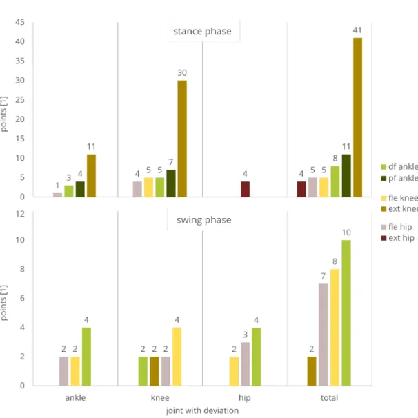
3.3.2 Deficit based support . . . 39
3.4 Essential requirements derived from pathological gait . . . 49
4 Discussion of essential requirements and design principles 53 4.1 Interface . . . 53
4.2 Provision of force . . . 55
4.3 Controls . . . 57
5 The Myosuit 59 5.1 The Myosuit concept . . . 60
5.2 Design . . . 62
5.2.1 Myosuit layers . . . 62
5.2.2 Tendon actuators . . . 64
5.2.3 Sensors . . . 65
5.2.4 Control unit . . . 65
5.3 Closed-Loop control algorithm . . . 66
5.3.1 Anti-gravity control against the force of gravity . . . 67
5.3.2 Anti-gravity control with the force of gravity . . . 69
5.4 Experimental protocol . . . 69
5.5 Data analysis . . . 70
5.5.1 Kinematics and kinetics . . . 70
5.5.2 Muscle activity . . . 70
5.5.3 Transition identification . . . 71
5.6 Results of the first testing . . . 72
5.6.1 Compression compensation . . . 72
5.6.2 Posture based anti-gravity control . . . 73
5.6.3 Joint kinematics and kinetics . . . 74
5.6.4 Muscle activity . . . 75
5.7 Discussion of first testing results . . . 76
5.7.1 Compression compensation and knee angle estimation . . . 76
5.7.2 Posture based anti-gravity control . . . 77
6 Separating Gait Phases 79 6.1 Segmentation options . . . 79
6.2 Study protocol . . . 82
6.3 Kinematics and ground reaction forces . . . 82
6.3.1 Sign changes of the angular velocity . . . 84
6.3.2 Constraint rule sets for event detection . . . 84
6.4 Study results . . . 85
6.4.1 Temporal progression of angular velocity . . . 85
6.4.2 Angular velocity and gait phase detection . . . 85
6.4.3 Transition timing . . . 87
6.5 Discussion of segmentation options . . . 89
6.5.1 Reliability . . . 89
6.5.2 Accuracy . . . 91
Contents xi
6.5.3 Limitations of detection methods . . . 91
6.6 Summary of IMU based gait segmentation . . . 93
7 Feasibility test 95 7.1 Myosuit alpha . . . 95
7.2 Test protocol . . . 97
7.3 Test results . . . 100
7.3.1 Practicability . . . 101
7.3.2 Acceptance . . . 103
7.3.3 Unstructured feedback . . . 103
7.3.4 Summary of results . . . 104
8 General Discussion and conclusion 105 8.1 Discussion . . . 105
8.2 Conclusion and outlook . . . 109
xiii
Preface
The work presented in this thesis was conducted at the Sensory-Motor Systems Lab at ETH Zurich in collaboration with MyoSwiss AG. I co-founded MyoSwiss AG as ETH Spin-off resulting from the positive results presented in chapter 5 and the patent appli- cation:
K. Schmidt, J.E. Duarte, R. Riener, ”Apparatus for supporting a limb of a user against gravity”, EP3342390, priority date 29.12.2016
Since the foundation of MyoSwiss in 2017, I act as Chief Technology Officer and board member, and since 2020, I act additionally as Chief Executive Officer for the European subsidiary based in Germany. The continued development of the Myosuit towards certi- fication as medical device and related intellectual property was completely managed by MyoSwiss AG.
Parts of the introduction and chapter 5 are based on the following scientific article that has been published:
K. Schmidt K, J.E. Duarte, M. Grimmer, A. Sancho-Puchades, H. Wei, C.S. Easthope, R. Riener, ”The Myosuit: Bi-articular Anti-gravity Exosuit That Reduces Hip Extensor Activity in Sitting Transfers”, Frontiers in Neurorobotics, Oct 2017
The scientific investigations reported in chapter 6 were conducted in close collaboration with Dr. Martin Grimmer. Parts of the introduction and chapter 6 are based on a publication with a shared first-authorship:
M. Grimmer, K. Schmidt K, J.E. Duarte, L. Neuner, K. Koginiv, R. Riener, ”Stance and Swing Detection Based on the Angular Velocity of Lower Limb Segments During Walking”, Frontiers in Neurorobotics, vol. 13, 2019
The feasibility test presented in chapter 7 was conducted in collaboration with licensed physiotherapist Nathalie Zwickl as part of her thesis to obtain the title Master of Science in Physiotherapy.
xv
List of Figures
2.1 Joints and bones of the lower limbs. . . . 6
2.2 Main muscles and muscle groups of the lower limbs. . . . . 7
2.3 Phases, periods and tasks of normal gait. . . . 8
2.4 Muscle activation during normal gait (data from [1]). . . 10
2.5 Redirection of the COM modeled as an inverted pendulum. . . 13
2.6 Horizontal and vertical COM velocity (data from [2]). . . 16
2.7 Main mechanism and features of normal gait. . . 18
2.8 Energy expenditure per distance traveled and per time (data from [3]). . 20
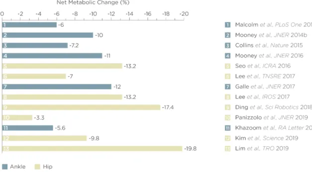
2.9 Devices that broke the metabolic cost barrier in normal gait. . . 21
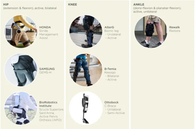
3.1 Compensation movements during walking based on data provided by [1]. 29 3.2 Active devices that have clinical data and compensate pathological walk- ing partially. . . 32
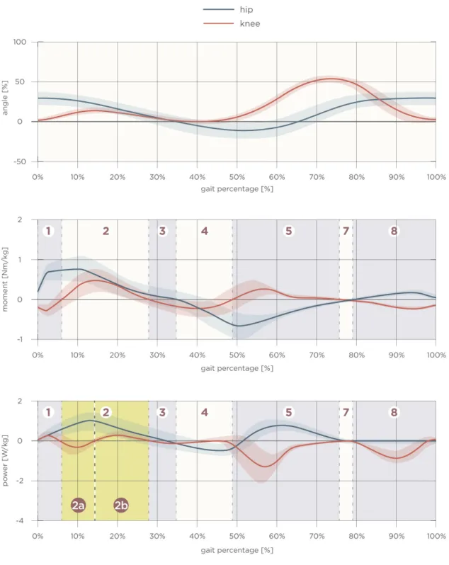
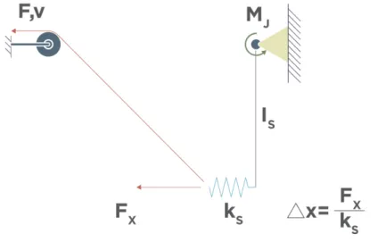
3.3 Angle, moment and power of the knee and hip joint during normal gait. Intervals have been established according to the method described by [4]. 35 3.4 Mechanical model of a single segment connected to a joint and to a cable actuator elastically. . . 37
3.5 Ankle joint deviations (excessive plantar-flexion, excessive dorsi-flexion) from normal gait, related deficits and potential support strategies. . . . 41
3.6 Knee joint deviations (inadequate flexion, excessive extension) from nor- mal gait, related deficits and potential support strategies. . . 43
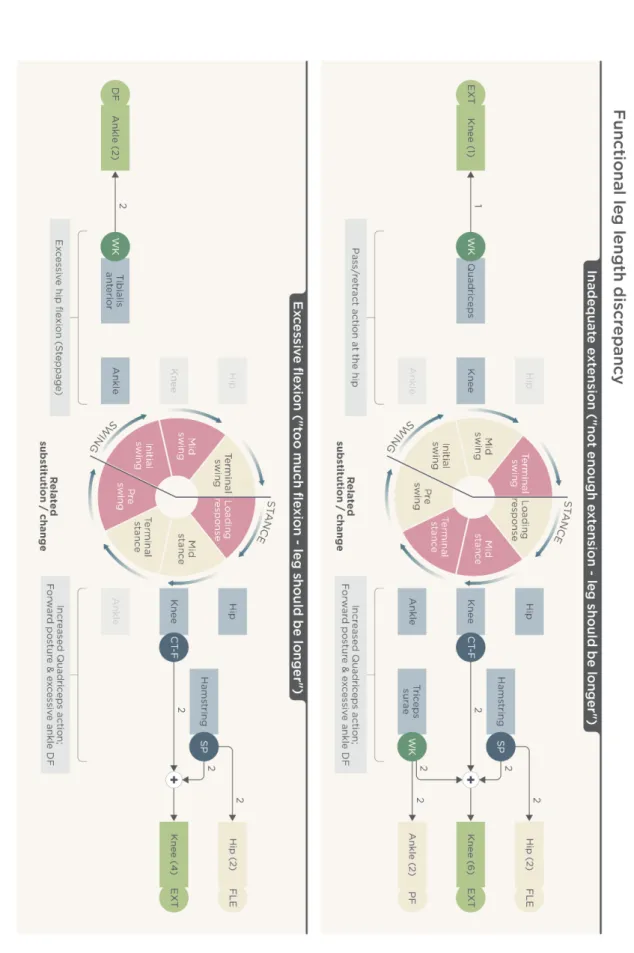
3.7 Knee joint deviations (inadequate extension, excessive flexion) from nor- mal gait, related deficits and potential support strategies. . . 44
3.8 Hip joint deviations (inadequate extension, excessive flexion) from normal gait, related deficits and potential support strategies . . . 47
3.9 Hip joint deviation (inadequate flexion) from normal gait, related deficits and potential support strategies . . . 48
3.10 Ranking of deficit targeting support methods according to the developed rating scheme. . . 50
4.1 Influence of different knee moment arm configurations at a constant output force of 400N at the actuator (assuming 40% transmission effi- ciency). . . 58
5.1 Three-layer architecture of the Myosuit. . . 60
5.2 Active and ligament layers of the Myosuit. . . 61
5.3 System architecture of the Myosuit . . . 62
5.4 Tendon actuator weighing 1070 g. . . 64
5.5 Control chart for the Myosuit. . . 66
5.6 Angles measured by the Myosuit. . . 67
5.7 Experimental setup for the sitting transitions. . . 70
5.8 Example hip kinematics and ground reaction forces used to segment the recorded data offline for the analysis. . . . 71
5.9 Myosuit characterization. . . 73
5.10 Moment-angle curve of hip and knee joints during sitting transfers. . . . 74
5.11 Experimental evaluation of the Myosuit during sit-to-stand (left) and stand-to-sit (right) transitions. . . 75
6.1 Lower limb segment models evaluated for the potential on stance and swing detection. . . 81
6.2 Lower limb segment model velocity based concept for stance and swing detection. . . 81
6.3 Measurement setup including an instrumented split-belt treadmill and a motion capture system. . . 83
6.4 Inertial measurement unit (IMU) setup for the thigh and the shank. . . 84
6.5 Angular velocities from motion capture (solid) and inertial measurement units (dashed) for the thigh (orange), shank (red), leg (blue) and ex- tended leg segment model (green) in level walking, inclined walking (10◦) and declined walking (10◦). Darker colors indicate higher speeds (0.9, 1.3 and 1.7 m/s for slopes, 0.5, 1.3 and 2.1 m/s for level walking). . . 86
6.6 Amount of zero crossings of the angular velocity per gait cycle for the thigh (orange), shank (red), leg (blue) and extended leg segment model (green) in level walking, inclined walking (10◦) and declined walking (10◦). Darker colors represent higher speeds (0.9, 1.3 and 1.7 m/s for slopes, 0.5, 1.3 and 2.1 m/s for level walking). Lines show the standard deviation. . . 88
6.7 Single subject angular velocities for one stride from motion capture (solid) and inertial measurement units (dashed) for the thigh (orange), shank (red), leg (blue) and extended leg (green) segment model. . . 89
6.8 Distance of the mean zero crossings of the angular velocity based on the inertial measurement unit to the heel-strike (left) and the take-off (right) identified by ground reaction forces (GRF). Evaluated segment angular velocities from the thigh (orange), shank (red), leg (blue) and extended leg (green). Distances are evaluated for three different speeds of level walking, walking inclines and walking declines. Darker colors indicate higher speeds (0.9, 1.3 and 1.7 m/s for slopes, 0.5, 1.3 and 2.1 m/s for level walking). . . 90
7.1 Myosuit alpha developed by MyoSwiss AG. . . 96
7.2 Control diagram for the Myosuit alpha. . . 97
7.3 Protocol of the feasibility test. . . 98
7.4 Results of the feasibility test (donning and doffing time, SUS, WEIMuS). 102 8.1 Commercial version of the Myosuit developed by MyoSwiss AG. . . 110
xvii
List of Tables
2.1 Requirements derived from exoskeletons that were able to break the metabolic cost barrier . . . 23 3.1 Requirements to support and improve pathological movements . . . 51 6.1 Rule sets applied to the different lower limb segment models with respect
to heel-strike (HS) and toe-off (TO). . . 85 7.1 Participant demographics. . . 100 7.2 Participant pathology, self-reported walking distance and mobility aid
needed to cover the distance. . . 101 7.3 Light adverse effects reported. . . 103
1
Chapter 1 Introduction
1.1 Impaired movements
Mobility and independence are key determinants for quality of life [5, 6]. When mobility is limited, a person’s quality of life can be impacted negatively. The main causes for mobility limitations are reductions in physical capacity with increasing age [7], diseases, or injuries. From the age of 60 to the age of 85, the mean steps per day decrease by about 77% [8]. This can negatively affect a person since a higher number of steps per day is associated with several positive health outcomes including reductions in body mass index [9], risk of cardiovascular disease [10], and mortality [11]. Along with aging, neurological injuries such as spinal cord injury (SCI) can also limit a person’s mobility.
For example, incomplete SCI patients (ASIA grades C and D; 30% of SCI patients) have full range of motion and the ability to move against gravity with at least half of the key muscles. However, walking and standing abilities, which are top priorities for this patient group [12], are clearly limited for most of the patients [13].
While exercise can help mitigate the reductions in strength [14] and stamina [15], the overall trend cannot be stopped [15, 16]. To be mobile and move independently, the best option for many people is the use of technologies that can compensate their impair- ment. These technologies range from passive devices, such as orthoses or wheelchairs, to powered devices, such as exoskeletons. Especially the wheelchair is an efficient tool to maintain mobility. But in terms of versatility and health related impact it cannot com- pete with bipedal locomotion. This might be the main reason why many patients report that recovering walking capacity is one of their top priorities [17, 18, 19, 20, 21, 22].
There is a great need for devices that can support and facilitate the rehabilitation process and help patients to reach their potential rapidly. But even if the rehabilitation poten- tial is exhausted and patients only improve incrementally, many still require assistance and support of locomotion tasks during their everyday life. In these cases, their most promising option to restore mobility and independence might be robotic exoskeletons.
1.2 Robotic exoskeletons
Given the great need for devices that can restore function related to specific movements, it is not surprising that there has been a multitude of exoskeleton developments. A recent systematic review identified 25 lower-limb exoskeletons that are intended for gait training in neuromusclular impairments [23]. Only six out of these 25 devices are commercially available. Yet two other systematic reviews found a total of 239 additional exoskeletons for the lower-limbs [24, 25]. Considering the high number of developments, it is surprising that exoskeletons are still not part of the daily life of patients. There seems to be a mismatch between the numerous developments and what is available and suitable for patients. Despite all the progress that has been made, exoskeletons are also still not recommended by official rehabilitation guidelines [26, 27]. There is a clear lack of evidence that these devices are are superior to conventional interventions. The great number of designs and the fact that these devices are not commonly used outside of research indicates that the requirements utilized to design these devices are insufficient and do not meet the user’s needs.
1.2.1 Compensation of impaired movements
Exoskeletons have the potential to help patients to achieve their goals within therapy and related to upright mobility. When exoskeletons are designed to fully drive the kinematic pattern of a user, controllers and actuators can rely on trajectories of unimpaired move- ments [28]. This approach is usually implemented in exoskeletons that are intended for paralysed or very weak patients that could not move without the device. However, de- sign decisions become more complicated when exoskeletons are designed to only partially support, and thus, provide assistance for residual functions. It can often be observed that patients with the same pathology move very differently. Patients usually have their own unique set of deficits that result in different manifestations of movement patterns. They substitute their lost functionality by using various compensatory movements. These in- dividual characteristics make it difficult to develop general approaches that can provide a benefit for many patients. This exacerbates the design process considerably. However, there seem to be general approaches that can support various pathologies. One such an approach is reflected in crutches. They provide partial weight-bearing that benefits a wide range of patients independent of their pathology. Similarly, patients seem to profit from support of toe-clearance to swing the leg forward by using ankle-foot orthoses (AFOs). These devices are recommended by the guidelines because they are simple, light-weight and efficient [26, 27, 29]. Exoskeletons could be also designed for basic functions while keeping their weight and level of complexity low. Nonetheless, it is not obvious which deficits exoskeletons should target and where they can be advantageous to conventional solutions.
1.3. Thesis goal and outline 3
1.2.2 Essential requirements for exoskeleton designs
The complexity due to the uniqueness of deficits and resulting movements require a structured design approach. Design requirements need to be derived from the targeted movements while also considering how deficits prevent their execution. Based on these insights, general support strategies could be deducted. Further requirements would also need to be elicited from the various exoskeleton designs that already provide data about their positive impact on movement economy. Such an approach would ensure that the device’s design targets relevant deficits that could provide a benefit to a multitude of patients, and thus, could serve for new developments. To be used as a basis for new developments, these requirements need be independent on any technical implementation.
That is why they are called essential requirements within the scope of this thesis. These requirements can potentially be used to derive design principles for implementations of exoskeletons that assist residual function. Simultaneously, the essential requirements could also be used as a framework during design validations. By following this approach, it becomes more likely that patients receive support that compensates their impairment which results in improved movement function.
1.3 Thesis goal and outline
The goal of the thesis is to find and validate general design approaches for exoskeletons which are reflected in essential requirements and design principles. They can serve for new developments of exoskeletons that are intended to support residual functions. This thesis presents a structured approach to derive these essential requirements. Chapter 2 identifies mechanisms and features that make walking efficient and devices that can improve movement economy. As pathological movements are likely to hinder these mechanisms, the underlying causes are systematically analysed and compared to existing devices that compensate such impairments in chapter 3. Due to the disparity of support strategies, this chapter develops a framework that allows to determine a support strategy that directly addresses the sources that hinder efficient and economical movements. The framework focuses on deviations and underlying causes in pathological gait. Based on the discussion of the essential requirements, additional design principles are derived in chapter 4 and implemented in the the design of the Myosuit in chapter 5. The Myosuit is an exoskeleton for partial support that combines active and passive actuation methods to enable the device to be light-weight and non-obtrusive while providing considerable assistance to the user. To use such a system during cyclic movements like walking, suitable phase segmentation methods are presented in chapter 6. Chapter 7 presents a feasibility test of the Myosuit with five patients. The feasibility test is used to validate the essential requirements. Eventually, the results of the validation are discussed in chapter 8.
5
Chapter 2 Normal Gait
To develop strategies to support impaired movements it is necessary to identify features that enable efficient and economical locomotion in unimpaired movements. People with limited mobility frequently report that regaining walking capacity is one of their top pri- orities [17, 18, 19, 20, 21, 22]. That is the reason why this chapter focuses on walking, and specifically on normal gait.
The distinct characteristics of normal gait and the associated functions of the biological joints and muscles are introduced in section 2.1. After establishing the basics and terminology of normal gait, this chapter focuses on the identification of mechanisms and features that enable an efficient and economical movement. That is why section 2.2 introduces three theories related to efficiency of normal gait. The theories include the six determinants of normal gait, dynamic walking and energy transfers in multi-segment systems. The derived insights serve as a basis to answer the question posed in section 2.3: What makes walking efficient? To find out how these mechanisms and features can be exploited in normal gait, section 2.4 presents assistive devices that are able to reduce the energy demand during normal walking beyond the energy demand without the device. The conclusions from the presented theories related to walking efficiency and the properties of these devices were used to derive the first essential requirements in section 2.5. These requirements include generic approaches that show high potential to be beneficial in assistive exoskeletons that aim to compensate impaired movements.
2.1 Basics of normal gait
2.1.1 Bones, joints and muscles of the lower limbs
This section establishes the terminology related to bones, joints and muscles of the lower limbs that are referred to in subsequent parts of the thesis. Figure 2.1 depicts the joints and main bones of the lower limbs. At the proximal part, the pelvis connects with the lumbar spine. Even though the pelvis consists of multiple bones that allow small relative movements, in gait analysis it is considered to be a single rigid structure. At its lower part, it composes the socket for the head of the femur creating the hip joint. The femur extends distally until the bone widens and forms a groove that articulates the patella and connecting to the tibia constituting the knee joint. The tibia articulates with the
femur at its proximal end and provides the upper and medial surfaces of the ankle joint.
On the lateral side of the tibia, there is the fibula which broadened lower end forms the lateral part of the ankle joint.
Figure 2.1: Joints and bones of the lower limbs.
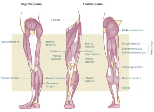
Figure 2.2 shows the muscles of the lower limbs. Gluteus maximus (extensor) and illiopsoas (flexor) are acting antagonistically across the hip. The quadriceps (vastus medialis, vastus intermedius, vastus lateralis, rectus femoris) are the main knee extensors including one bi-articular muscle (rectus femoris) that is also a hip flexor. Antagonistic to the quadriceps, the hamstrings act both as knee flexors and hip extensors. Gastrocnemius and plantaris are knee flexors as well. At the same time, these bi-articular muscles are also ankle plantar-flexors. Plantar-flexion is additionally supported by soleus which only spans the ankle joint. Antagonistic to the plantar-flexors, tibealis anterior is acting as dorsi-flexor.
2.1.2 Tasks, phases and periods of normal gait
Normal gait refers to the absence of pathological movement patterns that result in in- creased required energy expenditure. It consists of repetitive events and features that are optimized to ensure ambulation while conserving energy throughout the movement.
Due to the repetitiveness of the events and features, normal gait can be divided into distinct phases, and thus, it can be described as a repetitive cycle. Each gait cycle is defined as the interval between two successive instances observed on one leg at which the foot contacts the ground [1].
2.1. Basics of normal gait 7
Figure 2.2: Main muscles and muscle groups of the lower limbs.
The time shift between the cycles of the opposite legs is depicted in figure 2.3. After a foot contacts the ground there is always an interval of double support. During double support, both feet are on the ground to allow the weight shift from one leg to another.
One leg is leading meaning that the foot is placed anteriorly. The other leg is trailing with an posterior foot placement. The leading leg just finished swinging forward and the trailing leg finished single leg support. Single leg support allows the contralateral limb to swing forward while maintaining upright posture. Each cycle contains two intervals of single leg and double support while maintaining a constant time shift between the events at the same speed [1].
Figure 2.3 shows that one gait cycle equals one stride. One stride can be divided in different phases and periods by certain events. These events are:
• Initial contact
• Opposite toe off
• Heel rise
• Opposite initial contact
• Toe-off
• Feet adjacent
• Vertical tibia
One gait cycle consists of two phases, namely stance and swing phase. Initial contact and toe-off mark the transition between the two. Stance phase can be characterized by two main tasks: Weight acceptance and single limb support. The leg that started a new cycle and just touched the ground must bear the person’s weight without collapsing while maintaining static and dynamic balance. The main task of swing phase is limb advancement. Stance phase lasts about 60% of the cycle and swing phase for the remaining 40%.
The periods within stance phase are:
• Loading response
• Mid-stance
• Terminal stance
• Pre-swing
Swing phase is subdivided in:
• Initial swing
• Mid-swing
• Terminal swing
Each period can be described by the orchestration of muscles to ensure continuous and symmetric movements. The following two sections (2.1.3 and 2.1.4) describe the the periods and role of muscle groups in more detail.
Figure 2.3: Phases, periods and tasks of normal gait.
2.1. Basics of normal gait 9
2.1.3 Stance phase
Initial contact decelerates the body as the foot reaches the floor. This is achieved by simultaneous activity of the knee extensor and flexor muscles. The concurrent activation of antagonistic muscles stabilizes the knee and ensures that the subsequent loading of the leg does not collapse it. The hip extensors also contract to decelerate the thigh and to support knee extension in anticipation of the loading of the leg. The tibialis anterior begins a lengthening contraction (eccentric contraction) at the same time preventing the foot from slapping down on the floor. During loading response, the knee extensors contract and the leg accepts the body weight. The knee bends slightly and begins to extend under the shortening contraction (concentric contraction) of the knee extensors to soften the initial impact. This extension is supported by a lengthening contraction of the plantar-flexors. They move the contact point of the foot forward and shift the reaction force vector of the body anterior of the knee aiding with knee extension. At the same time, the gluteus medius contracts isometrically to stabilize the pelvis in the frontal plane. The center of gravity has reached its highest point in mid-stance. This potential energy is carried forward by momentum with little energy. While the knee remains extended with the foot on the floor the body weight falls forward. This trans- mission of energy is aligned with the direction of progression during normal gait. The lengthening contraction of the soleus muscle keeps the forefoot pressed against the floor.
The created force couple linkage supports knee extension without requiring muscle ac- tivation of the quadriceps. The knee stays passively extended in terminal stance. That is due to the force coupling created by knee extension and ankle plantar-flexion that continues to support the extended knee. Other than in the previous period, the ankle plantar-flexors begin a shortening contraction to accelerate the body forward generating a burst of energy. The created burst is responsible for most of the power generation during normal gait and propulses the body forward. In preparation of the unloading with the aim of swinging the leg forward, the hip flexors also become active. The opposite leg begins to accept weight in pre-swing which allows the leg to prepare for its own swing phase. The ankle plantar-flexors are no longer active and the hip flexors begin to lift the leg in preparation of swinging forward. This is achieved by shortening contractions of the hip flexors. Since the leg behaves predominantly as a passive pendulum in the subsequent swing phase, energy is only required for brief muscle activity of the hip flexors.
2.1.4 Swing phase
The passive pendulum characteristics affect the duration of swing and the length of the stride. There is a direct dependency on the length of the freely swinging leg and the dynamic friction of the knee joint and the associated tissue. Any prolongation of hip flexor activity or premature activity of the hamstrings changes the geometry of the swinging leg, and thus, affects swing duration and stride length. The hip flexors stop being active in initial swing as the leg is using the passive pendulum characteristics to swing forward. At the same time, the ankle dorsi-flexor begins a shorting contraction to allow the foot to clear the floor.
Figure 2.4: Muscle activation during normal gait (data from [1]).
The passive pendulum action of the leg and the dorsi-flexor activity for foot clearance continues during mid-swing until terminal swing. The leg begins to actively decelerate the swinging leg by contracting the hamstrings eccentrically or isometrically in terminal swing. This slows hip flexion and knee extension efficiently. At the same time, early quadriceps activity prepares the knee for weight acceptance. The tibialis anterior shows an isometric or lengthening contraction to ensure the foot is kept at a slightly dorsi- flexed position before touching the ground again. Detailed muscle activity patterns can be found in Figure 2.4.
2.2 Theories related to the efficiency of normal gait
This section introduces three theories related to the efficiency of normal gait. Insights from these theories are used in section 2.3 to summarize what mechanisms and features make walking an efficient task.
2.2.1 Six determinants of normal gait
It is hypothesized that the human body orchestrates motions of various segments and controls the activity of different muscles so that the metabolic energy required for a given task is minimized [3]. A theory first introduced by Saunders et al. described six determinants that make gait an efficient task [30, 31]. It is assumed that there are certain kinematic features that reduce the displacement of the body’s center of mass (COM) by transferring intersecting arcs to sinusoidal movements which in turn also reduces energy expenditure. This theory describes the determinants as
1. pelvic rotation,
2.2. Theories related to the efficiency of normal gait 11 2. pelvic obliquity (pelvic drop),
3. knee flexion during early stance, 4. ankle mechanism,
5. foot mechanism, and
6. lateral displacement of the body (hip abduction).
The determinants of gait have been generally accepted in the past without any empirical data or objective evaluation provided [32, 33, 34, 3]. However, more recent publications suggest that some of these kinematic features may not affect gait in the originally hy- pothesized way [35, 36, 37, 38, 39].
Pelvic rotation refers to to rotation of the pelvis around a vertical axis that brings the hip forwards during hip flexion and backwards during hip extension. The rotation reduces the required flexion and extension range at the hip for a given stride length. This reduction, in turn, results in a decreased vertical movement of the hip. Pelvic obliquity reduces the vertical trunk movement by tilting the pelvis about an anteroposterior axis.
When the hip of the stance phase leg is at its highest point, the pelvis drops down- wards on the contralateral side. This reduces the average height of the hip joint and decreases the vertical execution of the trunk. Knee flexion during early stance adjusts the effective leg length so that the hip joint does not rise as high if the knee would be kept extended. However, more recent studies suggest that the first three determinants do not reduce the vertical displacement during normal gait [35, 36, 38]. The second and third determinants are likely to support shock absorption that is caused by the discon- tinuities introduced by heel strike [37, 39]. The fourth and fifth determinants concern the foot and ankle complex and it is hypothesized that the related mechanisms create a nearly circular rocker [40]. At heel strike, the heel sticks out behind the ankle joint and lengthens the leg effectively. The forefoot lengthens it in a similar way at the end of stance phase. This behavior is also reflected in the center of pressure between the foot and the ground as it follows a circular trajectory. The kinematic features related to the foot and ankle complex are the only ones that appear to actually flatten the COM trajectories [3].
Even though the theory of the six determinants suggest that a minimization in ver- tical displacement is one of the main drivers to reduce energy expenditure, the human body does not seem to optimize the COM trajectory to approximate a flat line parallel to the direction of progression. It has been shown that energy expenditure increases by a factor of two or more when humans intentionally try to reduce vertical displacement of the COM while walking or when taking shorter steps [41, 42]. The human body exploits energy transfers from potential to kinetic energy. Vertical displacements are not necessarily inefficient and do not increase energy expenditure if the movement can be used for storing and returning mechanical energy. Nonetheless, this can only work suf- ficiently well if there are efficient transfers which could be enabled by certain kinematic features as described by the six determinants of gait. Even though the six determinants
are kinematic features that can be observed during normal gait, they are not necessarily the basic principles that make normal gait efficient.
2.2.2 Dynamic walking
Whereas the six determinants focus on certain kinematic features to reduce energy ex- penditure, the inverted pendulum model explains walking efficiency through energy con- versation. It suggests that it is energetically more beneficial for the leg to act like a pendulum carrying the trunk and adhere to the trajectory of an arc [43, 44]. There is a constant energy exchange between potential and kinetic energy requiring zero work for the COM to travel along the arc defined by the leg length. It is also assumed that the swing leg acts like a pendulum (non-inverted) when moving forward in line with the direction of progression [45]. Conserving energy by exchanging mechanical energy requires no work. Consequently, there is no additional force needed to cause movement.
This theory proposes an explanation why normal gait should not require any metabolic energy since the muscles don’t have to perform work. This, however, contradicts any- one’s experience that muscles have to contract during walking to cause a displacement of the COM. Even though the pendulum theory provides an analogy and indication how normal gait can be economical, the theory is incomplete.
McGeer developed an extension of the initial pendulum theory by including the heel- strike collision in the model. The theory is referred to as dynamic walking. It shows that the heel-strike collision can produce conditions for periodic gait without active control and minimal energy input [46]. Dynamic walking describes locomotion generated mainly by passive dynamics of the legs independent of any active powering or control. The original model was developed for two-legged machines that needed neither active control or energy input. Kuo et al. used this theory to explain the processes behind economical human gait and its clinical implications [47, 48].
This theory acknowledges that the single support leg behaves as an inverted pendu- lum to transport the COM with little energy. Although some metabolic energy is needed to activate the muscles ensuring that the leg is kept straight during the movement.
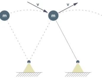
Dynamic walking considers step-to-step transitions in addition to the initial pendulum model. These transitions require energy because the COM velocity is redirected during double support phase. It is important to point out that the COM requires redirection and not propulsion or lifting. It can be best visualized when observing the COM velocity at the end of the arc when it is approximately perpendicular to the trailing leg. The associated motion is forward and downward. To continue moving the COM in the direc- tion of progression without falling, it requires redirection to the new arc spanned by to leading limp as shown in figure 2.5. This will result in an upward movement of the COM.
The redirection can be modeled as collision that dissipates energy. Since the leading leg is opposing the downwards path dictated by the trailing leg, the leading limb is per- forming negative work on the COM. Positive work is necessary to compensate for the dissipation. The work required can be performed by either ankle push-off or powering the hip [48]. Donelan et al. have shown that step-to-step transitions may require 60−70%
2.2. Theories related to the efficiency of normal gait 13
Figure 2.5: Redirection of the COM modeled as an inverted pendulum.
of overall metabolic energy for normal gait [49]. Grabowski et al. found a lower per- centage, i.e. 45%, for the metabolic cost of work performed on COM redirection . This is still a considerable portion especially when compared to the 28% of total metabolic cost that is required to generate the force to support the body weight [50].
A big part of of the remaining portion of required energy cost is believed to stem from forced leg motion. The dynamic walking model indicates that forced leg motion affects step-to-step transition cost significantly. Longer or wider steps increase step-to-step transition cost. This can be explained by increased COM velocity and greater directional change during redirection. The logical conclusion would be to use narrower, shorter and faster steps to mitigate the cost of step-to-step transitions. However, this is only a trade-off since work needs to be performed to move the leg relative the body. Even though shorter and faster steps reduce step-to-step transition cost, they increase the cost of moving the legs back and forth. There seems to be a trade-off between the two which also explains why humans increase step length and frequency proportionally [51].
Two of the six determinants of normal gait are also mentioned in the dynamic walking theory. The foot and ankle complex can reduce step-to-step transition cost when it is used to act like sections of a wheel. Dynamic walking models use arc-shaped feet to reduce the negative collision work [46]. During human normal gait, the feet seem to mimic this behavior as the center of pressure is progressing on the ground like a wheel.
This mechanism has been defined geometrically and is referred to as roll-over shapes (ROS) [40].
The dynamic walking model can also provide insights regarding stability during nor- mal gait. Walking is passively stabilized with no need for direct control over the leg motions. Small perturbations can be dissipated over subsequent steps due to the energy dissipation during heel-strike collision [48]. This highlights the importance of dynamics during normal gait. The stability of gait can be predominantly observed in the sagittal plane. Step length is relatively insensitive to anterior-posterior perturbations. The col- lision losses contribute to stability as well as the angle between the legs that increases
near the end of a step. Large perturbations, however, can change the step length and can cause substantially shorter steps that dissipate less energy. Insensitivity to lateral perturbations is less pronounced, and therefore, requires active feedback control. Kuo et al. suggested that medio-lateral stabilization can be achieved by [48]:
• moving the torso from side to side,
• producing active eversion/inversion moments at the ankle, or
• changing the foot placement actively by using hip abduction and adduction.
Since lateral stability requires active control, it also requires energy. Energy expenditure can be decreased if step width and its variability is reduced too [52]. The connection between step width variability and stability is in good agreement with the results of other research studies [53, 54, 55].
Dynamic walking demonstrates that the work performed on the COM for redirection, the work required to move the legs relative to the body and lateral stabilization are the main contributors to the cost of energy of normal gait. Metabolic energy is being used to perform work in order to make a system move. Also negative work requires a considerable amount of energy. Using such a significant amount of work and conserve energy at the same time is not necessarily a contradiction. It is possible for antagonistic muscles to perform an equal amount of positive and negative work on a pendulum. This would result in zero work being performed on that pendulum while the pendulum would be still able to conserve energy through the exchange of mechanical energy.
2.2.3 Energy transfers in a multi-segment system
The dynamic walking model demonstrated that work and energy flows are important to understand the causes of movements. Dynamic walking is mainly focusing on the COM and ignores energy transfers and storage between segments. This section will look more closely into energy transfers of a multi-segment system like the human body and the role of muscles.
A body segment can only change its energy if there is a flow of energy. There can be a change from potential energy (PE) to kinetic energy (KE) or vice versa, or, regarding mechanical energy, it means that work needs to be done. This is dictated by the law of conservation of energy that applies to all points in the body and at all instances in time.
The algebraic sum of all energy flows must equal the energy change of a segment. Even though work and energy have the same units, namely Joule (J), they are not the same.
Energy is a measure of the ability of a body to do work and work is a measure of an energy flow from one body to another. If work is done, there is the transfer of energy moving an object by a force and the object has covered a distance in a specific direction.
For example, a muscle can perform work on a segment if energy flows from the muscle to the segment. That means that the muscle provides a force to move the segment.
2.2. Theories related to the efficiency of normal gait 15 Joint power and work
Muscles are the main source of mechanical energy generation and absorption. The human body uses metabolic energy for the muscles to perform work and to produce force when there is no work performed. The total body energy increases when muscles do positive work. Consequently, the total body energy decreases when muscles perform negative work.
When considering the muscle power Pm in Watt, it can be calculated from the net muscle moment Mj in N m and the joint angular velocity ωj in rad/s.
Pm =Mjωj (2.1)
Therefore, concentric muscle contractions perform positive power if the muscle moment acts in the same direction as the angular velocity of the joint. If the muscle moment acts in the opposite direction to the angular velocity of the joint, negative power is performed.
Eccentric contractions usually happen if an external force is acting on a segment that creates a moment that exceeds the muscle moment.
The generated energy Wm transferred from a muscle to segments is reflected by the integral of the power over the time of the contraction. This shows the net work done by the muscles.
Wm =
∫ t2 t1
Pmdt (2.2)
The time interval needs to be chosen depending on the polarity ofPm to a calculate the total positive and negative work Wm. If done incorrectly, there could be a significant amount of work done that is not reflected in the total work calculated. When moving within a given time interval with an equal amount of positive and negative work, the net mechanical work is zero.
Calculating work from net joint moments includes any internal and external work that is done. Any external power is automatically reflected in increased joint moments.
Nonetheless, there is also a shortcoming. The net moment Mj results from all an- tagonist and agonist muscle activity, and thus, cannot account for co-contractions.
For example, if Mj = 50N m and ωj = 3rad/s, the joint power would result to 150W. In case of a co-contraction, an antagonist might provide a resisting moment of 20N m. To match the 150W in Joint power, the agonist would need to generate 150W + 3rad/s·20N m = 210W (Mj = 70N m) while the antagonist is absorbing energy at a rate of 3rad/s·20N m = 60W. This shows that the net power and work calculation underestimates positive and negative work done by the muscles groups at each joint. In order to overcome this problem the contribution to the net moment of each muscle would need to be known. Current measurement techniques do not allow for this task to be completely solved. For example, there are 15 major muscles responsible for the sagittal plane moments around the ankle, knee and hip joints. All three moments control the knee angle during stance phase because of the kinematic chain . This results in a indeterminacy when it comes to the relation of angle changes to moment patterns or combinations of muscle activity [2]. Another work component that is not accounted for are isometric contractions against gravity. These contractions require metabolic energy
but because there is no joint movement (ωj = 0rad/s), calculated power and work com- ponents result to zero. These effects must be accounted for in patients since their muscle activation patterns are disturbed and the occurrence of disregarded co-contractions could be significantly higher than in normal gait.
Energy flow and storage
Energy exists in three forms within body segments:
Es =P E+translational KE+rotational KE
=mgh+1
2mv2+ 1
2Iω2 (2.3)
A body or a segment can also exchange energy within itself and still maintain a constant total energy level. Body segments usually contain all three energies in different combi- nations any point in time during a movement. There can be energy exchanges within a segment or between adjacent segments. The pendulum conserving properties of the COM described before, can also be seen in the energy transfers of the head, arms and trunk (HAT) that has two peaks of potential energy at each stride. At these instances, the HAT shows the kinematic energy at its minimum. The body falls forward and the HAT accelerates while reducing its height, and thus, reducing its potential energy (figure 2.6).
Figure 2.6: Horizontal and vertical COM velocity (data from [2]).
The potential and kinetic energy approximate sinusoidal waves that are almost out of phase. According to Winter, the transfers of energy within body segments are character- ized by these opposite changes in potential and kinetic energy. To calculate the power balance within segments, all energy that enters or leaves the segment needs to be con- sidered. This includes active transfers by muscles including absorption and generation and passive transfers across joints at the proximal and distal ends. The passive transfer of mechanical energy between to adjacent segments is possible if there is a translational movement of the joints. Work is done if there is a force displacement through the prox- imal and distal joint center. The process is completely passive. This is reflected in the algebraic sum of all power flows that results to zero. Consequently, the total body energy remains constant. According to Winter, this mechanism plays an important role in the
![Figure 2.6: Horizontal and vertical COM velocity (data from [2]).](https://thumb-eu.123doks.com/thumbv2/1library_info/3908938.1525993/37.892.202.652.635.843/figure-horizontal-vertical-com-velocity-data.webp)
![Figure 3.1: Compensation movements during walking based on data provided by [1].](https://thumb-eu.123doks.com/thumbv2/1library_info/3908938.1525993/50.892.169.735.163.1052/figure-compensation-movements-walking-based-data-provided.webp)