Working Paper
Morphological study of embryonic Chd8+/- mouse brains using light-sheet microscopy
Author(s):
Gómez, Harold F.; Hodel, Leonie; Michos, Odyssé; Iber, Dagmar Publication Date:
2020-10-05 Permanent Link:
https://doi.org/10.3929/ethz-b-000445032
Originally published in:
bioRxiv , http://doi.org/10.1101/2020.10.05.326132
Rights / License:
Creative Commons Attribution-NonCommercial 4.0 International
This page was generated automatically upon download from the ETH Zurich Research Collection. For more information please consult the Terms of use.
ETH Library
Morphological study of embryonic Chd8
+/-mouse brains using
1
light-sheet microscopy
2
3
Authors:
4
Harold F. Gómez1,2#, Leonie Hodel1#, Odyssé Michos1,2, and Dagmar Iber1,2*, 5
6
Affiliations:
7
1 Department of Biosystems, Science and Engineering (D-BSSE), ETH Zurich, Mattenstraße 26, 4058 8
Basel, Switzerland 9
2 Swiss Institute of Bioinformatics (SIB), Mattenstraße 26, 4058 Basel, Switzerland 10
# These authors contributed equally.
11
* Corresponding authors.
12 13
Correspondences:
14
Harold F. Gómez: harold.gomez@bsse.ethz.ch 15
Leonie Hodel: leonie.hodel@gess.ethz.ch 16
Odyssé Michos: odysse.michos@gmail.com 17
Dagmar Iber: dagmar.iber@bsse.ethz.ch 18
Abstract
19
Objective 20
Autism spectrum disorder (ASD) encompasses a group of neurodevelopmental conditions that remain 21
poorly understood due to their genetic complexity. CHD8 is a risk allele strongly associated with ASD, and 22
heterozygous Chd8 loss-of-function mice have been reported to exhibit macrocephaly in early postnatal 23
stages. In this work, we sought to identify measurable brain alterations in early embryonic development.
24
Results 25
We performed light-sheet fluorescence microscopy imaging of N-cadherin stained and optically cleared 26
Chd8+/- and wild-type mouse brains at embryonic day 12.5 (E12.5). We report a detailed morphometric 27
characterization of embryonic brain shapes and cortical neuroepithelial apical architecture. While Chd8+/- 28
characteristic expansion of the forebrain and midbrain was not observed this early in embryogenesis, a 29
tendency for a decreased lateral ventricular sphericity and an increased intraocular distance in Chd8+/- brains 30
was found compared to controls. This study advocates the use of high-resolution microscopy technologies 31
and multi-scale morphometric analyses of target brain regions to explore the etiology and cellular basis of 32
Chd8 haploinsufficiency.
33 34
Keywords
35
Light-sheet microscopy, Chd8, Mouse brains, Autism, Tissue clearing, Morphometry, Lewis’ Law, Aboav- 36
Weaire’s Law.
37
38 39 40 41 42 43 44 45
Introduction
46
Autism spectrum disorder (ASD) is a poorly understood disease due to significant genetic complexity and 47
phenotypic heterogeneity. Despite our improved understanding of how ASD develops (1–5), mapping the 48
relative contribution of risk alleles to neuroanatomical abnormalities and clinically observed phenotypes like 49
macrocephaly remains challenging.
50
Genetic studies have implicated mutations in more than 800 genes of diverse function in the etiology of 51
ASD (1). One of the most strongly associated genes is the chromodomain helicase DNA-binding protein 8 52
CHD8, an ATP-dependent chromatin remodeler, and transcriptional repressor. Patients with loss-of- 53
function (LOF) mutations in CHD8 exhibit gene haploinsufficiency, display altered behavior, and region- 54
specific anomalies in brain morphology and physiology that manifest during early childhood (6–8). Similarly, 55
Chd8 haploinsufficiency in mice results in neonatal macrocephaly, increased brain weight, and craniofacial 56
abnormalities (9–11), mirroring clinical observations in patients and suggesting similar developmental 57
trajectories between species.
58
ASDs are largely hypothesized to originate in utero from profound perturbations in neural stem cell niche 59
regions of the developing brain (12). Gene expression profiling in the embryonic mouse cortex of Chd8 60
haploinsufficient mice shows a temporal modulation of Chd8 that peaks at E12, and helps negotiate the 61
complex balance between neuronal expansion (prior to E12.5) and differentiation (E12.5 to postnatal) (13).
62
As a result, dysregulations of Chd8 dynamics during embryonic cortical development in mice prematurely 63
deplete the neural progenitor pool, consistent with a lower density of neural cells and metabolic components 64
observed in children with ASD (14). In spite of novel neurodevelopmental evidence, it is unknown whether 65
these in utero perturbations manifest as distinctive anatomical dysmorphologies before the postnatal onset 66
of characteristic ASD phenotypes (15).
67
In this study, we investigated the morphological consequences of Chd8 haploinsufficiency during embryonic 68
mouse brains at the whole-organ and cellular level. To anticipate anatomical findings in a condition with 69
early-life onset, we leveraged N-cadherin staining, light-sheet fluorescence microscopy, and CUBIC tissue 70
clearing (16) to examine the neuroanatomical differences between E12.5 mouse brains with germline Chd8+/- 71
LOF mutations (10) and litter-matched wild-types. We report slight differences in intraocular distance and 72
ventricular sphericity and introduce a detailed approach for comparing cortical neuroepithelial apical 73
architecture. Taken together, these datasets provide a new avenue for querying the developmental role of 74
CHD8 and the cellular remodeling that is likely to precede associated post-birth brain malformations in 75
haploinsufficiency cases.
76 77
Methods and materials
78
Animals 79
Mice with loss-of-function mutations in Chd8 were generated using Cas9-mediated germline editing (10).
80
Immunofluorescence on embryonic brains 81
Following dissection, all E12.5 mouse brains were fixed with 4% paraformaldehyde in PBS. Samples were 82
then incubated with anti-N-cadherin antibody (BD Transduction Laboratories; Material No. 610920; 1:200) 83
at 4 °C for 3 days. After washing in D-PBS, brains were incubated with conjugated fluorescent secondary 84
Alexa Fluor 555 donkey anti-mouse IgG (H+L) (Abcam; Material No. ab150106; 1:250) for 2 days at 4 °C.
85
Optical clearing and light-sheet imaging 86
Whole-mount clearing was performed with the Clear Unobstructed Brain/Body Imaging Cocktails and 87
Computational Analysis (CUBIC) protocol (16). Delipidation and refractive index matching were carried 88
out with reagent-1 [25% (w/w) urea, 25% ethylenediamine, 15% (w/w) Triton X-100 in distilled water] and 89
reagent-2 [25% (w/w) urea, 50% (w/w) sucrose, 10% (w/w) nitrilotriethanol in distilled water], respectively.
90
Samples were incubated in 1/2 reagent-1 (CUBIC-1:H2O=1:1) for 1 day and then in 1X reagent-1 until 91
transparent. All samples were washed several times in PBS and treated with 1/2 reagent-2 (CUBIC- 92
2:PBS=1:1) for around 3 days. Lastly, incubation in 1X reagent-2 was done until transparency was achieved, 93
and the solution became homogeneous. All steps were performed on a shaker at room temperature.
94
Fluorescence images were acquired using a Zeiss Lightsheet Z.1 microscope. Acquisition optics included a 95
Zeiss 20x/1.0 Plan Apochromat water-immersion objective to acquire cell resolution data, and a Zeiss 96
5x/0.1 air objective lens for larger fields of view (whole brains). All image stacks were deconvolved using 97
Huygens deconvolution to improve contrast and resolution and further pre-processed in Fiji (17) to 98
accentuate feature boundaries.
99
3D surface reconstruction of whole mouse brains 100
3D segmentation of the embryonic ventricles and cerebral cortex was conducted with Imaris 101
MeasurementPro, a component of Imaris v9.1.2 (BitPlane, South Windsor, CT, USA). This enabled the 102
computational interpolation of planar 2D surface outlines from successive horizontal sections into 3D iso- 103
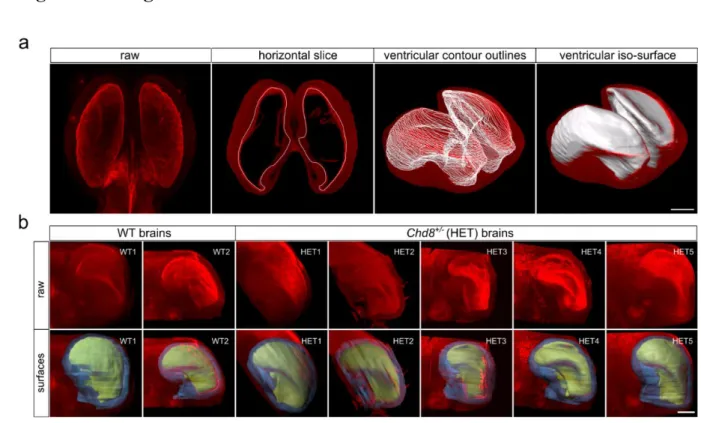
surface. Contours were drawn on magnified images to allow sub-voxel precision and faithful delineation of 104
small-scale features (Fig. 1). Quantified brain surface features included volume and surface area. Intraocular 105
distance was measured in 3D using measurement points placed in the center of the pupils.
106
As the cortex has broad irregular anatomical features that make absolute cortical thickness measurements 107
challenging, we considered the cortex to be a hollow cylinder with volume V and area A. In this way, the 108
cortical height of the neuroepithelial layer could be approximated as 109
ℎ𝑐𝑜𝑟𝑡𝑒𝑥 = 2𝑉𝑐𝑜𝑟𝑡𝑒𝑥 𝐴𝑜𝑢𝑡𝑒𝑟+ 𝐴𝑖𝑛𝑛𝑒𝑟 110
111
Furthermore, to derive ventricular sphericity, lateral ventricle iso-surfaces were separated at the septum 112
pellucidum. The sphericity of each 3D entity was then determined as 113
𝑆𝑣𝑒𝑛𝑡𝑟 =𝜋13(6𝑉𝑣𝑒𝑛𝑡𝑟)23 𝐴𝑣𝑒𝑛𝑡𝑟 114
115
Morphometric measurements of apical neuroepithelia 116
Cell morphology in the apical layer of the cortical epidermis was investigated using the open-source software 117
platform MorphoGraphX (18). A curved 2.5D image projection was constructed by meshing the apical 118
boundary and projecting 2-6 µm of the most apical signal onto it. Then, the Watershed algorithm was used 119
to segment all cell boundaries, some of which required manual curation. All border cells were excluded from 120
the analysis.
121
To characterize all projected polygonal apical lattices, quantifications on cellular areas and neighbour 122
numbers were imported into the R software platform. Apical packing was also explored via known 123
regularities of epithelial lattices. Termed Lewis’ Law, this property linearly relates the measured average cell 124
area 𝐴̅ and neighbour number n and has been previously described in all apical epithelia studied to date 125
(19,20).
126
𝐴̅𝑛
𝐴̅ =(𝑛 − 2) 127 4
As cells with small polygon numbers have the tendency to be in contact with cells of larger polygon numbers 128
and vice-versa, one also observes that the average number of neighbours of all n cells that border a cell with 129
n neighbours follows 130
𝑚(𝑛) = 5 −𝑛 131 8
a relationship termed Aboav-Weaire’s Law (21,22). Lastly, the cell aspect ratio was calculated using an in- 132
house algorithm that leverages MorphoGraphX’s modularity to fit an ellipse and extract major and minor 133
axes for each cell outline.
134 135
Results
136
ASD-associated craniofacial phenotypes in Chd8+/- mice 137
To determine whether Chd8 heterozygous mice exhibit structural and craniofacial ASD phenotypes during 138
embryonic development, we tested for differences in brain morphology between E12.5 Chd8+/- and control 139
animals. To this extent, user-assisted 3D segmentation software tools were used to derive iso-surface 140
representations from volumetric image stacks and enable tissue quantification measures of size, shape, and 141
asymmetry (Fig. 1).
142
We then characterized different anatomical features in both cortical and ventricular regions to reveal regional 143
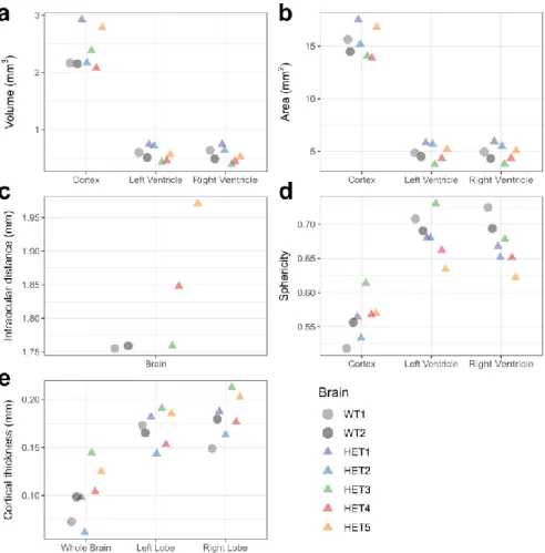
alterations (Fig. 2). Overall brain volume, including ventricles, showed no significant difference between the 144
two groups (wild-type [n=2] 3.41 mm3 and 3.15 mm3, Chd8+/- [n=5] 3.60 ± 0.56 mm3) (Fig. 2a). Similarly, 145
measured cortical volumes excluding the ventricular space showed no difference (wild-type [n=2] 2.17 mm3 146
and 2.15 mm3, Chd8+/- [n=5]: 2.47 ± 0.37 mm3). Individual ventricular volume was consistent within and 147
between groups (wild-type [n=4] 0.56 ± 0.07 mm3, Chd8+/- [n=10] 0.57 ± 0.14 mm3). Furthermore, we 148
observed no differences in brain surface area (wild-type [n=2] 15.6 mm2 and 14.5 mm2, Chd8+/- [n=5] 15.48 149
± 1.63 mm2) or in ventricular surface area (wild-type [n=4] 4.66 ± 0.30 mm2, Chd8+/- [n=10] 4.95 ± 0.83 150
mm2) between Chd8+/- and wild-type littermates (Fig. 2b).
151
CHD8 mutant patients often present craniofacial abnormalities (7). We report a slight increase in the 152
intraocular distance and variability within the Chd8+/- group (wild-type [n=2] 1.75 ± 0.002 mm, Chd8+/- 153
[n=3] 1.85 ± 0.10 mm) (Fig. 2c), which is consistent with mouse studies from similar genetic backgrounds 154
(10). Moreover, we observed a slight decrease in ventricular sphericity in Chd8+/- mice (wild-type [n=4] 0.70 155
± 0.01, Chd8+/- [n=10] 0.67 ± 0.03) (Fig. 2d). We also found a higher variability in whole-brain cortical 156
thickness in Chd8+/- mice (wild-type [n=2] 0.08 ± 0.02 mm, Chd8+/- [n=5] 0.10 ± 0.03 mm) (Fig. 2e). Thus, 157
only some craniofacial phenotypes were detected during early embryonic development.
158
Quantifying apical cell morphology in Chd8+/- mice 159
To identify cellular and tissue mechanics abnormalities preceding ASD-associated macrocephaly, we used 160
MorphoGraphX to isolate and mesh the apical boundaries of imaged epithelial cell patches in matching 161
regions of the cerebral cortex (18). By taking tissue curvature into account, we segmented a large number 162
of cell outlines from one Chd8+/- [n=3854] and one wild-type [n=1031] sample (Fig. 3a-b).
163
Apical organization was quantified by geometrical properties such as cell number of neighbours, areas, and 164
aspect ratio. We found similar hexagon and heptagon frequencies for the heterozygous (wild-type 29%
165
hexagons, 16% heptagons, Chd8+/- 31% hexagons, 20% heptagons) (Fig. 3c). The observed relation between 166
the polygon type of cells n and the average polygon type of their neighbours mn termed Aboav-Weaire’s Law 167
(21,22), recapitulated results in both the Drosophila wing disc and chicken neural tube epithelium (23) (Fig.
168
3d). Similarly, we compared area distributions per polygon type and found no significant difference between 169
groups. The average area per polygon type followed a linear dependency in both samples; a relationship 170
termed Lewis’ Law (19,20) (Fig. 3e). Moreover, considering local apical curvature, the aspect ratio was 171
determined by fitting an ellipse to each cell outline and extracting the major and minor axes. The aspect 172
ratio distribution of the Chd8+/- cells was minimally wider than the wild type (Fig. 3f).
173 174 175
Discussion
176
Chd8 haploinsufficient mice display various ASD-like phenotypes that parallel the clinical signature of 177
individuals with de-novo CHD8 mutations (10,11,13,24). Consistent with retrospective patient head 178
circumference data, mouse models for CHD8 haploinsufficiency suggest a postnatal onset of abnormal head 179
growth (10,11). In this study, we queried the neuroanatomy of Chd8+/- and litter-matched E12.5 control 180
mice using light-sheet microscopy to determine whether morphological anomalies in brain and cortical cell 181
shape could preindicate ASD-associated macrocephaly.
182
The results from a number of longitudinal studies of postnatal volumetric brain changes have implicated 183
neuroanatomical abnormalities in cortical thickness, ventricular morphology, cortical overgrowth, and 184
increased cortical surface area in the complex trajectory of brain development in individuals with ASD 185
(6,25–30). Accordingly, our work characterized cortical thickness along with volumetric features to confirm 186
whether analogous morphological alterations were observable in haploinsufficient mouse brains. We note 187
that in this particular instance, no significant discrepancies in cortical thickness, ventricular and cortical 188
volumes, or surface areas between groups could be determined. In line with the heterogeneous nature of 189
ASD, it is reasonable to assume that other brain regions may be affected instead. Notably, we provide 190
experimental evidence of dissimilarities in ventricular sphericity and intraocular distance that mirror known 191
phenotypes in haploinsufficient adult mice (Fig. 2) (10).
192
Similarly, a number of aberrations at the cellular scale have been reported in ASD during the establishment 193
of cortical microarchitecture (26). Consequently, we sought to ascertain differences in the cortical 194
organization between groups as defined by patterns of cell geometric features measured on the apical surface 195
(Fig. 3). Having quantified epithelial morphology according to the cell area, aspect ratio, neighbour topology, 196
and adherence to empirical laws such as Lewis’ and Aboav-Weaire’s (Fig. 3), our data showed no departure 197
in the cortical organization between haploinsufficient brains and controls, suggesting similar mechanical 198
behaviour (20,22).
199
In this work, we present a multi-scale assessment of the embryonic neuroanatomical implications of Chd8 200
haploinsufficiency in mice. We propose that an increased understanding of the identified organ-level 201
differences may shed light on the etiology of hypertrophic brain growth. What is more, our approach opens 202
exciting avenues to investigate the presence of cellular alterations in other implicated brain regions and 203
phenotypic differences across diverse Chd8 haploinsufficient mouse models, all of which have a wide range 204
of dosage-specific, dimorphic, and behavioural signatures (24,31).
205 206
Limitations
207
Underscoring the complexity of autism, our results did not show statistically significant differences in overall 208
morphology with the exception of slight deviations in ventricular sphericity and intraocular distance (Fig.
209
2). Furthermore, our study did not identify aberrations in cortical cellular architecture between groups (Fig.
210
3). We acknowledge that as only a small sample size could be studied (wild-type [n=2], and Chd8+/- [n=5]), 211
small morphological differences may have been missed due to the lack of statistical power.
212 213
Abbreviations
214
ASD: autism spectrum disorder 215
CHD8: chromodomain helicase DNA-binding protein 8 216
LOF: loss-of-function 217
CUBIC: clear unobstructed brain/body imaging cocktails and computational analysis 218
WT: wild type 219
HET: heterozygous 220
References
221
1. Abrahams BS, Geschwind DH. Advances in autism genetics: on the threshold of a new neurobiology.
222
Nature Reviews Genetics. 2008 May;9(5):341–55.
223
2. Iossifov I, Ronemus M, Levy D, Wang Z, Hakker I, Rosenbaum J, et al. De novo gene disruptions in 224
children on the autistic spectrum. Neuron. 2012 Apr 26;74(2):285–99.
225
3. O’Roak BJ, Deriziotis P, Lee C, Vives L, Schwartz JJ, Girirajan S, et al. Exome sequencing in sporadic 226
autism spectrum disorders identifies severe de novo mutations. Nature Genetics. 2011 Jun;43(6):585–9.
227
4. Parikshak NN, Luo R, Zhang A, Won H, Lowe JK, Chandran V, et al. Integrative Functional Genomic 228
Analyses Implicate Specific Molecular Pathways and Circuits in Autism. Cell. 2013 Nov 21;155(5):1008–21.
229
5. Sanders SJ, Murtha MT, Gupta AR, Murdoch JD, Raubeson MJ, Willsey AJ, et al. De novo mutations 230
revealed by whole-exome sequencing are strongly associated with autism. Nature. 2012 Apr 231
4;485(7397):237–41.
232
6. Courchesne E. Evidence of Brain Overgrowth in the First Year of Life in Autism. JAMA. 2003 Jul 233
16;290(3):337.
234
7. Bernier R, Golzio C, Xiong B, Stessman HA, Coe BP, Penn O, et al. Disruptive CHD8 Mutations Define 235
a Subtype of Autism Early in Development. Cell. 2014 Jul;158(2):263–76.
236
8. Xu Q, Liu Y, Wang X, Tan G, Li H, Hulbert SW, et al. Autism-associated CHD8 deficiency impairs axon 237
development and migration of cortical neurons. Molecular Autism. 2018 Dec 19;9(1):65.
238
9. Sugathan A, Biagioli M, Golzio C, Erdin S, Blumenthal I, Manavalan P, et al. CHD8 regulates 239
neurodevelopmental pathways associated with autism spectrum disorder in neural progenitors. Proceedings 240
of the National Academy of Sciences. 2014 Oct 21;111(42):E4468–77.
241
10. Platt RJ, Zhou Y, Slaymaker IM, Shetty AS, Weisbach NR, Kim J-A, et al. Chd8 Mutation Leads to 242
Autistic-like Behaviors and Impaired Striatal Circuits. Cell Reports. 2017 Apr 11;19(2):335–50.
243
11. Katayama Y, Nishiyama M, Shoji H, Ohkawa Y, Kawamura A, Sato T, et al. CHD8 haploinsufficiency 244
results in autistic-like phenotypes in mice. Nature. 2016 Sep;537(7622):675–9.
245
12. Corley MJ, Vargas-Maya N, Pang APS, Lum-Jones A, Li D, Khadka V, et al. Epigenetic Delay in the 246
Neurodevelopmental Trajectory of DNA Methylation States in Autism Spectrum Disorders. Front Genet.
247
2019 Oct 1;10:907.
248
13. Durak O, Gao F, Kaeser-Woo YJ, Rueda R, Martorell AJ, Nott A, et al. Chd8 mediates cortical 249
neurogenesis via transcriptional regulation of cell cycle and Wnt signaling. Nature Neuroscience. 2016 250
Nov;19(11):1477–88.
251
14. Dager SR, Friedman SD, Petropoulos H, Shaw DWW. Imaging Evidence for Pathological Brain 252
Development in Autism Spectrum Disorders. In: Autism [Internet]. Totowa, NJ: Humana Press; 2008 [cited 253
2019 Nov 6]. p. 361–79. Available from: http://link.springer.com/10.1007/978-1-60327-489-0_17 254
15. Fein D. The Neuropsychology of Autism. OUP USA; 2011. 559 p.
255
16. Susaki EA, Tainaka K, Perrin D, Yukinaga H, Kuno A, Ueda HR. Advanced CUBIC protocols for 256
whole-brain and whole-body clearing and imaging. Nat Protocols. 2015 Nov;10(11):1709–27.
257
17. Schindelin J, Arganda-Carreras I, Frise E, Kaynig V, Longair M, Pietzsch T, et al. Fiji: an open-source 258
platform for biological-image analysis. Nature Methods. 2012 Jun 28;9(7):676–82.
259
18. Barbier de Reuille P, Routier-Kierzkowska A-L, Kierzkowski D, Bassel GW, Schüpbach T, Tauriello G, 260
et al. MorphoGraphX: A platform for quantifying morphogenesis in 4D. eLife [Internet]. 2015 May 6 [cited 261
2017 Aug 25];4. Available from: http://elifesciences.org/lookup/doi/10.7554/eLife.05864 262
19. Lewis FT. The correlation between cell division and the shapes and sizes of prismatic cells in the 263
epidermis of cucumis. The Anatomical Record. 1928 May;38(3):341–76.
264
20. Kokic M, Iannini A, Villa-Fombuena G, Casares F, Iber D. Minimisation of surface energy drives apical 265
epithelial organisation and gives rise to Lewis’ law [Internet]. Biophysics; 2019 Mar [cited 2020 Feb 6].
266
Available from: http://biorxiv.org/lookup/doi/10.1101/590729 267
21. Aboav D. The arrangement of grains in a polycrystal. Metallography. 1970 Dec;3(4):383–90.
268
22. Vetter R, Kokic M, Gómez H, Hodel L, Gjeta B, Iannini A, et al. Aboave-Weaire’s law in epithelia results 269
from an angle constraint in contiguous polygonal lattices [Internet]. Biophysics; 2019 Mar [cited 2020 Feb 270
6]. Available from: http://biorxiv.org/lookup/doi/10.1101/591461 271
23. Sánchez‐Gutiérrez D, Tozluoglu M, Barry JD, Pascual A, Mao Y, Escudero LM. Fundamental physical 272
cellular constraints drive self‐organization of tissues. EMBO J. 2016 Jan 4;35(1):77–88.
273
24. Gompers AL, Su-Feher L, Ellegood J, Copping NA, Riyadh MA, Stradleigh TW, et al. Germline Chd8 274
haploinsufficiency alters brain development in mouse. Nature Neuroscience. 2017 Jun 26;20(8):1062–73.
275
25. Courchesne E, Pierce K, Schumann CM, Redcay E, Buckwalter JA, Kennedy DP, et al. Mapping Early 276
Brain Development in Autism. Neuron. 2007 Oct;56(2):399–413.
277
26. Donovan APA, Basson MA. The neuroanatomy of autism - a developmental perspective. J Anat. 2017 278
Jan;230(1):4–15.
279
27. Bigler ED, Tate DF, Neeley ES, Wolfson LJ, Miller MJ, Rice SA, et al. Temporal Lobe, Autism, and 280
Macrocephaly. American Journal of Neuroradiology. 2003 Nov 1;24(10):2066–76.
281
28. Herbert MR, Ziegler DA, Deutsch CK, O’Brien LM, Lange N, Bakardjiev A, et al. Dissociations of 282
cerebral cortex, subcortical and cerebral white matter volumes in autistic boys. Brain. 2003 May 283
1;126(5):1182–92.
284
29. Hazlett HC, Poe M, Gerig G, Smith RG, Provenzale J, Ross A, et al. Magnetic Resonance Imaging and 285
Head Circumference Study of Brain Size in Autism: Birth Through Age 2 Years. Archives of General 286
Psychiatry. 2005 Dec 1;62(12):1366.
287
30. Hazlett HC, Poe MD, Gerig G, Smith RG, Piven J. Cortical Gray and White Brain Tissue Volume in 288
Adolescents and Adults with Autism. Biological Psychiatry. 2006 Jan;59(1):1–6.
289
31. Jung H, Park H, Choi Y, Kang H, Lee E, Kweon H, et al. Sexually dimorphic behavior, neuronal activity, 290
and gene expression in Chd8-mutant mice. Nature Neuroscience. 2018 Sep;21(9):1218–28.
291 292
Declarations
293
Ethics approval and consent to participate 294
All animal experiments conducted in the USA followed Public Health Service (PHS) policy and guidelines 295
on humane care, and the use of laboratory animals was approved by the Massachusetts Institute of 296
Technology Committee for Animal Care (CAC).
297
Consent for publication 298
Not applicable.
299
Availability of data and materials 300
The datasets used and/or analyzed during the current study are available from the corresponding author on 301
reasonable request.
302
Competing interests 303
The authors declare that the research was conducted in the absence of any commercial or financial 304
relationships that could be construed as a potential conflict of interest.
305
Funding 306
This work has been supported through an SNF Sinergia grant to DI.
307
Authors’ contributions 308
The study was designed by DI. Staining was done by OM, clearing and imaging by HG. HG and LH 309
analyzed the data and wrote the manuscript. All authors read and approved the final version of the 310
manuscript.
311
Acknowledgments 312
We would like to thank Randall Platt and Ashwin S. Shetty for providing the embryos. Moreover, we 313
acknowledge Richard S. Smith for his expert advice on extending MorphoGraphX to enable aspect ratio 314
quantifications of segmented cell outlines.
315 316
Figures and Legends
317
318
Figure 1. Volumetric analysis of CUBIC-cleared wild-type and Chd8+/– samples, immunostained for N- 319
cadherin (red) to mark neuronal epithelial tissue. (a) Illustration of the processing steps in the creation of 320
manual surfaces. Sequential ventricular contours were drawn manually throughout the entire dataset to 321
extract 3D morphology (white outlines). Scale bar 500 µm. (b) Raw (top row) and overlays (bottom row) 322
of ventricular (yellow) and cortical (blue) iso-surfaces for each sample. Scale bar 400 µm.
323 324 325 326 327 328 329 330 331
332
Figure 2. Morphological characterization of E12.5 wild-type and Chd8+/- mouse brains. (a-c) Cortical and 333
ventricular (a) volume, (b) surface area, and (c) intraocular distance. (d) Sphericity of cortex and ventricles.
334
(e) Cortical thickness of whole brains and left and right lobes. Quantifications were extracted from 335
ventricular and cortical iso-surfaces.
336 337 338 339 340
341
Figure 3. Quantification of apical cell morphology in wild-type [n=1031] and Chd8+/- [n=3854] E12.5 342
mouse brains. (a, b) 2.5D segmentation overlay of the apical surface of (a) Chd8+/- (HET) and (b) wild- 343
type (WT) neurocortical epithelium. Scale bar: 20 µm. (c) Distribution of apical neighbour numbers per 344
sample. The average number of cell neighbours is 5.84 for WT and 5.80 for HET, which is close to the 345
topological requirement of 6. (d) Polygon type n times the mean polygon number of neighbours m of the 346
cell n follows a linear relationship termed Aboave-Weaire’s Law. (e) Average apical cell area by cell 347
neighbour number following a linear relationship termed Lewis’ Law (black line). (f) Cellular aspect ratios 348
between their longest and shortest axis.
349 350
![Figure 3. Quantification of apical cell morphology in wild-type [n=1031] and Chd8 +/- [n=3854] E12.5 342](https://thumb-eu.123doks.com/thumbv2/1library_info/5345347.1682245/17.892.221.664.96.712/figure-quantification-apical-cell-morphology-wild-type-chd.webp)
