Foot & Ankle International®
2016, Vol. 37(11) 1211 –1217
© The Author(s) 2016 Reprints and permissions:
sagepub.com/journalsPermissions.nav DOI: 10.1177/1071100716660824 fai.sagepub.com
Article
Introduction
Operative fracture treatment of irregularly shaped bones like the calcaneus, scapula, or scaphoid is demanding, starting with the need for high-quality imaging not only for classifi- cation of the fracture but also for planning of the procedure:
for example, which approach, how many fragments, where is the key fragment, and which implant to choose. Plain radiographs are not able to provide all this information.
Therefore, CT scans have become the criterion standard for the evaluation of complex fracture pathology.16-18
Calcaneal fractures are the most common fractures of the hindfoot, and CT scans have been emphasized for the evalua- tion of this fracture since the early 1990s.18,23 Moreover, clas- sification systems have been based on CT scans.18,24,26 However, none of these has reached general acceptance, as a result of low interrater reliability.9 A recent study confirmed these results with a large number of evaluators with different levels of experience.17 In this study, the proportion of intraob- server agreement was 82%. Cohen kappa was κ = 0.748, with P < .001. Overall interrater agreement was 61%. However,
only 42% of the evaluators were able to correctly classify the fracture according to Sanders. Sixty-nine percent of the evalu- ators agreed on the operative procedure necessary.
For this reason, the 3-dimensional (3D) reconstruction of CT scans was proposed to improve accuracy and precision of the evaluation of fracture pathology.25 With advancing technology in recent years, this has become available more readily. The large data sets obtained with spiral and multi- detector row CT have improved the potential for creating
1Department of Trauma and Reconstructive Surgery, University of Regensburg, Regensburg, Germany
2Department of Trauma and Reconstructive Surgery, Hospital Barmherzige Brüder Regensburg, Regensburg, Germany
3Department of Orthopaedic and Trauma Surgery, Robert-Bosch- Krankenhaus Stuttgart, Stuttgart, Germany
Corresponding Author:
Bernd Kinner, MD, PhD, Department of Orthopaedic and Trauma Surgery, Robert-Bosch-Krankenhaus Stuttgart, Auerbachstrasse 110, 70376 Stuttgart, Germany.
Email: bernd.kinner@rbk.de
Value of 3D Reconstructions of CT Scans for Calcaneal
Fracture Assessment
Christina Roll, MD, PhD
1, Johanna Schirmbeck, MD
1, Franz Müller, MD, PhD
2, Carsten Neumann, MD
1, and Bernd Kinner, MD, PhD
3Abstract
Background: The interpretation of CT scans for the evaluation of calcaneal fractures is difficult. Three-dimensional (3D) reconstruction (volume rendering technique [VRT]) has been valuable in the evaluation of irregularly shaped bones.
However, their value for the analysis of calcaneal fractures is still debated. Therefore, the objective of this study was to assess the effect of additional use of 3D CTs in calcaneal fractures.
Methods: In a prospective multicenter study, the CT data set of 5 different fractures was presented to 57 evaluators. First, the participating surgeons were asked to assess the fractures on the basis of axial, coronal, and sagittal reconstructions using a multiple-choice questionnaire. Second, 3D reconstructions (VRT) were presented. The CT scans were validated by the intraoperative findings and the results were compared to the model solution of 3 foot and ankle surgeons. Intra- and interrater reliabilities were calculated.
Results: The proportion of intraobserver agreement was 82%, with Cohen kappa of κ = 0.748 (P < .001). Interrater agreement varied between 0.772 (P = .006) for the assessment of concomitant fractures and 0.987 (P < .001) for the suggested approach. The evaluation of several items improved after presentation of the 3D CTs (Cochrane Q test, P < .001).
The benefit of 3D imaging was higher in inexperienced surgeons and complex fractures (Friedman test P < .001).
Conclusion: The evaluation of CT scans of calcaneal fractures was improved by the additional use of 3D images (VRT).
Level of Evidence: Level II, prospective comparative study.
Keywords: calcaneal fracture, CT scan, volume rendering technique, 3D reconstruction, reliability, validity
superior 3D images (3D CT). A variety of software pro- grams and rendering methods are available: “volume ren- dering,” “shaded surface rendering,” “maximum intensity projection,” and “multiplanar reformation.”
In “volume rendering,” the technique that we chose for this study, virtual light rays are emitted from a viewing point through the tissue of interest. These rays are attenu- ated according to their interaction with the tissues they encounter. To each tissue a color, brightness, and degree of opacity is assigned. The tissue of most interest is given the opacity 100%, the others only a fraction of it or even 0%.
This process is semiautomatic. Many parameters can be modified by the user to optimize the images.4,5 Finally, the virtual rays are cast from an infinite number of positions on a virtual sphere surrounding the CT data set. This last fea- ture enables the physician to look at the images from any desired perspective or to create a video, which shows the demonstrated organ or part of the body rotating in vertical and horizontal axes.4 Although this technique is used more and more in daily practice, only a few systematic evalua- tions of the benefits of this procedural method have been published. Thus, the objective of this study was to analyze the accuracy and precision of interpretation of CT scans of calcaneal fractures with additional 3D volume rendering.
Our specific research questions were as follows: (1) How is intra- and interobserver reliability affected using volume rendering technique (VRT)? (2) Can the accuracy (validity) of the identification of number of fragments, joints involved, dislocations present, osseous defects, and additional injuries be improved? (3) How does the level of experience affect the quality of interpretation and the pos- sible treatment decision proposed? Our hypothesis was that additional 3D reconstruction of the CT scans would be ben- eficial for the interpretation of calcaneal fractures.
Methods
After approval by the internal review board, the CT data set of 5 different patients/fractures (4 intraarticular and 1 extraarticular fractures; Table 1) was presented to a total of 57 evaluators. CT scans were obtained using spiral CT (Sensation 16; Siemens, Germany) with table-feed of 3 mm/s, slice collimation of 0.75 mm, pitch factor of 0.65, and slice thickness of 2 mm.
Axial, sagittal, and coronal reconstructions were obtained of each calcaneus. Axial views were recon- structed parallel to the sole of the foot, sagittal images relative to the ankle joint, and the coronal view parallel to the calcaneo-cuboid joint (CCJ). Three-dimensional reconstructions were obtained using a VRT as described above.
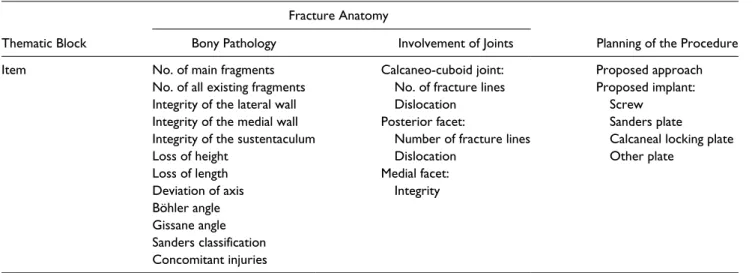
In a first run, the CT scans with the standard secondary reformations (axial, coronal, and sagittal) were presented to the evaluators in 4 different orthopedic trauma depart- ments (2 university hospitals, 2 nonacademic level 1 trauma centers) after a short introduction into the topic and the method of the study. The participating surgeons were asked to evaluate the calcaneal fractures on the basis of a multiple-choice questionnaire, which consisted of 25 items. The questionnaire was divided into 2 sections: frac- ture anatomy and preoperative planning. Table 2 summa- rizes the items. Time for evaluation was limited to 10 minutes. In a second run, immediately following the first one, the 3D reconstructions were presented as a video, where the calcaneus was rotating around 2 axes. A second questionnaire was completed. The evaluators had different experience in their field (Table 3). No foot and ankle spe- cialists were included.
All patients were treated by one experienced foot and ankle surgeon. The CT scans were validated by the intraop- erative findings, and discussed with 2 more experienced foot and ankle surgeons. A model answer for the question- naire was created. According to this model solution, the answers of the evaluators were assessed correct (1) or false (0). Additionally, the evaluators were asked to mark no answer if they did not understand the question, or if they were unsure about the answer.
Statistical Analysis
Statistical analysis was performed using SPSS 21.0.
Nonparametric statistical tests (Cochrane Q test) were used to determine a meaningful difference between the parame- ters and groups (2D vs. 3D). To assess intrarater reliability, symmetric cross-tables were constructed and Cohen kappa was calculated. Interrater reliability of the 57 raters was calculated using intraclass correlation coefficient (1-way random single measure).
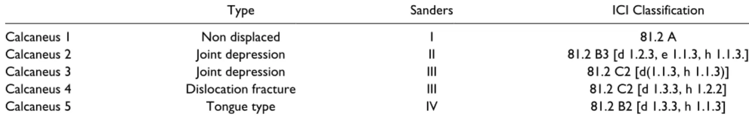
Table 1. Classification of the 5 Calcanei Presented, Provided by 3 Experienced Foot and Ankle Surgeons.a
Type Sanders ICI Classification
Calcaneus 1 Non displaced I 81.2 A
Calcaneus 2 Joint depression II 81.2 B3 [d 1.2.3, e 1.1.3, h 1.1.3.]
Calcaneus 3 Joint depression III 81.2 C2 [d(1.1.3, h 1.1.3)]
Calcaneus 4 Dislocation fracture III 81.2 C2 [d 1.3.3, h 1.2.2]
Calcaneus 5 Tongue type IV 81.2 B2 [d 1.3.3, h 1.1.3]
aClassification was validated by the intraoperative findings.
After the presentation of the 3D images, a 20% positive correction of the answers of the questionnaire was regarded as clinically relevant. Statistical significance was tested using a nonparametric 1-way analysis of variance (Friedman test). Statistical significance was assumed with P <.05.
Results
The participating evaluators were asked to subjectively judge image quality. Overall image quality of the presented CT scans was found to be good or excellent in 97%.
One item of the questionnaire asked whether the 3D pro- jection could help to more exactly analyze the fracture.
Fifty-seven percent of the inexperienced evaluators (≤6 years of experience) and 30% of the experienced evaluators (>6 years of experience) stated that the 3D reconstructions were helpful or very helpful for evaluation of the fracture (Spearman rank correlation coefficient ρ = −0.291, P < .01).
Fracture type had no significant influence on whether the 3D CT was regarded as helpful or not (P = .224).
Intra- and Interobserver Reliability
Three surgeons of one institution were asked to reevaluate 2 of the fractured calcanei (nos. 2 and 3) 9 months after the first evaluation to assess intraobserver reliability. The procedural
method was the same as before. Intraobserver reliability was calculated for items 1 to 13 (fracture pathology) of the ques- tionnaire. The proportion of agreement was 82% (Cohen kappa, κ = 0.748 with P < .001).
To assess interobserver reliability of the 57 raters, intra- class correlation coefficients for each item were calculated.
These were found to vary between 0.772 (P = .006) for the assessment of concomitant fractures and 0.987 (P < .001) for the suggested approach. No relevant difference was found between the ratings of 2D and 3D images.
Analysis of Fracture Pathology
Fracture pathology was analyzed by evaluation of the items 1 to 12. Table 4 shows the results after assessment of 2D and 3D CTs. Results are presented as percentage of raters with equal result compared with the model answer. For the items “number of fragments,” “fracture of the medial facet,”
“fracture line extending into the calcaneo-cuboid joint” and
“concomitant injuries” a significant “improvement” was found after 3D CT presentation (Cochrane Q test).
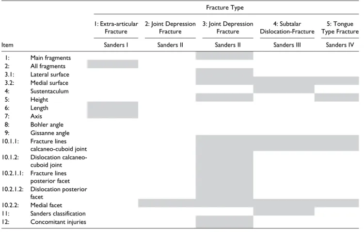
A correction of the ratings after presentation of 3D images of 20% or more was regarded clinically relevant. Table 5 shows the results after stratification per item of the question- naire. Figure 1 summarizes the results after stratification to fracture type (Figure 1A) and experience (Figure 1B).
Complex fractures demonstrated more benefit than fractures with lower complexity from 3D CT (Friedman test; P < .01).
All groups, except that of surgeons with more than 20 years of experience, benefited from 3D CTs (Friedman test; P < .01). Figure 2 shows a typical case.
Changes of Treatment Plan
The last 2 items of the questionnaire asked for the preferred approach and implants. After presentation of the 3D images, Table 2. Thematic Blocs and Items of the Questionnaire.
Fracture Anatomy
Planning of the Procedure
Thematic Block Bony Pathology Involvement of Joints
Item No. of main fragments Calcaneo-cuboid joint: Proposed approach
No. of all existing fragments No. of fracture lines Proposed implant:
Integrity of the lateral wall Dislocation Screw
Integrity of the medial wall Posterior facet: Sanders plate
Integrity of the sustentaculum Number of fracture lines Calcaneal locking plate
Loss of height Dislocation Other plate
Loss of length Medial facet:
Deviation of axis Integrity
Böhler angle Gissane angle Sanders classification Concomitant injuries
Table 3. Experience of the Evaluators.
Postgraduate Years n n (Surgeries/Year)
1-2 15 0 (0-1)
3-5 13 9 (0-1); 4 (2-5)
6-10 15 7 (0-1); 5 (2-5); 2 (6-10); 1 (>10) 11-20 9 1 (0-1); 2 (2-5); 3 (6-10); 3 (>10)
>20 5 1 (0-5); 1 (2-5); 2 (6-10); 1 (>10)
49% of the evaluators changed their plan in regard to the approach and 29% in regard to the implants.
Discussion
Analysis of fracture pathology of irregularly shaped bones is demanding, even when using CT scans with multiplanar reformation.17 Distinct information has to be extracted from the CT scans, and it affects our therapeutic decisions.
Recently we showed that interobserver agreement of the interpretation of 2-dimensional CT scans of calcaneal frac- tures is fair among not only inexperienced but also experi- enced surgeons. Therefore, our hypothesis was that additional 3D reconstruction of the CT scans would be ben- eficial for interpretation. Several studies have proposed that this might also be the case in calcaneal fractures. Böhmer et al2 described 3D CT as a useful instrument in evaluating calcaneal fractures and for preoperative planning because the topographic relationship of the fragments and the sur- rounding structures can be assessed more easily as the frac- ture can be seen from unusual perspectives. Likewise, Choplin et al4,5 proposed that 3D images are helpful in the diagnosis of all kinds of foot deformities because they improve the comprehension of the anatomy. Pretorius et al15 and Cotten et al6,15 recommended the use of 3D CT espe- cially for complex fractures. Allon and Mears1 compared plain radiography, 2D CT, and 3D CT of 30 fractured calca- nei and concluded that 3D CT improves preoperative
planning and the choice of an adequate approach. Pate et al13 evaluated 202 patients with musculoskeletal prob- lems and found that 3D CT was helpful for the analysis of fractures of bones with complex anatomy. Others came to similar results.12
However, recently the benefit of 3D CT was disputed.
Veltman et al22 concluded that the addition of 3D CT imag- ing does not increase inter- and intraobserver reliability for the classification of calcaneal fracture and should therefore not be a part of the routine workup of displaced intra-artic- ular calcaneal fractures. Others negated the beneficial effect of 3D CTs earlier.20,21 However, one must consider that at the time these studies were conducted, 3D reconstructions (volume rendering) was affected by artifact and strongly depended on the choice of threshold.
Intra- and interobserver reliability of classification sys- tems of calcaneal fracture has repeatedly been shown to be fair or low in 2D CTs.8,9,19 This is supported by our own studies.10,17 As expected, 3D imaging was not able to make a relevant difference in this respect in our study either. This is explained by the nature of the scoring systems; that is, Sanders classification is based on the coronal view of the 2D CT images. Three-dimensional CT cannot be helpful, espe- cially if the talus is not “exarticulated” virtually since the subtalar joint is not visualized, as in this and other studies.22
Yet the fate of 3D CT should not be reduced to inter- and intraobserver reliability of classification systems. Brunner et al3 concluded: “3D reconstructions may have other bene- fits not evaluated in the presented study and may give useful information not captured by current classification systems.”
Therefore, this study aimed to analyze the value of 3D reconstructions in a more holistic approach. Hence, we used a different methodological approach compared to other recent studies, namely, that of Veltmann et al.22 Besides ana- lyzing inter- or intrarater reliability, our main focus in this study was on the validity of 2D and 3D CTs. Therefore, the ratings obtained were matched to a model answer created by the treating surgeon who reevaluated the images in light of the intraoperative findings and after discussing his results with 2 additional foot and ankle surgeons. The model answer was created based on the consensus of the 3 foot and ankle surgeons. A second difference was the high number of evalu- ators. Prior investigations included 3 to 4 raters, whereas this study included 57 raters with various levels of experience in 4 different trauma departments.
One item asked for the subjective impression whether the 3D imaging helped when analyzing the fracture.
Predominantly inexperienced surgeons felt that they gained additional information with the 3D CT. Comparing the 2D and 3D results, we could demonstrate that the number of correct answers increased for most of the items, in some cases statistically significantly (Table 4). After stratification of the results to experience and fracture type, we found that the benefit of 3D CTs was higher for inexperienced surgeons Table 4. Analysis of Fracture Pathology.a
Item 2D, % 3D, % Pb
No. of main fragments 39 43 .262
No. of fragments 78 87 .018*
Lateral wall 56 57 .674
Medial wall 56 57 .674
Sustentaculum 65 69 .262
Height 61 67 .086
Length 55 56 .680
Axis 59 56 .492
Fracture line posterior facet 25 26 .886
Dislocation posterior facet 66 62 .199
Medial facet 42 52 .018*
Fracture line into calcaneo-cuboid
joint 31 47 <.001*
Dislocation of calcaneo-cuboid
joint 50 52 .475
Concomitant injuries 63 71 .002*
Gissane angle 71 68 .221
Bohler angle 47 49 .394
Sanders classification 27 32 .461
aPercentage of raters with equal result compared to the model answer.
bCochrane Q test.
*Statistically significant.
and complex fracture types. Similar to others, we found that some information was better extracted from conventional
image reformations than from the 3D images, especially concerning fracture classification. Therefore, in our daily Table 5. Changes of Rating After Presentations of the 3D Computed Tomographs.a
Item
Fracture Type 1: Extra-articular
Fracture 2: Joint Depression
Fracture 3: Joint Depression
Fracture 4: Subtalar
Dislocation-Fracture 5: Tongue Type Fracture
Sanders I Sanders II Sanders II Sanders III Sanders IV
1: Main fragments 2: All fragments 3.1: Lateral surface 3.2: Medial surface 4: Sustentaculum
5: Height
6: Length
7: Axis
8: Bohler angle 9: Gissanne angle 10.1.1: Fracture lines
calcaneo-cuboid joint 10.1.2: Dislocation calcaneo-
cuboid joint 10.2.1.1: Fracture lines
posterior facet 10.2.1.2: Dislocation posterior
facet 10.2.2: Medial facet 11: Sanders classification 12: Concomitant injuries
aItems with 20% or more corrected answers are shaded. Results are stratified to fracture type but not to evaluators’ experience.
Figure 1. Bar chart of the number of clinically relevant improved and declined items compared to the model answer. Clinically relevant was defined as an improvement or decline of 20% in each group. (A) Rating stratified to fracture type. Note the higher rate of improvements for complex fractures (calcaneus 4 and 5) (Friedman test, P < .01). (B) Rating stratified to experience of the raters (postgraduate years [PGYs]). All groups improved, except that of surgeons with more than 20 years of experience (Friedman test, P < .01).
practice we always use both 2D and 3D images to extract as much information from the CT scan as possible.22
Recent image-processing technology allows for easily subtracting adjacent bones or fragments overlapping the pathology of interest (Figure 2). Freud et al emphasized that 3D imaging without “exarticulation” is useless for the diag- nosis of calcaneal and talar fractures.7 According to our experience, exarticulation is not routinely necessary; how- ever, it may be helpful in special situations.
Prasartritha et al analyzed the diagnostic quality of 3D CT of 51 fractured calcanei. Important findings best seen on 3D CT images were the number and configuration of dis- placed posterior facet fragments, fracture lines separating the anterior process and the middle facet, and the extension of fracture lines into the calcaneo-cuboidal facet.14 These results compare well to those of this study.
One other point to be discussed is the possible costs associated with 3D CT. Almost all modern CTs provide the necessary software tools for volume rendering technology.
Therefore, no additional direct costs would be incurred.
Many hospitals also provide their users with special image- processing software, which enables to process the images in less than 10 minutes. Hence, the additional costs for 3D ren- dering are negligible. Moreover, in other fields, an overall cost reduction has been attributed to 3D volume rendering because of the superior information gained.11
Conclusion
The evaluation of CT scans of calcaneal fractures was improved by the additional use of 3D images (VRT).
Inexperienced surgeons benefited more than experienced surgeons and complex fractures more than simple fractures.
Specifically, regions of interest such as the middle facet and fractures extending into the calcaneo-cuboid joint were evaluated more precisely. However, the overall rate of “cor- rect” ratings was still low. Thus, 3D scans do not replace our teaching efforts emphasizing not only operative skills and novel implants but also the quality of interpretation of CT scans.
Declaration of Conflicting Interests
The author(s) declared no potential conflicts of interest with respect to the research, authorship, and/or publication of this article.
Funding
The author(s) received no financial support for the research, authorship, and/or publication of this article.
References
1. Allon SM, Mears DC. Three dimensional analysis of calca- neal fractures. Foot Ankle. 1991;11(5):254-263.
2. Bohmer G, Roesgen M, Hierholzer G. Three-dimensional computerized tomography in trauma surgery. A case presen- tation [in German]. Aktuelle Traumatol. 1992;22(2):47-56.
3. Brunner A, Heeren N, Albrecht F, et al. Effect of three-dimen- sional computed tomography reconstructions on reliability.
Foot Ankle Int. 2012;33(9):727-733.
4. Choplin RH, Buckwalter KA, Rydberg J, Farber JM. CT with 3D rendering of the tendons of the foot and ankle:
technique, normal anatomy, and disease. Radiographics.
2004;24(2):343-356.
Figure 2. Example of 21-year-old patient with calcaneal fracture. (A) Coronal, (B) sagittal, and (C) axial view of a CT scan. (D) Lateral and (E) medial aspect of the foot using VRT. (F) Lateral and (G) top view of the “exarticulated” calcaneus.
Note: Although the surface structures are more easily detected on the VRT images (eg, medial wall/middle facet) compared with the standard reconstructions, the huge central bone defect can only be appreciated on the coronal view (A). Additional information is gained when the calcaneus is
“virtually exarticulated.” However, this was beyond the aim of this study.
5. Choplin RH, Farber JM, Buckwalter KA, Swan S. Three- dimensional volume rendering of the tendons of the ankle and foot. Semin Musculoskelet Radiol. 2004;8(2):175-183.
6. Cotten AIS, Moise AB. Musculoskeletal System. Berlin, Heidelberg: Springer; 2002.
7. Freud M, Hohendorff B, Zenker W, Hutzelmann A, Heller M.
The CT of calcaneal fractures: 3-D reconstruction with elec- tronic disarticulation [in German]. Röfo. 1996;164(3):189- 195.
8. Howells NR, Hughes AW, Jackson M, Atkins RM, Livingstone JA. Interobserver and intraobserver reliability assessment of calcaneal fracture classification systems. J Foot Ankle Surg. 2014;53(1):47-51.
9. Humphrey CA, Dirschl DR, Ellis TJ. Interobserver reliabil- ity of a CT-based fracture classification system. J Orthop Trauma. 2005;19(9):616-622.
10. Kinner BJ, Best R, Falk K, Thon KP. Is there a reliable out- come measurement for displaced intra-articular calcaneal fractures? J Trauma. 2002;53(6):1094-1101; discussion 1102.
11. Lawler LP, Fishman EK. Multi-detector row CT of thoracic disease with emphasis on 3D volume rendering and CT angi- ography. Radiographics. 2001;21(5):1257-1273.
12. Pach M, Ditmar R, Herman M. Our experience with 3D CT.
Acta Univ Palacki Olomuc Fac Med. 1999;142:97-102.
13. Pate D, Resnick D, Andre M, Sartoris DJ, et al. Perspective:
three-dimensional imaging of the musculoskeletal system.
AJR Am J Röntgenol. 1986;147(3):545-551.
14. Prasartritha T, Sethavanitch C. Three-dimensional and two- dimensional computerized tomographic demonstration of cal- caneus fractures. Foot Ankle Int. 2004;25(4):262-273.
15. Pretorius ES, Fishman EK. Volume-rendered three-dimen- sional spiral CT: musculoskeletal applications. Radiographics.
1999;19(5):1143-1160.
16. Rammelt S, Zwipp H. Calcaneus fractures: facts, controver- sies and recent developments. Injury. 2004;35(5):443-461.
17. Roll C, Schirmbeck J, Schreyer A, et al. How reliable are CT scans for the evaluation of calcaneal fractures? Arch Orthop Trauma Surg. 2011;131(10):1397-1403.
18. Sanders R, Fortin P, DiPasquale T, Walling A. Operative treatment in 120 displaced intraarticular calcaneal fractures.
Results using a prognostic computed tomography scan clas- sification. Clin Orthop Relat Res. 1993;290:87-95.
19. Sayed-Noor AS, Agren PH, Wretenberg P. Interobserver reli- ability and intraobserver reproducibility of three radiological classification systems for intra-articular calcaneal fractures.
Foot Ankle Int. 2011;32(9):861-866.
20. Tanyu MO, Vinee P, Wimmer B. Value of 3D CT imag- ing in fractured os calcis. Comput Med Imaging Graph.
1994;18(3):137-143.
21. Vannier MW, Hildebolt CF, GiIula LA, et al. Calcaneal and pel- vic fractures: diagnostic evaluation by three-dimensional com- puted tomography scans. Digit Imaging. 1991;4(3):143-152.
22. Veltman ES, van den Bekerom MP, Doornberg JN, et al.
Three-dimensional computed tomography is not indicated for the classification and characterization of calcaneal fractures.
Injury. 2014;45(7):1117-1120.
23. Zwipp H. Reconstructive surgery of malunited joint fractures of the foot [in German]. Orthopade. 1990;19(6):409-415.
24. Zwipp H, Baumgart F, Cronier P, et al. Integral classification of injuries (ICI) to the bones, joints, and ligaments—applica- tion to injuries of the foot. Injury. 2004;35(suppl 2):SB3-SB9.
25. Zwipp H, Tscherne H, Thermann H, Weber T. Osteosynthesis of displaced intraarticular fractures of the calcaneus. Results in 123 cases. Clin Orthop Relat Res. 1993;290:76-86.
26. Zwipp H, Tscherne H, Wulker N, Grote R. Intra-articular frac- ture of the calcaneus. Classification, assessment and surgical procedures [in German]. Unfallchirurg. 1989;92(3):117-129.