Submitted to the
Combined Faculties for the Natural Sciences and for Mathematics of the Ruperto-Carola University of Heidelberg, Germany for the degree
of Doctor of Natural Sciences
presented by
Ramón Leonardo Serrano, M.S.
Tovar, Mérida, Venezuela
Oral examination: 9
thJuly, 2003
Structural and Functional Characterization of GAPR-1, a Mammalian Plant Pathogenesis-related Protein in Lipid-
enriched Microdomains of the Golgi Complex
Referees: Prof. Dr. Felix T. Wieland
Prof. Dr. Wilhelm Just
Abstract 1 Introduction
1 MICRODOMAINS IN BIOLOGICAL MEMBRANES
2 1.1 Lipid microdomains and signal transduction 5
1.1.1 Lipid microdomains and their potential role in immune cell activation
6 1.2 Membrane domains in the secretory pathway 7 1.3 Golgi apparatus as a signaling platform 10
2 PLANT PATHOGENS AND INTEGRATED DEFENCERESPONSES TO INFECTION
13
2.1 Systemic acquired resistance (SAR) 15 2.2 Mammalian PR-1 family members 17
3 PURPOSE OF THIS THESIS
17
Results
1 MEMBRANE ASSOCIATION OF GAPR-1
19 1.1 N-myristoylation of GAPR-1 in Escherichia coli 19 1.2 N-myristoylation of GAPR-1 in vivo 21 1.3 Association of GAPR-1 with Golgi membranes 23 1.4 GAPR-1 interaction with Caveolin-1 24 1.5 Phosphorylation of GAPR-1 in vivo. 26 1.6 Effect of phosphorylation on the partitioning of GAPR-1
to lipid-enriched microdomains. 27
2 STRUCTURAL CHARACTERISTICS OF GAPR-129 2.1 Large Scale Purification of GAPR-1 29 2.2 Crystal structure of GAPR-1 31 2.3 Role of conserved amino-acids in GAPR-1 34 2.4 Identification of proteins that bind to the GAPR-1 affinity column 36
2.4.1 Identification of proteins in CHO cytosol that bind GAPR-1
36
2.4.2 Identification of proteins present in complex pull down by
2.4.4 GAPR-1 interacting partner in vitro
42
3 STRUCTURAL CHARACTERIZATION OF GAPR-144 3.1 Characterization of Recombinant GAPR-1wt and GAPR-1mut 44 3.2 GAPR-1/GAPR-1 interaction in vivo 44 3.3 Circular Dichroism analysis of GAPR-1 46
4 INTERACTION TRAP OR TWO HYBRID SYSTEM49 4.1 Nucleolin- GAPR-1 interaction. 49
5 REGULATION OF GAPR-1 EXPRESSION52 5.1 Effect of serum starvation on GAPR-1 expression in CHO cells 52 5.2 Localization of Nucleolin to Golgi membranes 52
Discussion
1 BINDING OF GAPR-1 TO GOLGI MEMBRANES
55 1.1 GAPR-1 binding to membranes 55 1.2 A possible role of phosphorylation in membrane partitioning of GAPR-1? 57 1.3 Alternative roles of phosphorylation of GAPR-1 58
2 STRUCTURE FUNCTION-RELATIONSHIP OF GAPR-160 2.1 GAPR-1and the superfamily of the plant pathogenesis-related proteins 60 2.2 Effect of mutations on GAPR-1 structure 63
3 INTERACTION OF GAPR-1 WITH CYTOSOLIC PROTEINS 66 4 PERSPECTIVES OF GAPR-1 FUNCTION: RAFTS AND NUCLEOLIN 70Material and Methods Materials
1 CHEMICALS
73
1.1 Detergents 73
1.2 Inhibitors 73
1.3 Buffers 73
2.2 Secondary Antibodies 76
3 PLASMIDS
76
4 OLIGONUCLEOTIDES
77
5 EQUIPMENTS
Methods 78
6 METHODS IN CELL BIOLOGY AND IMMUNOLOGY 78
6.1 Cell culture 78
6.1.1 Passing of cells
78
6.1.2 Culture of CHO and HeLa cell in suspension
79
6.1.3 Transfection of cells
79
6.1.4 Isolation of Primary Hepatocytes
79
6.2 Synchronization of mammalian cells 80
6.2.1 Synchronization of mammalian cells by serum starvation
80
6.2.2 Synchornization of mammalian cells by drugs
81
6.2.2.1 Propidium Iodide Staining and Flow Cytometry
81
6.3 Immunofluorescence microscopy 81 6.4 Phosphorylation of GAPR-1 in vivo 82 6.5 Immunoprecipitation 83
7 METHODS IN MOLECULAR BIOLOGY
84
7.1 Polymerase Chain Reaction (PCR) 84
7.1.1 Polymerase chain reaction (PCR) for site-directed mutagenesis
84
7.1.2 Polymerase chain reaction (PCR) for Two Hybrid System
84
7.1.3 Subcloning
85
7.1.4 Lithium acetate transformation of EGY48-pSH18-34
86
8 METHODS IN BIOCHEMISTRY
87
8.1 Isolation of Golgi membranes 87
8.1.1 Golgi membranes from CHO cells
87
9 CYTOSOL PREPARATION FROM MAMMALIAN CELLS
90 9.1 Cytosol preparation from CHO and Hela Cells 90 9.2 Cytosol preparation from Rat liver 90 9.3 Cytosol fractionation 91 9.4 Cytosolic Protein complex denaturation 91
10 LARGE SCALE PURIFICATION OF GAPR-192 10.1 Size exclusion chromatography light scattering (SEC-LS) 93 10.2 Crystal structure determination 93
10.2.1 Data Collection
93
10.3 Circular Dichroism of GAPR-1 94 10.4 Coupling of GAPR-1 to CNBr-activated Sepharose 4B 94
11 GAPR-1 AFFINITY CHROMATOGRAPHY95
12 GAPR-1 LIGAND OVERLAY
95
13
SDS-PAGE AND WESTERN BLOT ANALYSIS96 13.1 SDS-PAGE for separation of proteins 96 13.2 Transfer proteins from SDS-PAGE to a PVDF membrane
or Nitrocellulose 96
13.3 Incubation of PVDF membranes with antibodies 96
14 PROTEIN DETERMINATION
97
14.1 Protein Determination by BCA 97 14.2 Protein determination by Lowry 98
15 PROTEIN PRECIPITATION
98
15.1 Chloroform-Methanol Precipitation 98 15.2 TCA precipitation 99
References 100
Acknowledgements 116
AA(aa) Amino acid
ARF ADP-ribosylation factor ATP Adenosine tri-phosphate BCA Bicinchonic acid
BFA Brefeldin A bp base-pair
BSA Bovine serum albumin
CHAPS 3-[(3-cholamidopropyl1)dimethylammonio]-1-propanesulfonate CHO Chinese hamster ovary
CNBr Cyanogen bromide COP Coat-protein DMSO Dimethyl sulfoxide
DRM Detergent-resistant membranes DTT Dithiothretiol
EDTA Ethylendiaminetetraacetic acid ER Endoplasmic reticulum
GDP Guanosine di-phosphate GPI Glycosylphosphatidylinositiol GTP Guanosine triphospahte hr hour
IF Immunofluorescence IP Immunoprecipitation kDa Kilo-Dalton
min Minute mut Mutant
MOPS Morpolinepropanesulfonic acid sodium salt NP-40 Nonidet® P40 (Nonylphenylpolyethylene glycol) NRK Normal rat kidney
nt Nucleotides
PBS-T Phosphate buffer saline + Tween 20
PIPES Piperazine-1,4-bis(2-ethanesulfonic acid) PMSF Phenylmethulsulfonyl fluoride
PR Plant Pathogenesis-related rpm Revolutions per minute
sec Second
SDS-PAGE Sodium dodecyl sulfate-polyacrylamide gel electrophoresis
SNARE Soluble N-ethylmaleimide sensitive factor attachment protein receptor TCA Trichloroacetic acid
TEMED N, N, N’, N’-Tetramethylethylenediamine v-ATPase vacuolar ATPase
wt wild type
During the characterization of lipid-enriched microdomains at the Golgi (GICs) (Gkantiragas, I. et al. 2001), a protein with an apparent molecular mass of 17 kDa was identified. Cloning and preliminary biochemical characterization identified a novel protein, GAPR-1, belonging to the superfamily of PR proteins.
Based on the primary amino acidic sequence of this protein, some potentially interesting characteristics were identified. It contains a consensus sequence for myristoylation, a putative caveolin-binding domain, a coiled-coil structure, and an isolelectric point (pI) of 9.4, suggesting that GAPR-1 is a highly hydrophilic protein (Eberle, H. B. et al. 2002).
In this thesis, this structural information, was used to i) study the interaction of GAPR-1 with membranes, ii) to obtain structural information on the protein, and iii) to identify proteins that interact with GAPR-1.GAPR-1 was shown to be myristoylated and to interact with Caveolin-1. Myristoylation, together with protein-protein or electrostatic interactions at physiological pH could explain its strong membrane association. The crystal structure of GAPR-1 showed strong structural similarities to other plant pathogenesis-related proteins. Substitution of the most conserved amino acids in GAPR-1 (His54, Glu65, Glu86 and His103) in the putative active center changed the protein behavior in solution. Size exclusion chromatography revealed that the major population of GAPR-1 mutant migrated as a dimer, whereas GAPR-1 wild type behaves predominantly as a monomer. The tendency of GAPR-1 to form dimers was confirmed by crosslink experiments and by the yeast two hybrid system. By affinity chromatography, GAPR-1 was shown to interact with three proteins: Nucleolin, Template activating factor α (TAFIα) and HSAPRIL. In the yeast two hybrid system, the interaction of GARP-1 with Nucleolin was confirmed and shown to be dependent on the most conserved amino acid residues in GAPR-1. The interaction between GAPR-1 and Nucleolin may represent a new mechanism of regulation of innate immunity in mammalian cells.
Introduction
Cell membranes are dynamic and fluid structures and their molecules are able to move in the plane of the membrane. A membrane provides a two- dimensional fluid support for proteins as well as a hydrophobic barrier to separate compartments. It is believed that a cell or plasma membrane similar to those of today's cells defined the boundary of the first cell nearly 4 billion years ago. Since then, cells have evolved in such a way that the plasma membrane and intracellular membranes now perform many functions: as a barrier to keep the contents of the cell together, allowing nutrients to pass in but keeping out many harmful substances; as a signaling platform to relay information about the surroundings of the cell to the inside and vice versa; as a scaffold to provide places where enzymes can be arranged in an assembly- line fashion; and as a compartmentalizing structure to separate different parts of the cell with different functions.
1 Microdomains in biological membranes
Progress in identifying and characterizing the constituents of membrane bound compartments has revealed a distinct level of cellular and sub-cellular compartmentation. Proteins and lipids are not uniformly distributed in the membrane of a given organelle as domains are formed by a combination of hierarchical assembly processes and protein and lipid segregation. This implies that membranes should not be considered as a random ocean of lipids (Singer, S. J. Nicolson, G. L. 1972), but rather the existence of domain structures in the bilayer is acknowledged that impose an organization on the distribution of proteins. One of the important features of these domains is that the composition and physical properties differ from the overall properties of the membrane (Brown, D. 2002). Lipid-based structures within the membranes have been designated as lipid microdomains or lipid rafts. These heterogeneous structures in membranes were postulated by Simons and van Meer (1988). The first experimental evidence for the existence of lipid- enriched microdomains was obtained by the finding that in non-ionic detergents (i.e. Triton X-100) in the cold, certain lipids such as cholesterol and sphingolipids are detergent-insoluble. In addition, due to the enrichment of
lipids, these detergent-insoluble complexes have a low density as observed by flotation experiments in 5-30% linear sucrose gradients (Brown, D. A.
Rose, J. K. 1992). Thus, the term raft refers to a domain in intact membranes, whereas the term detergent-resistant membrane (DRM) refers to the structure isolated by detergent insolubility. Due to their presence in the DRMs, many proteins are believed to be associated with lipid rafts, e.g. GPI-anchored proteins, transmembrane proteins, and dual acylated proteins such as tyrosine kinases (Src family) (reviewed in Simons, K. Ikonen, E. 1997). A wide variety of detergents other than Triton X-100 have been used to isolate low density detergent-insoluble membrane fractions, such as NP40, octylglucoside, CHAPs, lubrol and Brij96 (Ilangumaran, S. et al. 1999; Roper, K. et al. 2000; Bagnat, M. Simons, K. 2002; Drevot, P. et al. 2002). Detergent- free preparations of lipid microdomains and microdomain preparations in the presence of low TX-100 concentrations have also been reported (Song, K. S.
et al. 1996).
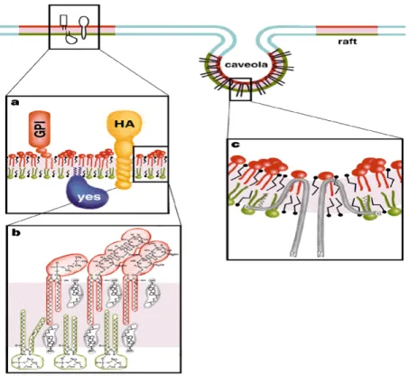
Lipid rafts or lipid-enriched microdomains can be defined as subdomains of the plasma membrane, containing high concentrations of cholesterol and sphingolipids (sphingomyelin and glycosphingolipids). In these domains, cholesterol condenses the packing of sphingolipid molecules by occupying the spaces between the saturated chains. Thus, a separate liquid-ordered phase (lo) is formed, which is dispersed in the liquid disordered phase (lc), the latter representing a freely packed fluid matrix of the membrane (Fig. 1). How the exoplasmic arrangement of sphingolipids and cholesterol is linked to the underlying cytoplasmic leaflet is currently not known. Lipid rafts incorporate distinct classes of proteins (Brown, D. A. London, E. 1998), such as glycosylphosphatidylinositol (GPI)-anchored proteins, dual acylated peripheral membrane proteins, cholesterol-linked proteins (Caveolin), and selected transmembrane proteins (Fig.1). Little is known about the targeting of proteins to lipid-enriched microdomains. A number of these proteins containing a combination of covalently attached fatty acids (myristate and palmitate) at their N-termini (Galbiati, F. et al. 1999b; Melkonian, K. et al. 1999; van’t Hof, W. Resh, M. 1997). This suggests that two saturated acyl chain can cause partitioning of proteins into lipid microdomains. Other reports show that a single prenyl group or myristate group alone can be sufficient to target a
protein to lipid microdomains (Song, K. S. et al. 1996). In these cases, N- terminal acylation, coupled with protein-protein interactions or protein-lipid interactions, can cause partitioning of a protein to DRMs (McCabe, J. B.
Berthiaume, L. G. 2001).
While there is abundant evidence that such microdomains exist and that they perform important functions, it has proven very difficult to obtain experimental evidence for their existence in vivo, including a description of their properties in terms of size, composition and dynamics. As mentioned above, the existence of different types of membrane (micro)domains adds to the difficulty in understanding their properties. For instance, Caveolae are one type of microdomain at the plasma membrane (Fig. 1). They are small (50-70nm in diameter) flask-shaped invaginations with an abundant membrane protein, Caveolin, associated with their structures (Kurzchalia, T. V. Parton, R. G.
1999). Caveolae can also be flat within the plane of the membrane, or be present as vesicles. These structures are cholesterol-rich, and when cells are treated in the cold with non-ionic detergents (i.e. Triton X-100), caveolae resist detergent-solubilization and can be recovered in low-density fractions on density gradients. Caveolae are enriched in molecules that play crucial roles in intracellular signal transduction. These molecules include the heterotrimeric G proteins, receptor tyrosine kinases, components of the MAP kinases pathway, and nitric oxide synthase (reviewed in Smart, E. et al.
1999). As a consequence, caveolae function as preassembled signaling complexes or chemical relays for integrating signal transduction. Interestingly, caveolae are only a minor fraction within DRMs (Kurzchalia, T. V. Parton, R.
G. 1999) and the existence of large amounts of non-caveolar (glycosphingolipids(GSL)-enriched) domains in the plane of the membrane (Iwabuchi, K. et al. 1998) adds to the complexity of lateral organizations in the plasma membrane for complex activities such as signal transduction (Anderson, R. 1998) (Vincent, J. 2003).
Figure 1. Model of a lipid-enriched domain at the plasma membrane. (a). A GPI- anchored protein is attached to the exoplasmic leaflet, and a doubly acylated Src-kinase to the cytoplasmic leaflet or a transmembrane protein (HA). Lipids in the raft are shown as red and green, and the lipids in the liquid disordered phase as blue. (b) The lipid bilayer in rafts is asymmetric with sphingomyelin (red) and glycosphiongolipid (red) enriched in the exoplasmic leaflet and phospholipids (green) e.g phophatidylserine and phosphatidylethanolamine.
Cholesterol (grey) is present in both faces of the membrane filling the space under the head groups of sphingolipids (c) Caveolae formed by caveolin molecules making a hairpin lop in the membrane, and the interaction with rafts may be mediated by binding of cholesterol and acylation of C-terminal cysteines. (Picture taken from Simons, K. and Ikonen, E. 1997).
1.1 Lipid microdomains and signal transduction
Experimental evidence suggests that there are several different mechanisms through which lipid microdomains may control cell signaling. Lipid rafts may contain complete signaling pathways that are activated when a receptor or other required molecule is recruited into the raft. In this view, rafts serve to co- localize the prerequisite components, facilitating their interaction and supporting signaling. Thus, receptors, coupling factors, effector enzymes and substrates would all be co-localized in a single microdomain, and specificity of signaling could be enhanced by restricting receptor localization to a particular class of microdomains that contains a specific subset of signaling components (Cary, L. Cooper, J. 2000). This restriction would limit access of the receptor to components of other signaling pathways and prevent non-specific signaling
(Langlet, C. et al. 2000 (Oh, P. Schnitzer, J. E. 2001;Prior, I. A. et al. 2001). In this model, microdomains could also contain a nearly complete signaling pathway that would be activated when a receptor or other require molecule, that is normally localized in the non-raft portion of the membranes, is recruited to the rafts (Roy, S. et al. 1999). As an alternative how rafts may control cell signaling, rafts could also limit signaling, either by physical sequestration of signaling components to block non-specific interactions or by suppressing the intrinsic activity of signaling proteins present within rafts (Mueller, G. Frick, W.
1999; Mettouchi, A. et al. 2001). In these scenarios, microdomains may provide regulation via compartmentalization of proteins that could otherwise interact, leading to unregulated activation of a pathway. Many receptor tyrosine kinases including the EGF receptor, the PDGF receptor, and the insulin receptor have been reported to control cell signaling by modulating their intrinsic activities due to lipid microdomain localization (Mineo, C. et al.
1996, Anderson, G. 1998). The involvement of lipid rafts in the function of proteins has been studied by different approaches such as depleting cells of cholesterol. Lipid rafts are held together via interactions between cholesterol and sphingolipids and the integrity can be disrupted by treatment with methyl- β-cyclodextrin that removes cholesterol (Kilsdonk, E. et al. 1995; Pike, L.
Miller, J. 1998). Cholesterol depletion e.g. impairs the ability of receptor tyrosine kinase to signal, diminishes insulin-stimulated phosphorylation of its receptor, or affects insulin-stimulated glucose uptake and oxidation (Vainio, S.
et al. 2002; Parpal, S. et al. 2001).
1.1.1 Lipid microdomains and their potential role in immune cell activation
One of the best described examples on the involvement of lipid rafts in cellular signaling processes are T cells. Cells of both the innate and adaptive immune systems express a variety of receptors that allow them to respond to the presence of foreign macromolecules in a highly discriminating and sensitive fashion. Multichain immune recognition receptors (MIRRs) in lymphocytes, for instance, are surface receptors formed by the association of immunoglobulin- like subunits (recognition subunits) and transducing subunits. The TCR, BCR and FcεRI are among the best studied MIRRS in terms of signal transduction
phosphorylation by the Src family protein tyrosine kinases such as LcK, Fyn or Lyn. Membranes lipid microdomains are enriched in Src proteins (Resh, M.
D. 1999) and upon activation of Lyn and Lck, engagement of FcεRI (Field, K.
et al. 1995) and TCR (Horejsi, V. et al. 1999) in membrane rafts is observed.
This implies that microdomain location is crucial for downstream signaling events. In the case of the TCR, upon ligand engagement, the microdomain- associated receptor complexes are highly enriched in hyperphosphorylated p23 ζ chains (Montixi, C. et al. 1998) and TCR-CD3 associated complexes.
This supports the idea that MIRRs directly transmit information via membrane rafts upon ligation. MIRR signaling in a restricted area of the membrane therefore permits a quick and efficient connection to signaling cascades upon receptor engagement. Still the implications of protein sequestering into lipid rafts are not completely clear. Membrane proteins in the lipid rafts could favor the formation and stabilization of supramolecular complexes by e.g.
sequestering some proteins away from the endocytic pathway. This could promote sustained signaling, a mechanism considered of vital importance during immune response (Langlet, C. et al. 2000). During the adaptive and innate immune response, microdomains could also be involved to establish functionally distinct signaling domains during antigen recognition.
1.2 Membrane domains in the secretory pathway
The primary function of the Golgi apparatus is the stepwise modification and sorting of cargo synthesized in the endoplasmic reticulum (ER) and destined for different cellular and extracellular locations (Rothman, J., Wieland, F.
1996). Many hypotheses have been proposed to understand the general concept of transport. Golgi anterograde transport may require vesicles, tubules and cisternal-mediated transport (reviewed in Marsh, B., and Howell, K. 2002). In the case of vesicular transport, a heptameric cytosolic protein complex called COPI (coatomer), in conjunction with the GTP binding protein ARF1, forms an electron-dense coat on Golgi membranes, facilitating membrane budding and fission events associated with Golgi membrane traffic (Nickel, W., Wieland, F. 1998). Recruitment of COPI onto Golgi membranes requires ARF1, which, like all GTPases, cycles between a GDP-bound, inactive, and a GTP-bound, active form. ARF-1-GTP assembles COPI onto
Golgi membranes, whereas GTP hydrolysis is thought to trigger membrane release of COPI into the cytosol (Donaldson, J. et al. 1992). This makes COPI available for repeated cycles of coat assembly and disassembly. ARF1 thus operates as a switch to control COPI assembly onto membranes and therefore to regulate its function (Rothman, J., Wieland, F. 1996; Helms, J.
and Rothman, J. 1992; Donaldson, J. G. et al. 1992). The binding of ARF and coatomer onto membranes creates a local domain, involved in sorting of cargo and budding of vesicles. Coatomer binds to the C-terminal KKXX motif of transmembrane proteins that cycle between the Golgi and ER interface (Sohn, K. et al. 1996). This carboxyl-terminal peptide functions as ER retrieval sequence (Nilsson, T. et al. 1989). By interaction of COPI-subunits with cytoplasmic tails of cargo proteins, resident proteins displaying the K(X)KXX- like sequence are recognized directly by the coat, and sorted into budding vesicles which then returns the resident proteins to earlier compartments in the pathway. COPI-coats therefore collect cargo into transport vesicles and mediate cargo sorting (Cosson, P. Letourneur, F. 1994). In addition to the KKXX sequence, many lumenal ER resident proteins contain a carboxyl terminal peptide with a KDEL-sequence, which functions also as retrieval signal returning lost ER proteins from as far away as the trans-Golgi network.
Both motifs are capable to retain certain molecules in the ER through constant retrieval from post ER compartments.
Many of the proteins going through the Golgi complex become modified by the action of enzymes present within the Golgi, followed by the sorting of the final product to its final destination. In mammalian cells, the Golgi is comprised of a ribbon of flattened stacks of cisternae that are interspersed by opening of various sizes, through which tubules project and vesicles can move (Ladinsky, M. S. et al. 1999). Proteins enter the stack at one face, the cis-Golgi network and eventually exit the stack at the other face, the trans- Golgi network. It has been recognized that individual cisternae include different set of proteins and that the lipid composition changes from one side of the Golgi stack to the other. For instance, the cis-Golgi contains the O- linked oligosaccharide-modification enzyme, N-acetylgalactosamine transferase; the medial Golgi contains N-aceylglucosamine transferase I;
enzyme activities between cisternae, each individual cisterna is segregated into domains by the assembly of transport vesicles: vesicles form exclusively at the cisternal rims and not in the middle of cisternal structures. The Golgi must retain resident proteins in one domain and catalyze vesicle formation in another (Warren, G. Malhotra, V. 1998; Shorter, J. Warren, G. 2002). Thus, as is postulated for the ER (see below), vesicle formation may represent a mechanism by which membrane domains are generated. When an ER retention signal is attached onto one of two medial Golgi enzymes, it was found that ER retention of one medial Golgi enzyme led to ER accumulation of the other untagged (Nilsson, T. Warren, G. 1994). In contrast, ER retention of a trans Golgi enzyme had no effect on the distribution of the medial Golgi enzymes (reviewed in Ward, T. H. et al. 2001). This suggests that the two medial Golgi enzymes are in domains as well, possibly a Golgi matrix.
A detergent-insoluble Golgi matrix was identified that binds specifically to medial-Golgi enzymes and contains the protein GM130 (Shorter, J. Warren, G. 2002). GRASP65, a cis-Golgi surface protein required for stacking of Golgi cisternae in vitro (Barr, F. et al. 1997) binds to GM130, and GM130 binds to the vesicle docking protein p115 and to Rab1, a GTPases needed for ER to Golgi transport. P115 also interact with Rab1 (Nakamura, N. et al. 1997).
Thus a cis-Golgi matrix, comprised minimally of GRASP65 and GM130, exists as an independent unit that can be recognized by vesicle docking and tethering machinery constituents (Rab1 and p115). Therefore, these proteins represent a mechanism in which the Golgi matrix could play a role for incoming vesicles recognizing the compartment at the Golgi and delivering the secretory cargoes.
The Golgi apparatus is also the major site of sphingolipid biosynthesis within the cell and acts as a buffer between the glycerolipid-rich ER and the sterol/sphingolipid-rich plasma membrane (van Meer, G. 1989). In the Golgi a gradient of cholesterol exists across the cisternae, with higher levels in the trans side (Pagano, R. E. et al. 2000). To explain this gradient, it was proposed that cholesterol-rich membrane domains are selectively transported forward through the Golgi toward the plasma membrane (Bretscher, M., Munro, S. 1993). Lipid rafts could also be involved in maintenance of the
distinct lipid compositions of the plasma membrane and organelles of the secretory pathway that are maintained in the face of membrane traffic in both directions (Mukherjee, S. Maxfield, F. 2000).
The importance of membrane microdomains in trafficking (Simons, K. Ikonen, E. 1997; Pfeffer, S. 2003) was shown at late stages of the secretory pathway and in the endocytic pathway. Membranes of the Golgi, TGN, and endocytic pathway can contain significant amounts of cholesterol and sphingolipid that may partition in microdomains as observed at the plasma membrane (Brown, D. A. London, E. 1998).Sorting of cargo proteins can be coupled to lipid sorting if proteins partition preferentially into lipid rafts (Simons, K. Ikonen, E.
1997). ER to Golgi transport of GPI-anchored proteins, for instance, is selectively retarded when sphingolipid synthesis is inhibited (Skrzypek, M. et al. 1997), suggesting that lipid microdomains form in the ER and that GPI- anchored proteins must partition into these domains for efficient transport (Sϋtterlin, C. et al. 1997). However, GPI-anchored proteins are detergent- soluble when present in the ER and only become detergent-insoluble in the medial-Golgi during biosynthetic transport (Brown, D. A. London, E. 2000).
Maybe microdomains in the ER are only held together by weak interactions that are disrupted by addition of Triton X-100.
1.3 Golgi apparatus as a signaling platform
Coatomer and Cdc42 interact at the Golgi apparatus and this interaction affects both secretory traffic and cellular growth control. This raises the possibility that the Golgi functions as a scaffold for cell signaling (reviewed in Donaldson, J. Lippincott-Schwartz, J. 2000). It is likely that mammalian cells have exploited Golgi membranes and their unique cellular setting to regulate several key cellular processes during evolution. The Golgi is situated between the ER and the plasma membrane (PM), at the intersection of a variety of membrane trafficking pathways. A variety of signaling molecules associate with Golgi membranes, including heterotrimeric G proteins, PI(3)kinase, eNOS and Cdc42 (McCalllum, S. et al. 1998; Garcia-Cardena, G. et al. 1997);
Godi, A. et al. 1999). Golgi membranes also interact with a variety of motor and cytoskeletal proteins, including p200/myosin II, myosin I, V, and VI,
facilitate the Golgi’s spatial control of membrane traffic but also might help to coordinate signaling pathways.
Signaling cascades at the Golgi complex may be regulated by lipid-enriched microdomains as well. The presence of such domains at this organelle is supported by several lines of evidence.
First, Lipid microdomains have been identified at several organelles along the late secretory pathway and endocytic pathway (including endosomes (Nichols, B. et al. 2001)), caveosomes (Pelkmans, L. et al. 2001) and phagosomes (Dermine, J. F. et al. 2001). Via membrane trafficking pathways, the Golgi complex is connected to these organelles and evidence exists that also raft components travel between the Golgi and these compartments.
Proteins such as TGN38 and STxB travel from the plasma membrane to the Golgi complex. They are taken up via clathrin-coated pits into transferrin- positive recycling endosomes and sorted for subsequent delivery to the Golgi complex (Gosh, R. et al. 1998; Mallard, F. et al. 1998). The folate receptor, a GPI-anchored protein, also recycles via this pathway and the recycling efficiency appears to depend on raft partitioning (Mayor, S. et al. 1998). Other rafts markers en route to the Golgi are, however, separated from the pathway followed by transferrin (Nichols, B. et al. 2001), implying that certain lipid microdomains continuously circulate between plasma membrane and Golgi pools, possibly via caveosomes.
Second, it has been observed that cholesterol is essential for the biogenesis of secretory vesicles from the TGN, suggesting that this process requires the formation of lipid rafts (Wang, Y. et al. 2001). At the TGN, assembly of glycosphingolipids together with cholesterol into lipid microdomains suggests a means of packaging glycosphingolipids into the curved lumenal membrane side of secretory vesicles. In this model, lipid rafts coincide with secretory vesicle formation, and this has been suggested for the formation of constitutive secretory vesicles to the apical membrane (Röper, K. et al. 2000).
Lipid rafts in the TGN may contribute to the driving force for the formation of secretory vesicles.
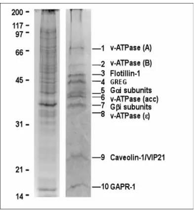
Third, microdomains have been isolated from early Golgi compartments (Gkantiragas, I. et al. 2001). These microdomains contain a unique subset of proteins and lipids as compared to other membrane rafts. These detergent- insoluble complexes in the Golgi have been denominated as GICs (Golgi- derived detergent-insoluble complexes). Similar to other types of DRMs, it reveals an enrichment of sphingolipids and cholesterol. This lipid-scaffold seems to be required for GICs integrity (Gkantiragas, I. et al. 2001).
Interestingly, GIC proteins and lipids are segregated for COPI vesicles, suggesting that they are not involved in the budding of COPI-vesicles at the early Golgi complex. Since the core complex travels through the early secretory pathway, an as yet unknown and distinct mechanism must exist by which GICs are transported. The presence in GICs of heterotrimeric G proteins (α and β subunits) opens the possibility that these microdomains are involved in signal transduction at the Golgi complex. GICs are composed of ten major proteins (Fig. 2): 1) Caveolin-1, which has been implied in regulating the function of heterotrimeric G proteins (Okamoto, T. et al. 1998) and which localizes predominantly to detergent-resistant complexes at the plasma membrane; 2-3) α and β subunits of heterotrimeric G proteins, implicated in signal transduction (Moffet, S. B., Deborah; Linder, Maurine.
2000), membrane fusion (Helms, J.B. et al. 1998) and maintenance of Golgi structure (Jamora, C. et al. 1997; Yamaguchi, T. et al. 2000); 4-7) 4 subunits of the V1 domain of the vacuolar H+ -ATPase, possibly involved in luminal pH regulation by association/dissociation of V1 and V0 subunits and in membrane fusion (reviewed in Gkantiragas, I. et al. 2001); 8) Flotillin-1, described as a component of DRMs (Volonte, D. et al. 1999) and as a component involved in insulin signaling by association with CAP-Cbl complex, directing the complex to the plasma membrane microdomain (Christian A.
Baumann, V. R., Makoto Kanzaki, et al. 2000); 9) GREG (referred to as p45 in Gkantiragas, I. et al. 2001), a GPI-anchored protein at the Golgi (Xueyi Li unpublished data), and involved in maintenance of the Golgi structure. GREG may also be responsible for the structural organization of GICs, and 10) GAPR-1. Cloning of GAPR-1 (Eberle, H. B. et al. 2002) allowed its classification as a mammalian homologue of plant pathogenesis-related proteins (PR proteins). Biochemical characterization revealed some unique
localization, myristoylation, membrane localization, and microdomain association. Its association to the microdomains and relationship to the immune response could help to understand and clarify its role in the cell as well as to understand the function of GICs in the Golgi apparatus.
Figure 2. Comparison of the protein components of DRM and GICs. GICs and total-DRM were isolated from CHO Golgi membranes and total cell lysates, respectively, as described in MATERIALS AND METHODS. Proteins were analyzed by SDS-PAGE (12%) gel and stained with Coomassie blue. Proteins identified in GICs are shown on the right (Gkantiragas, I. et al.
2001).
2 Plant pathogens and integrated defence responses to infection
The homology of GAPR-1 to the superfamily of PR proteins, group 1, suggests a possible role of GAPR-1 in the immune system. In plants, PR proteins have been implied in the immune system, which is reminiscent of the innate immune system in mammals. Innate immunity is an ancient form of defense against microbial infection that is shared by plants, insects and vertebrates. The discrimination of many potential pathogens from self is a formidable task for the innate immune system. Many plants respond to local attack by pathogens with a production of compounds reducing or inhibiting
further attack. Responses occur at the site attacked but also at distal parts. At the site of attack, the responses include an oxidative burst, leading to cell death. In this way the pathogen is killed and is prevented from spreading from the site of initial infection. Further local responses in the surrounding cells include changes in cell wall composition, de novo synthesis of phytoalexins and pathogenesis-related proteins (PR). PR proteins were detected and defined as being absent in healthy plants but accumulating in large amount after infection (Heil, M. Bostock, R. M. 2002). More than 17 protein families have been assigned to this superfamily of proteins (http://www.bio.uu.nl/~fytopath/PR-families.htm). PR-1 proteins play a central role in the defense system in plants during the manifestation of systemic acquired resistance (SAR). Fig. 3 shows the NMR structure of one member of the PR-1 group, p14a tomato, which exhibits a 35% amino acid sequence identity with the human glioma pathogenesis-related protein (GliPR).
Comparison of both proteins led to the identification of a common, partially solvent-exposed cluster of four amino acid residues in GLiPR-1 (His69, Glu88, Glu110, and His127) that is well conserved in all known plant PR proteins group 1 (Szyperski, T. et al. 1998). This could indicate the existence of a common active site for these proteins. In P14a, due to the arrangement of the amino acid residues at the potential active cluster, a possible role as a Zn protease has been suggested. However, a Zn2+ ion did not localize to the pocket of p14a when crystals were grown in presence of Zn+2 (Szyperski, T. et al. 1998). It has also been suggested that the arrangement of the two histidines at the active center represents the active site of a ribonuclease, but biochemical assays to detect such activity could not confirm this (Szyperski, T. et al. 1998). Interestingly, the same cluster of amino acid residues is observed in GAPR-1, which is the object of study in this experimental work.
Figure 3. P14a tomato drawing based on NMR information. Left view. Representation of structural elements in p14a, containing four α-helices, and four β-strands. Ribbon structure of p14a obtained by Dr. Matthew Groves using MolScript® (Kraulis, P. 1991), Raster3D®
(Bacon, D. Anderson, W. 1988). A similar structure has been described for Ves v5 from Vespula vulgaris (PDB 1QNX) and GAPR-1 isolated from GICs microdomains (see Discussion). Right view. Space-filling representation of P14a, showing the putative active site with histidinyl and glutamyl residues depicted in blue and red, respectively (Szyperski, T. et al.
1998).
2.1 Systemic acquired resistance (SAR)
Secondary to the responses at the site of infection, long range responses are induced to protect distal parts of the plants. These responses are described as systemic resistance of plants against pathogens (Hammerschmidt, R.
1999) or systemic acquired resistance (SAR). Many components associated with SAR, e.g. PR proteins, are expressed in response to a first infection, but the mechanisms of this response are not fully understood (Conrath, U. et al.
2002). For instance, salicylic acid (SA) in plants is a critical signaling molecule in the pathway(s) leading to local and systemic resistance and SA is synthesized in response to infection both locally and systematically (Thomma, B. et al. 1998). Thus, the actual sites of de novo production of SA in non-infected plant might therefore contribute to induction of SAR (Meuwly, P. et al. 1995). NO has been implicated in the activation of plant defenses as well. The immune response of plants also depends on reactive oxygen intermediaries (ROI) and reactive nitrogen intermediaries (RNI) for signaling and apoptosis, and possibly for necrosis and direct antimicrobial actions (Klessig, D. F. et al. 2000).
In mammalian cells, salicylates are potent scavengers of NO and its derivatives, and salicylates inhibit the activity and transcription of iNOS
(Farivar, R. S. Brecher, P. 1996). NO has previously been shown to serve as a key redox-active signal for the activation of various mammalian defense responses, including the inflammatory and innate immune responses (Schmidt, H. H. Walter, U. 1994;Mannick, J. B. et al. 1994; Stamler, J. S.
1994). Infections, microbial products, and cytokines readily induce expression of NO synthase in tissue macrophages from rodents. Reactive nitrogen intermediaries (RNI) are critical in host defense not only because they can damage pathogens but also because they are immunoregulatory (Bogdan, C.
et al. 2000). For instance, RNI can inhibit G proteins, activate or inhibit kinases, caspases, metalloproteases, transcription factors, and DNA methyltransferase, inhibit lymphocytes proliferation, alter cytokine and postaglanding production, and either cause or prevent apoptosis of the host cells (reviewed in Nathan, C. Shiloh, M. U. 2000).
Resistant plants often develop a hypersensitive response (HR), in which necrotic lesions form at the site(s) of pathogen entry (reviewed in Klessig, D.
F. et al. 2000). This localized cell death associated with the HR may help prevent the pathogen from spreading to uninfected tissues. Just before or concomitant with the appearance of a HR, an increased synthesis of several families of pathogenesis-related (PR) proteins is observed (Schneider, D. S.
2002). It has been proposed that the HR response by activation of R (resistance) genes is responsible for controlling the entire intracellular environment such as guarding the secretory apparatus (Schneider, D. S.
2002).
The function of PR proteins in both SAR and HR is not known. By looking for similarities between the immune responses in plant and animal cells, likely the innate immune system in mammals is the one that resembles HR-producing infections in plants. The similarities are not only based on molecular mechanisms of action, but also on other levels such as monitoring endosomal traffic and activation of programmed cell death if traffic is disrupted. These considerations may be important for defining a role for PR proteins in general and for GAPR-1 specifically. GAPR-1 as potential regulator in the innate immune response may monitor the integrity of the Golgi apparatus due to its location at this organelle.
2.2 Mammalian PR-1 family members
Since the identification plant PR-1 proteins, secretory proteins with a significant sequence homology have been identified in various other organisms including fruiting body proteins in fungi that are expressed during infection (Schuren, F. H. et al. 1993), insect allergens (Lu, G. et al. 1993;
Schreiber, M. C. et al. 1997), mammalian CRISP proteins possibly involved in sperm maturation or sperm-egg fusion (Kratzschmar, J. et al. 1996), human GliPR/RTVP-1, specifically expressed in glial tumors (Murphy, E. V. et al.
1995; Rich, T. et al. 1996), and in snake or lizard venoms, reported to block ryanodine receptors or cyclic nucleotide-gated ion channels (Morrissette, J. et al. 1995; Brown, R. L. et al. 1999). Together with plant PR-1 proteins, these proteins constitute a large PR-1 protein superfamily. Despite the diversity within this superfamily, very little is known about the function of the individual members and the plant PR proteins, described above, remain the most intensely studied family members.
3 Purpose of this thesis
During the characterization of lipid microdomains at the Golgi (GICs) (Gkantiragas, I. et al. 2001), a protein with an apparent molecular mass of 17 kDa was identified. Cloning and preliminary biochemical characterization identified a novel protein, GAPR-1, belonging to the superfamily of PR proteins. Based on the primary amino acidic sequence of this protein, some potentially interesting characteristics were identified (Fig. 4). It contains i) a consensus sequence for myristoylation (grey box); ii) a putative caveolin- binding domain (red box); iii) a coiled-coil structure (green box) and iv) an isolelectric point (pI) of 9.4, suggesting that GAPR-1 is a highly hydrophilic protein. Further characterization of GAPR-1 showed that the protein is differentially expressed in various tissues (Eberle, H. B. et al. 2002). For instance, GAPR-1 is highly expressed in immunocompetent organs and cells such as in lungs, spleen, and lymphocytes, but is absent in e.g. liver, heart, and brain. Here, this basic information was used to study the interaction of GAPR-1 with membranes (Section 1), to obtain structural information on the protein (in collaboration with Prof. Dr. Irmgard Sinning at the EMBL/BZH-
Heidelberg) (Section 2), and to identify proteins that interact with GAPR-1 (Section 2.4).
Figure 4. cDNA-derived sequence of GAPR-1. The consensus sequence for N- myristoylation is indicated by the grey box. The potential protein-protein interacting sites, i.e. a coiled-coil region and a caveolin-interacting region are indicated in green and red, respectively.
1 Membrane association of GAPR-1
1.1 N-myristoylation of GAPR-1 in Escherichia coli
The enzymology of the myristoylation reaction is well understood. Proteins that are destined to become myristoylated contain the N-terminal sequence: Met-Gly- X-X-X-Ser/Thr (for a review see Resh, M. 1999). The initiating methionine is removed co-translationally by methionine amino-peptidase, and myristate is linked to Gly-2 via an amide bond by an N-myristoyl transferase. To investigate whether the consensus sequence for N-myristoylation in GAPR-1 (Fig. 4) is a substrate of myristoyl-CoA:protein N-myristoyl-transferase (NMT1), an E .coli strain was used that contained two plasmids for simultaneous expression of GAPR-1 (expression vector pQE60, Eberle, H. B. et al. 2002) and yeast NMT1 (in expression vector pBB131 Duronio, R. J. et al. 1990) (Fig. 5 panel A). The vectors were designed so that they could be simultaneously maintained as stable episomal plasmids and that independent induction of transcription of their heterologous DNA sequences was possible. The expression of NMT was placed under the control of the isopropyl-β-D-thiogalactopyranoside (IPTG)-inducible tac promoter, and GAPR-1 under the control of the T5 promoter. The various plasmid combinations were compared for the efficiency of GAPR-1 myristoylation during expression of proteins upon induction with IPTG. To determine myristoylation efficiency of GAPR-1, [
3H]-myristic acid was added during the overexpression. Subsequently, lysates were prepared from the E. coli cultures, and subjected to SDS-PAGE (Fig. 5, middle panel B). Autoradiography showed that in the presence of NMT1 and GAPR-1, a labeled band of 17 kDa was observed and indicated that the radiolabel is readily incorporated into GAPR-1 (Fig. 5, lane 1, bottom panel C). Labeling of GAPR-1 was absolutely dependent on the presence of NMT1. E. coli that expressed NMT1 but lacked GAPR-1, or E.
coli that lacked NMT1 but expressed GAPR-1, failed to label the 17 KDa protein
with the tritiated fatty acid (Fig. 5, panel B and C, lanes 5 and 3, respectively).
Figure 5. [3H]-Myristate-labeled GAPR-1 in E. coli. (A) Schematic presentation of plasmid constructs used to express NMT1 and GAPR-1 in E. coli. KANR, gene for kanamycin resistance;
AMPR, gene for ampicilin resistance. (B) The radiolabeling conditions were compared for induced (IPTG) and non-induced bacteria, containing distinct plasmid combinations: transformed bacteria with GAPR-1 and NMT1 (lanes 1 and 2) were induced (lane 1) or not induced (lane 2) with IPTG (1mM) in the presence of [3H] myristic acid (50µCi/ml culture). Cells were lysed in SDS-gel loading buffer 1, heated for 5 min at 95o C, and after centrifugation (5 min, 14000 rpm), an aliquot of the supernatant (lysate) (20µl) was mixed with (5µl) SDS-gel loading buffer 2 (5X), and analyzed by SDS-PAGE (14% acrylamide). After staining with Coomassie Blue, the dried gel was exposed to a film. Lysates from bacteria containing only pQE60 (GAPR-1) (lanes 3 and 4) or pBB131 (NMT1) (lanes 5 and 6) were induced or not induced with IPTG (as indicated in figure) and analyzed as described above.
1.2 N-myristoylation of GAPR-1 in vivo
To determine whether the consensus sequence for N-myristoylation also results in myristoylation of GAPR-1 in vivo, proteins from a large-scale preparation of Golgi- derived detergent-insoluble complexes (GICs) were separated by SDS-PAGE.
After staining the gel with Coomassie Blue, the protein band at 17 kDa was excised from the gel and digested with trypsin. The resulting peptides were analyzed by electrospray ionization mass spectrometry (ESI-MS) (Fig. 6). Trypsinization of the GAPR-1 band generated 21 major peptides (Fig. 6A, upper panel). If native GAPR- 1 is myristoylated, then a myristoylated dipeptide [myrGK] with a theoretical [M+H]+ signal at m/z 414.33 is expected to be present. The survey spectrum of the tryptic digest of GAPR-1 showed a signal at m/z 414.327 (Fig. 6A, lower panel), consistent with the calculated m/z value for the myristoylated fragment. Also the isotopic
13C-peak at m/z 415.33 could be detected. The ion at m/z 414.327 was further analyzed by ESI tandem mass spectrometry, and the products obtained from this ion by fragmentation are displayed in Fig. 6B. All major fragment ions in this spectrum can be assigned to the myrGK sequence, as indicated in Fig. 6B (insert) and Table 1. In particular the fragment ion triplet at 211, 240 and 268 is indicative of the myrG structure. In summary, mass spectroscopic data show that native GAPR-1 is myristoylated at the N-terminus. All the ESI-MS data were obtained and analyzed in collaboration with Dr. Andreas Schlosser and Dr. Wolf Lehmann at the Cancer Center Heidelberg (DKFZ).
Table 1. Summary of positive nanoESI product ion spectrum of expected and calculated MS/MS signals for myrGK fragment of GAPR-1.
Figure 6. In vivo myristoylation of GAPR-1. (A) Positive nanoESI spectrum of peptides derived from an in-gel digest of native GAPR-1. The top panel shows the complete survey spectrum, and the lower panel the expanded view from m/z 413 to 417, showing the singly protonated molecular ion of the T1 fragment including the 13C isotope peak. (B) Positive nanoESI product ion spectrum of m/z 414.33. The spectrum shows the key fragments for the myrG structure at m/z 211, 240 and 268 and sequence–specific fragment ions, which identify the peptide as T1 fragment myrGK of GAPR-1.
1.3 Association of GAPR-1 with Golgi membranes
Myristoylation is necessary but not sufficient for membrane binding of myristoylated proteins (Peitzsch, R. M. McLaughlin, S. 1993). A second signal for stable membrane binding of N-myristoylated proteins has been defined as either a polybasic cluster of amino acids or a palmitate moiety (Resh, M. D. 1999). GAPR-1 is tightly associated with Golgi membranes, which is reflected by the absence of GAPR-1 from the cytosolic fraction: GAPR-1 could not be detected by immunoprecipitation from large amounts of cytosol (Fig. 7A). When the Golgi structure is disrupted with Brefeldin A and GAPR-1 is dispersed in the cell (Eberle, H. B. et al. 2002), GAPR-1 remains associated with the membrane fraction of these cells as determined by efficient immunoprecipitation the total membrane fraction. This indicates that GAPR-1 is absent from the cytosol (see, however section 1.6). The characteristics of this tight membrane association of GAPR-1 were further investigated by incubation of isolated Golgi membranes under various conditions. As shown in Fig. 7B, treatment of Golgi membranes with 1 M KCl did not strip GAPR-1 off the membranes, whereas NSF, a peripheral Golgi membrane protein (Block, M. R. et al. 1988) is affected by this treatment. Alkaline treatment of the membranes, which causes the dissociation of most peripheral membrane proteins, did affect the membrane binding of NSF and GAPR-1 (Fig. 7B). As a control, p23, a type I transmembrane protein of the Golgi complex (Sohn, K. et al.
1996), remains present in salt or alkaline-treated membranes (Fig. 7B). Biophysical
studies have established that the binding energy provided by myristate is relatively
low (with a K
dof 10
-4M ) (Peitzsch, R. M. McLaughlin, S. 1993) and not sufficient to
stably anchor a protein to a lipid membrane (Moffet, S. B. et al. 2000). To
determine whether the myristoyl moiety of GAPR-1 contributes to the salt-resistant
membrane binding, purified non-myristoylated GAPR-1 was bound to isolated
Golgi membranes (Fig. 7C). Upon salt treatment of the membranes, most of the
non-myristoylated GAPR-1 is stripped again from the membranes. These data
indicate that native GAPR-1 is bound to Golgi membranes not only by ionic
interactions, but also through the myristoyl moiety, which affects membrane
anchoring of the protein. The primary structure of GAPR-1 shows the presence of
basic residues in the N-terminal region. An amino-acidic stretch at the N-terminus (21 amino acids) has a theoretical pI 9.8, containing five positively charged amino acids [Arg-Lys]. The
151LPKK
154sequence at the C-terminus represents another potential positive structure that may play a role on membrane interaction. The high overall pI of GAPR-1 suggests that a contribution of several positively charged regions together with the myristoyl moiety allows the protein to bind strongly to the membrane.
1.4 GAPR-1 interaction with Caveolin-1
A number of studies support the hypothesis that caveolin provides a direct mean for proteins to be sequestered within lipid-enriched microdomains (Oh, P.
Schnitzer, J. E. 2001). Many proteins that interact with caveolin, including G- protein α subunits, Ha-Ras, Src family tyrosine kinases, endothelial NOS, 1 EGF-R and related receptor tyrosine kinases, and protein kinase C isoforms (Okamoto, T.
et al. 1998) do so via an interaction with a common putative caveolin-binding motif (Fig. 4, ΦXΦXXXXΦ, where Φ is aromatic amino acid Trp, Phe, or Tyr). GAPR-1 also contains this putative caveolin-binding motif (YnFqqpgF), which might contribute to its strong membrane-binding. To determine a direct interaction between caveolin-1 and GAPR-1, co-immunoprecipitation studies were carried out.
Under native conditions using various detergents caveolin could not be co- immunoprecipitated with GAPR-1 or vice versa (data not shown). For that reason, co-immunoprecipitation studies were performed after chemical crosslinking of proteins in Golgi membranes. As shown in Fig. 7D, crosslinking with N- Hydroxylsulfosuccinimidyl-4-azidobenzoate resulted in an irradiation-dependent crosslink product of caveolin-1 at 40-45kDa (Fig. 7D left panel).
When GAPR-1 was immunoprecipitated from similar incubations (10 fold upscale of incubations), caveolin-1 was found to co-immunoprecipitate with GAPR-1 in a crosslink product of 40-45kDa (Fig. 7D, lane 3). Co-immunoprecipitation of crosslinked Golgi membranes with Caveolin-1 antibody produced a similar result.
GAPR-1 was co-immunoprecipitated in a crosslinked product that runs at 40-
45kDa (Fig. 7D, lane 6), corresponding to a similar product observed for co-
immunoprecipitation with GAPR-1. These results indicate a direct interaction of GAPR-1 with caveolin-1. The crosslink products at high molecular weights (HMW) (Fig. 7D, lane 6) appeared only when immunoprecipitations were performed with caveolin-1 antibodies and western-blotting was performed with a GAPR-1 antibody.
This indicates that GAPR-1 can interact with caveolin-oligomers.
Figure 7. Interaction of GAPR-1 with Golgi membranes. The incubations were analyzed by SDS-PAGE and western blotting for the presence of the indicated proteins. (A) CHO cells were incubated for 30 minutes in the absence (lane 2 and 3) or presence of 5 µM Brefeldin A (lane 4 and 5).
After homogenization, the homogenate was centrifuged for 1 hour at 100,000 g to yield a total membrane (lanes 2 and 4) and a cytosolic fraction (lanes 3 and 5). GAPR-1 was immunoprecipitated from the membrane
fraction (2mg) or from the cytosolic fraction (2mg). As a control, GAPR-1 was immunoprecipitated from isolated CHO Golgi membranes (50µg) (lane 1). (B) 50µg of CHO Golgi membranes (lanes1-3) were incubated for 30 minutes on ice in the absence (lane 1) or presence of 1 M KCl (lane 2) or with 0.1 M Na2CO3, pH 11 (lane 3). After centrifugation through a 15% (w/v) sucrose cushion, equal amounts of membranes (29 nmol phospholipid) were analyzed. (C) CHO Golgi membranes (25 µg) were incubated for 30 minutes at 4oC in the absence (lane 1) or presence (lanes 2 and 3) of 3µl of bacterial expressed and purified, non-myristoylated GAPR-1 (5.3 mg/ml) in 25 mM Hepes/KOH, pH 7.2, 20 mM KCl, 2.5 mM magnesium acetate, 0.1 M sucrose, 1 mg/ml ovalbumine and 10 mM DTT. Subsequently, KCl (1 M final concentration) was added to one incubation (lane 3) and incubated further for 30 min at 4oC. Golgi membranes were re-isolated by centrifugation through a 15% (w/v) sucrose cushion. (D) CHO Golgi membranes (50µg) were incubated with N- Hydroxylsulfosuccinimidyl-4-azidobenzoate (5mM) in PBS for 30 minutes at RT and left on ice (lane1, and 4) or irradiated for 10 minutes at 254 nm (lane 2 and 5) and analyzed for crosslinked products. For immunoprecipitation (lane 3, and 6), 500 µg of Golgi membranes each were used for immunoprecipitation of GAPR-1 or Caveolin- 1. After western blotting, the PVDF membrane (left, immunoprecipitation using α-GAPR-1) was incubated first with α- Caveolin-1, followed by α-GAPR-1, or the PVDF membrane (right, immunoprecipitation using α-Caveolin-1) was incubated first with α-GAPR-1, followed by α-Caveolin-1
1.5 Phosphorylation of GAPR-1 in vivo.
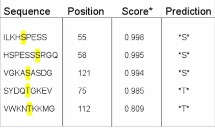
The partitioning of myristoylated proteins to cellular membranes can be sensitive to additional factors that affect the binding of these proteins to membranes. These factors can control the reversible translocation of myristoylated proteins onto membranes. The primary structure of GAPR-1 shows several predicted phosphorylation sites (NETPHOS 2.0 http://www.cbs.dtu.dk/services/NetPhos/) (Table 2). This program calculates a score based on the confidence of the prediction and similarity to known phosphorylation sites. To investigate whether GAPR-1 can be phosphorylated in vivo, CHO cells were incubated 4 hrs with radiolabeled inorganic phosphate (
32Pi). Cells were lysed, and cytosol and total membranes were isolated by centrifugation. The membrane fraction was solubilized in 1% SDS, quenched to 0.1% SDS with PEN buffer containing 1%
Triton X-100, and subjected to immunoprecipitation with an antibody against GAPR-1 (α-1852). Fig. 8 (lane 2) shows that the immunoprecipitated protein GAPR-1 (western blot) is radioactively labeled due to incorporation of
32P in the protein. These data indicate that GAPR-1 is phosphorylated in vivo. The extent of GAPR-1 phosphorylation seems not to be affected by treatment with phosphatase inhibitors (lane 2 and 3).
Figure 8. In vivo phosphorylation of GAPR-1. Confluent CHO wt cells were incubated for 4 hrs in DMEM medium (phosphate and serum free) at 37oC in the absence (lane 1) or presence (lanes 2 and 3) of [32P] 0.25mCi/ml. Cells were incubated in the absence (lanes 1 and 2) or the presence (lane 3) of phosphatase inhibitors.
Phophatase inhibitors were added during homogenization and IP. Cells were washed, harvested and lysed. The homogenate was centrifuged 1hr at 100,000 xg to isolate a total membrane fraction (pellet). The total membrane fraction was dissolved in 1%
SDS and incubated 5 min at 95oC. After incubation, the fraction was quenched to 0.1% SDS by 10-fold dilution in PEN buffer containing 1% Triton X-100. GAPR-1 was immunoprecipitated using α-GAPR-1 (α- 1852) antibody. Immunoprecipitated proteins were separated by SDS-PAGE and analyzed by western-blotting (upper panel) and autoradiography by exposure of the western blot to X-ray film (lower panel).
Table 2. Predicted phosphorylation sites present on GAPR-1. The program NETPHOS (2.0) was used to predict phosphorylation of amino acids in GAPR-1. Phosphorylation sites are sorted by their score. *Score indicates the likeliness of phosphorylation. Threshold score: 0.500.
1.6 Effect of phosphorylation on the partitioning of GAPR-1 to lipid- enriched microdomains.
Triton X-100 treatment of Golgi membranes or plasma membranes in the cold allows isolation of a detergent-insoluble fraction. The partitioning of proteins to the detergent-resistant fraction can depend on post-translational modifications (Moffet, S. B. et al. 2000). Phosphorylation therefore may have an effect on the partitioning of GAPR-1 into Golgi-derived microdomain complexes (GICs) as well. To analyze this possibility, total cell membranes, obtained from
32P-treated CHO cells, were resuspended in cold PEN buffer containing 1% Triton X-100 and detergent-soluble and insoluble fractions were analyzed for the presence of phosphorylated GAPR-1.
A major pool of GAPR-1 is insoluble in Triton X-100 (Fig. 9A, lane 1, upper panel),
and this fraction is highly phosphorylated (Fig. 9A, lane 1, bottom panel); a minor
pool of GAPR-1 is soluble in Triton X-100 (Fig. 9A, lane2, upper panel). This
soluble fraction shows a low level of phosphorylation (Fig. 9A, lane 2, bottom
panel). Interestingly, CHO cytosol of treated cells shows a barely detectable
fraction of GARP-1 by western blot analysis (Fig. 9A, lane 3, upper panel), but a
relatively high level of phosphorylation (radioactivity/amount of protein) is observed
in this sample (Fig. 9, lane 3, bottom panel). The radioactive signals were
quantified using the program Quantity One® (BioRad™). Fig. 9B shows that approximately 32.5% (calculated as percentage of total radioactive signal) of the incorporated Phosphate in GAPR-1 has a cytosolic localization. The soluble fraction in Triton X-100 represents approximately 11.6%. These data suggest that GAPR-1 can be phosphorylated in vivo and that phosphorylation of GAPR-1 can play a role on the dynamics of GAPR-1 association with Golgi membranes.
Figure 9. Phosphorylation of GAPR-1 and partitioning into lipid rafts. A) Confluent CHO wt cells were incubated for 4 hrs in DMEM medium (phosphate and serum free) at 37oC in the presence of [32P] 0,25mCi/ml. Cells were washed, harvested and lysed. The homogenate was centrifuged 1hr at 100,000 xg to isolate a total membrane fraction (pellet) and a cytosol fraction (supernatant). The total membrane fraction was dissolved in 1% TX-100/PEN buffer and incubated for 30 min in the cold. After incubation, the fraction was centrifuged 1 hr at 100,000 xg to yield soluble (lane 2) and insoluble (lane 1) fractions. GAPR-1 was immunoprecipitated from both fractions as well as from cytosol of lysated cells (lane 3). Proteins in the immunoprecipitates were separated by SDS-PAGE and analyzed for the presence of GAPR-1 by western-blotting (upper panel) and for phosphorylation of GAPR-1 by autoradiography (lower panel). B) Quantification of the radioactive signals by Quantity One® (Biorad).
2 Structural Characteristics of GAPR-1 2.1 Large Scale Purification of GAPR-1
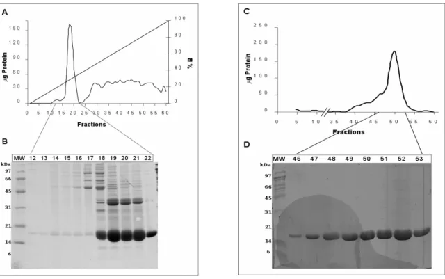
Structure determination of proteins is important in several ways. The function of a protein is linked to its three dimensional structure, and highly resolved structures can lead to a clear understanding of the possible function of a protein. Structures can also discriminate between evolutionary changes in the primary structure of related or non-related species. To obtain a crystal structure of GAPR-1, it was necessary to obtain protein crystals of suitable size and diffraction quality for X- ray analysis. GAPR-1 has several biochemical characteristics that can be used to obtain a highly purified protein fraction: an isoelectric point (pI) of 9.4, and a molecular weight of 17,2 KDa, useful in ion exchange and gel filtration chromatography, respectively.
The cDNA encoding GAPR-1 was cloned in the vector pQE60, and after transformation, GAPR-1 was overexpressed in M15/REP4 bacteria.
Recombinant wtGAPR-1 was purified from overexpression in 12 liters of LB- medium (Fig. 10). Diluted lysates were applied on to a DEAE-sepharose (anion exchange support) to remove most of the bacterial proteins in the lysate, followed by a High S-support column (cation exchange). GAPR-1 has a high pI (9.4), and the overexpressed protein was bound to the High S-support (Fig. 10A, lane 5).
After washing, proteins were eluted from the cation exchange column by a salt
gradient. The chromatogram and corresponding gels (Fig. 10B (Ieft part), panel A
and B) indicate that GAPR-1 eluted at a salt concentration of 250-300mM,
corresponding to fractions 18 to 22. These fractions were pooled and loaded onto
a Superdex 200 gel filtration column. Separation of proteins by gel filtration
produced several fractions (46-53) (Fig. 10B (right part), panel B) with GAPR-1,
purified to apparent homogeneity. These fractions were used for further studies,
including the generation of GAPR-1 crystals by Dr. Matthew Groves.
Figure 10A. Overexpression of GAPR-1 in bacteria (M15/REP4). Figure shows distinct steps during induction of GAPR-1 expression. Cells collected prior to induction (lane 1) by 1mM IPTG, and after 3 hrs of induction (lane 2). Cells were collected and lysed by French press. The homogenate (lane 3) was cleared after 100000 xg centrifugation, and diluted 1:6 with buffer Tris- HCl 50mM; pH 7.5; 50mM. 1.2 L of Lysate was loaded onto a DEAE column at a flow rate of 0.5 ml/min and the flow through (lane 4) was applied to a cation exchange column (High S-support), and washed Tris-HCl 50mM pH7.5; 50mM NaCl (lane 5, flow through). Proteins were eluted from the column with a NaCl gradient (50mM to 1000mM).
Figure 10B. Large scale purification of GAPR-1. Left panel: Cation exchange chromatography (High S-support cation exchange column). Panel A shows the chromatogram profile (amount of protein and conductivity versus fractions) obtained during elution of proteins from the High S column by a sodium chloride (NaCl) gradient (in 50 mM to 1M in Tris 50 mM, pH 7.5). Proteins eluted between 250 to 300mM NaCl were analyzed by SDS-PAGE (14%), and coomassie blue staining. Right panel: Gel filtration (Superdex 200). Upper panel (C) shows the chromatogram profile (µg protein vs fractions) of eluting proteins (Tris 50 mM, pH 7.5, 50 mM NaCl), and the bottom panel (D) shows the analysis of selected fractions by SDS-PAGE (14%) and coomassie blue staining.
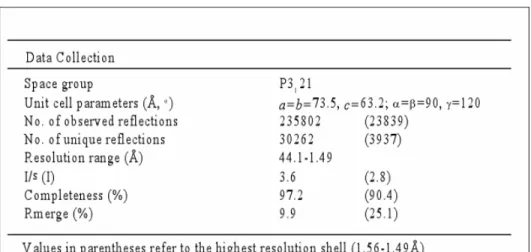
2.2 Crystal structure of GAPR-1
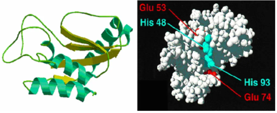
![Figure 5. [ 3 H]-Myristate-labeled GAPR-1 in E. coli. (A) Schematic presentation of plasmid constructs used to express NMT1 and GAPR-1 in E](https://thumb-eu.123doks.com/thumbv2/1library_info/5350851.1682749/28.918.164.772.187.804/figure-myristate-labeled-schematic-presentation-plasmid-constructs-express.webp)

![Figure 8. In vivo phosphorylation of GAPR-1. Confluent CHO wt cells were incubated for 4 hrs in DMEM medium (phosphate and serum free) at 37 o C in the absence (lane 1) or presence (lanes 2 and 3) of [ 32 P] 0.25mCi/ml](https://thumb-eu.123doks.com/thumbv2/1library_info/5350851.1682749/34.918.132.451.716.881/figure-phosphorylation-confluent-incubated-medium-phosphate-absence-presence.webp)
![Figure 9. Phosphorylation of GAPR-1 and partitioning into lipid rafts. A) Confluent CHO wt cells were incubated for 4 hrs in DMEM medium (phosphate and serum free) at 37 o C in the presence of [ 32 P] 0,25mCi/ml](https://thumb-eu.123doks.com/thumbv2/1library_info/5350851.1682749/36.918.227.705.338.783/figure-phosphorylation-partitioning-confluent-incubated-medium-phosphate-presence.webp)