Human intestinal spirochetosis – a review
Intestinale Spirochätose des Menschen – ein Review
Abstract
Human intestinal spirochetosis (IS) is a condition defined histologically by the presence of spirochetal microorganisms attached to the apical
Efstathia Tsinganou
1Jan-Olaf Gebbers
1cell membrane of the colorectal epithelium. Intestinal spirochetes comprise a heterogeneous group of bacteria. In humans,Brachyspira
aalborgi andBrachyspira pilosicolipredominate. Prevalence rates of 1 Institute of Environmental Medicine, Luzerner IS are low where living standards are high, in contrast to poorly de-
Kantonsspital, Luzern, Switzerland
veloped areas where IS is common. Homosexuals and HIV-infected in- dividuals are at high risk of being colonized. Clinical significance in indi- vidual cases has remained unclear up to now. A review of the literature assumes that invasion of spirochetes beyond the surface epithelium may be associated with gastrointestinal symptoms which respond to antibiotic treatment (metronidazole), whereas individuals lacking this feature may be mostly asymptomatic. Of unknown reason, homosexual and HIV-positive men as well as children are more likely to be sympto- matic irrespective of invasion. Rare cases of spirochetemia and multiple organ failure have been reported in critically ill patients with IS.
Keywords:human intestinal spirochetosis, microscopic colitis,
commensals, intestinal bacterial invasion, HIV-infection, spirochetemia
Zusammenfassung
Die intestinale Spirochätose des Menschen (IS) wird histologisch defi- niert als ein dichter Saum von Spirochäten, der an der apikalen Zell- membran des interkryptalen Epithels des Dickdarms haftet. Die intes- tinalen Spirochäten umfassen eine heterogene Gruppe von Bakterien.
Beim Menschen sind ganz überwiegendBrachispira aalborgiundBra- chispira pilosicolinachweisbar. Die Prävalenz der IS ist niedrig in Regio- nen hohen Lebens- und Hygienestandards im Gegensatz zu ärmeren Regionen, wo die IS häufig auftritt. Homosexuelle und HIV-positive Männer haben ein erhöhtes Besiedelungsrisiko. Die klinische Bedeutung der IS ist im Einzelfall bislang fraglich. Die Literaturübersicht lässt an- nehmen, dass bei der Schleimhautinvasion der Spirochäten klinische Symptome wahrscheinlich sind, die gut auf eine antibiotische Therapie (Metronidazol) ansprechen, während Personen ohne diesen Befund wohl meist symptomlos bleiben. Aus unbekannten Gründen leiden Ho- mosexuelle, HIV-positive Personen wie auch Kinder eher an Beschwer- den unabhängig von der Invasivität der Spirochäten. Spirochätämien und multiples Organversagen sind bei einzelnen, meist schwerkranken Patienten mit IS beschrieben worden.
Schlüsselwörter:humane intestinale Spirochätose, mikroskopische Kolitis, Kommensale, intestinale bakterielle Invasion, HIV-Infektion, Spirochätämie
Background
First recognized in humans by van Leeuvenhoek in his own diarrheal stool in the 17thcentury (named asanimal- cules), intestinal spirochetes in humans are still poorly
understood in their biology, origin, and state as commens- als or pathogens in the human large intestine. Originally found as a disease of economic devastation in veterinary medicine (e.g. in swine), intestinal spirochetes in humans
and its clinical significance have been debated for years [1], [2], [3], [4], [5], [6], [7], [8], [9], [10], [11], [12].
In 1967, Harland and Lee coined the term intestinal spirochetosis (IS), recognizing the adherence of spiro- chetes to colorectal epithelium in histology and electron microscopy, the characteristic appearance that is still considered pathognomonic for a possible capacity to cause human disease [2] (Figure 1, Figure 2, Figure 3, Figure 4, Figure 5). Despite improvements in the detection and identification of IS, it is still unclear whether this condition represents an actual disease process, or rather, the organisms represent interesting intestinal colonizers in men that does exclusively manifest in the large bowel.
Figure 1: Exfoliative cytology of the rectal mucosa in human spirochetosis with many spirochetes between rod-like
bacteriae. Warthin-Starry silver stain. x600.
Figure 2: Histology of human intestinal spirochetosis.
Hematoxylin-Eosin. x350.
Figure 3: Human intestinal spirochetosis in the vermiform appendix. Warthin-Starry silver stain. x350.
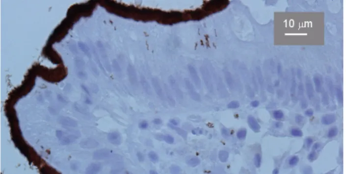
Figure 4: Immunohistochemical detection of human intestinal spirochetosis with signs of invasion. Strept-Avidin technique.
x680.
Figure 5: Human intestinal spirochetosis in transmission electron microscopy. x3500.
Epidemiology
In the veterinary world, IS has been linked to diarrheal illness in swine, poultry, dogs, cats, opossum, non-human primates, and guinea pigs. The disease causes significant economic losses when it affects large numbers of swine, leading to “porridge-like diarrhea”, malnutrition, de- creased food intake, and declining growth rates [3].
Human disease is less well understood, though the presence of intestinal spirochetes in stool has been documented microbiologically throughout Africa, Australia, India, Indonesia, and much of the Western world for decades (review: [4]).
The prevalence data strongly depend on the material and the detection methods used (direct histology or poly- merase chain reaction (PCR) of fecal samples or of colorectal biopsies). A large study in Chicago in the early 1900s revealed a 28% prevalence of intestinal spiro- chetes in the stools of healthy persons [5]. Studies of stools in West Africa found close to a 100% rate of spiro- chetes [6]. Prevalence rates in soldiers of Western Com- mand during the early 1900s reached 3.3% in their stools for those with previous bouts of dysentery [7]. It is note- worthy that the presence of spirochetes in the stool might not be associated with IS and with clinical symptoms.
In more recent times, the prevalence of intestinal spiro- chetes in stools appears to correspond with habitation in a developing region. Prevalence rates of 32.6% are
seen in Australian aboriginal children. In contrast, spiro- chaetes were only recovered from 8 of 695 (1.2%) fecal samples that were obtained from other mainly non-Abori- ginal children and adults in Western Australia or the Northern Territory of Australia, even though most of these individuals were suffering from gastrointestinal disturb- ances [8]. Villages in India have shown rates as high as 64.3% in otherwise healthy individuals [6]. A study looking at hospitalized and healthy persons in Oman found pre- valence rates of 11.4 and 26.7%, respectively [9]. A study in Bali in 2002 examined 992 fecal samples from people living in rural, urban, and suburban areas. In contrast to the rural predominance seen in earlier studies, prevalence in Bali varied from 3.3 to 23.4%, with the highest percent- ages in the suburban areas [10]. Other studies reported rates from 1.1 to 5% in most developed countries [4], [6], [13], [14].
The highest rates of colonization of stools with intestinal spirochetes in developed countries are found in homo- sexual males and in human immunodeficiency virus (HIV)- infected individuals. In the United States, homosexual males have shown rates of colonization as high as 20.6 to 62.5% [4], [15], [16], [17], [18], [19]. The reason for this increased colonization in homosexual men is specu- lative at best but has caused proponents to ponder whether IS is sexually transmitted [17], [18], [19]. For those with IS and HIV, there appears to be no correlation with degree of immunodeficiency and extent of disease [19].
Colonization of the colonic mucosa with intestinal spiro- chetes (IS) is not limited to the homosexual population in developed countries, as cases in heterosexual adults have been reported in the U.S., Japan [20], Australia [21], Denmark, Sweden, Switzerland, Norway, England, France [22], Italy, Spain and Brasil [23] (review: [4]). Particularly in children, IS may be associated with severe clinical symptoms [24], [25], [26], [27], [28]. Intestinal spiro- chetes have been documented in second trimester fetuses while infections by Treponema pallidum, Lyme and relapsing fever Borrelia and Leptospira were ruled out. Fetal tissues showed a brisk lymphocytic-plasmacytic response in the intestinal mucosa. In all instances the placenta had chorioamnionitis and severe chronic villitis.
The placental findings suggest an ascending transamni- otic infection [29].
Microbiology
As the modern classification of bacteria came to rely on morphologic differences at the level of DNA and RNA, the spirochetes were divided into three phylogenetic groups.
The family Spirochaetaceae includes Borrelia, Spiro- chaeta, Spironema, and Treponema; Leptospiraceae containsLeptonemaandLeptospira; and the intestinal spirochetes of Brachyspira (Serpulina) are in the Bra- chyspiraceaefamily (Table 1) [30].
Traditionally,BrachyspiraandSerpulinawere referred to as separate genera; however, a lack of significant phylo-
genetic differences has led to the unifying classification underBrachyspira, with the two genus names considered interchangeable [31].
The two members of the Brachyspiraceaefamily most commonly associated with human IS are Brachyspira aalborgiandBrachyspira pilosicoli.B. aalborgiwas first identified in the stool of a patient from Aalborg, Denmark in 1982 [32]. In the years that followed, subsequent cases of IS were assumed to be caused byB. aalborgion the basis of similar histologic appearance. However, studies published in 1994 and 1996 reexamined the stools using multilocus enzyme electrophoresis and yiel- ded a predominance ofB. pilosicoli[33], [34]. Addition- ally, a study by Trivett-Moore et al. [35], published in 1998, looked at rectal biopsy specimens in homosexual men and found onlyB. pilosicoli. Following these studies, most subsequent cases of IS were attributed to B.
pilosicoli. More recently, PCR-based assays have been used to identify these fastidious organisms (see below).
Members of the familyBrachyspiraceaeare morphologic- ally similar to other spirochetes. The characteristic of all spirochetes, movement through fluid environments, is performed by rotation of flagellae. A central cylinder en- closed by a cytoplasmic membrane is the basic morpho- logic structure. The periplasmic space contains the axial fibrils, the number of which varies for individual species.
Characteristics of B. aalborgi are the length: 2–6 μm;
diameter: 0.2 μm; slender, tapered point (causative agent of diarrhea in humans) [11], [12], [36]. Characteristics of the weakly beta hemolytic B. pilosicoli are length:
4–20 μm; diameter: 0.2–0.5 μm; slender, tapered point (causative agent of diarrhea in humans, pigs, dogs, poultry) [3].
BothB. aalborgiandB. pilosicoliare slowly growing fas- tidious anaerobes, with estimated growth times of 6 days forB. pilosicoliand up to 2 weeks forB. aalborgi[36], [21], [31], [35].B. aalborgiis difficult to grow on artificial culture media. The first reported isolation of the organism from human feces was on brain heart infusion agar with 10% bovine blood and spectinomycin plus polymyxin B.
Incubation in an anaerobic jar allowed growth of larger colonies, and growth was slightly improved at 38.5°C than at 37°C [37], [38].
A report on antimicrobial susceptibility testing of B.
pilosicoliwas published in 2003 [39]. Antimicrobial sus- ceptibility was determined using Clinical and Laboratory Standards Institute (formerly National Committee for Clinical Laboratory Standards, or NCCLS) breakpoints for anaerobes, with isolates determined to be susceptible to ceftriaxone, chloramphenicol, meropenem, metronidazole, and tetracycline. An arbitrary breakpoint was established for ciprofloxacin, yielding a 60% resistance rate. A slightly better response rate to moxifoxacin was exhibited.
Erythromycin was not active againstB. pilosicoli, but ap- proximately 30% of erythromycin-resistant isolates were susceptible to clindamycin.
The physiology of ruminal and intestinal spirochetes has been reviewed by Stanton in 1998 [40].
Table 1: Classification of Spirochetes [30]
Clinical presentation, diagnosis, and treatment
In many cases, the histological findings of IS are simply an incidental discovery during a screening colonoscopy.
Symptomatic IS is most commonly accompanied by complaints of chronic (watery) diarrhea and vague abdom- inal pain without other apparent cause [4].
Though mild to moderate disease symptoms predominate, disease severity can range from asymptomatic to invasive and rapidly fatal. Several cases of invasive disease have been reported [16], [17], [18], [19], [20], [22], [23], [41], [42], [43], [44], [45], [46], [47], [48], [49], [50], [51], [52], [53], [54]. Infected children usually complain of diarrhea and may also present with nausea, weight loss, and failure to thrive [24], [25], [26], [27], [28].
Co-infection with other enteric pathogens, includingEn- terobius vermicularis,Helicobacter pylori,Shigella flexn- eri, andNeisseria gonorrhoeae, is common, making the question of clinical significance of IS a difficult one to answer [17], [27], [54].
The endoscopic appearance of the colon lends very little to the diagnosis. A literature review by Alsaigh and Fogt [51] examined the documented endoscopic appearance of 15 biopsy specimens that were histologically consistent with IS. A “polypoid” appearance was noted in seven pa- tients, an “erythematous” area was seen in one patient, a “lesion” was documented in another patient; and nor- mal-appearing mucosa was noted in six patients. Hence, the endoscopic appearance seems to contribute estab- lishing the diagnosis of IS. But probably the spirochetes were found coincidentally in biopsies taken from mucosal areas with irregular appearance, while in most cases the mucosa colonized with spirochetes does not reveal any gross irregularities. Colonic involvement has been docu- mented from the proximal to the distal colon, including the rectum. Involvement of the vermiform appendix has also been reported [36], [45], [55].
The diagnosis of IS is traditionally based on the histo- logical appearance of a diffuse blue fringe (seen in hem- atoxylin-eosin stain), which is approximately 3 to 6 μm thick, along the border of the intercryptal epithelial layer.
This finding is referred to as the “false brush border” [2], [4] (Figure 2). When IS is suspected on the basis of finding a blue fringe, Warthin-Starry or Dieterle silver impregna-
tion stains can be used to highlight the spirochetes in fixed tissue samples (Figure 3) [4], [36], [43], [44], [45], [46], [47], [48], [49], [50], [51], [52]. Recently, an anti- body againstBorrelia burgdorferihas been applied in the immunohistochemical detection of IS [53] (Figure 4). This is a great diagnostic advantage.
On electron microscopy, the spirochetes are seen to dock perpendicularly to the intestinal epithelium [2], [4], [43], [44], [45], [46], [47], [48] (Figure 5). Even with significant diarrhea, the organisms have been described for a long time to be typically non-invasive mainly seen docking onto the cell surface mostly without actually penetrating the membrane [2], [3], [11], [12], [44], [45], [46]. This view has been challenged for the first time in electron micro- scopic studies; additionally, a particular intraepithelial mast cell and IgE plasma cell reaction has been found [47], [48]. The epithelium undergoes changes, such as blunting and loss of the microvilli, defects of the glycoca- lyx, and swelling of the mitochondria [11], [12], [47]. Cell- membrane destruction can occur with the spirochetes found in the intercellular spaces, within the surface epi- thelial cytoplasm as intact organisms, or in phagolyso- somes of macrophages as morphological altered spiro- chetes [47], [48]. The amount of cell destruction usually parallels the degree of invasion microscopically and clin- ically, with more diarrhea typically seen in those with a greater degree of microvillus destruction and a heavier burden of spirochete attachment [16]. The diarrhea is hypothesized to be a result of decreased resorptive areas of the damaged brush border [46], [48].
Two reports that reviewed histological changes in symp- tomatic HIV-infected patients with IS noted a higher de- gree of epithelial invasion, as well as more pronounced loss of microvilli, in this population compared with non- HIV-infected patients [16], [44]. Because diarrhea is common in the HIV-infected population, subtle histologic changes can be easily overlooked, with diarrhea attributed to a cause other than IS. Diagnosis of IS in the HIV-infec- ted population requires pathologists to have a high level of expertise in evaluating biopsy material from HIV-infec- ted individuals [41].
Although the diagnosis of IS is usually made from histo- logical examination of colorectal biopsy material, newer methods for identifying the etiologic organism are being explored but are not yet available commercially. PCR has become one of the more reliable methods, targeting the
16S rRNA, NADH-oxidase, and the 23rDNA gene specific for B. pilosicoli, B. hyodysenteriae, and S. intermedia [56], [57]. Novel techniques such as immunomagnetic separation show promise for the future [58]. Additionally, fluorescent in situ hybridization with oligonucleotide probes targeting 16S or 23S rRNA ofB. aalborgiandB.
pilosicolihas been reported to be applicable in formalin- fixed, paraffin-embedded intestinal biopsy specimens [59], [60].
Nevertheless, although molecular genetic techniques separate the different spirochetal species specifically, the diagnosis of IS has still to be ascertained
morphologically in the biopsy material.
Nine cases of bacteremia caused byB. pilosicoli, mostly in immunocompromised or critically ill patients have been reported in the English literature [61], [62], and a specific antibody toB. aalborgicould be obtained from the serum of a patient with IS [63].
Response to antibiotic therapy for IS has varied. While some patients may have complete remission of diarrhea and normalization of the colorectal mucosa, others con- tinue to have diarrhea with or without persistence of the
“false brush border”. We suggest that the amount of in- vasiveness could correlate to the clinical signs and symptoms and that patients with invasion of spirochetes beyond the surface epithelium may be more apt to re- spond to antibiotic therapy [4]. Generally, a trial of antibi- otic therapy is warranted, most commonly with met- ronidazole. Eradication of symptoms has been reported with metronidazole administered at 500 mg q.i.d. for 10 days [50]. However, there are relatively little data pub- lished on recommended treatments other than successful case reports [12], [16], [46], [47], [53], [54]. Symptomatic improvement with the use of other antibiotics, including clindamycin and macrolides, has been reported as well [27].
New perspectives
As the genome sequence of the major swineBrachyspira hyodysenteriaewas recently deciphered [64] the genome sequences of other pathogenic and non-pathogenicBra- chyspiraspecies are becoming available. This data will facilitate to reveal how these species have evolved and adapted to the varied lifestyles in the complex and changing nutritional and polymicrobial environment of large bowels of different species, and why some but not others can induce colitis and diarrhea [65].
Also, it will be possible to know what survival advantages are gained byBrachyspiraspecies through lateral gene transfer events that seemed to be a dominant evolution- ary force in several pathogens [66]. Particularly detailed functional genomic analysis ofBrachyspiraspecies may reveal the association with chemotaxis, motility, invasive- ness, proteases, hemolysins and other potential virulence factors and allow a differentiation between pathogenic and non-pathogenic strains.
Conclusions
As advances in techniques for the detection of intestinal spirochetes emerge, experts continue to argue about the significance of this condition. Although treatment with effective antibiotic does lead to symptomatic remission and histological clearance in some patients, it is still un- certain whether it was the elimination of the spirochetes that led to the symptomatic improvement. Yet, other pa- tients have no symptomatic relief with treatment despite clearance of spirochetes. Diagnosis of apparent IS made on the basis of histologic examination of biopsy speci- mens from asymptomatic patients add further to the confusion. On the other hand, the reported cases of IS with septicemia and dissemination provide some validity to the possible consequences of the condition. As IS is more consistently recognized with better identification techniques, it is hoped that the clinical significance of this condition, particularly that of different strains and their potential of invasiveness, will soon become evident.
Notes
Conflicts of interest
None declared.
References
1. van Mook WN, Koek GH, van der Ven AJ, Ceelen TL, Bos RP.
Human intestinal spirochetosis: any clinical significance? Eur J Gastroenterol Hepatol. 2004;16(1):83-7. DOI:
doi:10.1097/00042737-200401000-00013
2. Harland WA, Lee FD. Intestinal spirochaetosis. Br Med J.
1967;16:718-23. DOI: 10.1136/bmj.3.5567.718
3. Lee JI, McLaren AJ, Lymbery AJ, Hampson DJ. Human intestinal spirochetes are distinct from Serpulina hyodysenteriae. J Clin Microbiol. 1993;31(1):16-21.
4. Körner M, Gebbers JO. Clinical significance of human intestinal spirochetosis--a morphologic approach. Infection.
2003;31(5):341-9.
5. Parr LW. Intestinal spirochetosis. J Infect Dis. 1923;33:369-83.
6. Teglbjærg PS. Intestinal spirochaetosis. Curr Top Pathol.
1990:81:247-56.
7. Fantham HB. Remarks on the nature and distribution of the parasites observed in the stools of 1305 dysenteric patients.
Lancet. 1916;187:1165-6. DOI: 10.1016/S0140- 6736(00)53413-8
8. Lee JI, Hampson DJ. Intestinal spirochaetes colonizing aborigines from communities in the remote north of Western Australia.
Epidemiol Infect. 1992;109(1):133-41.
9. Barrett SP. Intestinal spirochaetes in a Gulf Arab population.
Epidemiol Infect. 1990;104(2):261-6. DOI:
10.1017/S0950268800059434
10. Margawani KR, Robertson ID, Brooke CJ, Hampson DJ.
Prevalence, risk factors and molecular epidemiology of Brachyspira pilosicoli in humans on the island of Bali, Indonesia.
J Med Microbiol. 2004;53(Pt 4):325-32. DOI:
10.1099/jmm.0.05415-0
11. Takeuchi A, Jervis HR, Nakazawa H, Robinson DM. Spiral-shaped organisms on the surface colonic epithelium of the monkey and man. Am J Clin Nutr. 1974;27(11):1287-96.
12. Nielsen RH, Orholm M, Pedersen JO, Hovind-Hougen K, Teglbjaerg PS, Thaysen EH. Colorectal spirochetosis: clinical significance of the infestation. Gastroenterology. 1983;85(1):62-7.
13. Lindboe CF. The prevalence of human intestinal spirochetosis in Norway. Anim Health Res Rev. 2001;2(1):117-9.
14. Tompkins DS, Foulkes SJ, Godwin PG, West AP. Isolation and characterisation of intestinal spirochaetes. J Clin Pathol.
1986;39(5):535-41. DOI: 10.1136/jcp.39.5.535
15. McMillan A, Lee FD. Sigmoidoscopic and microscopic appearance of the rectal mucosa in homosexual men. Gut.
1981;22(12):1035-41. DOI: 10.1136/gut.22.12.1035 16. Cooper C, Cotton DW, Hudson MJ, Kirkham N, Wilmott FE. Rectal
spirochaetosis in homosexual men: characterisation of the organism and pathophysiology. Genitourin Med. 1986;62(1):47- 52.
17. Surawicz CM, Roberts PL, Rompalo A, Quinn TC, Holmes KK, Stamm WE. Intestinal spirochetosis in homosexual men. Am J Med. 1987;82:587-92. DOI : 10.1016/0002-9343(87)90104-5 18. Lafeuillade A, Delbeke E, Benderitter T, Dhiver C, Gastaut JA,
Chaffanjon P, Quilichini R. Spirochetose intestinale chez les homosexuals infectes par le virus de l'immunodeficience humaine. Trois observations [Intestinal spirochetosis in homosexuals infected with HIV. 3 cases]. Ann Med Interne (Paris).
1990;141(5):464-7.
19. Kostman JR, Patel M, Catalano E, Camacho J, Hoffpauir J, DiNubile MJ. Invasive colitis and hepatitis due to previously uncharacterized spirochetes in patients with advanced human immunodeficiency virus infection. Clin Infect Dis.
1995;21(5):1159-65.
20. Nakamura S, Kuroda T, Sugai T, Ono S, Yoshida T, Akasaka I, Nakashima F, Sasou S. The first reported case of intestinal spirochaetosis in Japan. Pathol Int. 1998;48(1):58-62. DOI:
10.1111/j.1440-1827.1998.tb03829.x
21. Mikosza AS, La T, de Boer WB, Hampson DJ. Comparative prevalences of Brachyspira aalborgi and Brachyspira (Serpulina) pilosicoli as etiologic agents of histologically identified intestinal spirochetosis in Australia. J Clin Microbiol. 2001;39(1):347-50.
DOI: 10.1128/JCM.39.1.347-350.2001
22. Lambert T, Goursot G. Diarrhee aigue avec homocultures et coprocultures positives a Treponema. Med Mal Infect.
1982;12:276-8. DOI : 10.1016/S0399-077X(82)80028-0 23. De Brito T, Sandoval MP, Silva AG, Saad RC, Colaiacovo W.
Intestinal spirochetosis: first cases reported in Brazil and the use of munohistochemistry as an aid in histopathological diagnosis. Rev Inst Med Trop Sao Paulo. 1996;38(1):45-52.
24. da Cunha Ferreira RM, Phillips AD, Stevens CR, Hudson MJ, Rees HC, Walker-Smith JA. Intestinal spirochaetosis in children. J Pediatr Gastroenterol Nutr. 1993;17(3):333-6. DOI:
10.1097/00005176-199310000-00020
25. White J, Roche D, Chan YF, Mitchell EA. Intestinal spirochetosis in children: report of two cases. Pediatr Pathol. 1994;14(2):191- 9. DOI: 10.3109/15513819409024252
26. Heine RG, Ward PB, Mikosza AS, Bennett-Wood V, Robins-Browne RM, Hampson DJ. Brachyspira aalborgi infection in four Australian children. J Gastroenterol Hepatol. 2001;16(8):872-5. DOI:
10.1046/j.1440-1746.2001.t01-1-02543.x
27. Marthinsen L, Willén R, Carlén B, Lindberg E, Värendh G.
Intestinal spirochetosis in eight pediatric patients from Southern Sweden. APMIS. 2002;110(7-8):571-9. DOI: 10.1034/j.1600- 0463.2002.11007809.x
28. Koteish A, Kannangai R, Abraham SC, Torbenson M. Colonic spirochetosis in children and adults. Am J Clin Pathol.
2003;120(6):828-32. DOI: 10.1309/G7U6BD85W4G3WJ0J 29. Abramowsky C, Beyer-Patterson P, Cortinas E. Nonsyphilitic
spirochetosis in second-trimester fetuses. Pediatr Pathol.
1991;11(6):827-38. DOI: 10.3109/15513819109065480 30. Paster BJ, Dewhirst FE. Phylogenetic foundation of spirochetes.
J Mol Microbiol Biotechnol. 2000;2(4):341-4.
31. Ochiai S, Adachi Y, Mori K. Unification of the genera Serpulina and Brachyspira, and proposals of Brachyspira hyodysenteriae Comb. Nov., Brachyspira innocens Comb. Nov. and Brachyspira pilosicoli Comb. Nov. Microbiol Immunol. 1997;41(6):445-52.
32. Hovind-Hougen K, Birch-Andersen A, Henrik-Nielsen R, Orholm M, Pedersen JO, Teglbjaerg PS, Thaysen EH. Intestinal spirochetosis: morphological characterization and cultivation of the spirochete Brachyspira aalborgi gen. nov., sp. nov. J Clin Microbiol. 1982;16(6):1127-36.
33. Lee JI, Hampson DJ. Genetic characterisation of intestinal spirochaetes and their association with disease. J Med Microbiol.
1994;40(5):365-71. DOI: 10.1099/00222615-40-5-365 34. Trott DJ, Stanton TB, Jensen NS, Hampson DJ. Phenotypic
characteristics of Serpulina pilosicoli the agent of intestinal spirochaetosis. FEMS Microbiol Lett. 1996;142(2-3):209-14.
DOI: 10.1111/j.1574-6968.1996.tb08432.x
35. Trivett-Moore NL, Gilbert GL, Law CL, Trott DJ, Hampson DJ.
Isolation of Serpulina pilosicoli from rectal biopsy specimens showing evidence of intestinal spirochetosis. J Clin Microbiol.
1998;36(1):261-5.
36. Gebbers JO, Marder HP. Human intestinal spirochetosis: unusual findings. Microecol Therap. 1989;18:214-52.
37. Brooke CJ, Riley TV, Hampson DJ. Evaluation of selective media for the isolation of Brachyspira aalborgi from human faeces. J Med Microbiol. 2003;52(Pt 6):509-13. DOI:
10.1099/jmm.0.05105-0
38. Koneman EW. Processing of cultures. In: Koneman EW, editor.
Color Atlas and Textbook of Diagnostic Microbiology. Philadelphia:
Lippincott-Raven; 1997. p.93.
39. Brooke CJ, Hampson DJ, Riley TV. In vitro antimicrobial susceptibility of Brachyspira pilosicoli isolates from humans.
Antimicrob Agents Chemother. 2003;47(7):2354-7. DOI:
10.1128/AAC.47.7.2354-2357.2003
40. Stanton TB. Physiology of ruminal and intestinal spirochaetes.
In: Hampson DJ, editor. Intestinal Spirochaetes in Domestic Animals and Humans. Wallingford : CAB International; 1997. p.7- 46.
41. Orenstein JM, Dieterich DT. The histopathology of 103 consecutive colonoscopy biopsies from 82 symptomatic patients with acquired immunodeficiency syndrome: original and look- back diagnoses. Arch Pathol Lab Med. 2001;125(8):1042-6.
42. Minio F, Tinietti G, Torsoli A. Spontaneous spirochete infestation in the colonic mucosa of healthy men. Rendic Gastroenterol.
1973;5(3):183-95.
43. Antonakopoulos G, Newman J, Wilkinson M. Intestinal spirochaetosis: an electron microscopic study of an unusual case. Histopathology. 1982;6(4):477-88. DOI: 10.1111/j.1365- 2559.1982.tb02744.x
44. Nielsen RH, Orholm M, Pedersen JO, Hovind-Hougen K, Teglbjaerg PS, Thaysen EH. Colorectal spirochetosis: clinical significance of the infestation. Gastroenterology. 1983;85(1):62-7.
45. Henrik-Nielsen R, Lundbeck FA, Teglbjaerg PS, Ginnerup P, Hovind-Hougen K. Intestinal spirochetosis of the vermiform appendix. Gastroenterology. 1985;88(4):971-7.
46. Rodgers FG, Rodgers C, Shelton AP, Hawkey CJ. Proposed pathogenic mechanism for the diarrhea associated with human intestinal spirochetes. Am J Clin Pathol. 1986;86(5):679-82.
47. Gebbers JO, Ferguson DJ, Mason C, Kelly P, Jewell DP.
Spirochaetosis of the human rectum associated with an intraepithelial mast cell and IgE plasma cell response. Gut.
1987;28(5):588-93. DOI: 10.1136/gut.28.5.588
48. Gebbers JO, Ferguson DJ, Mason C, Crucioli V, Jewell DP. Lokale Immunreaktion bei intestinaler Spirochätose des Menschen [Local immune reaction in human intestinal spirochetosis].
Schweiz Med Wochenschr. 1987;117(29):1087-91.
49. Zerpa PR, Rivera J, Huicho L. Un cas de rectocolite hémorragique associée à la présence de spirochètes intestinaux [A case of hemorrhagic rectocolitis associated with the presence of intestinal spirochetes]. Bull Soc Pathol Exot. 1996;89(4):287- 90.
50. Peghini PL, Guccion JG, Sharma A. Improvement of chronic diarrhea after treatment for intestinal spirochetosis. Dig Dis Sci.
2000;45(5):1006-10. DOI: 10.1023/A:1005597729899 51. Alsaigh N, Fogt F. Intestinal spirochetosis: clinicopathological
features with review of the literature. Colorectal Dis.
2002;4(2):97-100. DOI: 10.1046/j.1463-1318.2002.00284.x 52. Körner M, Gebbers JO. Spirochaetes within the cysts of
pneumatosis coli. Histopathology. 2004;45(2):199-200. DOI:
10.1111/j.1365-2559.2004.01840.x
53. Koopmans NG, Kwee WS, Grave W, Stals FS. Ernstige diarree met invasieve intestinale spirochetose [Severe diarrhoea with invasive intestinal spirochaetosis]. Ned Tijdschr Geneeskd.
2005;149(51):2873-6.
54. Guccion JG, Benator DA, Zeller J, Termanini B, Saini N. Intestinal spirochetosis and acquired immunodeficiency syndrome:
ultrastructural tudies of two cases. Ultrastruct Pathol.
1995;19(1):15-22. DOI: 10.3109/01913129509014599 55. Yang M, Lapham R. Appendiceal spirochetosis. South Med J.
1997;90(1):30-2.
56. Leser TD, Møller K, Jensen TK, Jorsal SE. Specific detection of Serpulina hyodysenteriae and potentially pathogenic weakly beta- haemolytic porcine intestinal spirochetes by polymerase chain reaction targeting 23S rDNA. Mol Cell Probes. 1997;11(5):363- 72. DOI: 10.1006/mcpr.1997.0129
57. Mikosza AS, La T, Brooke CJ, Lindboe CF, Ward PB, Heine RG, Guccion JG, de Boer WB, Hampson DJ. PCR amplification from fixed tissue indicates frequent involvement of Brachyspira aalborgi in human intestinal spirochetosis. J Clin Microbiol.
1999;37(6):2093-8.
58. Corona-Barrera E, Smith DG, La T, Hampson DJ, Thomson JR.
Immunomagnetic separation of the intestinal spirochaetes Brachyspira pilosicoli and Brachyspira hyodysenteriae from porcine faeces. J Med Microbiol. 2004;53(Pt 4):301-7. DOI:
10.1099/jmm.0.05500-0
59. Jensen TK, Boye M, Ahrens P, Korsager B, Teglbjaerg PS, Lindboe CF, Møller K. Diagnostic examination of human intestinal spirochetosis by fluorescent in situ hybridization for Brachyspira aalborgi, Brachyspira pilosicoli, and other species of the genus Brachyspira (Serpulina). J Clin Microbiol. 2001;39(11):4111-8.
DOI: 10.1128/JCM.39.11.4111-4118.2001
60. Schmiedel D, Epple HJ, Loddenkemper C, Ignatius R, Wagner J, Hammer B, Petrich A, Stein H, Göbel UB, Schneider T, Moter A.
Rapid and accurate diagnosis of human intestinal spirochetosis by fluorescence in situ hybridization. J Clin Microbiol.
2009;47(5):1393-401. DOI: 10.1128/JCM.02469-08 61. Trott DJ, Jensen NS, Saint Girons I, Oxberry SL, Stanton TB,
Lindquist D, Hampson DJ. Identification and characterization of Serpulina pilosicoli isolates recovered from the blood of critically ill patients. J Clin Microbiol. 1997;35(2):482-5.
62. Bait-Merabet L, Thille A, Legrand P, Brun-Buisson C, Cattoir V.
Brachyspira pilosicoli bloodstream infections: case report and review of the literature. Ann Clin Microbiol Antimicrob. 2008;7:19.
DOI: 10.1186/1476-0711-7-19
63. Abe Y, Hirane A, Yoshizawa A, Nakajima H, Adachi Y. The specific antibody to Brachyspira aalborgi in serum obtained from a patient with intestinal spirochetosis. J Vet Med Sci. 2006;68(10):1089- 91. DOI: 10.1292/jvms.68.1089
64. Bellgard MI, Wanchanthuek P, La T, Ryan K, Moolhuijzen P, Albertyn Z, Shaban B, Motro Y, Dunn DS, Schibeci D, Hunter A, Barrero R, Phillips ND, Hampson DJ. Genome sequence of the pathogenic intestinal spirochete brachyspira hyodysenteriae reveals adaptations to its lifestyle in the porcine large intestine.
PLoS One. 2009;4(3):e4641. DOI:
10.1371/journal.pone.0004641
65. Hampson DJ, Ahmed N. Spirochaetes as intestinal pathogens:
Lessons from a Brachyspira genome. Gut Pathog. 2009;1(1):10.
DOI: 10.1186/1757-4749-1-10
66. Ahmed N, Dobrindt U, Hacker J, Hasnain SE. Genomic fluidity and pathogenic bacteria: applications in diagnostics,
epidemiology and intervention. Nat Rev Microbiol. 2008;6(5):387- 94. DOI: 10.1038/nrmicro1889
Corresponding author:
Prof. Dr. med. Jan-Olaf Gebbers
Institute of Environmental Medicine, Luzerner Kantonsspital, CH-6000 Luzern 16, Switzerland janolaf.gebbers@ksl.ch
Please cite as
Tsinganou E, Gebbers JO. Human intestinal spirochetosis – a review. GMS Ger Med Sci. 2010;8:Doc01.
DOI: 10.3205/000090, URN: urn:nbn:de:0183-0000907
This article is freely available from
http://www.egms.de/en/gms/2010-8/000090.shtml
Received:2009-10-29 Revised:2009-12-13 Published:2010-01-07
Copyright
©2010 Tsinganou et al. This is an Open Access article distributed under the terms of the Creative Commons Attribution License
(http://creativecommons.org/licenses/by-nc-nd/3.0/deed.en). You are free: to Share — to copy, distribute and transmit the work, provided the original author and source are credited.