Insights into the evolutionary conserved regulation of Rio ATPase activity
Robert Kn ¨ uppel
1, Regitse H. Christensen
2, Fiona C. Gray
2, Dominik Esser
3,
Daniela Strauß
4, Jan Medenbach
4, Bettina Siebers
3, Stuart A. MacNeill
2,5, Nicole LaRonde
6and S ´ebastien Ferreira-Cerca
1,*1
Biochemistry III – Institute for Biochemistry, Genetics and Microbiology, University of Regensburg, Universit ¨atsstraße 31, 93053 Regensburg, Germany,
2Department of Biology, University of Copenhagen, Copenhagen Biocenter, University of Copenhagen, Ole Maaløes Vej 5, 2200 Copenhagen N, Denmark,
3Molecular Enzyme Technology and Biochemistry, Biofilm Centre, Faculty of Chemistry, University of Duisburg-Essen, Universit ¨atsstraße 5, 45141 Essen, Germany,
4Biochemistry I – Institute for Biochemistry, Genetics and Microbiology, University of Regensburg,
Universit ¨atsstraße 31, 93053 Regensburg, Germany,
5School of Biology, University of St Andrews, North Haugh, St Andrews KY16 9ST, UK and
6Department of Chemistry and Biochemistry, University of Maryland, College Park, MD 20742, USA University of Maryland Marlene and Stewart Greenebaum Cancer Center, Baltimore, MD 21201, USA
Received March 28, 2017; Revised November 27, 2017; Editorial Decision November 28, 2017; Accepted December 01, 2017
ABSTRACT
Eukaryotic ribosome biogenesis is a complex dy- namic process which requires the action of numer- ous ribosome assembly factors. Among them, the eukaryotic Rio protein family members (Rio1, Rio2 and Rio3) belong to an ancient conserved atypical protein kinase/ ATPase family required for the matu- ration of the small ribosomal subunit (SSU). Recent structure–function analyses suggested an ATPase- dependent role of the Rio proteins to regulate their dynamic association with the nascent pre-SSU. How- ever, the evolutionary origin of this feature and the detailed molecular mechanism that allows controlled activation of the catalytic activity remained to be de- termined. In this work we provide functional evidence showing a conserved role of the archaeal Rio pro- teins for the synthesis of the SSU in archaea. More- over, we unravel a conserved RNA-dependent regu- lation of the Rio ATPases, which in the case of Rio2 involves, at least, helix 30 of the SSU rRNA and the P- loop lysine within the shared RIO domain. Together, our study suggests a ribosomal RNA-mediated regu- latory mechanism enabling the appropriate stimula- tion of Rio2 catalytic activity and subsequent release of Rio2 from the nascent pre-40S particle. Based on our findings we propose a unified release mechanism for the Rio proteins.
INTRODUCTION
Protein synthesis is performed by a universally conserved macromolecular machine: the ribosome. Ribosomes are composed of two ribosomal subunits, the small ribosomal subunit (SSU) and large ribosomal subunit (LSU) that arise from the intricate assembly of several ribosomal RNAs (rRNAs) and ribosomal proteins (r-proteins) (1–4). In every growing cell, protein synthesis-homeostasis is achieved by virtue of an efficient, lean-manufacturing-like, growth-rate- dependent synthesis of the ribosomal subunits (5–7). More- over, ribosome synthesis is one of the most energy consum- ing processes during growth (5–7). As such, it is not surpris- ing that (non-lethal) mutations affecting ribosome biogene- sis are associated with the onset of various human disorders (8–10).
Thus far, principles of ribosome synthesis have been best characterized in the bacterial and eukaryotic model organ- isms: Escherichia coli and Saccharomyces cerevisiae, respec- tively (1–4). In contrast, ribosome synthesis in archaea is still poorly understood (11–13). Despite, the universal con- servation of the core ribosomal subunits, several aspects of the bacterial and eukaryotic ribosome biogenesis differ sub- stantially (14,15). These striking variabilities within the dif- ferent domains of life have largely hampered the transfer of knowledge between different groups of organisms, and lim- ited the definition of general evolutionary conserved molec- ular principles that enable ribosome assembly.
Eukaryotic ribosome biogenesis is characterized by a large increase of accessory factors dedicated to the proper
*
To whom correspondence should be addressed. Tel: +49 941 943 2539; Fax: +49 941 943 2474; Email: sebastien.ferreira-cerca@ur.de Present addresses:
Regitse H. Christensen, TrygFondens Center for Aktiv Sundhed, Rigshospitalet 7641, Blegdamsvej 9, 2100 Copenhagen Ø, Denmark.
Dominik Esser, Boehringer-Ingelheim RCV GmbH & Co KG, Dr. Boehringer Gasse 5–11, 1120 Vienna, Austria.
C
The Author(s) 2017. Published by Oxford University Press on behalf of Nucleic Acids Research.
This is an Open Access article distributed under the terms of the Creative Commons Attribution License (http://creativecommons.org/licenses/by-nc/4.0/), which
permits non-commercial re-use, distribution, and reproduction in any medium, provided the original work is properly cited. For commercial re-use, please contact
assembly and modification of both nascent ribosomal sub- units. Moreover, additional molecular requirements have emerged to accommodate eukaryotic cellular compartmen- talization. In yeast, ribosome biogenesis is initiated by a dedicated RNA polymerase, the RNA polymerase I, that transcribes the rDNA locus, which is located in a sub- nuclear structure - the nucleolus (1,3,4). The emerging pri- mary transcript, which encodes three of the four mature rRNAs, is matured by a series of relatively well-defined steps that include numerous RNA folding, modification and mat- uration events (1,3,4). Simultaneously, r-proteins are assem- bled with the nascent pre-rRNA in an apparent subdomain- dependent hierarchical order (1,15,16). In addition, a large amount of transiently acting protein factors, also called ri- bosome assembly factors, have been proposed to facilitate and guide the formation of the ribosomal subunits (1,3,4).
Recently, highly sophisticated approaches combining the power of yeast genetics and biochemistry, in addition to the recent flourishing of high resolution structural analysis of pre-ribosomal subunits and isolated factors, have provided very detailed description of the ribosome assembly pathway (17–20). However, understanding the exact molecular func- tion of the individual assembly factors, and how these indi- vidual functional entities are integrated together to ensure a thermodynamically efficient and coordinated assembly of the evolving ribosomal subunits, remains a grand challenge.
Among the numerous ( > 200) ribosome biogenesis fac- tors present in eukaryotic cells, the Rio protein family, which contains two ubiquitous family members (Rio1 and Rio2) and a third family member (Rio3) found only in mul- ticellular eukaryotes, has been the focus of several studies (21–33). On the basis of structural and sequence similari- ties, the Rio protein family members have been originally classified as atypical protein kinases (22,25,26) However, recent structural and functional analysis of the eukaryotic Rio proteins have challenged this view (23,28,33). Remark- ably, the Rio proteins belong to an ancient protein family with sequence homologs present in all the different domains of life (22,25). Whereas, all eukaryotic Rio protein mem- bers studied to date have been implicated in the last steps of SSU biogenesis, the in vivo role(s) of the bacterial (RioB) and archaeal (Rio1 and Rio2) Rio (aRio) protein family members remains poorly characterized (22,25,26,34). The Rio proteins share a catalytic domain, the RIO domain, which is structurally related to the canonical eukaryotic protein kinase (ePK) domain. However, the RIO domain differs substantially from the ePK domain and lacks the
‘classical’ ePK subdomains involved in enzyme activation and substrate binding (22,25,26). Moreover, the RIO do- main is characterized by sub-family specific structural ele- ments, such as the flexible loop and the phosphate-binding loop (P-loop) (22,25,26). Previous mutational analysis of these structural elements suggested their involvement in pre-40S binding (23,33). In addition, the Rio protein fam- ily contains additional, subfamily-specific, conserved struc- tural features flanking the RIO domain. However, the ex- act functions of these additional domains is currently not well understood (25). Recent functional and structural anal- ysis of eukaryotic Rio1 and Rio2 have provided impor- tant clues that predict how the Rio proteins achieve their function on the pre-ribosomal subunits (23,28,33). In sum-
mary, these studies have revealed the existence of an ADP and phospho-aspartate (P-Asp) intermediate, a characteris- tic shared among P-type ATPases (23,33,35). Furthermore, these findings correlated with robust ATPase activity mea- sured for Rio1 and Rio2 in vitro. Finally, Rio2 binding to the pre-40S particle with its active site orientated towards the rRNA moiety led to the suggestion that Rio2, and most likely Rio1 also, function as ATPases in the nascent pre- ribosomal subunit context (23,28,33). However, the molec- ular mechanism(s) that allow(s) appropriate regulated acti- vation of the Rio catalytic activity and its subsequent appro- priate release from the nascent ribosomal subunit remain to be determined.
In this work, we provide functional evidence supporting a conserved role of the Rio proteins in the synthesis of the archaeal SSU. We show that the common functional core of the Rio proteins is dedicated to the production of ribo- somal subunits, from archaea to human. Moreover, our re- sults suggest a unified release mechanism of the Rio pro- teins from the pre-SSU. Remarkably, this molecular mecha- nism takes advantage of dynamic structural rearrangements events which control correct activation and release of the Rio proteins. Based on these findings, we also discuss a con- ceptual framework where interaction dynamics is used as a means to perform rRNA-mediated cross-talk between ri- bosome assembly factors, thereby allowing real-time moni- toring of assembly states within the pre-ribosomal subunits and ultimately triggering progression of the ribosome as- sembly process.
MATERIALS AND METHODS Strains, plasmids and growth conditions
Strains, plasmids, and oligonucleotides used in this study are listed in Supplementary Tables S1, S2 and S3, respec- tively. Yeast culture and genetic manipulation were essen- tially performed according to standard procedures, and have been described earlier (23,33). H. volcanii strains were grown, unless specified, at 42
◦C under vigorous agitation in Hv-YPC medium (36).
Total cDNA from Drosophila melanogaster embryo- extract was performed using SuperScript III and poly-dT (Invitrogen) as suggested by the manufacturer. Dm Rio3 (Q9VR42) coding region was amplified by PCR. Molecu- lar cloning and amplification of plasmids were performed according to standard molecular biology methods.
Construction of rio1, rio2 and rio1 rio2 in H. volcanii In-frame deletions were performed using the pop-in / pop- out strategy (37). Single rio1 and rio2 deletion mutants were generated independently in the MacNeill and Ferreira- Cerca laboratories. Double rio1 rio2 strains were gener- ated in the MacNeill laboratory.
The Ferreira-Cerca laboratory rio1 and rio2 deletions
were performed as follows: 500 bp of the upstream (us)
and downstream (ds) regions spanning the target open
reading frames (Hv Rio1 = HVO 0135 and Hv Rio2 =
HVO 0569) were amplified by PCR using the following
primers (Supplementary Table S3): Hv Rio1(us): oHv 029
and oHv 030; Hv Rio1(ds): oHv 031 and oHv 032;
Hv Rio2(us): oHv 025 and oHv 026; Hv Rio2(ds):
oHv 027 and oHv 028, and cloned into the integrative vector (pTA131) (36) generating pTA131-Rio1usds and pTA131-Rio2usds. For marked-deletion, the selectable markers trpA (HVO 0789) (BamHI-BamHI fragment from plasmid pTA298) was cloned between the upstream and downstream regions of Rio1 and Rio2, respectively.
The resulting integrative plasmids pTA131-rio1::trpA and pTA131-rio2::trpA, were transformed in H53 cells using the spheroplast/PEG transformation protocol (36).
Positive transformants were first selected on Hv-Ca
+plates lacking uracil (pop-in). Recombination events generating rio1 and rio2 were selected on Hv-Ca
+containing 5-FOA (pop-out). Knock-out candidates (correct deletion and absence of the targeted ORF) were verified by PCR and Southern blot analysis.
The MacNeill laboratory rio1, rio2 and rio1 rio2 dele- tions were performed as follows. For rio1 (HVO 0135), 500 bp of the 5
and 3
flanking regions were amplified from H. volcanii H26 (1) genomic DNA using oligonucleotide primers HfxRio1-U5E and -U3B (5
flanking region) and HfxRio1-D5B and -D3S (3
flanking region), restricted with EcoRI and BamHI (5
flanking region) and BamHI and SpeI (3
flanking region) and cloned together into the EcoRI and SpeI sites of plasmid pTA131 to generate plasmid pTA131-HfxRio1-EBSX. For rio2 (HVO 0569), a similar strategy was used, except that the 5
flanking region was only 300 bp in length. The primers for amplification were HfxRio2-U5E and -U3B for the 5
flanking region and HfxRio2-D5B and -D3S for the 3
flanking region); the re- sulting plasmid was designated pTA131-HfxRio2-EBSX.
The selectable markers trpA (BamHI–BamHI fragment from plasmid pTA298) and hdrB (BamHI–BglII fragment from plasmid pBB187) (38) were then ligated into the cen- tral BamHI sites of pTA131-HfxRio1-EBSX and pTA131- HfxRio2-EBSX to generate plasmids pTA131-HfxRio1 - trpA, pTA131-HfxRio1-hdrB, pTA131-HfxRio2-trpA and pTA131-HfxRio2 -hdrB.
Individual rio1 and rio2 gene deletions were per- formed using the pop-in / pop-out method in H. vol- canii H99 strain ( trpA hdrB pyrE2) (36) with transformants being selected on Hv-Ca medium supple- mented with 50 g / ml tryptophan (pTA131-HfxRio1 - hdrB and pTA131-HfxRio2-hdrB transformations) or 40 g / ml thymidine / hypoxanthine (pTA131-HfxRio1 - trpA and pTA131-HfxRio2-trpA transformations). After 8 days at 45
◦C, transformant colonies were re-streaked on the same medium, before single colonies were picked, resus- pended in 18% SW and plated onto Hv-Ca medium sup- plemented with 10 g / ml uracil, 50 g / ml 5-fluoroorotic acid (5-FOA) and either 50 g/ml tryptophan or 40 g/ml thymidine / hypoxanthine, as necessary. After 8 days at 45
◦C, 5-FOA-resistant colonies were picked and re-streaked, with single colonies subsequently being analysed by PCR using primers internal to the rio1, rio2, trpA, hdrB and mcm ORFs (see Supplementary Table S3 for primer sequences).
Double deletions were constructed by pop-in / pop-out and also by mating (see following section). For pop- in / pop-out, single rio1 and rio2 deletion strains were trans- formed as above. After 8 days incubation at 45
◦C, transfor- mants were re-streaked and ultimately plated onto Hv-Ca
medium supplemented with 10 g / ml uracil and 50 g / ml 5-fluoroorotic acid (5-FOA) only. Following purification, colonies were subjected to diagnostic PCR as above.
Strain construction by mating
To construct rio1 rio2 deletion double strains by mating, cultures of cells for mating (strains SMH666 rio1::trpA, SMH668 rio2::trpA, SMH670 rio1::hdrB, SMH672 rio2::hdrB, plus H53 and H98 as controls) were grown in YPC at 45
◦C to an OD
650nmof 0.5. Next, 0.5 ml of each mating partner were mixed and filtered onto a Whatman 0.45 m cellulose nitrate filter. The filter was then placed on the surface of an YPC plate (supplemented with 40 g/ml thymidine and hypoxanthine) at incubated overnight at 45
◦C. Cells were then washed off with 1 ml of YPC medium before being plated on Hv-Ca medium supplemented with 50 g / ml uracil. Following 8 days incubation at 45
◦C, single colonies were picked and re-streaked on the same medium prior to DNA preparation for diagnostic PCR.
Growth analysis of H. volcanii
Semi-automated growth analysis was performed as previ- ously described (39). In brief, exponentially growing cells (Hv-YPC) were diluted in Hv-YPC and aliquoted into a 96-well plates. Growth (OD
612nm) at 41.5
◦C ( ± 0.3
◦C) was monitored every 20 min for at least 2 days, using a TECAN Infinite F500 reader. Optical density values were corrected with the average background optical density mea- surement of abiotic medium. Growth analysis for each strains were performed in sextuplicate. Growth rate were calculated according to the following formula: growth rate
= ln[OD
612 (t2)/OD
612 (t1)]/(t
2– t
1) and normalized to H53 growth rate.
RNA extraction and detection of RNA
Total RNA was extracted using the hot-phenol extraction procedure as previously described and separated by dena- turing agarose gel electrophoresis (39,40). For radioactive detection of steady-state rRNA the following radiolabeled probes oHv189, oHv190 and oHv194.
32P labeling of oligo probes, blot hybridization, and radioactive signal acqui- sition and quantitation were performed as previously de- scribed (39,40). Experiments were performed in biological triplicate.
Expression and Purification of Recombinant proteins Purification of Ct Rio1 and Ct Rio2 was performed as de- scribed earlier, except that E. coli cells were lysed using zir- conia beads.
For purification of Hv Rio, Saci Rio and Dm Rio3 pro-
teins, induction was performed overnight at 20
◦C in pres-
ence of 0.1 mM IPTG. For the Hv Rio protein expressions,
cells were resuspended in K2000 high-salt buffer (20 mM
Tris–HCl pH 7.5, 2 M KCl, 50 mM MgCl
2, 10 mM imi-
dazole, 2 mM -mercaptoethanol, 10% glycerol) and lysed
with zirconia beads.
For the Saci Rio and Dm Rio3 protein expressions, cells were resuspended in K200 buffer and purified as de- scribed for the Ct Rio proteins (23,33), except that for the Saci Rio purifications whole cell lysates were incubated at 65
◦C for 20 min to denature most E. coli proteins prior to clarification by centrifugation.
His-tagged proteins were then immobilized on Talon- beads (Clontech) and extensively washed. The eluted pro- teins were buffer-exchanged and concentrated on ultra- filtration column to reduce the amounts of imidazole, aliquoted and stored at –80
◦C.
Single-turnover experiments
Unless otherwise specified, all single-turnover experiments were performed with 1 M of purified proteins and with a final concentration of 50 nM ATP supplemented with 750 nCi of ␥
32P-labeled ATP (Hartman analytic 6000 Ci / mmol), as described earlier (23,33).
For H. volcanii and S. acidocaldarius single-turnover as- says were performed in the corresponding protein purifica- tion buffer at 42
◦C and 75
◦C, respectively.
Total RNA was extracted from the indicated organisms as described above. Short RNA fragments used in this study were both obtained by in vitro transcription (T7 polymerase – NEB) and RNA synthesis (Biomers). Short RNA frag- ments were first heat-denatured for 10 min and slowly an- nealed in a thermocycler. RNA and recombinant proteins were pre-incubated for 5 min at the corresponding reac- tion temperature prior addition of pre-warmed ATP/␥
32P- labeled ATP mixture.
For the ADP and P
irelease analysis (Figure 8) a fi- nal concentration of 50 nM ATP supplemented with equal amounts of ␣
32P-labeled ATP (750 nCi) and of ␥
32P-labeled ATP (750 nCi) (Hartman analytic 6000 Ci / mmol) was used.
Unless otherwise stated, single-turnover experiment quantifications and standard deviation were derived from at least a biological replicate (two different purifications) and a technical replicate.
Electro mobility shift assay
RNAs were transcribed in vitro using T7 RNA polymerase (NEB) according to the manufacturer in a buffer containing 0.5 mM of ATP, UTP, GTP, 0.1 mM CTP and 20 Ci ␣
32P- labeled CTP for 2 h at 37
◦C. Labeled RNAs were purified by gel exclusion chromatography (Microspin 6 Biorad). For formation of dsRNA h30, equal amounts of single stranded RNA were mixed, denatured for 5 min at 75
◦C, and allowed to annealed by slowly cooling down to room temperature.
RNA/protein complex formation was performed typically with 500–1000 c.p.m. of labeled RNA and the indicated amounts of recombinant proteins for 20 min at 23
◦C in 20 mM Tris–HCl pH 7.5, 200 mM KCl, 5 mM MgCl
2,10%
glycerol. Free RNA and RNA / protein complexes were sep- arated by native polyacrylamide gel electrophoresis (8–10%
in 0.5 × TBE) run at 100 V in 1 × TBE buffer. Gels were dried and exposed to phosphoimager screen. Quantifica- tions and standard deviation were derived from triplicates.
Additional biochemical methods
Polysome profiles were analyzed by sucrose gradient centrifugation as previously described (23,33). Affinity- purifications of TAP-tagged bait proteins were performed in a buffer containing 50 mM Tris–HCl, pH 7.5, 100 mM NaCl, 10 mM MgCl
2, 5% glycerol, and 0.1% NP-40. Im- mobilized pre-ribosomal subunits (Fast flow Sepharose- IgG beads––GE Healthcare) were extensively washed, prior to addition of 1 mM of the respective nucleotide (Sigma- Aldrich) and incubated for 1 h at 16
◦C. Flow-through (re- leased material) were collected and the immobilized riboso- mal subunits were further washed with 20 beads volumes.
Washes were collected and pooled with the initial flow- through. Immobilized ribosomal subunits were eluted by tobacco etch virus (TEV) protease cleavage. Eluates (TEV eluate and release fractions) were precipitated by the addi- tion of TCA and dissolved in SDS-sample buffer (10-fold concentrated), before separation on NuPAGE SDS 4–12%
gradient polyacrylamide gels and Western blotting. West- ern blot analysis was performed using the following primary antibodies: ␣-Rio2 (1:500––Rio2 Y-220, sc-98828, Santa- Cruz) (33), ␣− Pno1 and ␣ -Tsr1 (all 1:2000) (41). Salt- dependent RNA co-immunoprecipitation of TAP-tagged Rio2 variants were performed in buffer K200 containing 50 mM Tris–HCl, pH 7.5, 200 mM KCl, 5 mM MgCl
2as described previously (42). Bait protein associated to pre-ribosomal subunits were immobilized on Fast flow Sepharose-IgG beads (GE Healthcare) for 90 min at 4
◦C and washed with 40 beads volumes. Beads were split in two equal parts and further washed with 60 beads volumes of either K200 or K400 buffer (same as K200 except that fi- nal concentration of KCl was 400 mM). Equal amounts of beads were directly used for RNA extraction, and proteins were directly eluted with SDS-sample buffer containing 6 M Urea at 95
◦C for 15 min and separated on a NuPage system as described previously (23,33). Northern blot and 20S rRNA detection was performed as described previously (23,33). ProtA-tagged bait proteins were detected by west- ern blotting using Peroxidase Anti-Peroxidase soluble com- plex antibody as described previously (23,33).
RESULTS
The archaeal Rio proteins are non-essential ATPases involved in 16S rRNA processing
Sequence homologs of the Rio protein family members have
been found in the vast majority of the archaeal genomes an-
alyzed to date (12,22). However, the in vivo function of these
proteins, in archaea, has not been fully addressed. To ana-
lyze the functional role of these proteins we took advantage
of established archaeal genetic systems (36,37). Initially, we
attempted to replace the rio1 and rio2 genes (HVO 0135
and HVO 0569 respectively) of the halophilic euryarchaeon
Haloferax volcanii individually with both the trpA and
hdrB selectable markers (36) using the pop-in / pop-out tech-
nique (43) (see Material and Methods). All four deletion
strains (rio1::trpA, rio1::hdrB, rio2::trpA, rio2::hdrB) were
obtained by this method, with genotypes being confirmed
by diagnostic PCR (Figure 1A). In each case growth of the
rio1 deletion strains was mildly reduced in comparison to
Figure 1.
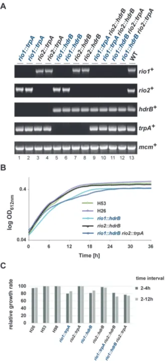
Both archaeal Rio proteins are dispensable for cell viability in H. volcanii. (A) Diagnostic PCR of wild-type, single and double dele- tion strains using primer pair’s specific for rio1, rio2, mcm, trpA and hdrB genes (Supplementary Table S3). Strains tested: rio1::trpA (lanes 1 and 2, strains SMH666 and SMH667, respectively), rio2::trpA (lanes 3 and 4, strains SMH668 and SMH669), rio1::hdrB (lanes 5 and 6, strains SMH670 and SMH671), rio2::hdrB (lanes 7 and 8, strains SMH672 and SMH673), rio1::trpA rio2::hdrB (lanes 9 and 10, strains SMH674 and SMH675), rio1::hdrB rio2::trpA (lanes 11 and 12, SMH676 and SMH677) and wild-type DS70 (lane 13). (B) Growth analysis of single and dou- ble deletion strains. Exemplary growth analysis for wild-type (H26 and H53), rio1 and rio2 single deletion (SMH670 and SMH672) and rio1 rio2 double deletion (SMH676) strains are depicted. The depicted curves correspond to the average growth of 6 independent measurements. (C) Relative growth rate of rio1 and rio2 mutant strains. Relative growth rate of strains rio1::trpA (SMH666), rio2::trpA (SMH668), rio1::hdrB (SMH670), rio2::hdrB (SMH672), rio1::trpA rio2::hdrB (SMH674), rio1::hdrB rio2::trpA (SMH676) at two time intervals (2–4 h and 2–12 h) are provided (see Materials and Methods).
wild-type (Figure 1B and C). Thus, in contrast to their eu- karyotic counterparts, rio1 and rio2 are individually non- essential for H. volcanii cell viability.
Next we sought to analyze possible genetic interaction/redundancy in the archaeal cellular con- text. To this end, we attempted to make rio1 rio2 double deletions by replacing rio1 with trpA in the rio2::hdrB single deletion strain and rio2 with trpA in the rio1::hdrB single deletion strain, again using the pop-in / pop-out method.
In addition, we generated rio1 rio2 double deletions by exploiting the previously described mating system for H.
volcanii (44,45) (see Material and Methods). Remarkably, double deletions (rio1::trpA rio2::hdrB and rio1::hdrB rio2::trpA) were readily obtained by both methods (Fig- ure 1A). Growth of the rio1 rio2 double deletions was comparable to the single rio1 deletion strains (Figure 1B and C), indicating that the genes do not share an essential overlapping function.
Next, we analyzed the effect of Rio1 and Rio2 deletions on the rRNA maturation pathway in H. volcanii. In con- trast, to bacteria and eukarya, the exact order and nature of the rRNA maturation events are poorly characterized in archaea (11–13). Therefore, by analogy to the processing de- fects observed after depletion of Rio1 or Rio2 in eukaryotes [defective processing at the 3
end of the SSU rRNA (29,30)], we focused on this particular rRNA region. Similarly, to the phenotype observed in eukaryotic cells, deletion of either H.
volcanii Rio protein (Hv Rio) affected processing at the 3
end of the 16S rRNA, whereas the levels of 5
extended 16S pre-rRNA intermediates were similar in wild-type and rio1 or rio2 deleted strains (Figure 2A and data not shown).
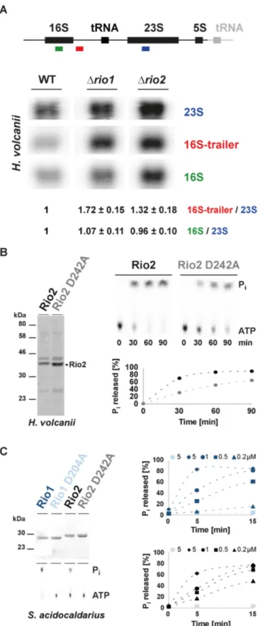
Previous structural and functional studies (23,33) sug- gested that the Rio proteins use an unusual ATP hydroly- sis cycle, which includes an autophosphorylation step at an invariant aspartate (phospho-aspartate––P-Asp) (kinase- like function) and a subsequent release of the phosphate moiety (ATPase-like function). To assess the evolution- ary conservation of this characteristic, we purified recom- binant Hv Rio1 and Hv Rio2 expressed in E. coli. Un- fortunately, Hv Rio1 solubility, yield and purity was not sufficient to perform accurate biochemical characteriza- tion of its activity (data not shown). In contrast, the yield of Hv Rio2 was comparably higher but still con- tained substantial amounts of impurities (Figure 2B). De- spite this limitation, we performed single-turnover exper- iments using recombinant Hv Rio2 and Hv Rio2 D242A (Mg
2+coordinating / P-Asp residue) (Supplementary Fig- ure S1). As shown in Figure 2B, and similarly to its eu- karyotic counterpart (23,33), Hv Rio2 hydrolyzed ATP and released P
i. To confirm this result, we purified re- combinant Rio1 and Rio2 derived from the crenarchaeon Sulfolobus acidocaldarius (Saci). Both proteins were puri- fied in large amounts (Figure 2C and data not shown).
As shown in Figure 2C, both proteins exhibited protein
concentration-dependent ATP hydrolysis activity. Finally,
we also extended our analysis of the Rio-dependent AT-
Pase activity by analyzing the catalytic activity of an ex-
emplary eukaryotic Rio3 family member in vitro (Supple-
mentary Figure S2). Likewise, Rio3 protein exhibited pro-
tein concentration-dependent ATP hydrolysis activity (Sup-
plementary Figure S2 and data not shown). Finally, we at-
Figure 2.
The archaeal Rio proteins are ATPases involved in 16S rRNA processing. (A) Analysis of rRNA maturation in H. volcanii strains deleted of Rio1 or Rio2. The schematic rDNA operon organization from H.
volcanii is depicted (upper panel). Note that H. volcanii contains two nearly identical rDNA operons. For simplicity, the main difference (the presence of an additional tRNA at the 3
end of one of the operons) is
tempted to confirm the presence of a P-Asp intermediate using hydroxylamine sensitivity of this intermediate as pre- viously described (23,33). However, owing to the stringent conditions (high-salt or high-temperature) required for the archaeal Rio protein activity, we could not reliably detect stabilized P-Asp intermediates under the conditions tested (data not shown).
Together, our results suggest that the aRio proteins are non-essential for cell viability under laboratory growth con- ditions. In addition, both aRio proteins participate in the in vivo 3
end maturation of the 16S rRNA. Finally, both aRio proteins showed ATPase activity in vitro.
Evolutionary conservation of RNA-dependent regulation of the Rio catalytic activity
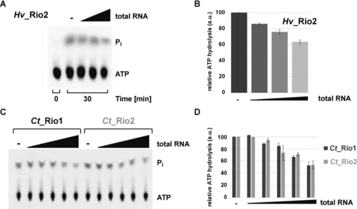
In eukaryotes, the Rio proteins, like many other ribosome assembly factors, are thought to be associated to distinct, relatively long-living, pre-ribosomal particles (1,3,4). This feature suggests a spatial and temporal regulation of the Rio functions. Therefore, we sought to analyze the molecu- lar requirements that allow regulation of the Rio-dependent ATP hydrolysis. To this end, we analyzed the influence of adding increasing amounts of total RNA on the Rio- dependent ATP hydrolysis-rate. As depicted in Figure 3, the ATPase activity of archaeal Rio2 was reduced in an RNA concentration-dependent manner (Figure 3A and B). To confirm and extend this observation we turned to the better-established eukaryotic Rio1 and Rio2 proteins, and specifically, those encoded by the thermophilic fungus Chaetomium thermophilum (Ct) (23,33). Remarkably, the ATPase activities of the eukaryotic Ct Rio1 and Ct Rio2 were also regulated in an RNA-dependent manner (Figure 3C and D). Collectively, our results are in good agreement with the evolutionary conservation of the catalytic RIO do- main shared within the Rio protein family, and suggest that the conserved Rio protein activity can be regulated by the presence of nucleic acids.
Rio2’s P-loop lysine facilitates ATP hydrolysis reaction Next, we aimed to determine the functional relevance of this RNA-dependent regulation for the maturation of the
←−−−−−−−−−−−−−−−−−−−−−−−−−−−−−−−−−−−−−−−−−−−
indicated in gray. Northern analysis was performed with the indicated color-coded probes (oHv189–16S, oHv190–16S trailer and oHv194–23S rRNA). Quantifications and standard deviation were derived from bio- logical triplicate. (B) Hv Rio2 acts as an ATPase in vitro. ATPase ac- tivity of purified Hv Rio2 and catalytic mutant Hv Rio2 D242A (Mg
2+coordinating
/putative P-Asp) (Coomassie - left panel) was analyzed by
single-turnover experiments (right panel). Released free phosphate (P
i)
separated from ATP by thin-layer chromatography and detected by phos-
phorimaging is shown. Hv Rio2 and Hv Rio2 D242A are depicted in black
and grey, respectively. (C) S. acidocaldarius Rio proteins act as ATPase in
vitro. Same as in (B) purified Saci Rio1 and Rio2 and their corresponding
catalytic mutants [D204A and D242A, respectively (Mg
2+coordinating
/putative P-Asp) - Coomassie upper left panel] were subjected to single-
turnover analysis with the indicated concentrations of recombinant pro-
teins. Saci Rio1
/Saci Rio1 D204A are depicted in dark and light blue,
respectively and Saci Rio2
/Saci Rio2 D242A are depicted in black and
gray, respectively. For simplicity, standard deviation are not represented in
(B) and (C). Note that the dashed-lines connecting the different time points
are only depicted to facilitate reading of the figure.
Figure 3.
RNA-dependent regulation of the RIO catalytic activity. (A and B) Exemplary RNA-dependent regulation of an archaeal Rio family member.
ATPase activity of purified Hv Rio2 in presence of increasing amounts of H. volcanii total RNA (up to 2
g
/l) is shown in (A) and the quantitation of four independent experiments is shown in (B). (C and D) RNA-dependent regulation of Ct Rio1 and Ct Rio2 ATPase activity. Single-turnover experiments (
≈0.5
M protein) performed in presence of increasing amounts of yeast total RNA (up to 2
g
/l) are shown in (C) and the quantitation of four independent experiments is shown in (D).
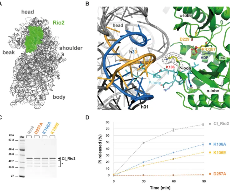
nascent small ribosomal subunit. To get insight into the molecular basis of the observed RNA-dependent regula- tion of the Rio catalytic activity, we went back to previously published data that provided a relative topological orienta- tion of the eukaryotic Rio2 bound to the nascent pre-40S (Figure 4) (33,46). On the basis of these studies, we pos- tulated that the invariant P-loop lysine [S. cerevisiae (Sc) Rio2 K105 / Ct Rio2 K106] could play a role in the Rio2 catalytic cycle’s regulation by coordinating rRNA binding and promoting catalytic activity (Figure 4 and Supplemen- tary Figure S1).
We previously showed that Sc Rio2 invariant P-loop ly- sine K105 is part of the binding surface involved in Rio2- pre-40S binding (33). Interestingly, this lysine is also part of the conserved P-loop motif GxGxxGKES (22,25,26) (Sup- plementary Figure S1). P-loop motifs are structurally flex- ible motifs that participate in the catalytic process by en- suring proper coordination of the nucleotide (47). Together these observations suggested that the Rio2 P-loop lysine can potentially adopt, at least, two possible conformations: an RNA-bound conformation (hereafter called K-bd) and an RNA-unbound conformation (hereafter called K-un) stim- ulating ATP hydrolysis.
To test this hypothesis, we first analyzed the role of the Rio2 P-loop lysine in stimulating ATP hydrolysis (K105 / K106 in S. cerevisiae and C. thermophilum, respectively).
To this end we took advantage of the previously devel- oped Ct Rio2 in vitro ATPase assay (33). Recombinant
Ct Rio2 K106A and K106E mutant proteins were gener- ated and their respective in vitro ATPase activities were an- alyzed. As shown in Figure 4 both Ct Rio2 K106A and K106E have reduced ATPase activity in vitro as compared to wild-type Ct Rio2. However, the observed reduction in catalytic turnover-rate was not as strong as that observed after mutating the critical catalytic residue Ct Rio2 D257 (Mg
2+coordinating / phospho-Asp residue) (Figure 4C-D and Supplementary Figure S1). Together, these results sug- gest that the integrity of the P-loop lysine is necessary for full activation of the enzyme.
Rio2’s P-loop lysine is necessary for optimal growth at low temperature and SSU biogenesis
Previous work demonstrated that impaired Rio2 catalytic
activity confers cold sensitivity to yeast strains (33). To con-
firm the role of the P-loop lysine in stimulating ATP hy-
drolysis we analyzed the effect of mutating the P-loop on
cell growth and ribosome biogenesis. As described previ-
ously, cells solely expressing Sc Rio2 K105E are inviable,
owing to its inability to establish stable contact with the
nascent pre-40S (33). In contrast, cells expressing Sc Rio2
K105A supported growth and showed an intermediate
cold-sensitivity phenotype as compared to mutation affect-
ing other critical catalytic residues (Figure 5). This interme-
diate cold-sensitivity is in good agreement with our single-
turnover measurements showing that Ct Rio2 K106A only
Figure 4.
P-loop lysine facilitates ATP hydrolysis reaction. (A) Relative positioning of Ct Rio2 into the yeast pre-40S structural context. Ct Rio2 (in green) docked on the yeast 40S particle (in grey), based on the cryo-electron microscopy map of the pre-40S particle as described previously (33), is depicted from the inter-subunit face. (B) Detailed view of the Ct Rio2 binding interface with the pre-40S. Ct Rio2 is depicted in green, the P-loop lysine (Ct Rio2 K106
/Sc Rio2 K105), the catalytic aspartate (Ct Rio2 D229
/Sc Rio2 D229), and the coordinating Mg
2+/P-Asp (Ct Rio2 D257
/Sc Rio2 D253) are indicated.
The 18S rRNA helix h30, in close proximity of Ct Rio2 K106 (Sc Rio2 K105), which is formed by two 18S rRNA elements (1169–80 and 1458–70 Sc numbering) are depicted in blue and yellow respectively and h31 in black (see Supplementary Figure S3 and below). The flexible loop (F-loop) of Rio2, known to be important for the interaction with the pre-40S, is shown in cyan sticks. (C) Purification of Ct Rio2 and P-loop mutants. Purified Ct Rio2, coordinating Mg
2+/P-Asp mutant (D257A), and P-loop lysine mutants (K106A and K106E) are shown. * Ct Rio2 indicates degradation products. (D) ATPase activity of P-loop lysine mutants. ATPase activity of Ct Rio2, D257A, K106A and K106E are depicted. Quantitation and standard deviation were derived from four independent experiments. Note that the dashed-lines connecting the different time points are only depicted to facilitate reading of the figure.
mildly affects ATP hydrolysis. In addition, this suggests that Sc Rio2 K105A is still able to bind, to some ex- tent (see below), to the pre-40S particles, and partially fulfill its function. Moreover, similar to the catalytic mu- tant Sc Rio2 D253A (Ct Rio2 D257A; Mg
2+coordinating / phospho-Asp residue), Sc Rio2 K105A leads to pre-40S biogenesis defects as shown by polysome profile analy- sis (Figure 5C). Finally, in good agreement with a P- loop lysine-dependent small ribosomal subunit progression, cells expressing Sc Rio2 K105A mutation showed synthetic growth defect in combination with loss of Ltv1, a non- essential pre-40S biogenesis factor (48,49) (Figure 5D). To-
gether our results suggest that the Rio2 P-loop lysine partic- ipates in apparently two (mutually exclusive) steps: (i) bind- ing of Rio2 to pre-40S and (ii) stimulation of ATP hydroly- sis.
Rio2’s P-loop lysine modifies the Rio2 binding affinity land- scape to the pre-40S
To further substantiate the P-loop lysine dual function, we
turned to genetic analysis in S. cerevisiae. Previously, we
showed that overexpression of catalytically impaired Rio2
mutants are dominant negative provided that the integrity
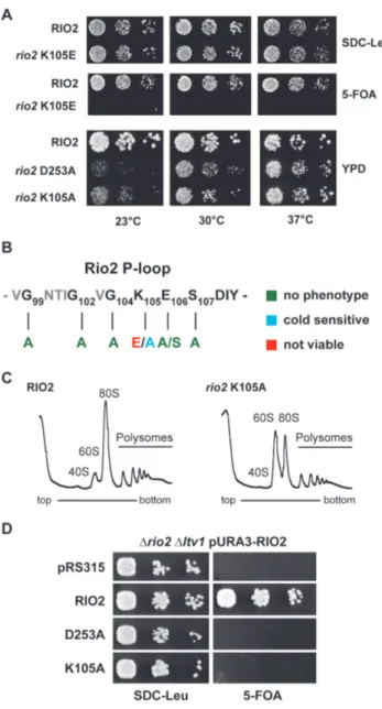
Figure 5.
P-loop lysine is necessary for optimal growth at low tempera- ture and SSU biogenesis. (A) In vivo analysis of P-loop lysine mutants. Se- rial dilutions of yeast strains expressing wild-type RIO2 or the indicated mutations were spotted on the indicated plates and incubated at the indi- cated temperatures. RIO2 shuffle strain transformed with LEU2-carrying plasmids harboring wild-type Sc RIO2 or Sc rio2 K105E, were spotted on SDC-Leu and SDC plus 5-FOA plates (to shuffle out wild-type pURA3- RIO2). Yeast strains solely expressing wild-type RIO2 or the indicated mu- tations Sc rio2 D253A and K105A were spotted on YPD. (B) Summary of phenotypes observed after P-loop mutagenesis. The phenotypes observed after individually mutating the following Sc Rio2 P-loop residues G99A, G102A, G104A, K105A, K105E, E106A, E106S, S107A and S107E are summarized. (C) Polysome profile analysis of the Sc rio2 K105A mutant.
Whole cell lysates from cells incubated for 4 h at 23
◦C were separated on a 5–40% sucrose gradient. The A
260nmprofiles of the derived sucrose gradi- ent fractions are depicted. (D) Genetic interaction of Sc rio2 K105A allele with the non-essential ribosome biogenesis factor Ltv1. Serial dilutions of yeast strains expressing wild-type RIO2 or the indicated mutations were spotted on the indicated plates and incubated at the indicated tempera- tures. RIO2 shuffle strain deleted for LTV1 and transformed with LEU2- carrying plasmids harboring wild-type and mutated Rio2, were spotted on SDC-Leu and SDC plus 5-FOA plates.
of the Rio2 interaction surface with the pre-40S is main- tained (33). On the basis of our observation that the P-loop lysine participates in pre-40S binding, we hypothesized that Sc Rio2 K105A affinity to pre-40S might be reduced. Ac- cordingly, we rationalized that (i) owing to reduced catalytic turnover rates and its reduced binding capacity to the pre- 40S, overproduction of Sc Rio2 K105A should have an in- termediate dominant negative effect as compared to over- production of Sc Rio2 D253A. Moreover, (ii) owing to the intrinsic reduced affinity of Sc Rio2 K105A, overproduc- tion of an Sc Rio2 K105A D253A double mutant protein should have a weaker dominant negative phenotype as com- pared to the overexpressed Sc rio2 D253A mutation alone (intragenic suppressor). As shown in Figure 6, overexpres- sion of Sc rio2 K105A results in an intermediate dominant negative phenotype as postulated above. Moreover, in good agreement with its dual function in pre-40S binding and ATP hydrolysis stimulation, the K105A mutation acts as a partial intragenic suppressor of Sc rio2 D253A (Figure 6A––23
◦C and 30
◦C panel).
To confirm this genetic observation, and to test the rela- tive binding affinity of Rio2 to the pre-40S in a more direct way, we analyzed salt-dependent interaction of Rio2 vari- ants with the 20S pre-rRNA as described previously (42).
As shown in Figure 6B, the apparent interactions of Rio2, Rio2 D253A and Rio2 K105A with pre-20S rRNA were similar at lower salt concentration (200 mM KCl), whereas increased salt concentration (400 mM KCl) revealed a re- duced affinity of the Rio2 K105A variant toward the 20S pre-rRNA in comparison to the Rio2 wild-type and Rio2 D253A (Figure 6B and below).
Collectively, our genetic and biochemical analyses strengthen the idea that the Rio2 P-loop lysine mutants have both a lower affinity to pre-40S and a reduced ATPase activity (Figures 4 and 6, respectively) (33). Moreover, our results also indicate that Rio2 might adopt at least two discrete conformational states a K-bd/ K-un. Therefore, we conclude that the P-loop lysine plays a central role in regulating the molecular events contributing to the Rio2 binding and release steps from the pre-40S.
Rio2’s P-loop lysine contributes to the RNA-dependent regu- lation of Rio2 ATPase activity
To further support our findings, we performed single- turnover measurements of wild-type Ct Rio2, Ct Rio2 K106E and K106A in presence of yeast total RNA. As shown in Figure 7, whereas wild-type Ct Rio2 ATPase ac- tivity was inhibited by the presence of total RNA, the Ct Rio2 K106E and K106A mutants were less sensitive to RNA-dependent inhibition of ATPase activity.
Next, we aimed to identify the rRNA counterpart
that could recapitulates RNA-dependent inhibition of the
Ct Rio2 ATPase activity in vitro. Previous work demon-
strated that Rio2 crosslinks with helix 31 (h31) of the 18S
rRNA (50). In good agreement, mutational analysis and
docking of Rio2 in the pre-40S cryo-EM map suggested that
the flexible loop which is part of the RIO domain might es-
tablish contact with this rRNA helix (33,46) (Figure 4). In
addition, previous analyses suggested that Rio2 K106 is in
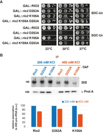
Figure 6.
P-loop lysine modifies the Rio2 binding affinity landscape to the pre-40S. (A) Sc rio2 K105A is a partial suppressor of the dominant- negative Sc rio2 D253A mutant. Serial dilutions of a wild-type yeast strain carrying the same plasmid with galactose-inducible GAL::RIO2, GAL::rio2 D253A, GAL::rio2 K105A and GAL::rio2 K105A D253A (in- tragenically combined), respectively, were spotted on SDC-Ura (glucose, repressed) and SGC-Ura (galactose, induced) plates and incubated at the indicated temperatures. (B) Salt-dependent interaction of Sc Rio2 variants with 20S pre-rRNA. Wild-type yeast cells (BY4741-Euroscarf) were trans- formed with a plasmid carrying the indicated bait proteins fused with a C-terminal TAP-tag and were grown in SDC-Leu to OD
600 nm≈0.8. The indicated bait proteins were purified as described in Material and Meth- ods. To test salt-dependent interaction of the Sc Rio2 variants with 20S pre-rRNA, immobilized proteins were extensively washed with a buffer containing either 200 or 400 mM KCl and the associated pre-20S rRNA was analyzed by Northern blot (see Materials and Methods). For North- ern blot (NB) analysis membrane were hybridized with a probe antisense to the ITS1 region between D-A
2and exposed to a Phosphorimager screen.
Quantitation and standard deviation were derived from 3 independent ex- periments. Immobilized bait proteins were analyzed by Western blotting (WB).
close proximity of other rRNA residues belonging to h30 (Figures 4 and 7B) (33,46).
In order to test the possible effects of these RNAs on the Ct Rio2 ATPase activity we performed in vitro transcrip- tion (and chemical synthesis) of a ‘native’ h31, a stabilized h31-like (h31*) and an h30-like rRNA (Figure 7 and Sup- plementary Figure S3).
In vitro transcripts mimicking the h31, h31* and h30 rRNAs were able to interact with an apparent weak affin- ity to Ct Rio2, as analyzed by electro mobility-shift assay (Supplementary Figure S3 and data not shown). Despite binding to Ct Rio2, the h31 or h31* RNA mimic did not in- terfere with the Ct Rio2 ATPase activity within the concen-
tration range tested (Figure 7C and data not shown). Next, we tested the effect of adding a mimic of the double stranded rRNA region forming h30, two well conserved rRNA ele- ments bridging the 5
and 3
rRNA parts forming the SSU head domain, on the Ct Rio2 ATPase activity (Figure 7 and Supplementary Figure S3). Interestingly, Rio2 ATPase ac- tivity was reduced in presence of h30 rRNA mimic (Figure 7D).
To further elucidate the molecular basis of the h30 RNA- dependent inhibition of Ct Rio2 ATPase activity we per- formed single-turnover experiments in presence of a ‘fully assembled’ h30 or of the individual single stranded RNA forming h30. Interestingly, RNA-dependent inhibition of Ct Rio2 ATPase activity was only observed in presence of the double stranded RNA mimic forming h30 (Figure 7E and data not shown). Finally, we analyzed the ability of h30 to inhibit Ct Rio2 ATPase activity in absence of a func- tional P-loop lysine (Ct Rio2 K106A). As shown in Fig- ure 7F, Ct Rio2 K106A was resistant to h30-dependent Ct Rio2 ATPase activity inhibition and was marginally af- fected by the presence of h30 RNA mimic in comparison to the wild-type situation (Figure 7F). Moreover, loss of h30 RNA-dependent Rio2 ATPase activity regulation corre- lated with a reduced binding affinity of Ct Rio2 K106A to- ward h30 RNA mimic, but not h31* (Supplementary Figure S3). Interestingly, and in contrast to its ATPase activity in- hibition, h30 RNA mimic interaction with Ct Rio2 K105A was not completely abolished and only slightly weakened.
These results suggest that additional structural regions of Rio2 (e.g. RIO domain/ wHTH domain) might participate in rRNA binding, however, without significant effect on the regulation of Ct Rio2 ATPase activity in the in vitro condi- tions tested (Figure 7F and Supplementary Figure S3).
Together, these results suggest that the P-loop lysine–
rRNA interaction contributes to the formation of a K-bd conformation leading to stabilization of a catalytically in- competent intermediate, and presumably ensures accurate control of Rio2 release from the pre-40S particle. More- over, our results also suggest that additional, not yet identi- fied, rRNA / Rio2 structural element interactions might ad- ditively contribute to the full Rio2 ATPase activity regula- tion in the context of the pre-40S particle.
ADP-dependent release of Rio2 from the pre-40S
Together, our work suggests a model where Rio2 binds to
the pre-ribosome in a K-bd conformation. Upon structural
rearrangement and/ or r-proteins assembly events, the P-
loop lysine might disengage from its interaction with the
rRNA and stimulate the formation of a catalytically ac-
tive conformation (K-un). This conformational transition
might allow proper positioning of the P-loop and / or the
catalytic center, thereby stimulating the ATP hydrolysis re-
action and the formation of an ADP::phospho-aspartate in-
termediate as previously suggested (33). However, the de-
creased affinity reached in this condition is presumably not
sufficient to directly trigger release of Rio2, since additional
Rio2 structural elements also contribute to the Rio2-pre-
40S interaction (33). In agreement with this idea is the ob-
servation that the Sc Rio2 K105A mutant already adopts
a lower binding affinity-state but still binds to the pre-40S
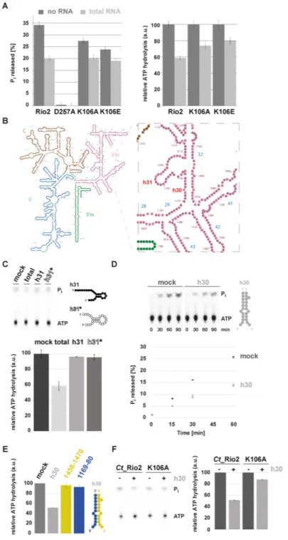
Figure 7.
P-loop lysine and h30 participate in the RNA-dependent regulation of Rio2 ATPase activity. (A) RNA-dependent regulation of Ct Rio2 ATPase activity. Single-turnover experiments using the indicated recombinant proteins (
≈0.5
M protein) were performed in presence of 2
g
/l of yeast total RNA. Percentage of released P
i(left panel) and the deduced relative ATPase activity (right panel) are shown. (B) Yeast 18S rRNA 2D structure. 2D structure prediction were obtained from
http://apollo.chemistry.gatech.edu/RibosomeGallery(72). The 5
domain (blue), Central domain (brown), 3
Major domain (magenta) and 3
minor domain (green) are depicted. A close-up of the h30 and h31 region is depicted in the right panel. (C) ATPase activity analysis in the presence of an h31 rRNA-mimic. Relative ATPase activity was monitored during 30 min by single-turnover experiment in presence of 2
g
/l total yeast RNA or 10
M of h31 derivatives (
≈0.5
M protein). Helix 31* is a stabilized version of h31 to which a series of A–U base pairing to stabilize the stem of this helix was added (see also Supplementary Figure S3). 2D RNA structure predictions are schematically represented (see Supplementary Figure S3 for further information). (D) ATPase activity analysis in the presence of an h30 rRNA-mimic. Time-dependent P
ireleased (in %) as obtained with Ct Rio2 (
≈0.5
M) incubated in presence of an excess (10
M) of an annealed h30-mimic is shown. 2D RNA structure predictions are schematically represented (see Supplementary Figure S3 for further information). (E) Double strand RNA integrity of h30 is required for RNA-dependent regulation of Ct Rio2 ATPase activity. Relative ATPase activity was monitored during 30 min by single-turnover experiment as obtained with Ct Rio2 (
≈0.5
M) incubated in presence of an excess (10
M) of an annealed h30-mimic and (20
M) of each individual single strand RNA region forming h30 (18S rRNA 1169–80 and 1458–70 Sc numbering, depicted in blue and yellow respectively). All quantitation and standard deviation were derived from 4 independent experiments.
(F) Ct Rio2 P-loop lysine integrity is required for efficient h30-dependent Rio2 ATPase activity inhibition. Relative ATPase activity was monitored during
30 min by single-turnover experiment as obtained with Ct Rio2 and Ct Rio2 K105A (
≈0.5
M) incubated in presence of an excess (10
M) of annealed
h30-mimic is shown.
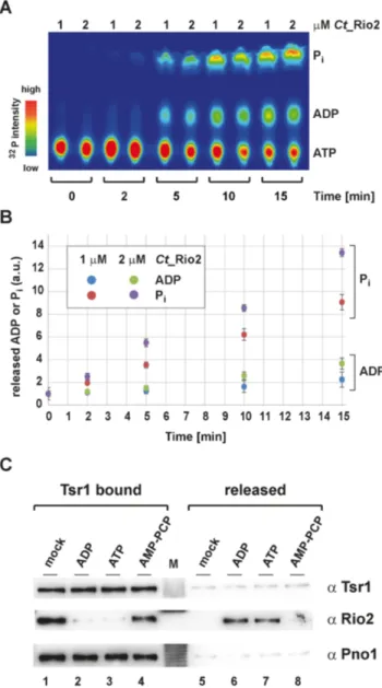
Figure 8.
ADP-dependent release of Rio2 from the pre-40S. (A) Time- and concentration-dependent release of ADP and P
i. Separation of ATP, ADP and P
iby thin-layer chromatography analysis is shown as a heat-map (
32P relative intensity). (B) Relative release of ADP and P
i. Quantitation and standard deviation derived from four independent single-turnover re- action measurements as shown in the exemplary panel (A) and obtained with the indicated concentration of Ct Rio2 are depicted. Note that the total amounts of
32P in the supernatant was constant over the time-course provided. (C) Nucleotide-dependent release of Rio2 from pre-ribosomal subunit. Nucleotide-dependent release of Rio2 from immobilized (Tsr1 bound) pre-ribosomal subunit were analyzed by Western blotting using the indicated antibodies.
(Figures 5 and 6). Finally, the catalytic cycle of other P- type ATPases is characterized by the formation of several discrete catalytic intermediates that correlates with differ- ent structural and functional states (35).
To further clarify the Rio2 catalytic cycle and its con- tribution to the Rio2 release from the pre-40S particle, we first analyzed the apparent release kinetics of ADP and P
iin vitro. To test whether ADP and P
ireleases are ki- netically distinguishable, we applied a modified version of our single-turnover assay. In this case, we incubated im-
mobilized Ct Rio2 with equal amounts of ␣
32P- and ␥
32P- labeled ATP. At each time points, Ct Rio2 bound to beads was spun down and an aliquot of the supernatant was col- lected ( = released material). The released material (ATP, ADP and P
i) was then analyzed by thin-layer chromatogra- phy. As shown in Figure 8, short-time kinetics demonstrated that P
i(P-Asp) is released with a faster kinetic than ADP, in a protein concentration manner in vitro. This result sug- gests that Rio2, unlike classical P-type ATPases (35), adopts an ADP bound state(s), as one of the last steps of its cat- alytic cycle. In good agreement with these results, whereas Rio2 nucleotide binding is not essential for pre-40S binding, a mutation affecting nucleotide binding residue (Sc rio2 K123A) affected yeast growth more strongly than the cat- alytic inactive mutants (Sc rio2 D229A, D253A, catalytic aspartate and Mg
2+coordinating / P-Asp, respectively) (33) (Supplementary Figure S1), thereby underscoring the im- portance of the nucleotide load for Rio2 biology in vivo. Ac- cordingly, we hypothesized that Rio2 release-competence from the nascent pre-40S is stimulated when Rio2 adopts an ADP-loaded state.
To analyze this possibility, we purified pre-ribosomes from yeast containing Sc Rio2 and analyzed its putative nucleotide-dependent release in vitro. As shown in Figure 8, in the absence of nucleotide (mock) or in presence of non- hydrolysable ATP, or GTP (data not shown) no significant Rio2 release was observed. In contrast, addition of ATP (51) or ADP stimulated Rio2 release from the pre-40S par- ticle (Figure 8C and data not shown). Together, these results strongly suggest that Rio2-binding to the pre-40S is desta- bilized when reaching an ADP-loaded state.
DISCUSSION
The Rio proteins are involved in ribosome maturation from archaea to human
Ribosome biogenesis in archaea is poorly characterized
(13,52). Interestingly, phylogenetic analyses have revealed
that the information-processing machineries (DNA replica-
tion, transcription, and translation) are from a sequence,
structural and functional point of view, more closely re-
lated to their eukaryotic counterparts (53). Early struc-
tural analysis of ribosomal subunits by negative staining
electron microscopy and more recent cryo-EM analysis re-
vealed structural features shared among archaea and eu-
karya (54,55). Moreover, archaea and eukarya share an
evolutionary related machinery, the snoRNP/sRNP, which
performs the vast majority of the rRNA modifications (2
-
O-methylation and pseudouridylation) (56). Furthermore,
whereas the set of ribosome assembly factors are not con-
served between bacteria and eukarya, some sequence ho-
mologs of eukaryotic-like ribosome biogenesis factors can
be found in most archaeal genomes (11,12). Finally, recent
metagenomic and phylogenetic analyses suggested an ar-
chaeal origin of the eukaryotic lineage (55,57–60). Together,
these observations indicate that part of the eukaryotic ribo-
some biogenesis pathway evolved on the basis of a simpli-
fied ancestral eukaryotic-like archaeal ribosome biogenesis
pathway. However, the in vivo functional similarities of this
pathway have not been firmly analyzed in the archaeal cellu-
lar context. Remarkably, our in vivo genetic analysis demon- strates that the H. volcanii rio1 and rio2 genes are neither individually essential for cell viability nor do they share an essential function, as cells deleted for both rio1 and rio2 are viable and display little growth impairment. The non- essentiality of the individual Rio proteins is not confined to the euryarchaeon H. volcanii: a recent ‘kinome-wide’ dele- tion analysis in the crenarchaeon S. acidocaldarius demon- strated that Saci Rio1 and Saci Rio2 are non-essential in this organism as well (61). The fact that the Rio proteins are individually non-essential in these two highly divergent archaeal organisms suggests that this pattern is likely to be conserved across most archaea and is in striking contrast to the essential role of the Rio proteins in eukarya (29,30).
Despite their non-essentiality, our evidence clearly indicates that the H. volcanii Rio proteins contribute to SSU matu- ration in a similar manner to their eukaryotic counterparts.
In doing so, they may provide a growth advantage to cells growing in sub-optimal (non-laboratory) conditions, thus explaining why they have been retained through evolution despite being non-essential in laboratory conditions. More- over, it suggests that both Rio proteins, like their eukaryotic counterparts, do not have essential overlapping functions, and indicates that the Rio functional specialization / split has probably occurred at an early stage during the evolution process, presumably at the level of the last archaea-eukarya ancestor.
In bacteria, the vast majority of ribosome assembly fac- tors are non-essential for cell viability and bacterial ribo- somes, as opposed to their eukaryotic counterparts, can be assembled in vitro from their purified structural com- ponents (2,15). Strikingly, archaeal ribosomal subunits can also be reconstituted in vitro (62,63). Whether archaeal ribosome synthesis takes advantage of a facilitated self- assembly process similar to bacteria remains to be de- fined in future work. Finally, deciphering the contribu- tions of bacterial- and eukaryotic-like features, in addition to archaeal-specific features required for efficient ribosome synthesis in archaea, will provide a deeper understanding of the evolution history of this fundamental pathway.
The Rio catalytic-cycle as a molecular clock to monitor and control pre-40S progression.
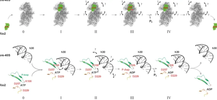
In this work, we also unravel a cross-talk regulatory mech- anism enabling the appropriate stimulation of the Rio2 catalytic activity and subsequent release of Rio2 from the nascent pre-40S particle. On the basis of our results [this work and (33)], we propose a model where the Rio2 protein first associates with the pre-40S particle in a catalytically inactive conformational-state where the P-loop lysine most likely interacts with h30 of the 20S pre-rRNA. In the course of the SSU maturation, several structural rearrangements within the SSU head domain might facilitate the disruption of catalytically inhibitory Rio2 / rRNA interactions (includ- ing the P-loop lysine / h30 contact). As a consequence, the free P-loop adopts or relays a structural conformation state within the Rio2 catalytic center that contributes to pro- mote ATP hydrolysis and to the subsequent formation of a Rio2::ADP::P-Asp intermediate. Finally, subsequent P-
Asp hydrolysis allows the formation of a release-competent Rio2::ADP intermediate (Figure 9).
The principle of the regulatory mechanism described above, which utilizes a conserved structural element of the shared RIO domain, is likely to be preserved within the Rio protein family. Whether this mechanism also holds true for RioB, a family member usually found in bacteria and some archaea (22), remains to be determined in future work. The current mechanism described in this study is also likely to apply to Bud32, a structural mimic of Rio1 (64). Bud32 is a subunit of the KEOPS complex which is involved in tRNA modification (65). Remarkably, Bud32 also acts as an AT- Pase (65). Based on our findings, it is tempting to speculate that structural rearrangements within the KEOPS::tRNA complex might control the ATPase activity of Bud32 and control the proper dissociation of the KEOPS complex from its substrate tRNA. In agreement with this idea the Bud32 P-loop orientation is not compatible with ATP hy- drolysis when bound to Kae1 (66).
Finally, the exact molecular determinants responsible for the controlled release of the other Rio proteins members remain to be fully characterized. Moreover, the respective contribution of the additional structural features, either flanking the RIO domain and / or within the RIO domain itself (e.g. the wHTH found in Rio2, and / or the shared flex- ible loop), to the regulation of the Rio catalytic cycle remain to be fully analyzed.
Integrated molecular-conformational dynamics: a pacemaker for ribosomal subunit progression?
The vast majority of ribosome biogenesis factors, includ- ing enzymes, are stably associated with defined set of pre- ribosome population. In addition, some of these factors are engaged in apparent long-living intermediates. How- ever, the exact molecular determinants responsible for the spatial and temporal regulation of the individual ribosome biogenesis factors activity remain poorly understood.
Our analysis of the Rio proteins provides additional hints of how this problem might be solved at the molecular level.
Interestingly, our work provides evidence for a dynamic in-
teraction process that coordinates and controls the affinity
and activity landscapes of the individual ribosome biogen-
esis factors. Together, local and long distance structural re-
arrangements induced by r-proteins assembly and rRNA
folding events presumably modifies the ribosome biogene-
sis factor assembly landscape. In agreement with this idea
is the gradual stable incorporation of the r-proteins which
also correlates with major maturation events, and presum-
ably correlates with the destabilization of ribosome biogen-
esis factors (42,67,68). Moreover, ribosome assembly is ac-
companied by various, sometimes dramatic, rRNA / protein
structural rearrangements which correlate with assembly
events (18,46,68–70). Finally, structural snapshots of pre-
ribosomal particles recently suggested that ribosome bio-
genesis factors, including Rio2, can adopt different confor-
mation on the pre-ribosomal particles (33,46). As such, ri-
bosome assembly efficiency relies on an integrated coopera-
tive molecular network utilizing conformational changes to
monitor and relay the current global and local pre-ribosome
assembly states.
Figure 9.