Future
Medicinal Chemistry
Future Med. Chem.
Systematic Review
913
2017
Aim: Resistances to antibiotics employed for treatment of infectious diseases have increased to alarming numbers making it more and more difficult to treat diseases caused by microorganisms resistant to common antibiotics. Consequently, novel methods for successful inactivation of pathogens are required. In this instance, one alternative could be application of light for treatment of topical infections.
Antimicrobial properties of UV light are well documented, but due to its DNA- damaging properties use for medical purposes is limited. In contrast, irradiation with visible light may be more promising. Method: Literature was systematically screened for research concerning inactivation of main oral bacterial species by means of visible light. Results: Inactivation of bacterial species, especially pigmented ones, in planktonic state showed promising results. There is a lack of research examining the situation when organized as biofilms. Conclusion: More research concerning situation in a biofilm state is required.
First draft submitted: 1 March 2017; Accepted for publication: 8 May 2017; Published online: 9 August 2017
Keywords: antimicrobial • bacteria • light • oral • visible
Far back in 1945, Sir Alexander Fleming already mentioned in his noble prize speech that there may be the ability of bacteria to get resistant to antibiotics
[1]. Since then, antimicrobial resistances of microorganisms have become an increasing problem to public health making it more difficult to treat dis- eases caused by resistant pathogens in lack of proper treatment modalities. This can lead to severe infections or even death of patients
[2]. In recent years, it has been reported that some bacterial strains have become resistant to all available antibiotics
[3]. Thus, development of novel strategies for treatment of infections caused by antibiotic-resistant pathogens has to be a major research goal in life sciences.
Also in the field of dentistry, administra- tion of systemic and topic antibiotics should be reduced as much as possible for reduc- ing the risk of inducing new resistances.
Furthermore, there are more and more
refractory infections of endodontic and peri- odontal origin caused by antibiotic-resistant pathogens
[4,5].
In this instance, the antimicrobial photo- dynamic therapy (aPDT) may be a promising alternative for topical killing of bacteria
[6,7]. Usually, aPDT consists of application of an external substance, the so-called photosen- sitizer (PS), and subsequent irradiation with light of an appropriate wavelength, result- ing in generation of reactive oxygen species (ROS) that kill bacteria by oxidative pro- cesses
[8]. Pronounced inactivation rates of oral biofilms have already been shown for aPDT
[9].
On the other hand, it is known that some bacteria are sensitive to irradiation with light only without the application of a light-sensitive molecule
[10,11]. For instance, irradiation with UV light, whose spectrum is subdivided into three sections (UV-C: 100–280 nm,
Antimicrobial efficacy of irradiation with visible light on oral bacteria in vitro:
a systematic review
Andreas Pummer*,1, Helge Knüttel2, Karl-Anton Hiller1, Wolfgang Buchalla1, Fabian Cieplik1 & Tim Maisch3
1Department of Conservative Dentistry
& Periodontology, University Medical Center Regensburg, 93053 Regensburg, Germany
2University Library, University of Regensburg, 93053 Regensburg, Germany
3Department of Dermatology, University Medical Center Regensburg, 93053 Regensburg, Germany
*Author for correspondence:
Tel.: +49 941 944 6016 Fax: +49 941 944 6025 andreas.pummer@ukr.de
For reprint orders, please contact reprints@future-science.com
UV-B: 280–315 nm and UV-A: 315–380 nm), proved to be able to inactivate bacteria
[12]. It was shown that its maximum bactericidal effect occurs within the UV-C range between 240–280 nm
[13]. However, there are several disadvantages of using UV light in patients.
It has been reported that absorption of UV-A light might result in damage of major biomolecules includ- ing DNA and membrane lipids in eukaryotic cells
[14]. It is also known that UV light leads to different classes of mutagenic and cytotoxic DNA lesions
[15]. High amounts of UV rays can also lead to skin cancer and eye conditions such as cataracts
[16]. Thus, the usage of UV light for topical killing of pathogens may not be a proper alternative for treatment of oral infections.
Consequently, application of visible light could be an alternative for inactivation of bacteria. Visible light corresponds to wavelengths between 380 and 750 nm reflecting a color range from violet to red
[17]. There are reports suggesting that light from the visible spectrum might lead to an autophotosensitization process induc- ing production of ROS in pathogens as a result of an accumulation of endogenous substances already present within biofilms or tissue that can act as PS
[11]. For con- trol of oral infections, irradiation with visible light may be a favorable approach due to the easy accessibility of the oral cavity compared with other parts of the body.
The aim of this systematic review was to assess the results of studies investigating the efficacy of treatment with visible light without external application of a PS in vitro on relevant bacteria occurring in the oral cavity.
Methods
As this systematic review does not study any health- related outcome of direct patient or clinical relevance, it was not considered eligible for registration in the International Prospective Register of Systematic Reviews PROSPERO.
The focused question of this systematic review was:
‘Is irradiation with visible light capable of inactivating oral bacteria in vitro?’
Identification of studies
Studies were identified by searching electronic data- bases and scanning the reference lists of eligible articles and relevant reviews. Search strategies were developed by a subject specialist and medical librar- ian who is trained and experienced in conducting sys- tematic literature searches (H Knüttel). Although we had no opportunity to have the search strategies peer- reviewed, we strived to design, carry out and report the literature search according to current checklists and recommendations
[18,19].
In the research question, we identified three search concepts that were combined using the Boolean
operator AND: ‘photoinactivation,’ ‘bacteria’ and
‘oral.’ For each of the concepts, search terms includ- ing synonyms were compiled and combined using the Boolean operator OR in order to compose a highly sensitive search strategy. We selected feasible search terms, relevant subject headings and appropriate syn- tax according to the databases and search interfaces.
No limits such as for date and language were imposed at the time of searching.
We selected databases by thematic relevance and accessibility. On 22 December 2016, we searched MEDLINE (Ovid: Epub Ahead of Print, In-Process &
Other Non-Indexed Citations, Ovid MEDLINE[R]
Daily and Ovid MEDLINE[R] 1946 to present), Embase (Ovid: Embase 1974 to 21 December 2016) and Web of Science (Science Citation Index Expanded 1965 to present; data last updated: 21 December 2016).
A detailed documentation of the searches allowing for replication is attached in Appendix 1. References were exported from the databases and deduplication was carried out using the method of Bramer et al.
[20].
The reference lists of eligible articles were scanned independently by two of the authors (subject special- ists) for additional relevant articles (A Pummer, F Ciep- lik). Occasional articles encountered by serendipity in other sources were also included.
Inclusion criteria
In vitro studies examining inactivation of bacteria by means of visible light irradiation without addition of an exogenous light-sensitive substance were taken into consideration. Bacteria could be organized in plank- tonic state or in biofilm state. Only studies treating bacteria typically occurring in the oral cavity were chosen. Studies published in English or German were included as a consequence of lack of proper expertise in other languages considering scientific issues. Neverthe- less, manual screening of different databases leads to the assumption that there were no relevant studies on this topic published in other languages.
Exclusion criteria
Systematic or nonsystematic reviews were excluded.
However, eligible studies found in thematically rel- evant reviews were included. Studies treating other microorganisms than bacteria such as fungi or viruses or studies where exogenous light-sensitive substances were added to bacteria as well as studies in which illu- mination was performed by means of nonvisible light were not taken into consideration. Non-in vitro stud- ies were not taken into consideration as we aimed to show susceptibility of bacteria to light itself like they occur in clinical practice without any outer influences.
Moreover, studies examining bacteria that were not of
oral origin or relevance were not included. Conference abstracts without full data or experimental details were excluded.
Data organization
A standard document, which included author(s) and year of publication, investigated microorganisms, orga- nization of microorganisms (planktonic or biofilm), type of light source, irradiation parameters (output power and intensity, wavelength, applied energy) as well as a summary of the main outcomes was used in order to systematize data received from each report
(Table 1).
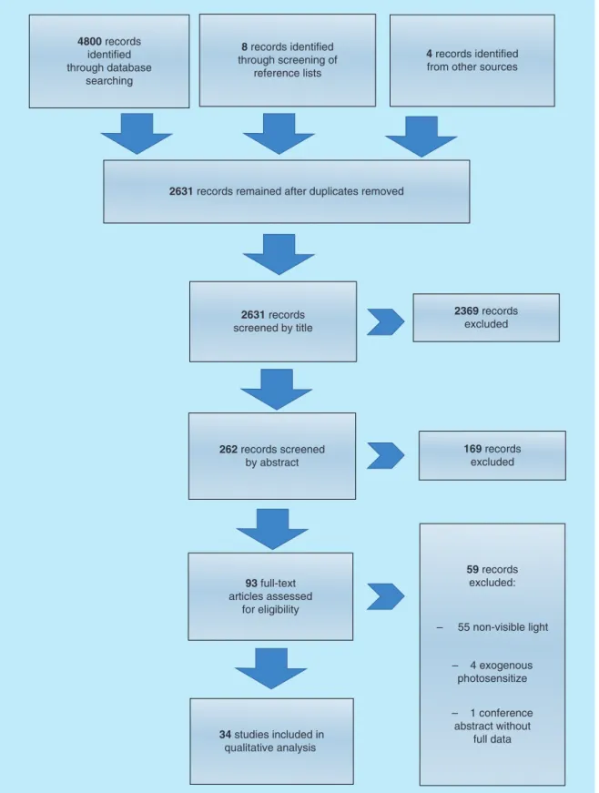
Results & discussionStudy identification
Searching of three electronic databases yielded a total of 4800 records (MEDLINE: 1411; Embase: 2010, Web of Science: 1379). After elimination of duplicates with the Bramer method, 2619 records remained
[20]. Eight additional studies were found in the reference lists of the included studies and another four in other sources.
Study selection
In the first step, studies were filtered by title screening by two independent subject specialists (A Pummer, F Cieplik). Studies not relevant by topic were excluded in this step. Second, the abstracts of all of the remaining studies were read and a decision considering suitability was made. In a third step, the full-text articles were read.
Only studies that were regarded as suitable after this step were included (
Figure 1for reasons of exclusion).
Screening by title left 262 records that were screened by abstract. 93 articles remained that were screened by reading the full text. Finally, a total of 34 articles relat- ing to the same number of studies (22 articles from the database searches plus 12 studies identified in the refer- ence lists and other sources) were considered as eligible for the review (
Figure 1for a PRISMA [Preferred report- ing items for systematic reviews and meta-analyses] flow diagram of the process
[54]).
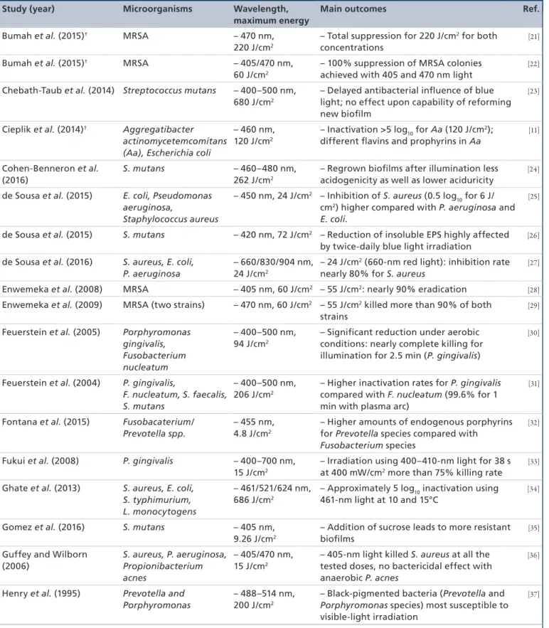
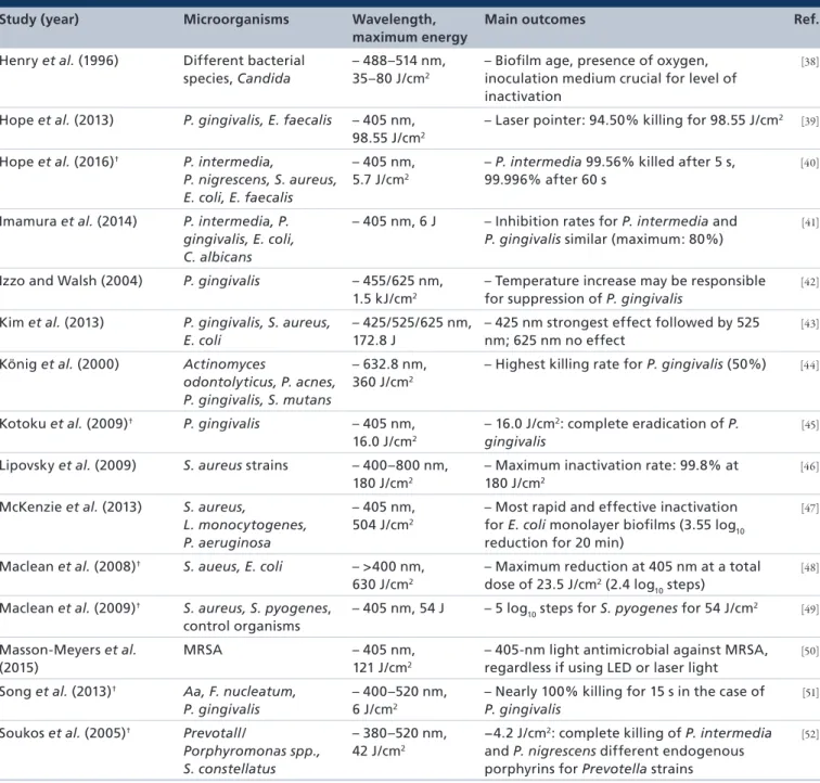
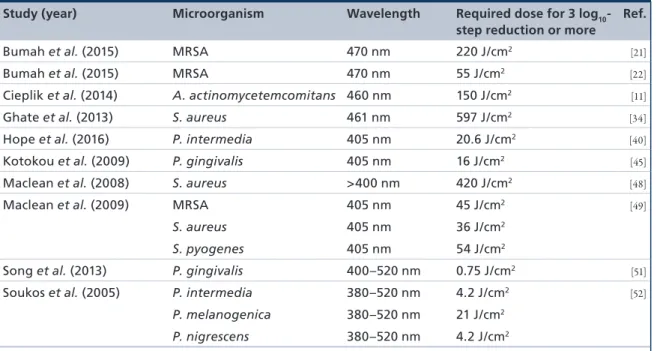
Table 1shows a summary of the 34 studies selected.
Table 2lists a subset of 9 of these 34 studies, where an inactivation of 3 log
10steps (99.9%) or more could be achieved. The microorgan- ism and wavelength tested is listed, as well as the light dose (J/cm
2) required to achieve a 3 log
10-step reduc- tion. An inactivation rate of 3 log
10steps was chosen as it is declared as a biologically relevant antimicrobial activity according to infection control guidelines
[53].
Actinomyces actinomycetemcomitansKönig et al. used planktonic suspensions of Actino- myces odontolyticus among others and exposed them to red light (60 mW helium–neon laser; 632.8 nm;
total energy density: 360 J/cm
2)
[44]. The killing rate
(CFU [colony forming units] values of laser-exposed bacteria compared with CFU values of nonexposed bacteria) for A. odontolyticus was 70 ± 4%, whereas it was 42 ± 10% for Propionibacterium acnes and 50 ± 10% for Porphyromonas gingivalis. In contrast, no effect could be observed upon illumination of Streptococcus mutans. As a result, it can be concluded that there was no antibacterial effect (99.9% reduction or more
[53]) for neither of the tested microorganisms. Additional fluorescence measurement proved existence of intracel- lular protoporphyrin IX for A. odontolyticus as well as for P. acnes indicating that the observed phototoxicity might be due to an autophotosensitization process
[44].
Aggregatibacter spp.In a study from our group, planktonic suspensions of Aggregatibacter actinomycetemcomitans were irradiated with a light-emitting diode (LED) light-curing unit with an emission peak at 460 nm at a total energy dose of 150 J/cm
2(1250 mW/cm
2; 120 s)
[11]. This resulted in a reduction of more than 5 log
10steps (antibacterial effect
[53],
Table 2) for A. actinomycetemcomitans, whereas there was no effect for blue-light irradiation of Escherichia coli, which was used as a control organism
[11].
Spectroscopic investigations showed presence of intracellular porphyrins and flavins. Excitation at the emission peak of the light source used in the experiments (460 nm) showed that particularly fla- vins may have been causative for inactivation of A. actinomycetemcomitans by irradiation with blue light
(Tables 1 & 2)[11].
However, these results are in marked contrast to those of Song et al., who examined the effect of blue-light irradiation on A. actinomycetemcomitans in planktonic suspensions as well as in biofilm state
[51]. A halogen lamp (
λ= 400–520 nm; output power of 500 mW/cm
2) was used for the experiments. Samples were exposed to light for 15–120 s (7–60 J/cm
2). There was no effect for blue-light illumination neither in planktonic nor in biofilm state for A. actinomycetemco- mintans, whereas blue light was strongly bactericidal to P. gingivalis and Fusobacterium nucleatum in planktonic state (
∼100% killing after 15 s of irradiation in the case of P. gingivalis, 99.1% following 60 s for F. nucleatum, respectively
[51]). Effect upon illumination of bacteria organized in biofilm state, however, was statistically significant only for P. gingivalis (
∼1 log
10reduction for 120 s, no antibacterial effect
[53]).
As these two studies showed totally different
results for phototoxicity of A. actinomycetemcomitans
toward blue light, no final conclusion on suscepti-
bility of A. actinomycetemcomitans to visible light
illumination can be drawn so far. The observed dif-
ferences may be due to different light sources (LED
Table 1. Summary of all 34 selected studies sorted by authors.
Study (year) Microorganisms Wavelength, maximum energy
Main outcomes Ref.
Bumah et al. (2015)† MRSA – 470 nm,
220 J/cm2
– Total suppression for 220 J/cm2 for both concentrations
[21]
Bumah et al. (2015)† MRSA – 405/470 nm,
60 J/cm2
– 100% suppression of MRSA colonies achieved with 405 and 470 nm light
[22]
Chebath-Taub et al. (2014) Streptococcus mutans – 400–500 nm, 680 J/cm2
– Delayed antibacterial influence of blue light; no effect upon capability of reforming new biofilm
[23]
Cieplik et al. (2014)† Aggregatibacter actinomycetemcomitans (Aa), Escherichia coli
– 460 nm, 120 J/cm2
– Inactivation >5 log10 for Aa (120 J/cm2);
different flavins and prophyrins in Aa
[11]
Cohen-Benneron et al.
(2016)
S. mutans – 460–480 nm,
262 J/cm2
– Regrown biofilms after illumination less acidogenicity as well as lower aciduricity
[24]
de Sousa et al. (2015) E. coli, Pseudomonas aeruginosa,
Staphylococcus aureus
– 450 nm, 24 J/cm2 – Inhibition of S. aureus (0.5 log10 for 6 J/
cm2) higher compared with P. aeruginosa and E. coli.
[25]
de Sousa et al. (2015) S. mutans – 420 nm, 72 J/cm2 – Reduction of insoluble EPS highly affected by twice-daily blue light irradiation
[26]
de Sousa et al. (2016) S. aureus, E. coli, P. aeruginosa
– 660/830/904 nm, 24 J/cm2
– 24 J/cm2 (660-nm red light): inhibition rate nearly 80% for S. aureus
[27]
Enwemeka et al. (2008) MRSA – 405 nm, 60 J/cm2 – 55 J/cm2: nearly 90% eradication [28]
Enwemeka et al. (2009) MRSA (two strains) – 470 nm, 60 J/cm2 – 55 J/cm2 killed more than 90% of both strains
[29]
Feuerstein et al. (2005) Porphyromonas gingivalis, Fusobacterium nucleatum
– 400–500 nm, 94 J/cm2
– Significant reduction under aerobic conditions: nearly complete killing for illumination for 2.5 min (P. gingivalis)
[30]
Feuerstein et al. (2004) P. gingivalis,
F. nucleatum, S. faecalis, S. mutans
– 400–500 nm, 206 J/cm2
– Higher inactivation rates for P. gingivalis compared with F. nucleatum (99.6% for 1 min with plasma arc)
[31]
Fontana et al. (2015) Fusobacaterium/
Prevotella spp.
– 455 nm, 4.8 J/cm2
– Higher amounts of endogenous porphyrins for Prevotella species compared with Fusobacterium species
[32]
Fukui et al. (2008) P. gingivalis – 400–700 nm, 15 J/cm2
– Irradiation using 400–410-nm light for 38 s at 400 mW/cm2 more than 75% killing rate
[33]
Ghate et al. (2013) S. aureus, E. coli, S. typhimurium, L. monocytogens
– 461/521/624 nm, 686 J/cm2
– Approximately 5 log10 inactivation using 461-nm light at 10 and 15°C
[34]
Gomez et al. (2016) S. mutans – 405 nm,
9.26 J/cm2
– Addition of sucrose leads to more resistant biofilms
[35]
Guffey and Wilborn (2006)
S. aureus, P. aeruginosa, Propionibacterium acnes
– 405/470 nm, 15 J/cm2
– 405-nm light killed S. aureus at all the tested doses, no bactericidal effect with anaerobic P. acnes
[36]
Henry et al. (1995) Prevotella and Porphyromonas
– 488–514 nm, 200 J/cm2
– Black-pigmented bacteria (Prevotella and Porphyromonas species) most susceptible to visible-light irradiation
[37]
For further details, see Supplementary Table 2.
†In these studies, a reduction of 3 log10 steps or more (antibacterial effect [53]) was achieved. Details are presented in Table 2.
EPS: Extracellular polysaccharide; LED: Light-emitting diode; MRSA: Methicillin-resistant Staphylococcus aureus.
light source in the first study
[11], halogen lamp in the second study
[51]) or due to different strains (ATCC 43718
[11]and ATCC 33384
[51], respectively) used for experiments.
As the first study clearly showed presence of fla- vins and porphyrins, killing by means of visible light could be a proper treatment modality for controlling infections caused by A. actinomycetemcomitans. In order to maintain the best results possible, light units showing broad overlap with the absorption spectra
of both flavins and porphyrins should be used for irradiation.
Fusobacterium spp.
Song et al. tested F. nucleatum in planktonic state as well as in biofilm state
[51]. Each bacterial sample was exposed to light from a halogen lamp (
λ= 400–
520 nm; output power of 500 mW/cm
2) for 15–120 s (7.5–60 J/cm
2). 99.1% reduction for planktonic bac- teria was achieved after illumination for 60 s (<3 log
10,
Study (year) Microorganisms Wavelength, maximum energy
Main outcomes Ref.
Henry et al. (1996) Different bacterial species, Candida
– 488–514 nm, 35–80 J/cm2
– Biofilm age, presence of oxygen, inoculation medium crucial for level of inactivation
[38]
Hope et al. (2013) P. gingivalis, E. faecalis – 405 nm, 98.55 J/cm2
– Laser pointer: 94.50% killing for 98.55 J/cm2 [39]
Hope et al. (2016)† P. intermedia,
P. nigrescens, S. aureus, E. coli, E. faecalis
– 405 nm, 5.7 J/cm2
– P. intermedia 99.56% killed after 5 s, 99.996% after 60 s
[40]
Imamura et al. (2014) P. intermedia, P.
gingivalis, E. coli, C. albicans
– 405 nm, 6 J – Inhibition rates for P. intermedia and P. gingivalis similar (maximum: 80%)
[41]
Izzo and Walsh (2004) P. gingivalis – 455/625 nm, 1.5 kJ/cm2
– Temperature increase may be responsible for suppression of P. gingivalis
[42]
Kim et al. (2013) P. gingivalis, S. aureus, E. coli
– 425/525/625 nm, 172.8 J
– 425 nm strongest effect followed by 525 nm; 625 nm no effect
[43]
König et al. (2000) Actinomyces
odontolyticus, P. acnes, P. gingivalis, S. mutans
– 632.8 nm, 360 J/cm2
– Highest killing rate for P. gingivalis (50%) [44]
Kotoku et al. (2009)† P. gingivalis – 405 nm, 16.0 J/cm2
– 16.0 J/cm2: complete eradication of P.
gingivalis
[45]
Lipovsky et al. (2009) S. aureus strains – 400–800 nm, 180 J/cm2
– Maximum inactivation rate: 99.8% at 180 J/cm2
[46]
McKenzie et al. (2013) S. aureus,
L. monocytogenes, P. aeruginosa
– 405 nm, 504 J/cm2
– Most rapid and effective inactivation for E. coli monolayer biofilms (3.55 log10 reduction for 20 min)
[47]
Maclean et al. (2008)† S. aueus, E. coli – >400 nm, 630 J/cm2
– Maximum reduction at 405 nm at a total dose of 23.5 J/cm2 (2.4 log10 steps)
[48]
Maclean et al. (2009)† S. aureus, S. pyogenes, control organisms
– 405 nm, 54 J – 5 log10 steps for S. pyogenes for 54 J/cm2 [49]
Masson-Meyers et al.
(2015)
MRSA – 405 nm,
121 J/cm2
– 405-nm light antimicrobial against MRSA, regardless if using LED or laser light
[50]
Song et al. (2013)† Aa, F. nucleatum, P. gingivalis
– 400–520 nm, 6 J/cm2
– Nearly 100% killing for 15 s in the case of P. gingivalis
[51]
Soukos et al. (2005)† Prevotall/
Porphyromonas spp., S. constellatus
– 380–520 nm, 42 J/cm2
−4.2 J/cm2: complete killing of P. intermedia and P. nigrescens different endogenous porphyrins for Prevotella strains
[52]
For further details, see Supplementary Table 2.
†In these studies, a reduction of 3 log10 steps or more (antibacterial effect [53]) was achieved. Details are presented in Table 2.
EPS: Extracellular polysaccharide; LED: Light-emitting diode; MRSA: Methicillin-resistant Staphylococcus aureus.
Table 1. Summary of all 34 selected studies sorted by authors (cont.).
Figure 1. Flowchart of the search strategy as well as study selection and data management procedure.
4800 records identified through database
searching
8 records identified through screening of
reference lists
4 records identified from other sources
2631 records remained after duplicates removed
2631 records screened by title
2369 records excluded
169 records excluded 262 records screened
by abstract
59 records excluded:
– 55 non-visible light
– 4 exogenous photosensitize
– 1 conference abstract without
full data 93 full-text
articles assessed for eligibility
34 studies included in qualitative analysis
no antibacterial effect
[53]), whereas there was no effect on F. nucleatum organized as biofilm for any irradiation period tested. Inactivation rates were slightly higher for
P. gingivalis under the same conditions (antibacterial
effect
[53]), whereas A. actinomycetemcomitans was not
susceptible to visible light irradiation in any case
(Table 1).
In another study, the effect of blue light from three different light sources was investigated for inactivation of planktonic F. nucleatum – two halogen lamps com- bined with filters (400–500 nm), a filtered xenon light source (plasma arc; 450–490 nm) and an LED (450–
480 nm)
[31]. Samples of bacteria in suspension and single bacteria on agar were exposed to light from the halogen lamps (260 and 416 mW/cm
2; 16–75 J/cm
2), the LED (520 mW/cm
2; 31–94 J/cm
2) and the plasma arc (1144 mW/cm
2; 69–206 J/cm
2). Corresponding irradiation periods for every sample were 1–3 min. As a result, higher inactivation rates could be detected when irradiation on agar plates was done in compari- son to irradiation of planktonic cultures: 2.5 min with halogen lamp 2 resulted ‘in nearly zero survival’
[31]when irradiated on agar (no antibacterial effect
[53]), whereas the survival rate was determined to be 40%
when performed in suspension. Irradiation of P. gin- givalis yielded higher inactivation rates compared with F. nucleatum (99.6% for 1 min with the plasma arc). Inactivation rates were below 99.9% in all cases meaning that no antibacterial effect was achieved
[53].
In addition, in this study the effect of blue light was evaluated on a bacterial lawn (minimal fluence required for inhibiting bacterial lawn from growing into biofilm: minimal inhibitory dose [MID]). The MID for P. gingivalis and F. nucleatum was deter- mined as 16–62 J/cm
2, whereas MID for S. mutans and S. faecalis was 159–212 J/cm
2(
∼3 min), indicating
that the Streptococcus strains tested were less susceptible to blue-light irradiation.
In a another study, the same group tested blue-light inactivation of bacteria under aerobic and anaerobic conditions as well as in presence of scavengers of ROS (dimethylthiourea, superoxide dismutase, ascorbic acid or a ‘cocktail’ of all three scavengers) on planktonic bacteria (for light sources, see
[31])
[30]. There were no significant reduction rates for illumination under anaerobic conditions, whereas significant reduction could be detected in aerobic environment (nearly com- plete eradication for illumination for 2.5 and 3 min with LED for P. gingivalis and F. nucleatum; 3 min corresponding to 94 J/cm
2; no exact data presented, only figures). Effect for F. nucleatum was smaller com- pared with P. gingivalis for all experiments, suggesting that P. gingivalis might be more susceptible to killing by blue light (no antibacterial effect in any case
[53]).
Addition of the scavengers only led to virtually smaller inactivation. For these experiments, only the halogen lamps and the plasma arc were used. The authors assumed that protection was incomplete due to inefficient access of the scavengers into the bacterial cells and inactivation might be due to photodynamic processes. In addition, poor inactivation under anaer- obic conditions indicate that oxygen is necessary for inactivation of bacteria or that production of endog- enous light-sensitive molecules may be downregulated under anaerobic conditions.
Table 2. Subset of nine studies, where an inactivation of ≥3 log10 steps† was achieved, sorted by the author.
Study (year) Microorganism Wavelength Required dose for 3 log10- step reduction or more
Ref.
Bumah et al. (2015) MRSA 470 nm 220 J/cm2 [21]
Bumah et al. (2015) MRSA 470 nm 55 J/cm2 [22]
Cieplik et al. (2014) A. actinomycetemcomitans 460 nm 150 J/cm2 [11]
Ghate et al. (2013) S. aureus 461 nm 597 J/cm2 [34]
Hope et al. (2016) P. intermedia 405 nm 20.6 J/cm2 [40]
Kotokou et al. (2009) P. gingivalis 405 nm 16 J/cm2 [45]
Maclean et al. (2008) S. aureus >400 nm 420 J/cm2 [48]
Maclean et al. (2009) MRSA 405 nm 45 J/cm2 [49]
S. aureus 405 nm 36 J/cm2
S. pyogenes 405 nm 54 J/cm2
Song et al. (2013) P. gingivalis 400–520 nm 0.75 J/cm2 [51]
Soukos et al. (2005) P. intermedia 380–520 nm 4.2 J/cm2 [52]
P. melanogenica 380–520 nm 21 J/cm2
P. nigrescens 380–520 nm 4.2 J/cm2
†Antibacterial effect [53].
MRSA: Methicillin-resistant Staphylococcus aureus.
Fontana et al. used different Fusobacterium spp.
for their experiments (F. nucleatum ss. nucleatum, F.
nucleatum ss. polymorphum, F. nucleatum ss. vincentii, F. periodonticum)
[32]. Irradiation of bacteria was per- formed using LED light at a wavelength of 455 nm at 80 mW/cm
2. Irradiation time was 60 s corresponding to an energy fluence of 4.8 J/cm
2. Killing rates were 46.9% for ss. nucleatum, 66.6% for ss. peridonticum, 67.4% for ss. vincentii and 93.6% ss. polymorphium, respectively. No antibacterial effect (3 log
10-steps reduction or more
[53]) could be shown for any of the tested species.
These studies suggest that different Fusobacterium strains might be susceptible to blue-light irradia- tion, but inactivation rates are lower compared with more pigmented genera (Porphyromonas, Prevotella).
This might be due to lower amounts of endog- enous porphyrins, as it was shown by the group of Fontana et al.
[32].
Porphyromonas spp.
Kim et al. evaluated the efficacy of three different wavelengths for inactivating P. gingivalis (three-in- one mounted LED package, peaks at 425, 525 and 625 nm corresponding to blue, green and red light, respectively)
[43]. Irradiation was performed for 0–24 h with an average output power of 6 mW/cm
2per hour (48–144 J/cm
2). At a wavelengths of 425 and 525 nm, viability was decreased by 40–60% and 10–20%, respectively. No bactericidal effect was observed for 625 nm. ‘For the suspensions containing 2 × 10
8CFU/ml, the optical density (OD)
600was decreased at 425 nm by 90–100% at all time points’
[43]and 40–70% by 525-nm light after 8 h, whereas viability increased when irradiated with 625-nm light for 8 h.
These results suggest that 425 nm is most effective in inactivating and suppressing P. gingivalis, followed by 525-nm light. 625-nm light did not show any effect for any case tested. As there is no accurate descrip- tion concerning exact percentage of reduction, the observed effect cannot be declared as an antibacterial effect
[53].
Feuerstein et al. investigated the phototoxic- ity of light from three different light sources (halo- gen lamp with a mounted filter,
λ= 400–500 nm, 260 mW/cm
2; a filtered xenon light source,
λ= 450–490 nm, 1144 mW/cm
2; LED,
λ= 450–480 nm, 520 mW/cm
2)
[30]. Using the LED, samples of F. nucleatum and P. gingivalis were irradiated for 1–3 min, corresponding to fluences of 31–94 J/cm
2, respectively. Inactivation only occurred under aero- bic conditions (P. gingivalis nearly 80% for 1 min; for further details, see
Table 1and the Fusobacterium spp.
chapter; no antibacterial effect
[53]in any case).
In another study
[31], the same group performed irradiation experiments using two halogen lamps com- bined with filters (Halogen 1 and Halogen 2; 260 and 416 mW/cm
2;
λ= 400–500 nm), an LED and a filtered plasma arc (xenon light source; for details, see
[30]).
Irradiation periods were 1, 1.5, 2, 2.5 or 3 min, result- ing in fluences of 16–75 J/cm
2for the halogen lamps, 31–94 J/cm
2for the LED and 69–206 J/cm
2for the plasma arc. As a result, inactivation was higher with the LED and plasma-arc light source compared with the halogen lamp, which probably was due to the higher output powers (1144 and 520 mW/cm
2for plasma arc and LED, respectively; 260 and 416 mW/cm
2for halogen lamps; 99.6% killing efficacy in suspen- sion for 1 min with the plasma arc for P. gingivalis; no antibacterial effect according to
[53]; compare
Table 1).
Henry et al. performed their experiments using an argon laser (
λ= 488–514 nm; 20–200 J/cm
2)
[37,38]. They were able to inactivate different Prevotella spp.
(P. intermedia ATCC 15033 and 49046; P. denticola ATCC 33184) and P. gingivalis (strains ATCC 33277 and ATCC 350406)
[37]. P. intermedia ATCC 15033 was most sensitive to irradiation ‘with no cells surviv- ing fluences of 70 J/cm
2or greater’
[37]. Because no exact data are presented, it was not possible to decide if there was an antibacterial effect
[53]. In addition, they tested if replacement of hemin in the medium against hemoglobin had any influence in susceptibility to laser irradiation. They could show that one P. gin- givalis strain (ATCC 33277) became tolerant to light irradiation in this case, indicating that hemin could be an essential factor for production of endogenous light-sensitive molecules. Also, the authors concluded that environmental oxygen is required for visible light inactivation. In general, nonblack-pigmented bacteria were much less sensitive to irradiation compared with black-pigmented bacteria
In a second study, the same group tested the sus-
ceptibility of bacteria organized in a biofilm grown on
agar medium using the same argon laser
[38]. Fluences
of 35–80 J/cm
2were able to inhibit biofilm growth
of P. endodontalis, P. gingivalis as well as of different
Prevotella spp, while there was no effect on the genera
Bacillus, Candida, Enterobacter, Proteus, Psuedomonas,
Staphylococcus and Streptococcus applying a fluence
of 70 J/cm
2 (Table 1). The level of inactivation was
affected by different parameters such as the biofilm
age, presence of atmospheric oxygen or medium used
for bacterial inoculation. Overall, the results were in
accordance with the first study, suggesting that inac-
tivation of different Prevotella and Porphyromonas
species might be possible in planktonic as well as in
biofilm state using an argon laser (no antibacterial
effect
[53]in any case).
Hope et al. irradiated a P. gingivalis suspension in 96-well plates
[39]. Irradiation was performed using the LEDs from a ‘toothcare device’ (
λ= 405 nm; power output: 3.2 mW) as well as a laser pointer (
λ= 405 nm;
power output: 42.7 mW) for 30, 60 and 300 s, cor- responding to fluences of 0.34, 0.68 and 3.42 J/cm
2, respectively, for the toothcare device, whereas in the case of the laser 0.5, 1 and 5 min, corresponding to 9.86, 19.71 and 98.55 J/cm
2, respectively, were chosen.
Irradiation with the LEDs was performed using E. fae- calis as control organism under the same conditions as described for P. gingivalis. Irradiation at a fluence of 0.34 J/cm
2with the LEDs resulted in a killing effi- cacy of 63.41%, whereas the killing rate at a fluence of 3.42 J/cm
2was 94.11%. Using the laser pointer, kill- ing rates were 90.21% for 9.86 J/cm
2and 94.50% for 98.55 J/cm
2. There was no effect on E. faecalis upon irradiation. In neither case, there was an inactivation rate of 3 log
10steps or more
[53].
Song et al. used a halogen lamp (dental-curing unit;
3M Curing Light XL3000, 3M ESPE, MN, USA;
λ= 400–520 nm; 500 mW/cm
2) for irradiation of bacteria both in planktonic as well as in biofilm state (6.5 J/cm
2up to 60 J/cm
2)
[51]. The killing rate for P. gingivalis in planktonic state was below detection limit with an irradiation time of only 15 s (antibacterial effect
[53],
Table 2
). When investigating visible light susceptibil- ity of bacteria organized in a biofilm, only P. gingivalis could be killed using the same irradiation procedure as it was used with planktonic bacteria. A. actinomy- cetemcomitans and F. nucleatum were not susceptible to inactivation by visible light irradiation when organized as biofilms (no antibacterial effect
[53]in any case).
Soukos et al. used a halogen lamp (
λ= 380–520 nm;
70 mW/cm
2; 0–42 J/cm
2) for irradiation of differ- ent black-pigmented bacteria
[52]. Survival fractions for P. gingivalis were 77.25% (4.2 J/cm
2), 12.55%
(21 J/cm
2) and 1.48% (42 J/cm
2). Killing rates for the tested Prevotella species (P. intermedia, P. nigres- cens, P. elanogenica) were higher compared with those of P. gingivalis. In contrast, S. constellatus, a nonpig- mented species, was not killed under the same condi- tions, suggesting that pigmentation of bacteria is an important factor for visible light phototoxicity. No antibacterial effect
[53]occurred in any case.
Fukui et al. aimed to determine the most effective wavelength for bacterial growth inhibition
[33]. In course of this, planktonic P. gingivalis was exposed to monochromatic light using an Okazaki large spectrograph (
λ= 400–700 nm in 10–20-nm steps;
50–400 mW/cm
2; 18 J/cm
2). Significant inhibition of P. gingivalis occurred between 400 and 410 nm, whereas no significant growth inhibition could be found when irradiation was performed at wavelengths
longer than 500 nm. For example, irradiation using 400–410-nm light for 38 s at 400 mW/cm
2resulted in a killing rate of more than 75% compared with nonirradiated control (no antibacterial effect
[53]).
Fontana et al. exposed P. gingivalis to an LED emit- ting light at 455 nm (80 mW/cm
2)
[32]. Irradiation time was 60 s corresponding to an energy fluence of 4.8 J/cm
2. The killing rate for P. gingivalis was 20.3%
(no antibacterial effect
[53]).
Furthermore, Kotoku et al. also performed photo- toxicity experiments on P. gingivalis using a violet laser diode module of oscillating wavelength at 405 nm (200–800 mW/cm
2; 2.0–16.0 J/cm
2)
[45]. An energy density of 4 J/cm
2led to growth inhibition of more than 97%, while 16 J/cm
2(20 s of irradiation) resulted in nearly complete eradication of P. gingivalis (anti- bacterial effect
[53],
Table 2). In general, higher output powers resulted in higher inactivation rates (400 mW for 5 s resulted in significant higher inactivation rates than 200 mW for 10 s; 2 J/cm
2, respectively).
Another study examined bacteria in suspension exposed to light from a helium–neon laser (
λ= 632.8 nm red light; light intensity of 100 mW/cm
2; fluence of 360 J/cm
2)
[44]. The killing rate for P. gingivalis was 41 ± 10% (70% ± 4% for A. odontolyticus; 42 ± 9%
for P. acnes; no effect for S. mutans). Additional test- ing of susceptibility of wild-type P. gingivalis isolated from plaque samples showed a killing rate of 41 ± 26%
(Prevotella spp. 58 ± 14%; A. actinomycetemcomitans 35
± 17%). These results suggest that it is also possible to kill bacteria using red light, although the fluences used in the study were much higher than those used in most of the studies using blue light. In all cases, there was no antibacterial effect
[53].
In contrast to the studies mentioned before, Izzo et al. assumed that killing of P. gingivalis was due to increasing temperatures, not due to a phototoxic effect. In this study, P. gingivalis was grown in a broth containing hemin and vitamin K
[42]. For irradiation experiments, two different LEDs with peaks at wave- lengths of 455 ± 20 or 625 ± 20 nm were used. Tubes containing bacteria were irradiated with a total dose of 978 J/cm
3(625 ± 20 nm) or 1.5 kJ/cm
3(455 ± 20 nm) in a temperature-controlled shaker. Temperature during irradiation was 39–40°C for blue light and 41–42.5°C for red light. Consequently, the authors suggested that the increase of temperature was responsible for sup- pression of P. gingivalis and not a phototoxic effect due to excitation of endogenous PS. However, it has to be considered that these light doses were very high which may explain the observed temperature increases.
Overall, these results show that P. gingivalis was
susceptible to blue- and red-light irradiation in most
of the studies, although an antibacterial effect
[53]was
observed in only two studies
[45,51](Tables 1 & 22). Inac- tivation rates for blue light were higher in general, but red-light irradiation could be particularly useful as it is known that light of longer wavelengths is able to penetrate tissues to a deeper extent
[55].
Prevotella spp.
Imamura et al. investigated the effect of blue light on P. intermedia
[41]. Irradiation was performed using a 405-nm monochromatic laser (300 s; 0.05–60 J).
Inhibition rates were 40% for 15 J,
∼70% for 30 J and
∼
80% for 45 and 60 J (no antibacterial effect
[53]).
Inhibition of P. intermedia was similar to the results found for P. gingivalis, while there was no effect for suspensions of E. faecalis. There was no antibacterial effect
[53]in any case.
Another group used planktonic P. intermedia, P. elanogenica and P. nigrescens grown anaerobically for their experiments (halogen lamp; 70 mW/cm
2; 0–42 J/cm
2)
[52]. Irradiation for 1 min (4.2 J/cm
2) resulted in an inactivation below detection limit for P. intermedia and P. nigrescens, while P. melanogenica was reduced by 70%. Inactivation below detection limit of the latter was achieved by 5 min of irradiation (21 J/cm
2; antibacterial effect
[53],
Table 2). Survival fractions for P. gingivalis were 77.25% (4.2 J/cm
2), 12.55% (21 J/cm
2) and 1.48% (42 J/cm
2), while there was no effect in the case of S. constellatus. HPLC anal- ysis showed endogenous porphyrins in P. intermedia, P. nigrescens and P. melanogenica
[52].
Hope et al. tested strains of P. intermedia and P. nigrescens
[40]. Irradiation was performed using an LED (19.1 mW/cm
2,
λ= 405 nm) and a laser pointer (346.2 mW/cm
2,
λ= 405 nm). Irradiation periods were 5–60 s for the laser (1.7–20.8 J/cm
2) and 10–300 s for the LED (0.19–5.7 J/cm
2). Control organisms (E.
coli, Staphylococcus aureus, E. faecalis) were irradiated for 300 s using the LED (5.7 J/cm
2). Killing rates for P. nigrescens for the LED were 64.1% after 30 s and 94.26% after 300 s. In the case of P. intermedia, even higher killing rates were observed using the LED (75.62% after 10 s, 96.51% after 60 s, 99.75% after 300 s). Using the laser pointer, 99.56% of bacteria were killed after 5 s and 99.996% after 60 s (antibacterial effect [Lit],
Table 2). Higher inactivation rates using the laser might be due to the much higher output power compared with the LED (346.2 mW/cm
2as compared with 19.1 mW/cm
2). With respect to the control organ- isms, only in the case of S. aureus, a statistically signifi- cant antimicrobial effect could be observed (36.73%
after 300 s, no antibacterial effect
[53]).
Fontana et al. used different Prevotella spp. for their experiments (P. intermedia, P. melanogenica, P. nigrescens; LED;
λ= 455 nm; 80 mW/cm
2; 60 s;
4.8 J/cm
2)
[32]. Survival fractions were 46.2% for P. ntermedia, 32.5% for P. nigrescens and 21.3% for P. melanogenica, respectively. The lowest survival rate was shown for F. nucleatum ss. polymorphum (6.4%), while the highest survival rate was shown for P. gin- givalis (79.7%; no antibacterial effect
[53]in any case).
Additional examination of endogenous porphyrin pro- duction showed 80- to 200-times higher amounts for Prevotella spp. compared with Fusobacterium spp.
Henry et al. performed experiments using an argon laser (
λ= 488–514 nm; 20–200 J/cm
2) for inactivation experiments of different Prevotella and Porphyromo- nas strains (please see
Table 1and Porphyromonas spp.
chapter)
[37,38].
König et al. evaluated the effect of red light on wild- type germs, isolated from patients with periodontal dis- ease (helium–neon laser,
λ= 632.8 nm, 360 J/cm
2)
[44]. The killing rate for Prevotella spp. was determined to be 58 ± 14% (no antibacterial effect
[53]).
Overall, these results show that Prevotella spp. might be susceptible to irradiation with blue and red light, although no antibacterial effect
[53]could be found in any of the studies. Visible light could be an adjunctive method in the treatment of diseases associated with the presence of these germs. As it was shown that inactiva- tion might be possible using blue light and also red light, a combination of blue and red light could be use- ful for inactivation of Prevotella species as this com- bination would combine maximum bactericidal effect and maximum tissue penetration, which is known to be higher for light of longer wavelengths
[55].
Staphylococcus spp.
de Sousa et al. investigated the effect of blue light emit- ted from a laser (
λ= 450 nm; 70 mW) on strains of S. aureus
[25]. Growth was inhibited at fluences higher than 6 J/cm
2(reduction of 22% for 6 J/cm
2; no higher inhibition rates detected for 12, 18 and 24 J/cm
2; no antibacterial effect
[53]in any case). In general, inhibi- tion of S. aureus was higher than inhibition observed for the control organisms Pseudomonas aeruginosa and E. coli.
Maclean et al. observed a significant bacteri-
cidal effect with a 5 log
10-step reduction (antibacte-
rial effect
[53]) upon illumination of S. aureus with a
broadband xenon light source (30 min; 350 mW/cm
2,
630 J/cm
2; 3 log
10reduction for 420 J/cm
2)
[48]. Fur-
ther experiments showed no effects for wavelengths
longer than 430 nm, suggesting that only a small
fraction within the wavelength range emitted by the
broadband light source was responsible for inactiva-
tion. Maximum reduction in dependence of applied
wavelength was observed at 405 nm at a fluence of
23.5 J/cm
2(2.4 log
10steps). Besides, they tested two
different MRSA strains (a laboratory strain and a clini- cal strain obtained from a wound infection) for suscep- tibility under the same conditions. Results were simi- lar to those obtained with S. aureus
[48](no detailed data presented). According to the study, there was an antibacterial effect
[53]with the broadband xenon light for 420 J/cm
2. For E. coli, which was used as reference strain, there was no inactivation at all for irradiation up to 30 min.
In another study by the same group, selected medi- cal pathogens (please see
Table 1) were illuminated with light from an arrow of LEDs (10 mW/cm
2) at a wavelength of 405 nm
[49]. An approximately 5 log
10- step reduction of CFU counts was achieved following exposure for 60–90 min (36–54 J/cm
2). As a result of all the tested bacteria, they concluded that lower doses are sufficient for inactivation of Gram-positive bacte- ria, with the exception of E. faecalis, which was least susceptible to irradiation performed at 405 nm. They concluded, that light emitted from an arrow of LEDs at 405 nm may be able to completely eradicate bacteria at high-population densities. For methicillin-suceptible S. aureus, 36 J/cm
2was needed for a 5 log
10-step reduc- tion, whereas 45 J/cm
2was necessary in order to reach the same effect for MRSA (antibacterial effect
[53]).
Using violet and blue light (
λ= 405 and 470 nm) from superluminescent diodes at different fluences (1–15 J/cm
2), Guffey et al. observed nearly 90% of bacteria killing for S. aureus strains at a fluence of 15 J/cm
2at 405 nm (160 mW)
[36]. 405-nm light killed S. aureus at all the tested fluences, whereas 470 nm (150 mW) showed an effect at 10 and 15 J/cm
2only exhibiting a 62% killing rate at 15 J/cm
2. Irra- diation of P. aeruginosa resulted in higher inactivation rates at all doses (95.1% maximum killing rate for 405 nm, 96.5% for 470 nm), suggesting that P. aeru- ginosa might be slightly more susceptible (no antibac- terial effect
[53]) to violet- and blue-light irradiation.
In contrast, no effect was reported upon irradiation of P. acnes, which is in contrast to several other in vitro and in vivo studies
[56,57].
Kim et al. performed experiments using 425, 525 and 625 nm light on P. gingivalis, S. aureus and E.
coli
[43]. Results suggest that 425 nm as well as 525 nm light might be able to inactivate bacteria with higher susceptibility for P. gingivalis and E. coli compared with S. aureus (for details, please see
Table 1and Porphyromonas spp. chapter).
In another study, irradiation with different wave- lengths from the visible and near-infrared spectrum (660 nm red light, 830 and 904 nm near-infrared) emitted from a laser was tested for its effect in inhib- iting growth of S. aureus. Inhibition of growth was observed for fluences higher than 12 J/cm
2 [27]. At a
fluence of 24 J/cm
2(660 nm red light; 30 mW out- put power), the inhibition rate was nearly 80% (no antibacterial effect
[53]). Red light was more effective than infrared light. S. aureus was more susceptible to irradiation at all tested wavelength regardless of the applied fluence than P. aeruginosa and E. coli (no antibacterial
[53]effect in any case).
Liposvky et al. tested two different strains of S. ureus for their susceptibility to visible light emit- ted from a halogen lamp (300 mW/cm
2; 1, 5, 10 min;
18–180 J/cm
2), one methicillin-sensitive and one methicillin-resistant strain
[46]. Results showed that the methicillin-sensitive strain was more susceptible to white light irradiation than the methicillin-resistant strain, with a maximum inactivation rate of 99.8%
at a fluence of 180 J/cm
2compared with 55.5% (no antibacterial effect
[53]in any case). Measurements of hydroxyl and superoxide radical production in illumi- nated bacteria as well as porphyrin synthesis showed higher amounts for the methicillin-sensitive strain, whereas the resistant strain was able to adapt to oxi- dative stress to a higher extent. Carotinoid produc- tion was also measured, exhibiting higher values for the resistant strain. These findings may explain the higher susceptibility of the methicillin-sensitive strain to visible light irradiation.
Emwemeka et al. used a 470-nm SLD (superlu- minescent diode) phototherapy device (30 mW/cm
2; 1–60 J/cm
2) for experiments with two strains of MRSA
[29]. At a fluence of 3 J/cm
2, nearly 30% inacti- vation was found for both strains. 55 J/cm
2led to more than 90% killing rate for both strains (no antibacterial effect
[53]in any case).
In another study performed by the same group, two strains of MRSA were irradiated with violet light
[28]. As a light source, a cluster of 36 SLDs emit- ting light ranging from 390 to 420 nm with a 405-nm peak was used (average power: 500 mW; irradiance:
100 mW/cm
2; 1–60 J/cm
2). Maximum eradication was achieved applying light for 9.2 or 8.4 min with 55 J/cm
2resulting in a killing rate of nearly 90% (no antibacterial effect
[53]in any case).
The same group used MRSA spread on tryptic soy agar for irradiation experiments with a 470-nm blue light-emitting LED (30 mW/cm
2; 55–220 J/cm
2)
[21]. 55 J/cm
2resulted in 86–92% inactivation while 110 J/cm
2and 220 J/cm
2resulted in total suppres- sion regardless of the cell concentration (antibacterial effect
[53],
Table 2).
Bumah et al. used an MRSA isolate for experiments with violet and blue light (405 and 470 nm)
[22]. Bacte- ria were spread on agar plates before irradiation. 100%
of MRSA colonies at a concentration of 3 × 10
6cells/ml
were suppressed by a single exposure to 55 or 60 J/cm
2of 470-nm light; double exposure to 405-nm light with a 6-h interval at 50, 55 or 60 J/cm
2showed the same result. Furthermore, this double treatment resulted in suppression of a cell density of 5 × 10
6CFU/ml, which was below detection limit (antibacterial effect
[53]). 7 × 10
6CFU/ml density in contrast had to be illuminated either once with 220 J/cm
2of 470-nm blue light or twice with 220 J/cm
2using 405-nm blue light in order to achieve the same effect. The authors concluded that repeated illumination may be necessary for com- plete eradication of denser bacterial concentrations, especially when applying lower fluences
(Tables 1 &2).
Masson-Meyers et al. compared the effect of 405-nm blue light emitted from an LED (100 mW/cm
2) with blue light emitted from a laser (135 mW/cm
2) on MRSA (5 × 10
6cells/ml)
[50]. Irradiation was performed once, twice or thrice with either light from the LED or from the laser at fluences of 40, 54, 81 or 121 J/cm
2. Time intervals in between were 15, 30 or 240 min. Results showed significant growth suppression for each fluence for both light sources with no ‘statistical difference’
for LED and laser ‘in 35 of the 36 experimental sam- ples’
[50]. Irradiation in two or three intervals increased bacterial suppression, especially when the treatment interval was 15 or 30 min. 54 J/cm
2triple irradiation with laser (intervals of 15 min) resulted in suppres- sion, which was below detection limit (antibacterial effect
[53],
Table 2).
Testing susceptibility of monolayer biofilms formed by different bacteria on glass surfaces, McKenzie et al.
performed irradiation for 5, 10 and 20 min using a 405-nm LED (141.48 mW/cm
2)
[47]. Inactivation rates for S. aureus were 0.61, 1.87, 2.75 log
10steps for 5, 10 and 20 min (0.7, 1.4, 2.8 J/cm
2), respectively (no anti- bacterial effect
[53]). Most rapid and effective inactiva- tion was observed for E. coli monolayer biofilms with a 2.52 log
10-step reduction upon 10 min of exposure to light, and 3.55 log
10-step reduction following 20 min of exposure (antibacterial effect
[53]). In the case of mixed-species biofilms formed by strains of S. aureus and E. coli, a 2.19 log
10-step reduction (total viable counts) after 30 min of irradiation was achieved.
Ghate et al. tested the antibacterial effect of LEDs emitting light in the visible region (7.5 h; 461 nm, 16 mW/cm
2, 432 J/cm
2; 521 nm, 22,1 mW/cm
2, 557 J/cm
2; 642 nm, 25,4 mW/cm
2, 640 J/cm
2) on S.
aureus as well as selected foodborne pathogens (E. coli, S. typhimurium, L. monocytogens)
[34]. Irradiance was performed at three different temperatures (20, 15 and 10°C). An approximately 5 log
10-step inactivation (antibacterial effect
[53]) for all the tested strains was observed using 461-nm light at 10 and 15°C, while inactivation rates for 521 nm light were 1–2 log
10steps only and there was no antibacterial effect
[53]for 642-nm light. In general, inactivation rates were higher at 10 and 15°C compared with 20°C.
It can be concluded that Staphylococcus spp. may be susceptible to visible light irradiation to a certain extent. Especially in the case of strains resistant to common antibiotics (MRSA), this might be a useful additional treatment modality.
Streptococcus spp.