Kathrin Abstiens
Heteromultivalent Nanoparticles for Selective Targeting of Renal Cells
Heteromultivalent Nanoparticles for Selective Targeting of Renal Cells
Dissertation zur Erlangung des Doktorgrades der Naturwissenschaften
(Dr. rer. nat.)
der Fakultät für Chemie und Pharmazie der Universität Regensburg
vorgelegt von Kathrin Abstiens aus Schwäbisch Gmünd
November 2018
Diese Arbeit entstand in der Zeit von November 2013 bis September 2017 am Lehrstuhl für Pharmazeutische Technologie der Universität Regensburg.
Die Arbeit wurde von Herrn Prof. Dr. Achim Göpferich angeleitet.
Promotionsgesuch eingereicht am: 30.11.2018 Tag der mündlichen Prüfung: 18.01.2019
Prüfungsausschuss: Prof. Dr. Jens Schlossmann (Vorsitzender) Prof. Dr. Achim Göpferich (Erstgutachter) Prof. Dr. Rainer Müller (Zweitgutachter) Prof. Dr. Frank-Michael Matysik (Drittprüfer)
Meiner Familie
Table of Contents
Heteromultivalent Nanoparticles for Selective Targeting of Renal Cells ... 1
Chapter 1 General Introduction ... 3
Chapter 2 Goals of the Thesis ... 19
Chapter 3 Microfluidic manufacturing improves polydispersity of multicomponent polymeric nanoparticles ... 23
Chapter 4 Interaction of functionalized nanoparticles with serum proteins and its impact on colloidal stability and cargo leaching ... 41
Chapter 5 Ligand density and linker length are critical factors for multivalent nanoparticle-receptor interactions ... 65
Chapter 6 Development and characterization of virus-mimetic heteromultivalent nanoparticles with enhanced selectivity towards mesangial cells ... 89
Chapter 7 Gold-tagged polymeric nanoparticles with spatially-controlled composition for enhanced detectability in biological environments ... 113
Chapter 8 Summary and Conclusion ... 133
References ... 139
Appendix ... 165
Supplementary Data ... 167
Abbreviation ... 175
Curriculum Vitae ... 179
List of Publications ... 181
Acknowledgements ... 183
Statement in Lieu of an Oath ... 185
Heteromultivalent Nanoparticles for Selective Targeting of Renal Cells
Es ist nicht genug zu wissen, man muss es auch anwenden.
Es ist nicht genug zu wollen, man muss es auch tun.
Johann Wolfgang von Goethe (1749-1832)
Chapter 1
General Introduction
Chapter 1: Introduction
4
1 Fundamental concepts of nanomedicine
1.1 Nanoparticle-based therapeutic systems
Nanomaterials have received growing attention as therapeutic systems due to their favorable pharmacokinetic and pharmacodynamic profiles [1-4] which are owed to their unique characteristics and their targeting capability. The increasing interest in the development of colloidal drug delivery systems is driven by the need for safer and more efficient therapeutics [5,6]. The use of conventional small-molecular drugs is often accompanied by undesired side effects deriving from their intrinsic toxicity and their non-specificity, which significantly limit their applicable dose and frequency of treatment [7,8]. In contrast, colloidal drug delivery systems provide essential advantages, such as improved drug solubility, controlled drug release, prolonged circulation times, reduced adverse effects and their potential to specifically target distinct tissues [1,9-11]. Nanoparticle-based therapeutic systems (Figure 1) are typically degradable, biodegradable or bioeliminable structures with a colloidal size up to 150 nm [12]. They can be loaded with a variety of payloads (e.g. small molecular drugs, peptides and proteins) which are usually entrapped within the particle core [13].
Figure 1. Parameters for the design of colloidal therapeutic systems. Adapted from [11].
Fundamental concepts of nanomedicine
An additional polymer shell can be used to endow nanocarriers with “stealth”
properties and prolong their circulation time in the systemic blood stream [14,15].
Furthermore, these polymers can be functionalized with ligands or imaging modalities and can be used to control surface charge and hydrophobicity [11,16-18]. Their physicochemical properties, including composition, size, surface charge and chemistry, hydrophobicity or shape mainly determine their biodistribution and drug release profiles and have to be taken carefully into consideration for the development of nanoparticle-based therapeutic systems [19,20].
1.2 Biodistribution and pharmacokinetics of colloids
Upon intravenous administration, NPs enter the systemic blood circulation and are either excreted from the body via different clearance pathways or distributed to organs and peripheral tissues. NPs smaller than approximately 8 to 10 nm in size can pass the glomerular filtration barrier relatively unhindered and are rapidly removed from the blood via the urine [21-23]. In contrast, larger particles are subjected to biliary excretion and biodistribution into tissues [22,24,25]. Their circulation time in the blood stream mainly depends on the colloidal size and their ability to evade the reticuloendothelial system (RES) [11,26]. The RES, or mononuclear phagocyte system (MPS), consists of immune cells, including macrophages and monocytes, which are responsible for engulfing and eliminating pathogens, xenobiotic products or cell debris from the blood. Sequestration of NPs by these immune cells is mediated by presentation of distinct plasma complement factors and immunoglobulin proteins, also referred to as
“opsonins” [27,28]. Upon entering the blood stream, numerous biomolecules instantly adsorb onto the colloidal surface forming a protein corona that bestows NPs with a new biological identity [29]. The identity and degree of opsonization is thereby dictated by physicochemical characteristics of colloids such as surface chemistry, material composition, size, charge, shape, hydrophobicity, or roughness [30-34]. For instance, larger particle sizes, correlating with larger radii of curvature, and a cationic surface charge favor opsonin adsorption and consequently enhance clearance by the RES [35].
To reduce recognition and uptake of colloids by immune cells of the RES it is essential to “stealth” NPs using highly hydrophilic antifouling polymers such as poly(ethylene glycol) (PEG) [26]. PEGylation significantly reduces adsorption of biomolecules and therefore, prolongs their circulation time which is considered a prerequisite for efficient accumulation of NPs in target tissues [14,15].
Chapter 1: Introduction
6
2 Targeting strategies
2.1 Passive tissue targeting
Passive targeting is widely exploited for the treatment of tumors because it facilitates accumulation of long-circulating NPs due to the pathophysiological differences compared to healthy tissues [36]. The basement membrane of pathological neovasculature is discontinuous or even absent, allowing macromolecules and NPs with a size smaller than 200 nm to extravasate and accumulate in the interstitium of the diseased tissue [37-40]. Furthermore, solid tumors lack a lymphatic system resulting in reduced drainage of accumulated colloids [38,41,42]. This ability of nanoscaled colloids to accumulate selectively and to be retained for a prolonged period of time in tumor tissue or other infarcted areas is commonly referred to as “enhanced permeation and retention” (EPR) effect [36]. As efficient EPR-mediated targeting requires a prolonged circulation half-life that allows time for NPs to passage from the systemic circulation into the diseased and permeable regions, passively targeted colloidal drug delivery systems are usually modified with hydrophilic polymers, such as PEG [15]. However, PEGylation at the same time, impedes endocytic uptake and endosomal escape of NPs in target cells, which consequently results in a significant loss in efficacy of the drug delivery system, also referred to as “PEG dilemma” [43,44].
Recent studies have indicated, that the EPR effect offers less than 2-fold increase in NP accumulation in the diseased tissue compared with critical healthy organs which often achieves no adequate dose for efficient therapeutic treatment [45].
2.2 Active tissue targeting
In contrast to passive targeting, which has been introduced in the previous section, active targeting relies on the affinity of ligands presented on the surface of long-circulating colloids for matrix proteins or antigen receptors expressed on target tissues [46]. The rationale for specific NP-cell interactions is based on significantly higher expression levels of antigenic receptors on the cellular membrane and the high specificity and high binding affinity of the ligand [11]. A general scheme of the active targeting concept is depicted in Figure 2. In the case of off-target cells, the receptor expression is low or even absent, impeding specific interactions with targeted colloids, whereas ligand-decorated NPs can specifically bind to the target receptors resulting in NP accumulation on the cellular membrane. Moreover, receptor-mediated targeting
Targeting strategies
can be used to trigger endocytosis into cells, which can enhance therapeutic efficacy as it can significantly increase intracellular concentration of drug carriers [47,48].
Internalized NP-receptor complexes end up in vesicles of endosomes, which progressively acidify and mature into late endosomes. Unless NPs are not able to escape the endosome, they will be subsequently degraded in lysosomes [49].
Figure 2. Schematic concept of active targeting using ligand-decorated NPs. Targeted NPs can selectively recognize and bind to their target cells followed by endocytosis. Concomitantly, interactions with off-target cells that express lower levels of the receptor can be minimized.
Active targeting can be further subdivided into homomultivalent, i.e., targeting using only a single type of ligand, and heteromultivalent targeting, i.e., targeting using multiple types of ligand which are simultaneously presented on a single particle. A complex example for heteromultivalent binding is the targeting sequence of viruses which often also includes time-dependent effects. All of these concepts will be introduced in the following sections.
2.2.1 Homomultivalent ligand presentation
In contrast to small molecular drugs, ligand-decorated NPs are capable of multivalent binding to their targets [50]. Multivalency describes simultaneous interactions of multiple ligands of one entity with multiple receptors of another one, which results in an increase in avidity compared to a single ligand-receptor interaction [51,52]. This mechanism can also be used to transform low-affinity monovalent ligands into a high-avidity multivalent construct [53]. A general obstacle originating from covalent ligand attachment is the considerable loss in binding affinity which can, on the one
Chapter 1: Introduction
8
hand, be attributed to steric hindrance deriving from the polymeric linker [54,55] or, on the other hand, be a result of chemical transformation of distinct functional groups that are peculiarly important for the interaction of the ligand with the complementary receptor binding-sites [56]. Therefore, multivalent ligand presentation can benefit from the avidity gain that was previously reported to overcompensate the loss in affinity deriving from ligand conjugation to PEG linkers [54].
Moreover, multivalency enhances selectivity towards the target site since multivalent binding requires a high receptor density, whereas the binding affinity is substantially alleviated with decreasing densities of the target receptor [57]. As a consequence, the intrinsic multivalent nature of colloids allows for discrimination between target surfaces with different receptor expression levels [58]. In the case of pathological tissues with upregulated receptor expression, the binding of multiligand-decorated NPs is enhanced while the interaction with healthy organs and cells expressing basal levels of the respective receptor is minimized [57,58]. For this reason, an adequate manufacturing process that allows for precise control over particle characteristics is key for the successful development of multivalent binding NPs. Pivotal design factors include the colloidal ligand density [59-61], the flexibility and length of the linker [62,63], and the size [64,65]of the particle that dictates the interfacial contact area.
Ligands that are employed for active targeting include a myriad of different synthetic and natural compounds including monoclonal antibodies (mAbs) and their fragments, proteins, peptides, aptamers, sugars and small molecular drugs [11,66].
Monoclonal antibodies are among the most commonly used targeting ligands and have been successfully utilized to enhance targeting efficiency of polymeric NPs. For example, decoration of NPs with rituximab and trastuzumab resulted in a 6-fold higher particle uptake compared to non-targeted NPs [67,68]. However, mAb-decorated NPs still encounter diverse challenges and limitations since antibodies show insufficient stability in organic solvents, are potentially immunogenic, severely affect surface hydrophobicity and “stealth” properties upon conjugation to the colloid, which enhances recognition by the RES and promote rapid NP clearance [69,70]. Moreover, due to their large size of approximately 150 kDa, corresponding to an average hydrodynamic size of about 15 nm, surface conjugation of mAb-moieties leads to a significant increase in overall colloidal size by up to 30 nm [47,71]. To overcome these limitations, antibody fragments, such as antigen-binding fragments and single chain variable fragments are preferably employed [72].
Targeting strategies
Many endogenous proteins can be used to accomplish selective binding to specific receptors on cell membranes, followed by receptor-mediated endocytosis. For example, the epidermal growth factor (EGF), which binds specifically to the EGF receptor, has been attached to different types of NPs for imaging or therapeutic purposes [73-75]. However, protein-decorated NPs suffer from severe drawbacks, such as an enhanced immunogenicity as well as a rapid clearance from the systemic circulation through several mechanisms in the body, which drastically limit their in vivo performance [11]. Moreover, cognate receptors are commonly expressed on healthy off-target tissues, increasing the chance of undesired side-effects [76].
Peptides have gained increasing popularity as targeting moieties because they are small in size, can be chemically synthesized and therefore, provide tailorable chemical functions which facilitate specific conjugation to polymers, lipids and colloids [77,78].
Moreover, peptides are significantly less immunogenic compared to proteins and possess a higher stability in both aqueous and organic solvents [79]. One of the most studied peptide sequences is Arg-Gly-Asp, also known as RGD, which targets ανβ3 integrin receptors highly upregulated in tumor cells or angiogenic endothelial cells [80-83]. However, ανβ3 integrin receptors are also expressed in normal tissues including endothelial cells of the healthy vascular system [11] or renal cells such as podocytes [84,85].
Aptamers are nucleic acid ligands [86] composed of either DNA, RNA or oligonucleotides folded into unique secondary structures which endows them with high specificity against their targets [87]. However, a change in this secondary structure due to heat or endonuclease and exonuclease degradation severely impairs their stability and targeting capability [88].
Small molecules constitute another class of targeting ligands. Due to their small size (< 1000 Da), high ligand densities can be obtained. Moreover, a wide range of different ligands are available possessing variable solubility and different functional groups that can be used for conjugation [17]. Their synthesis is scalable and requires less effort compared to e.g. the production of antibodies and moreover, they possess only low or even no immunogenic potential. For these reasons, small targeting molecules, such as folic acid [89,90] or methotrexate [91,92], which both target the folate receptor, have become a standard tool for targeted drug delivery systems.
Chapter 1: Introduction
10
2.2.2 Heteromultivalent ligand presentation
Owed to the massive increase in binding affinity, multivalent NPs can still interact with off-target tissues expressing lower levels of the complementary receptor if a certain density threshold is surpassed. Recent approaches have tried to overcome this limitation by simultaneous presentation of two different types of ligand [93-97].
Kluza and colleagues [97] prepared paramagnetic liposomes, decorated with cyclic RGD and anginex (Anx) for selective targeting of galectin-1 and ανβ3 integrin receptors, respectively. In contrast to their single-targeted counterparts decorated with either Anx- or RGD-ligands, dual-targeted liposomes displayed enhanced intracellular uptake and higher inhibitory effects for angiogenesis in vitro. In a further study by Kluza et al. [98], Anx/RGD-targeted liposomes were administered into melanoma-bearing mice. Interestingly, although the specificity for melanoma endothelium was increased, the tumor targeting efficacy of dual-targeted colloids was lower compared to liposomes decorated with RGD ligands which could be attributed to the cationic charge of Anx-ligands that facilitated recognition and clearance of dual targeted colloids. These findings emphasize that colloidal stability and clearance in vivo have great impact on the targeting efficacy of colloidal drug delivery systems.
Rangger and colleagues [99] constructed dual-targeted liposomes by combining RGD-ligands with substance P for concomitant targeting of ανβ3 integrins and neurokinin-a, respectively, aiming at increased specificity towards tumor tissue.
Although the in vitro cellular uptake study displayed specific binding of single-targeted NPs towards two different cancer cell lines, decreased cellular binding was observed for heteromultivalent targeted liposomes. Moreover, in vivo biodistribution studies in an animal tumor model revealed only low accumulation in tumor xenografts using a dual-targeting approach. These results clearly demonstrate that the arrangement of two ligand types with regard to their densities and position play a pivotal role in synergistic targeting approaches and have to be optimized for each type of target.
These studies demonstrate that for the development of heteromultivalent targeted systems a variety of design parameters have to be considered to enhance targeting specificity and efficacy in vivo. These considerations particularly include an appropriate combination of ligands and optimized ligand densities on the colloidal surface, as well as a surface composition diminishing interactions with serum proteins
Targeting strategies
that potentially enhances NP clearance from the blood. Moreover, it is often overlooked that by involving multiple types of ligands also the number of off-target sites in vivo is increased. This owed to the fact that circulating colloids can potentially encounter a higher number of cells or tissues that express only one of the respective target motifs but are earlier accessible than the target site.
2.2.3 Advanced targeting strategies of viruses
Viruses constitute prominent examples that have developed advanced strategies of recognizing and invading their host cells in a superselective fashion [100-102]. Virus binding to the cell surface and host cell entry is a highly orchestrated process that involves the consecutive binding of different receptor types, or even enzymatic on-site activation of a pro-ligand, followed by membrane fusion or receptor-mediated endocytosis [103-105].
For instance, the entry process of hepatitis C virus (HCV) requires a whole series of binding events to specific host factors [102,105,106]. The target cell is initially recognized through binding to two attachment factors: low-density lipoprotein receptors and glycosaminoglycans which are expressed on the basolateral surface of hepatocytes. The close proximity to the membrane allows the virus to subsequently bind to the four receptors scavenger receptor class B type I, tetraspanin CD81, the tight junction proteins claudin 1 and occludin followed by binding to several entry factors such as the EGF receptor, the ephrin receptor A2, the transferrin receptor 1 and cholesterol transporter Niemann-Pick C1-like 1.
Another prominent example is the influenza A virus [107,108], which possesses two envelope glycoproteins: hemagglutinin (HA) and neuraminidase (NA). While NA facilitates elution of virus replicates from the infected host cell, the trimeric glycoprotein HA serves as an anchor and mediates target cell adherence and virus entry in a unique fashion. The first step of target cell infection is the recognition of the target receptor molecule terminal α-sialic acid by HA [107]. Thereby, multivalent binding of HA moieties to multiple sialic acid receptors is mandatory to enable sufficiently tight attachment to the host membrane [109], because the monovalent interaction a single HA with sialic acid is known to be of low affinity [110]. In contrast, high-avidity binding of several receptor-ligand interactions results in strong binding of the virus particle to the host membrane. In a next step, HA must be cleaved by membrane-bound proteases to expose a distinct amino acid sequence that allows the
Chapter 1: Introduction
12
virus to fuse with the host membrane and enter the cytoplasm [111]. Cleavage of HA is a prerequisite for hijacking of host cells and serves as a checkpoint to confirm target cell identity [112]. For this reason, influenza A infection and replication in human is exclusively limited to the respiratory tract, since that is the only place where the respective protease is expressed.
Few studies have tried to transfer this advanced targeting strategy to colloidal drug delivery systems [113-116], yet the successful development of virus-mimicking NPs is still in its infancy.
3 The kidney as novel target for nanomedicines
NPs have been extensively investigated as therapeutic drug delivery systems aiming at specific targeting of diseased tissue sites. The majority of effort is dedicated to the development of nanoparticle-based therapeutic systems for cancer treatment due to the inherent tendency of NPs to accumulate in tumor tissues [1,22]. Although the kidney is severely affected in various diseases, such as diabetes mellitus [117], it has been largely overlooked as valuable target organ for NP-mediated drug delivery.
Diabetes mellitus has become one of the most common chronic diseases worldwide.
Due to a change in lifestyle, e.g. low physical activity and increased obesity, the number of people suffering from this condition is expected to continuously raise [118].
Approximately one-third of all patients suffering from diabetes type 1 and 2 are affected by diabetic nephropathy (DN) [119], which has become the leading cause of end-stage renal disease (ESDR) and is predicted to become the seventh leading cause of death by 2030 [120,121]. Current therapies such as glycaemic control, blood pressure control and interception of the renin-angiotensin-aldosteron system (RAAS) aim at decelerating disease progression, however, the incidence of ESRD under therapy still remains unacceptably high [120]. For these reasons, there is a need for alternative therapeutic approaches with higher efficacy and fewer side effects. Targeted nanoparticle-based therapeutic systems would therefore be a valuable platform to improve treatment of various kidney diseases. However, due to the complex composition of the glomerular filtration barrier, colloids must possess distinct attributes that allows them to enter or transit distinct kidney areas [12,22].
The kidney as novel target for nanomedicines
3.1 Kidney anatomy and renal function
The kidneys are bean-shaped organs located at the posterior abdomen. They perform a variety of essential functions such as osmoregulation, maintenance of the fluid balance and secretion of hormones [12]. In addition, kidneys filter the plasma and thereby excrete metabolic waste products and toxins with the urine [122].
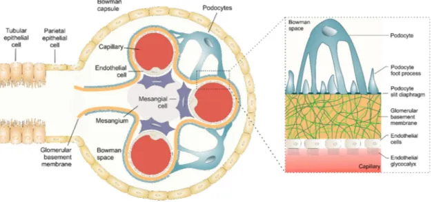
The functional unit of the kidney is the nephron. Each human kidney contains approximately one million nephrons [123]. The nephron is composed of a renal corpuscle and a hairpin shaped tubular system, consisting of the proximal convoluted tubule, the loop of Henle, a distal convoluted tubule and the collecting duct [122]. The renal corpuscle is composed of the glomerulus, a capillary tuft, and the Bowman’s capsule that surrounds the glomerulus. The renal corpuscle compromises glomerular endothelial cells, a glomerular basement membrane (GBM), mesangial cells, podocytes and parietal epithelial cells [12,124]. Approximately 180 liters of plasma are filtered each day [125]. The primary urine is generated in the renal corpuscle by selective filtration of water, ions and small molecules from the blood and the filtrate is subsequently reabsorbed and concentrated in different sections of the tubule system to produce 1.5 liters of urine. The filtrate has a similar composition as plasma but is largely devoid of proteins due to the selective filtration of the glomerular barrier [125].
3.2 Components of the glomerular barrier
The glomerular filtration barrier has been intensively studied for decades, since it constitutes the most complex biological membrane being not only size- and but also charge-selective [125]. It is composed of three sequential layers that physically separate the vasculature from the urinary space and selectively filter blood components. These layers are (1) the glomerular endothelium [126], (2) the glomerular basement membrane (GBM) [127] and (3) podocytes with their foot processes [128] (Figure 3).
The glomerular endothelial cells form the capillaries within the glomerulus. They are unusually flattened with a height of 50 to 150 nm [125]. Moreover, they possess large fenestrated areas with a pore size of 50 to 100 nm [129], constituting 20 to 50% of the entire endothelial surface [130,131]. These fenestrations allow water and small solutes to pass unhindered from the vasculature into the glomerulus, but they effectively restrict the passage of blood cells [125]. Endothelial cells are coated by a negatively charged glycocalyx, consisting of proteoglycans and glycosaminoglycans, that restricts
Chapter 1: Introduction
14
negatively charged biomolecules (e.g. albumin) from entering the GBM by virtue of electrostatic repulsion [132].
The GBM is a 300 to 350 nm thick basal lamina which separates the endothelial from the epithelial layer [22]. It is mainly composed of type IV collagen and laminin, crosslinked with heparan sulfate and other charged proteoglycans such as agrin, perlecan and nidogen/entactin [22,125,133,134]. Due to its highly negative charge and the average pore size of 3 nm, the GBM selectively filters small molecules and colloids by their size and charge [135].
The third and outermost filtration barrier is represented by visceral epithelial cells also known as podocytes [136]. Podocytes and their extended foot processes enwrap the outer aspects of glomerular capillaries facing the urinary space [128,137]. Thereby, foot processes form filtration slits with an average width of 32 nm [22]. Adjacent foot processes are interconnected by a thin slit diaphragm, a specialized intercellular junction composed of unique membrane proteins (e.g. nephrin, podocin) and additional adherens junction proteins (e.g. P-cadherin) [138]. Both structures, podocyte foot processes and the slit diaphragm serve as the final filtration barrier that restricts proteins and macromolecules from reaching the urinary space.
Figure 3. The kidney glomerulus and its filtration barrier consisting of the glomerular endothelium, the glomerular basement membrane (GBM), and podocytes with their foot processes and the slit diaphragm.
Adapted from [12].
The kidney as novel target for nanomedicines
Although mesangial cells are not directly assigned to the glomerular filtration barrier, they are an essential part of glomerulus and mandatory to maintain its filter function [139]. Mesangial cells and their matrix provide substantial structural support for glomerular capillary loops [140]. Moreover, they are responsible for regulation of the filtration surface area and therefore are essential for the integrity of the renal filter [12,139].Mesangial cells, podocytes and endothelial cells constitute a functional unit and communicate with each other via cytokine cross-talk [141]. Alteration in any of these three cell types can have severe impact on the others. For example, mesangial cell injury can result in podocyte foot process fusion and proteinuria, whereas podocyte injury can induce mesangial cell proliferation [139].
Impairment of either of these glomerular components can lead to proteinuria, which designates the presence of increased protein levels in the urine (> 300 mg in 24 hours) and constitutes a hallmark of glomerular diseases such as diabetic nephropathy [125].
3.3 Selective permeability
The overall effective size cutoff for molecules and colloids to pass the filtration barriers of the renal corpuscle and enter the urinary space is approximately 8 to 10 nm, referring to the hydrodynamic diameter [21-23]. Therefore, most of therapeutic colloids are not subject to kidney filtration as their size exceeds the size cutoff unless the filtration barrier is impaired by disease or particles are degraded into smaller fragments that fall below the kidney filtration threshold [12,22].
Colloids with a hydrodynamic diameter below 8 to 10 nm can pass the renal filter and are excreted into the urine [21-23]. In contrast, larger particles can enter the glomerulus from the afferent arteriole through the endothelial fenestrae, however their accumulation within the glomerulus is restricted to the mesangium and the extracellular matrix. Choi and colleagues [142] have recently investigated glomerular deposition of intravenously administered PEGylated gold NPs as a function of size.
The total deposition of NPs below 100 nm was increased with larger particles size and was primarily controlled by NP uptake into mesangial cells, whereas larger particles (> 100 nm) were increasingly restricted from entering the mesangium due to the size limitation of endothelial fenestrations.
Charge selectivity is a second crucial parameter that influences the behavior of colloids within the glomerulus [125]. Choi and colleagues [142] have shown that anionic
Chapter 1: Introduction
16
PEGylated gold NPs do not deposit in the GMB or podocytes which could be attributed to the repulsive electrostatic forces of the negatively charged GBM. In contrast, positively charged polymer-based siRNA NPs of the same size (80 nm) were reported to accumulate within the GBM and the mesangium [143]. These findings were supported by Bennett et al. [144] who demonstrated that cationic ferritin-decorated NPs accumulate in the GBM after intravenous injection in rats. However, the concept of charge selectivity remains highly controversial. Kodaira and colleagues [145] found that negatively charged polyvinylpyrrolidone (PVP) derivatives could accumulate in the glomerulus after systemic administration. Paradoxically, a more pronounced negative charge of carboxylated PVP colloids resulted in enhanced accumulation in the kidney, with renal levels reaching approximately 30% of the administered dose.
Interestingly, sulfonated PVP NPs could pass the glomerular filtration barrier and were rapidly excreted in the urine.
Another criterion for kidney filtration is particle geometry. Carbon nanotubes with high aspect ratios have been shown to paradoxically being filtered by the glomerulus [22,146,147] despite their average length of 100 to 500 nm, which exceeds the renal filtration threshold by a factor of 10 to 50. Ruggiero et al. [147] concluded that the long axis of single-walled carbon nanotubes was coordinated perpendicular to the GBM through hydrostatic forces, thereby orienting their short axis into the pores of the GBM. These results were further confirmed by Lacerda and colleagues [146] who used transmission electron microscopy (TEM) to demonstrate that the orientation of carbon nanotubes within the glomerulus corresponded to the predicted model.
The filtration threshold can be influenced by further particle characteristics including tensile strength and young’s modulus which are primarily dictated by the NP composition. The role of flexibility on renal clearance was investigated using dendrimers possessing different degrees of intramolecular flexibility. Soft and flexible dendrimers with a hydrodynamic size of 18 nm showed renal excretion, whereas dendrimers with a rather rigid core but smaller size could not pass the glomerular filtration barrier [148-150].
In summary, particle size, surface charge, shape, and flexibility are all major determinants of NP deposition within the glomerulus. However, permeability restrictions only apply for healthy tissues and can be greatly altered in a diseased state.
The kidney as novel target for nanomedicines
3.4 Pathophysiological changes in diabetic nephropathy and targeting opportunities
DN constitutes a life-threatening complication and affects one-third of all patients suffering from diabetes [119]. Pathophysiological changes in DN progress rapidly and severely affect components of the glomerular filtration barrier [119]. For example, the thickness of the GBM increases by about 30% [151] and further worsens during disease progression. The number of podocytes is drastically reduced and podocytes start to detach from the GBM, leading to a loss of foot process and fusion [152]. Mesangial cells start to change their gene expression [153] and are also subject to cell proliferation and hypertrophy [140]. The relative increase in volume of the mesangium drastically impairs other structures within the glomerulus, resulting in reduction of the glomerular filtration area and segmental occlusion of glomerular capillaries [125].
Because pathological changes in the mesangium are among the earliest and most critical events found in DN [140], it would be highly beneficial to consider mesangial cells as therapeutic target for the treatment of renal diseases [154].
Due to the fact that mesangial cells are separated from the systemic blood stream only by fenestrated endothelial cells, they are accessible for colloids that fall below the endothelial pore size of 50 to 100 nm [129]. As aforementioned, recent studies have demonstrated that colloids can enter the mesangium via size-selective passive targeting which is similar to the EPR effect. For example, Choi and colleagues [142] found that PEGylated gold NPs with a size of 50 to 100 nm accumulated in the mesangium and were partly internalized by mesangial cells within 24 hours after intravenous injection, whereas larger particles (> 100 nm) were not able to access the mesangium due to steric restrictions at the endothelial fenestrations. In a different study by Pollinger and coworkers [155], it was demonstrated that PEGylated quantum dots (Qdots) with a hydrodynamic diameter of approximately 20 nm and distinct negative charge (-14.5 to -15.6 mV) displayed pronounced enrichment in the mesangium of healthy mice one hour after systemic administration. Although these studies provide important information about glomerular deposition of colloids as a function of their attributes (i.e. size), these inorganic nanomaterials are not suited as drug delivery system as their solid cores cannot be loaded with therapeutic drugs. Recently, Li and coworkers [156]
combined the advantages of polymeric NPs and the size selectivity of renal glomerulus.
To this end, they prepared dexamethasone acetate-loaded polymeric NPs which were injected into healthy rats. They found that drug-loaded polymeric NPs with a
Chapter 1: Introduction
18
hydrodynamic size of 90 nm accumulated in the kidney to a greater extent than free DiD solution. Thereby, drug-loaded NPs were primarily deposited within the glomerular mesangium.
These studies emphasize the feasibility of nanomedicines to target the mesangium, however, they exclusively rely on passive targeting strategies. Multivalent targeting would be the perfect strategy to enhance target selectivity and, in addition, trigger uptake into renal cells by receptor-mediated endocytosis. Scindia and colleagues [157]
have used anti-α8-integrin ligands to enhance glomerular deposition of liposomal nanocarriers with a size of 70 to 130 nm in lupus glomerulonephritis susceptible mice.
In contrast to control immunoglobulin-decorated liposomes, targeted colloids were capable of specific accumulation in the mesangium of diseased mice.
Furthermore, receptor expression of glomerular cells can be altered in a diseased state providing a promising opportunity for selective targeting. For example, ανβ3 integrin receptors are increasingly expressed on mesangial cells during the progression of diabetic nephropathy [158,159], whereas E-selectin expression in endothelial cells is upregulated at the onset of glomerulonephritis [160].
Biodegradable polymeric NPs constitute the ideal system for multivalent targeting of mesangial cells because they possess highly tunable physicochemical properties, which allows for exact tailoring of NP size, they can be modified with multiple targeting ligands of defined density, and provide the capacity to accommodate high amounts of hydrophobic drugs inside their colloidal core [10,11].
4 Conclusion
Multivalent NPs have shown tremendous potential for selective targeting because they can accumulate at target sites while sparing healthy off-target tissues. Although the kidney is severely affected in various diseases, such as diabetes mellitus, it has largely been overlooked as target for NP-mediated therapeutic intervention. In the diseased state, the renal filter integrity is severely impaired, allowing colloids to enter or even transit the glomerular filtration barrier. Precise tailoring of NP characteristics in terms of kidney retention and binding to distinct cell populations which are affected during disease progression (i.e. mesangial cells) can be exploited to develop novel therapeutic approaches for the treatment of renal diseases.
Chapter 2
Goals of the Thesis
Chapter 2: Goals of the Thesis
20
Maximizing drug concentration at the site of disease while minimizing systemic drug effects is regarded the ideal conception for pharmacotherapy. This is of particular importance when the therapeutic target is confined to a defined tissue which is the case for various cancers, vitreoretinal diseases or diabetic nephropathy. Targeted drug delivery is widely regarded the most potent tool to make this vision come true. In particular, polymeric nanoparticles (NPs) have tremendous potential for targeted delivery as they are biocompatible, degradable, possess unique and highly tunable physicochemical properties and can be loaded with high amounts of cargo.
As they provide extremely high flexibility in their design, polymeric NPs can be precisely engineered to suit a wide range of therapeutic applications. Although polymeric NPs possess tremendous potential, their bench to bedside transition proceeds slowly. This is attributed to several limiting factors such as: (1) lack of manufacturing platforms that allow for a controlled and scalable synthesis of NPs with high batch-to-batch consistency, (2) inadequate knowledge of events at the nano-bio interface both in vitro and in vivo, (3) the limited understanding of their fate in the body, organs and at cellular levels due to their poor detectability in tissues and biological media, (4) incomplete knowledge of critical particle attributes with regards to targeting, and (5) insufficient selectivity and efficiency of targeted NPs in vivo.
The aim of this work was to design and characterize core/shell structured multivalent polymeric NPs intended for selective targeting of mesangial cells with special focus on addressing the abovementioned challenges.
Development of therapeutic colloids intended for selective targeting requires the precise control of NP attributes. Particle size is widely considered a key factor since it determines the interfacial contact area and therefore, has a major impact on multivalent binding to target cells. Moreover, with respect to targeting of kidney cells, size also constitutes a restricting parameter in terms of glomerular entrance. In Chapter 3, microfluidic synthesis of multicomponent polymeric colloids was evaluated and compared to bulk nanoprecipitation, which is considered the gold standard in the field.
Three NP formulations with varying core to shell ratios were optimized by adjusting critical process and formulation parameters and the influence on particle size and size distribution was investigated and analyzed by dynamic light scattering and transmission electron microscopy.
Goals of the Thesis
A profound understanding of NP behavior at the nano-bio interface is essential for the design of therapeutic systems. Adsorption of serum proteins onto the colloidal surface can substantially enhance recognition and clearance of NPs by the reticuloendothelial system. Moreover, formation of a protein corona can severely impair colloidal stability and therefore, has a major impact on the in vivo performance of colloidal drug delivery systems. To gain a better understanding of the nano-bio interface, the interaction of functionalized NPs with serum proteins was systematically studied as a function of surface charge and hydrophobicity. Moreover, the impact of protein corona formation on NP integrity was assessed (Chapter 4).
Surface functionalization with high-affinity ligands is commonly considered the major determinant for controlling the uptake efficiency in vitro and in vivo. However, it is often not sufficiently taken into consideration that cellular uptake of targeted colloids is governed by a delicate interplay between different particle features. In Chapter 5, the combinational influence of cRGD ligand density and poly(ethylene glycol) (PEG) linker length on NP uptake into αvβ3 integrin overexpressing U87MG glioblastoma cells was investigated by flow cytometry and fluorescence microscopy.
The therapeutic concept of targeted drug delivery systems relies on the specific interaction of targeting ligands presented on the colloidal surface with cognate receptors of the target cell. However, cell surface receptors are not exclusively located on target cells but can also be expressed by healthy off-target tissues. To overcome premature NP uptake into off-target cells and minimize systemic toxicity, new targeting strategies with enhance selectivity towards target cells are needed. Inspired by viruses, which are capable of sequential recognition and entry of their host cells, heteromultivalent binding NPs were designed. The advanced specificity of virus mimicking dual-targeted NPs towards rat mesangial cells, which are affected in a number of severe renal diseases, was assessed by flow cytometry and fluorescence microscopy (Chapter 6).
Polymeric NPs suffer from poor visibility in biological environments. Reliable and simultaneous qualitative and quantitative detectability in cells and tissues is a largely unmet need. Because profound knowledge of pharmacokinetics and biodistribution are key to the successful development of nanomedicines, additional labels are required to evaluate their in vivo performance and track their journey from the point of administration to the target site. To overcome this limitation, small gold tags were introduced as a contrast agent at the core/shell interface of polymeric NPs and
Chapter 2: Goals of the Thesis
22
polymer/gold hybrid NPs were characterized for their physicochemical characteristics and their toxicity in vitro. Moreover, the enhanced detectability in cells was evaluated by fluorescence and transmission electron microscopy (Chapter 7).
Chapter 3
Microfluidic manufacturing improves
polydispersity of multicomponent polymeric
nanoparticles
Chapter 3: Microfluidic manufacturing of multicomponent nanoparticles
24
Abstract
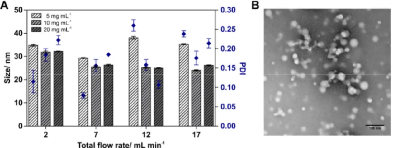
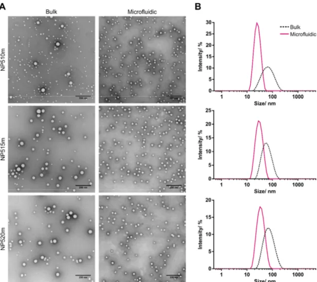
A key challenge in manufacturing of multicomponent polymeric colloids is obtaining monodisperse nanoparticles (NPs) with reproducible characteristics. Herein, NP formulations with varying core to shell ratios consisting of different poly(lactic acid)-poly(ethylene glycol) (PLA-PEG) copolymers (10k-b-5k, 15k-b-5k, 20k-b-5k) and poly(lactic-co-glycolic acid) (PLGA) at varying copolymer mass ratios (70/30, 65/35 or 60/40) were either prepared by bulk nanoprecipitation or microfluidic synthesis and evaluated regarding their size and polydispersity. Microfluidic process parameters, such as the total flow rate (TFR) and the flow rate ratio (FRR) of the aqueous phase and the organic polymer solution, were varied to tune particle size and further improve size distribution. Z-average hydrodynamic diameter and polydispersity index (PDI) were analyzed by dynamic light scattering (DLS) and selected formulations were further examined by transmission electron microscopy (TEM). In general, bulk nanoprecipitation resulted in formation of larger particles (52 - 65 nm) with a wide size distribution, whereas particles were significantly smaller in size (24 - 43 nm) and displayed a rather monodisperse size distribution when they were manufactured with a microfluidic system. The FRR was identified to be the key factor for improving the size distribution of NPs with a high ratio of PLA-PEG to PLGA, whereas it had only minor impact on particle characteristics of formulations with a lower copolymer ratio.
Polydispersity of different NP formulations was successfully improved by microfluidic manufacturing, demonstrating that this technology is a valuable tool that allows for reproducible and scalable manufacturing of multicomponent NPs with precisely tunable quality attributes.
Introduction
1 Introduction
The use of core/shell structured nanoparticles (NPs) as drug delivery system holds tremendous promise for improving the treatment of several diseases in terms of therapeutic efficacy and reduced side effects [161]. Especially, biocompatible and biodegradable NPs composed of poly(lactic acid)-poly(ethylene glycol) (PLA-PEG) and poly(lactic-co-glycolic acid) (PLGA) have gained enormous attention since they provide the possibility to combine various useful features such as hosting high amounts of hardly soluble drugs, controlled drug release with tunable release profiles and specific receptor targeting [162-164]. Different block copolymers are often combined to optimize NP composition and engineer their characteristics, such as particle size, charge, surface functionalization or the core to shell ratio [60,61].A fast and simple technology to prepare PLGA-blended PLA-PEG nanoparticles is bulk nanoprecipitation by solvent exchange [165], which involves dropwise addition of a water-miscible organic polymer solution to a larger quantity of an aqueous non-solvent under stirring. As a consequence of diffusion, the solvent is exchanged leading to interfacial deposition of hydrophobic or amphiphilic polymers and self-assembling into core/shell structured NPs in order to reduce the free energy of the system [11,166].
However, in a bulk nanoprecipitation approach, phase mixing proceeds slowly and uncontrolled, which favors formation of hetero-sized nanocolloids due to the unequal distribution of polymer blocks and consequently, results in an insufficient batch-to-batch consistency. Hence, precise engineering, optimization and testing of core-blended NPs is severely impaired. As the in vivo performance of NPs strongly depends on their physicochemical characteristics it is crucial to enhance the reproducibility and controllability of NP synthesis [11,167,168]. Microfluidics has recently emerged as a new technology that offers precisely controlled reaction environments and has been harnessed to manufacture polymeric NPs with defined properties and excellent batch-to-batch consistency [61,168]. Furthermore, microfluidics can tackle the limited options provided by bulk nanoprecipitation for down- and up-scaling which makes this technology highly valuable for high-throughput screenings during the development of new nanomedicines as well as for clinical applications, where sufficient quantities of NPs with consistent properties are needed [61,169]. For these reasons, microfluidics was considered as an ideal platform to optimize size distribution of multicomponent polymeric NPs with distinct core to shell ratios. In this study, three NP formulations composed of PLA-PEG
Chapter 3: Microfluidic manufacturing of multicomponent nanoparticles
26
copolymers with increasing molecular weights of the PLA block (10k-b-5k, 15k-b-5k, 20k-b-5k) and varying PLA-PEG to PLGA copolymer mass ratios (70/30, 65/35 or 60/40) were prepared by bulk nanoprecipitation and microfluidic synthesis. Blends of PLGA and PLA-PEG copolymers were used to control the ratio between the hydrophilic PEG shell and the hydrophobic PLA/PLGA core of polymeric NPs. Optimization of these NP formulations was proceeded by adjusting process and formulation parameters and the influence of copolymer concentration, total flow rate (TFR) and the flow rate ratio (FRR) of aqueous to organic phase on particle size and size distribution was investigated and analyzed by dynamic light scattering (DLS) and transmission electron microscopy (TEM).
2 Materials and methods
2.1 Materials
1,8-Diazabicyclo[5.4.0]undec-7-ene (DBU) solution, 3,6-Dimethyl-1,4-dioxane- 2,5-dione (Lactide), 3-(Trimethylsilyl)propionic-2,2,3,3-d4 acid sodium salt (TMSP), anhydrous dichloromethane (DCM), anhydrous ethyl acetate, deuterated chloroform (CDCl3), deuterium oxide (D2O), methoxy poly(ethylene glycol) with a molecular mass of 5000 g mol-1 (MeO-PEG5k-OH), Resomer® RG 502 (PLGA) and Tetramethylsilane (TMS) were received from Sigma-Aldrich (Taufkirchen, Germany). All other materials were reagent grade and obtained from Merck KGaA (Darmstadt, Germany). Ultrapure water was obtained by using a Milli-Q water purification system (Millipore, Schwalbach, Germany).
2.2 Synthesis of functionalized PLA-PEG block copolymers
Poly(lactic acid)-poly(ethylene glycol) (PLA-PEG) block copolymers with different polymerization degrees of the PLA block (10k-b-5k, 15k-b-5k, 20k-b-5k) were synthesized by ring-opening polymerization as previously described by Qian et al. [170] with slight modifications. Prior to use, racemic 3,6-dimethyl-1,4- dioxane-2,5-dione (D,L-lactide) was purified by recrystallization from anhydrous ethyl acetate and dried under vacuum for at least 12 h. Linear methoxy poly(ethylene glycol) with a molecular mass of 5000 g mol-1 was used as macroinitiator for the ring-opening polymerization of lactide using DBU as catalyst. After quenching the polymerization reaction with benzoic acid, block copolymers were subsequently precipitated in diethyl
Materials and methods
ether and dried under vacuum at 45 °C overnight. 1H-NMR spectra were recorded in CDCl3 at 295 K using a Bruker Avance 300 spectrometer (Bruker BioSpin GmbH, Rheinstetten, Germany). The number-average molecular weight of PLA-PEG block copolymers was determined by integration of the methylene signal of PEG residues based on molecular weight (provided by the manufacturer) and calculation of the methine and methylene proton signals of PLA.
2.3 Bulk nanoprecipitation of nanoparticles
Polymeric NPs were prepared by bulk nanoprecipitation as previously described [165].
In general, PLA-PEG and PLGA were diluted with acetonitrile to a final polymer concentration of 5, 10 or 20 mg mL-1. The polymerization degree of the PLA-PEG copolymer (10k-b-5k, 15k-b-5k, 20-b-5k) as well as the mass ratio of PLA-PEG to PLGA (70:30, 65:35 or 60:40) was varied depending on the NP formulation. A detailed description of the respective NP compositions with either high (NP510m), medium (NP515m) or low PEG content (NP520m) is given in Figure 1. The polymer solution was added dropwise into 10 volumes of stirred ultrapure water and NP dispersions were stirred for 3 hours to evaporate the organic solvent. NPs were filtered through a 0.45 µM Rotilabo® PES syringe filters (Carl Roth, Karlsruhe Germany) and concentrated by ultrafiltration using Pall Macrosep® Advance Centrifugal Devices (Pall GmbH, Dreieich, Germany) with 100 kDa MWCO.
2.4 Microfluidic synthesis of nanoparticles
Polymeric NPs were also prepared via microfluidics using a benchtop NanoAssemblr™ instrument (NanoAssemblr™, Pecision NanoSystems Inc., Vancouver, Canada) and process parameters were controlled using the corresponding NanoAssemblr™ Controller software (v1.0.5). Ultrapure water was injected into the first inlet and the organic polymer solution in the second inlet of the microfluidic mixer.
The TFR was varied from 2 to 17 mL min-1 and the FRR of the aqueous phase and the organic polymer solution was either 10:1 or 5:1. NP formulations were collected at the outlet channel and stirred for 1 h to remove the organic solvent. Afterwards, NP dispersions were filtered through 0.45 µM Rotilabo® PES syringe filters (Carl Roth, Karlsruhe Germany).
Chapter 3: Microfluidic manufacturing of multicomponent nanoparticles
28
2.5 PEG quantification
The PEG content was determined by 1H-NMR spectroscopy according to a method described by Bertrand et al. [171] with slight modifications. To determine the total PEG content in NP formulations, NP510m, NP515m and NP520m were prepared in ultrapure water and 600 µL of the concentrated solutions (approx. 4 to 5 mg mL-1) were lyophilized for 48 h. The exact NP mass of each sample was determined gravimetrically (n=3) and samples were subsequently dissolved in 600 µL of CDCl3 containing 0.05%
(v/v) TMS as an internal standard. Solutions of MeO-PEG5k-OH at defined concentrations (0.1-5.0 mg mL-1) in CDCl3 containing 0.05% (v/v) TMS were used for calibration. Methylene protons of PEG (3.65 ppm) were normalized to the proton signal of the internal standard (TMS) and used for calculation of the PEG concentration in respective samples. To determine the concentration of hydrated PEG in the NP shell, NP510m, NP515m and NP520m were prepared in ultrapure water and the solvent was exchanged with D2O supplemented with 0.05% (m/v) TMSP as an internal standard using Microsep® Advance Centrifugal Devices (Pall GmbH, Dreieich, Germany) with 100 kDa MWCO. The exact polymer concentration of samples in D2O was determined gravimetrically after lyophilization (n=3). The ratio of the methylene protons of PEG (3.72 ppm) to the integral of the methyl protons of the internal standard (TMSP) was then compared with the calibration curve of MeO-PEG5k-OH (0.1 - 5.0 mg mL-1) in D2O to calculate the PEG content in the NP shell. All measurements were conducted on a Bruker Avance 300 spectrometer (Bruker BioSpin GmbH, Rheinstetten, Germany) at 295 K.
2.6 Dynamic Light Scattering
Z-average hydrodynamic size and polydispersity index (PDI) of NP formulations were determined on a Zetasizer Nano ZS device (Malvern, Herrenberg, Germany). DLS measurements were performed in ultrapure water at a constant temperature of 25 °C using semi-micro PMMA disposable cuvettes (Brand, Wertheim, Germany). The position from the cuvette wall was set to 4.65 mm and the attenuator was optimized by the device. Data were collected and analyzed using the Malvern Zetasizer software version 7.11 (Malvern Instruments, Worcestershire, United Kingdom).
![Figure 1. Parameters for the design of colloidal therapeutic systems. Adapted from [11]](https://thumb-eu.123doks.com/thumbv2/1library_info/3735454.1508997/12.892.243.618.681.1058/figure-parameters-design-colloidal-therapeutic-systems-adapted.webp)