C ONTRIBUTIONS TO THE A SYMMETRIC
C ATALYSIS OF C - C C OUPLINGS ,
AND TO THE C HEMICAL I NDUCTION OF
C ARDIOMYOGENESIS FROM E MBRYONIC S TEM C ELLS
Inaugural Dissertation
zur Erlangung des Doktorgrades
der Mathematisch-Naturwissenschaftlichen Fakultät
der Universität zu Köln
vorgelegt von
Diplom-Chemikerin Bianca Seelig
aus Hilden
Köln 2009
Gutachter: Pror. Dr. A. Berkessel
Prof. Dr. H.-G. Schmalz
Tag der mündlichen Prüfung: 06. Juli 2009
“Organic chemistry just now is enough to drive one mad.”
F. Wöhler, in a letter to his mentor J. J. Berzelius
dedicated to B.
Acknowledments
Research work reported in this thesis was carried out from March 2006 until Mai 2009 at the Institute for Organic Chemistry of the University of Cologne under the supervision of Prof. Dr. Albrecht Berkessel. Additionally, a part of the research was done at the Graduate School of Pharmaceutical Sciences of the University of Tokyo, Japan, during October 2008 - February 2009, in the research group of Prof. Dr. Masakatsu Shibasaki.
First of all, I would like to acknowledge Prof. Dr. Albrecht Berkessel for providing me with those challenging and rewarding thesis projects. His constant help, critical advices and active encouragement were very important for the good outcome of this work.
Shibasaki-sensei deserves my sincerest gratitude for giving me the opportunity to work under his guidance at the tôdai. どうもありがとうございます。
I would like to thank Prof. Dr. Hans Günther Schmalz for reviewing the thesis.
I would also like to mention the people who have been collaborating on the projects described in this thesis: Prof. Dr. J. Hescheler, Prof. Dr. A. Sachinidis and Dr. Silke Schwengberg as part of the stem cell project and the people at the Shibasaki group, especially Mastunaga-sensei and Morimoto-san, contributed in great extent to the success of this work.
I am very grateful to all my colleagues for providing a wonderful working atmosphere, for their help, patience and support. A standing ovation is to Ilona Jurkiewicz for being much more then the best lab mate one can imagine. I am thankful to my practical students, particulary for Jan Krämer, for sharing some of my experimental work. Thanks are also due to all of my Japanese colleagues, for the wonderful time in Japan.
For the critical reading of this work, I would like to thank Dr. Burkhard Koch, Dr. Nicolas Leconte, Angela Heinsch, Ilona Jurkiewicz, Eva Leitterstorf, David Müller, Mei Ching Ong and Dr. Silke Schwengberg.
My special thanks are to all the employees of the Institute for Organic Chemistry:
Sarwar Aziz (HPLC), Dr. Nils Schlörer (NMR), Christoph Schmitz (GCMS), Dr. Johann
Lex and Dr. Jörg Neudörfl (X-ray), Michael Neihs and Dr. Mathias Schäfer (MS). I also
want to thank Susanne Geuer and Dr. Wolfgang Klug for their help in organisational
Hartmann and Peter Küpper for their kind help with technical problems.
A big hug goes to all my dear friends for being there to remind me of the more important aspects of life.
I am gratefull for my parents for giving me the freedom to find my own interests and goals and for their support and tolerance.
B., thank you for your encouragement, your support, and for always being there when I
needed you.
TABLE OF CONTENTS
1 Summary... 1
1.1 Chemically Induced Cardiomyogenesis of mES Cells ...1
1.1.1 Substance Screening ...1
1.1.2 Identification of Signalling Cascades Involved in Cardiomyogenesis ...3
1.2 A Simplified Synthesis of Takemoto’s Catalyst...3
1.3 La-linked BINOL Catalysed Asymmetric aza-BH reaction ...4
2 Introduction ... 7
3 Chemically Induced Cardiomyogenesis of mES Cells ... 11
3.1 Background ...11
3.2 Concept ...14
3.2.1 Forward Chemical Genetics Approach ...14
3.2.2 Selection of Substrates...16
3.3 Results and Discussion ...16
3.3.1 Substance Screening ...16
3.3.2 Identification of Signalling Cascades Involved in Cardiomyogenesis ...27
3.3.3 Synthesis of the Test Substrates ...30
3.4 Experimental Part...39
3.4.1 Physiological ES cell screenings ...39
3.4.2 Synthesis of the Test Substrates ...40
3.4.3 Synthesis of (1S,2R,4S,5R)-2,5-Dihydroxybicyclo[2.2.1]heptane (13) ....41
3.4.4 Synthesis of (1S,4S)-Bicyclo[2.2.1]heptane-2,5-dione (14) ...42
3.4.5 Synthesis of (1S,2S,4S,5S)-2,5-Dibenzylaminobicyclo[2.2.1]hep- tane (15) ...43
3.4.6 Synthesis of (1S,2S,4S,5S)-2,5-Diaminobicyclo[2.2.1]heptane (9) ...44
3.4.7 Synthesis of (2R,3R)-2,3-Bis(benzoyloxy)butanedioic acid (1S,5R)-(5- amino-1,3,3-trimethylcyclohexyl)-methaneamine salt (1:1) (17) ...45
3.4.8 Synthesis of (1R,3S)-3-Aminomethyl-3,5,5-trimethylcyclohexyl- amine (10) ...47
3.4.9 Synthesis of (1R,2R)-1,2-Diaminocyclohexane (11)...48
3.4.10 Synthesis of N-Phenyl-N'-[(1R,3S)-3-({[(phenyl)amino]-thioxomethyl}- amino)methyl-3,5,5-trimethylcyclohexyl]thiourea (III-2) ...49
3.4.11 Synthesis of 1,1’-[(1S,2S,4S,5S)-Bicyclo[2.2.1]heptane-2,5-diyl]bis- {3-[3,5-dimethoxyphenyl]urea} (III-3) ...50
3.4.12 Synthesis of N-[3,5-Di(methoxy)phenyl]-N’-[(1R,3S)-3-{[({[3,5-bis(me- thoxy)phenyl]amino}oxomethyl)amino]methyl}-3,5,5-trimethylcyclo- hexyl]urea (III-4) ...51
3.4.13 Synthesis of 1,1’-[(1S,2S,4S,5S)-Bicyclo[2.2.1]heptane-2,5-diyl]bis-
phenylurea (III-5) ...52
3.4.15 Synthesis of N-[3,5-Bis(trifluoromethyl)phenyl]-N'-[(1R,3S)-3-{[({[3,5- bis(trifluoromethyl)phenyl]amino}thioxomethyl)amino]methyl}-3,5,5-
trimethylcyclohexyl]thiourea (III-7) ... 55
3.4.16 Synthesis of 1-[3,5-Bis(trifluoromethyl)phenyl]-3-{(1R,2R)-2-[3-[3,5- bis(trifluoromethyl)phenyl)thioureido]cyclohexyl}thiourea (III-8) ... 56
3.4.17 Synthesis of 9-(Hydroxymethyl)-(1,8-R;4,5-S)-1,2,3,4,5,6,7,8-octahy- dro-1,4:5,8-dimethanoanthracene (26) ... 57
3.4.18 Synthesis of 9-(Chloromethyl)-(1,8-R;4,5-S)-1,2,3,4,5,6,7,8-octahydro- 1,4: 5,8-dimethanoanthracene (24)... 58
3.4.19 Synthesis of 9-O-Methylquinidine (20)... 59
3.4.20 Synthesis of 1-N-[9-((1,8-R;4,5-S)-1,2,3,4,5,6,7,8-Octahydro-1,4:5,8- dimethanoanthracenyl)methyl]-9-O-methylquinidinium chloride (IV-1) .... 61
3.4.21 Synthesis of 10,11-Dihydroquinidine (21) ... 62
3.4.22 Synthesis of 1-N-[9-((1,8-R;4,5-S)-1,2,3,4,5,6,7,8-Octahydro-1,4:5,8- dimethanoanthracenyl)methyl]10,11-dihydroquinidinium chloride (IV-2) . 63 3.4.23 Synthesis of 6’-Hydroxy-cinchonine (22) ... 65
3.4.24 Synthesis of 1-N-[9-((1,8-R;4,5-S)-1,2,3,4,5,6,7,8-Octahydro-1,4:5,8- dimethanoanthracenyl)methyl]- 6’-hydroxycinchoninium chloride (IV-4).. 66
3.4.25 Synthesis of 1-N-[9-((1,8-R;4,5-S)-1,2,3,4,5,6,7,8-Octahydro-1,4:5,8- dimethanoanthracenyl)methyl]-6’-isopropoxy-cinchoninium chlori- de (IV-5)... 67
3.4.26 Synthesis of 1-N-(9-Anthrylmethyl)quinidinium chloride (IV-6) ... 68
3.4.27 Synthesis of 1-N-(1-Naphthylmethyl)quinidinium chloride (IV-7) ... 70
3.4.28 Synthesis of 1-N-(Benzyl)quinidinium chloride (IV-8)... 71
3.4.29 Synthesis of 1-N-[9-((1,8-R;4,5-S)-1,2,3,4,5,6,7,8-Octahydro-1,4:5,8- dimethanoanthracenyl)methyl]quininium chloride (IV-9) ... 72
3.4.30 Synthesis of N-Methyl-quinidinium iodide (38)... 73
3.4.31 Synthesis of N-Methyl-quinidinium chloride (V-1) ... 75
3.4.32 Synthesis of N-1-Butyl-quinidinium chloride (V-2)... 76
3.4.33 Synthesis of 4-(Trifluoromethyl)benzyl chloride (30)... 77
3.4.34 Synthesis of 1-N-(4-Trifluoromethylbenzyl)quinidinium chloride (V-3) ... 78
3.4.35 Synthesis of 1-N-(4-Fluorobenzyl)quinidinium chloride (V-4)... 79
3.4.36 Synthesis of 4-Iodobenzyl chloride (32)... 80
3.4.37 Synthesis of 1-N-(4-Iodobenzyl)quinidinium chloride (V-5)... 81
3.4.38 Synthesis of 1,2,3,4,5,6,7,8-Octahydro-1,4:5,8-diethano-9-anthracene- methanol (37) ... 82
3.4.39 Synthesis of 9-(Chloromethyl)-1,2,3,4,5,6,7,8-octahydro-1,4:5,8-di- ethanoanthracene (35) ... 83
3.4.40 Synthesis of (9S)-9-Hydroxy-6’-methoxy-1-[(1,2,3,4,5,6,7,8-octahydro- 1,4:5,8-diethano-9-anthraceny)methyl]quinidinium chloride (V-6) ... 84
3.4.41 Synthesis of 2,4-Difluorobenzyl chloride (34) ... 85
3.4.42 Synthesis of 1-N-(2,4-Difluorobenzyl)quinidinium chloride (V-7) ... 86
3.4.43 Synthesis of 6’-Cyclopentyloxy-cinchonine (27) ... 87
3.4.44 Synthesis of 1-N-[9-((1,8-R;4,5-S)-1,2,3,4,5,6,7,8-Octahydro-1,4:5,8- dimethanoanthracenyl)methyl]-6’-(cyclopentyloxy)cinchonine (V-8) ... 88
4 A Simplified Synthesis of Takemoto’s Catalyst ... 90
4.2 Concept ...92
4.3 Results and Discussion ...96
4.4 Experimental Part ...99
4.4.1 General Experimental Conditions ...99
4.4.2 Preparation of trans-4,5-Tetramethyleneimidazolidine-2-thione (81)...100
4.4.3 Preparation of 1-(3,5-Bis-trifluoromethyl-phenyl)-3-{(1R,2R)-2-[3- (3,5-bis-trifluoromethyl-phenyl)-thioureido]-cyclohexyl}-thiourea (63)...102
4.4.4 Preparation of 1-[(1R,2R)-2-Aminocyclohexyl]-3-[3,5-bis(trifluoro- methyl)phenyl]thiourea (62)...103
4.4.5 1-[3,5-Bis(trifluoromethyl)phenyl]-3-[(1R,2R)-2-(dimethylamino)cyclo- hexyl]thiourea (51, Takemoto’s Catalyst) ...105
5 La-linked BINOL Catalysed Asymmetric Aza-BH Reaction ... 108
5.1 Background ...108
5.1.1 Mechanistic Studies...108
5.1.2 Substrate Diversity ...110
5.1.3 Development of Asymmetric Aza-Baylis-Hillman Reactions...112
5.1.4 Use of Aza-Baylis-Hillman Adducts in Synthesis ...119
5.2 Concept ...120
5.3 Results & Discussion...123
5.4 Outlook...128
5.5 Experimental Part ...130
5.5.1 General Experimental Conditions ...130
5.5.2 Syntheses of the Substrates for the Aza-BH Reaction ...131
5.5.2.1 Synthesis of P,P-Diphenylphosphinic amide (109)
128...131
5.5.2.2 Synthesis of P,P-Diphenyl-N-(phenylmethylene)phosphinic amide (98a)
128...132
5.5.2.3 Synthesis of P,P-Diphenyl-N-(4-chlorophenylmethylene)phos- phinic amide (98b)
128...133
5.5.2.4 Synthesis of N-[Cyclohexyl[(4-methylphenyl)sulfonyl]methyl]- P,P-diphenylphosphinic amide (rac-112) ...134
5.5.2.5 Synthesis of N-(Cyclohexylmethylene)-P,P-diphenylphosphinic amide (98c)...135
5.5.3 Syntheses of Racemic Reference Substances...136
5.5.3.1 Synthesis of Methyl ( β S)- β -[(diphenylphosphinoyl)amino]- α -me- thylenebenzenepropanoate (rac-100aa) ...136
5.5.3.2 Synthesis of Phenyl ( β S)- β -[(diphenylphosphinoyl)amino]- α -me- thylenebenzenepropanoate (rac-100ab) ...137
5.5.3.3 Synthesis of 1,1-Dimethylethyl ( β S)- β -[(diphenylphosphinoyl)- amino]- α -methylenebenzenepropanoate (rac-100ac)...138
5.5.3.4 Synthesis of ( β S)- β -[(Diphenylphosphinoyl)amino]- α -methylene-
benzenepropanenitrile (rac-100af)...139
amino]- α -methylenebenzenepropanoate (rac-100ba)... 140
5.5.3.6 Synthesis of Phenyl ( β S)-4-chloro- β -[(diphenylphosphinoyl)- amino]- α -methylenebenzenepropanoate (rac-100bb)... 141
5.5.3.7 Synthesis of 1,1-Dimethylethyl ( β S)-4-chloro- β -[(diphenylphos- phinoyl)amino]- α -methylenebenzenepropanoate (rac-100bc) ... 142
5.5.3.8 Synthesis of ( β S)-4-Chloro- β -[(diphenylphosphinoyl)amino]- α - methylenebenzenepropanenitrile (100bf)... 143
5.5.4 General Procedure for the (R,R)-La-Linked BINOL Catalysed Aza- Baylis-Hillman Reaction of N-Diphenylphosphinoyl-Protected Imines with Olefins ... 144
5.5.4.1 Methyl ( β S)- β -[(diphenylphosphinoyl)amino]- α -methyleneben- zenepropanoate (100aa) ... 144
5.5.4.2 Phenyl ( β S)- β -[(diphenylphosphinoyl)amino]- α -methyleneben- zenepropanoate (100ab)... 145
5.5.4.3 1,1-Dimethylethyl ( β S)- β -[(diphenylphosphinoyl)amino]- α -me- thylenebenzenepropanoate (100ac) ... 146
5.5.4.4 2-Naphthalenyl ( β S)- β -[(diphenylphosphinoyl)amino]- α -me- thylenebenzenepropanoate (100ad) ... 146
5.5.4.5 2,2,2-Trifluoro-1-(trifluoromethyl)ethyl ( β S)- β -[(diphenylphos- phinoyl)amino]- α -methylenebenzenepropanoate (100ae)... 147
5.5.4.6 ( β S)- β -[(Diphenylphosphinoyl)amino]- α -methylenebenzene- propanenitrile (100af) ... 148
5.5.4.7 Methyl ( β S)-4-chloro- β -[(diphenylphosphinoyl)amino]- α -me- thylenebenzenepropanoate (100ba). ... 148
5.5.4.8 Phenyl ( β S)-4-chloro- β -[(diphenylphosphinoyl)amino]- α -me- thylenebenzenepropanoate (100bb). ... 149
5.5.4.9 1,1-Dimethylethyl ( β S)-4-chloro- β -[(diphenylphosphinoyl)amino]- α -methylenebenzenepropanoate (100bc). ... 150
5.5.4.10 2-Naphthalenyl ( β S)-4-chloro- β -[(diphenylphosphinoyl)amino]- α -methylenebenzenepropanoate (100bd)... 150
5.5.4.11 ( β S)-4-Chloro- β -[(diphenylphosphinoyl)amino]- α -methyleneben- zenepropanenitrile (100bf) ... 151
5.5.5 Hydrolytic C,N Terminal Deprotection
104... 152
5.5.5.1 Synthesis of ( β S)- β -Amino- α -methylenebenzenepropionic acid hydrochloride (106a) ... 152
5.5.5.2 ( β S)-4-Chloro- β -amino- α -methylenebenzenepropionic acid hydrochloride (106b) ... 153
6 Appendix... 154
6.1 Abbrevations... 154
6.2 References ... 157
6.3 Abstract ... 162
6.4 Erklärung ... 163
1 Summary
This thesis deals with
• the investigation of chemically induced cardiomyogenesis of mouse ES cells,
• a simplified procedure for the synthesis of Takemoto’s catalyst,
• the development of the first Lewis acid catalysed asymmetric aza-BH reaction.
1.1 Chemically Induced Cardiomyogenesis of mES Cells
Cardiomyocytes are used as donor cells in cell replacement therapies to treat serious heart damage. Despite the significant demand for heart cells, there exists up to now no efficient method for the production of these donor cells. The most promising approach so far is the in vitro cardiomyogenesis from embryonic stem (ES) cells induced by small molecules. However, with this method, only a small ratio of stem cells evolve into car- diomyocytes. The design of new and highly selective small molecules is therefore of great interest.
11.1.1 Substance Screening
For the identification of a cardiomyogenesis inducing substance, a rational drug de- sign (RDD) was performed with a preliminary diversity orientated screening (DOS) focused on the synthesis and screening of (thio)urea and cinchona alkaloid based libraries. Derivatives of these (thio)ureas and cinchona alkaloids are known to exhibit bioactivities correlated to decreased proliferation rates of cancer cells
2or cardiac action potential,
3respectively.
A transgenic murine ES cell lineage, expressing enhanced green fluorescence pro- tein (EGFP) under the control of the heart specific promoter α-myosine heavy chain (pα-MHC), was used to investigate the effects of the test substrates on cardiomyo- genesis.
Diversity Orientated Screening (DOS). For a preliminary DOS, two substance libraries I and II containing (thio)urea (I, 19 compounds) or cinchona-alkaloid (II, 20 compounds) derivatives with extensively modified substitution patterns were synthesised and tested for their cardiomyogenesis inducing efficiency.
Among the substrates tested, especially compounds I-6, I-15 and II-16 induced an
increase of 80 %, 50 % or 60 %, respectively, of the EGFP fluorescence which corre-
lates with a positive cardiomoygenic effect (Scheme 1-1, p. 2).
I-6 I-15 II-16
Scheme 1-1 Influence of the compounds I-6, I-15 and II-16 on the expression of EGFP. Both a significant increase and decrease of values, compared to controls, represent an effect on the generation of cardiac cells.
Rational Drug Design (RDD). On the basis of the three lead structures (I-6, I-15, II-16), a focused (thio)urea (III, 8 compounds) and a focused cinchona-alkaloid based library (IV, 9 compounds) were created, tested for their cardiomyogenic activity, and analysed in a RDD approach.
The results obtained with the (thio)urea based library did not provide a sufficiently solid and reliable basis for a meaningful structure-activity relationship (in this case of small molecule induced cardiomyogenesis).
In contrast, RDD analysis of the physiological screening of the cinchona alkaloid based library IV showed that especially variations on the alkoxy group (IV-5) and the quarternary ammonium moiety (IV-8) lead to an increased formation of cardiomyocytes (Scheme 1-2).
IV-5
II-16
IV-8
Scheme 1-2 Influence of the variations on the alkoxy and quarternary ammonium substituents modified
N-substituent
0 20 40 60 80 100 120 140 160 180 200
1 E-09 1 E-08 1 E-07 1 E-06 1 E-05 1 E-04 concentration [M]
% of control
0 20 40 60 80 100 120 140 160 180 200
1 E-09 1 E-08 1 E-07 1 E-06 1 E-05 1 E-04 concentration [M]
% of control .
0 20 40 60 80 100 120 140 160 180 200
1 E-09 1 E-08 1 E-07 1 E-06 1 E-05 1 E-04 concentration [M]
% of control
0 20 40 60 80 100 120 140 160 180 200
1 E-09 1 E-08 1 E-07 1 E-06 1 E-05 1 E-04 concentration [M]
% of control
0 20 40 60 80 100 120 140 160 180 200
1 E-09 1 E-08 1 E-07 1 E-06 1 E-05 1 E-04 concentration [M]
% of control
HN HN NH O NH O F3C
CF3 CF3
CF3
NH Me Me
Me H O N
NH
NH O
N CH2
N OMe
OH
Cl
N CH2
N OMe
OH
Cl
N CH2
N OiPr
OH
Cl
N CH2
N OMe
OH
Cl
modified alkoxy group
Based on these positively tested substances (compounds IV-5 and IV-8), a new quinidine based substance library V (8, compounds) was developed and screened for its cardiomyogenesis inducing effectivity. This library contains substances which differ either in the alkoxy group attached to the quinidine moiety, or in the quinuclidine N-sub- stituent compared to the lead structures IV-5 and IV-8. However, these structural modifi- cations did not improve the cardiomyogenesis inducing effect of the lead structures.
Conclusion. The substance screenings led to the identification of cardiomyogenesis inducing compounds with good (IV-5) to very good activities (II-16, IV-8) determined by a 50 to 80 % increase of the EGFP fluorescence compared to untreated cells.
1.1.2 Identification of Signalling Cascades Involved in Cardiomyogenesis
Verapamil rac-6, a cardiomyogensis inducing agent found in previous studies,
4and the most active compound identified in the substance screening II-16 were used for the identification of signalling cascades involved in the cardiomyogenesis by a time-depen- dent screening approach. In this method, an increased cardiomyogenesis indicates in- teraction of the compound tested with a target expressed at a certain point in time du- ring the application time-frame of the test substrate.
rac-6
.
II-16Scheme 1-3 Cinchona alkaloid II-16 (left), verapamil rac-6 (racemic mixture, right).
Since both substrates exhibited different cardiomyogenesis inducing profiles in each test series, the cardiomyogenesis inducing effect of the tested substrates appeared to rely on an interaction of the tested substances with several target proteins expressed at different points of time. In this case, time-dependent correlations between cardiomyo- genesis and concentrations of the substrates are not suitable to limit the number of potential targets associated to the various developmental stages.
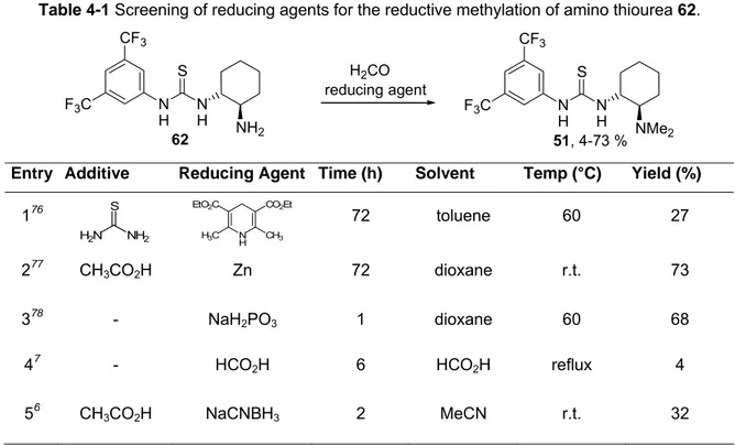
1.2 A Simplified Synthesis of Takemoto’s Catalyst
A significant number of C-C couplings can be catalysed enantioselectively by Take- moto’s bifunctional amino thiourea derivative 51.
5The original synthesis of the amino
N CH2
N OMe
OH
Cl
N MeO
MeO
OMe OMe iPr Me
NC
thiourea catalyst 51 consists in the facile addition of isothiocyanate 43 to N,N-dimethyl- trans-1,2-diaminocyclohexane 11. The preparation of the N,N-dimethyl-trans-1,2-diami- nocyclohexane building block 52 is a laborious four step procedure (Scheme 1-4).
6,7NH2
NMe2 N
H NMe2 NH
S CF3
F3C NCS
CF3
F3C NH2
NH2 2 h, THF, r.t.
51, 63 %
11 52
43
Scheme 1-4 Synthesis of the amino thiourea 51 developed by Takemoto.
An improved synthesis of Takemoto’s catalyst 51 was realised consisting of only two steps: The thiourea moiety was obtained by condensation of 3,5-bistrifluoromethyl- aniline 68 with phenyl chlorothioformate 71 and subsequent reaction with trans-1,2- diaminocyclohexane 11, according to a modified reaction protocol of Nagasawa.
8Subsequent reductive dimethylation using formaldehyd / zinc afforded Takemoto’s catalyst in an overall yield of 36 % (Scheme 1-5).
N H NH2 N
H S CF3
F3C N
H NMe2 NH
S CF3
F3C NH2
CF3
F3C PhO
S Cl
62, 50 % 51, 73 %
Zn / H2CO / AcOH dioxane, r.t., 72 h +
1) pyridine 2) diamine 11, Hünig's base CH2Cl2, r.t., 15 min
68 71
Scheme 1-5 Improved synthesis of Takemoto’s catalyst 51.
1.3 La-linked BINOL Catalysed Asymmetric aza-BH reaction
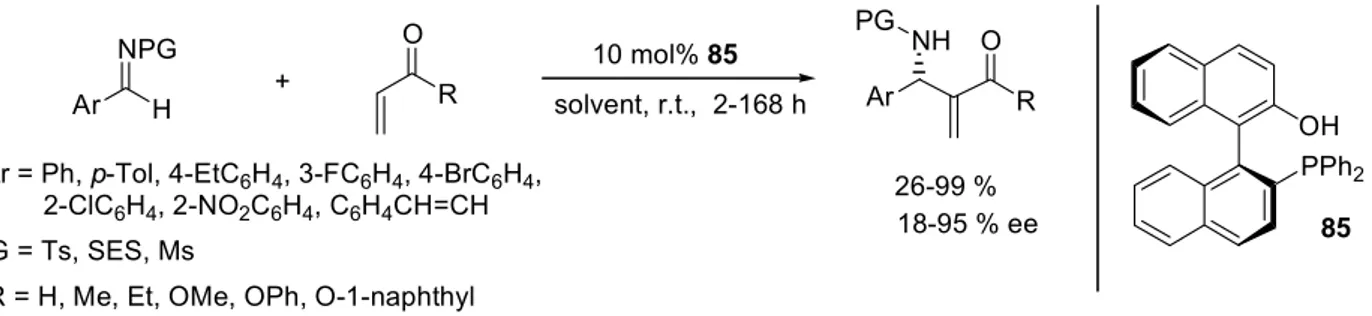
The asymmetric aza-Baylis-Hillman (aza-BH) reaction of achiral imines with acrylates provides a direct access to chiral β -amino acid esters, which have gained considerable interest due to their important biological properties, their occurrence in natural products, and as potential precursors for β -lactams.
9In this work, the first aza-Baylis-Hillman reaction catalysed by a chiral Lewis acidic complex was developed. Diphenylphosphinoyl- (dpp-) protected imines were employed as electrophiles, due to the ease of removal of this activating and protecting group.
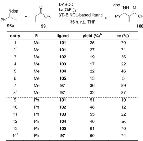
In an initial ligand screening with La(OiPr)
3and various (R)-BINOL-based ligands (Scheme 1-6, p. 5), high enantioselectivity was achieved with linked-(R,R)-BINOL 97.
Since raising the ligand to metal ration for (R)-BINOL 101 from 1:1 to 2:1 showed no be-
discussed as effect of its higher stability compared to the La-(R)-BINOL complex. The introduction of sterically more demanding substituents in the (R)-BINOL 3,3’ position lead to catalysts with low reactivity and enantioselectivity.
Ph H
Ndpp
OR
O DABCO
Ph OR
NH O dpp
CF3 F3C
Pr
Pr iPr H
OH OH R
R
O OHHO OHHO La(OiPr)3
(R)-BINOL-based ligand 35 h, r.t., THF +
98a 99 100
R =
i
i 105 102
97
103 101 104
Scheme 1-6 Screening of (R)-BINOL-based ligands.
The La-linked-(R,R)-BINOL complex is well known for its dual reactivity as Lewis acid and Brønsted base. However, increasing the basicity of the catalyst by deprotonation with KHMDS, NaHMDS or n-BuLi did not result in higher yields or enantioselectivities relative to the non-metallated species.
A variation of the catalyst’s Lewis-acidity by complexation of linked-(R,R)-BINOL 97 with various M(OiPr)
x, identified Ln-complexes as the most efficient catalysts, whereas the more Lewis-acidic Ti-complex as well as the more Lewis basic Sr-complex led to decreased yields and enantioselectivities.
An extensive screening of the reaction parameters was performed. The optimised conditions are shown in Scheme 1-7.
Ph H Ndpp
OMe
O 20 mol% DABCO
Ph OMe
NH O
dpp O
OH OH
HO HO 10 mol% La(OiPr)3 / ligand 97
35 h, 40 °C, THF +
1.0 eq 98a 4.0 eq 99a 100aa 50 %, 90 % ee 97
Scheme 1-7 Optimised reaction conditions for the aza-BH reaction catalysed by La-linked-BINOL
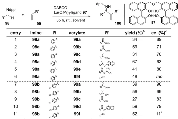
In a substrate screening, among the acrylates tested, the highest yields were
achieved with 2-naphthyl acrylate 99d (up to 67 %), while methyl acrylate 99a led to the
highest enantioselectivities (up to 90 % ee). Comparison of dpp-protected benzald-
imine 98a with its para-Cl-substituted analogue 98b revealed in most cases higher
enantioselectivities at virtually unchanged yields for the para-Cl-substituted and dpp-
protected benzaldimine 98b. Aliphatic imines were also tested, but only traces of the
corresponding aza-BH adducts were detected.
Hydrolytic deprotection of the aza-Baylis-Hillman products 100 provided single step access to the corresponding β -amino acids 106 in up to 75 % yield (Scheme 1-8).
Ar OR
NH O dpp
Ar OR
NH2O
100ab R = Ph, Ar = Ph 100ba R = Me, Ar = pClC6H4
75 % 106a Ar = Ph 62 % 106b Ar = pClC6H4 20 % HCl .
reflux, 6 h
HCl
Scheme 1-8 Single step conversion of aza-BH adducts 100 to β-amino acids 106.
Conclusions. It was shown for the first time that the aza-Baylis-Hillman reaction can be
catalyzed enantioselectively by a chiral Lewis acidic metal complex. Using the La-
linked-(R,R)-BINOL complex as catalyst and dpp-protected imines as substrates the
corresponding aza-BH adducts could be obtained in up to 68 % yield and up to
90 % ee. Hydrolytic deprotection of the highly enantio-enriched aza-Baylis-Hillman
products thus obtained grants access to α -methylene- β -aminoacids in a simple one pot-
procedure.
2 Introduction
In modern sciences, interdisciplinary fields become more and more important. For example, organic chemistry and biology are linked by the building blocks of life.
Chemically, such building blocks of life are small organic molecules (i.e. amino acids, sugars and nucleic bases) or polymers (i.e. proteins, nucleic acids, carbonhydrates).
Biologically, the building blocks of life are cells (Scheme 2-1).
ORGANIC CHEMISTRY
AGROCHEMICALS FOOD CHEMISTRY
PHARMACEUTICAL CHEMISTRY
herbicide, fungicide, insecticide
flavours pharmaceuticals
genetically modified
BIOLOGY
yeast soybeans
ES cells
BUILDING BLOCKS OF LIFE sugars, amino acids,
nucleic bases cells
Scheme 2-1: Building block of life as interdisciplinary field of organic chemistry and biology and their relations to applied chemistry.
Cellular biology, studied since the 1800s, is the basis of current stem cell research. The advent of the microscope provided scientists a first look at human cells. At this time cells were identified as the building blocks of life, capable of reproducing and different- tiating into all the cell types that make up the living body.
Although the history of stem cell research started in the early 1900s with the disco- very that the various types of blood cells all came from a particular ‘stem cell’, the po- tential of these stem cells has been discovered in the late 1960s: McCulloch and Till detected self-renewing cells in mouse bone marrow
10and Altman as well as Das showed that adult neurogenesis was accompanied by stem cell activity in the brain.
11Stem cell research has basically focused on bone marrow transplants. Since 1950,
theses transplants have been used in patients additionally treated with radiation and
chemotherapy.
12However, first the pioneering discovery by Dausset in 1958, which identified the first human histocompatibility antigens, enabled the establishment of this stem cell theraphy
.13Developments in biotechnology in the 1980s and 1990s afforded techniques for targeting and altering genetic material as well as methods for growing human cells in the laboratory. These advances opened the doors for stem cell research.
14In 1998, Thompson isolated cells from the inner cell mass of early embryos, and developed the first embryonic stem cell lines.
15The blastocysts used for human stem cell research typically originate from in vitro fertilization procedures.
These discoveries stimulated further research. The growing interest in stem cell research is evident from the exponential increase of publications in this research area.
The possibilities for stem cell research are indeed endless, but yet unpredictable. If scientists can master the biochemistry behind stem cell development, stem cell techno- logy could be used, e.g. to produce replaceable tissues or organs.
A contribution to the stem cell development is the subject of the present work. In the first part of this thesis, the in vitro development of cardiomyocytes from ES cells induced by small molecules is described.
Organic compounds such as pharmaceuticals, agrochemicals, flavors and fragrances play an improtant role in modern life. These compounds possess biological activity which arises from the interaction of the organic compound with a biological target.
These biomolecules are single enantiomers of chiral compounds since they are con- structed from chiral building blocks such as amino acids or carbohydrates, which are present in nature as a single enantiomer.
Besides ex chiral pool syntheses (Scheme 2-2 (a)), there are two general methods for obtaining enantiomerically enriched compounds: (Scheme 2-2 (b)) they may be syn- thesised in racemic form and resolved into their optical antipodes, or (Scheme 2-2 (c)) the synthesis may be directly performed in an enantioselective manner.
(b) An example for the separation of enantiomers by chemical resolution based on
the selective crystallisation of one enantiomer by diastereomeric salt formation. Al-
though this method is not generally applicable, it is still quite popular for the large scale
preparation of enantiomerically pure acids or amines. Other approaches of resolution
involve the formation of covalent bonds between the racemic substrate and an enan-
tiomerically pure compound. The resulting diastareomeric compounds can be separated
appropriate diastereomer by chemical transformations. However, these methods yield only up to 50 % of the desired enantiomer.
(c) For asymmetric syntheses, i.e. the generation of enantio-enriched compounds from achiral precursors, chiral auxilaries, chiral reagents or chiral catalysts, all in enantio-pure form, can be employed. There are prominent auxiliar-based synthesis, e.g.
Ender’s SAMP/RAMP
16or Schöllkopf’s bislactim ether
17syntheses. However, the covalent binding to and cleaving from the substrate makes these procedures less efficient. Chiral enantio-pure reagents, e.g. BINAL-H,
18must be employed in stoichio- metric quantities, this likewise decreases the efficiency of the synthesis. Hence, enantioselective catalyses with small amounts of “chiral information” are the most efficient procedures to obtain enantiopure chiral compounds. There are three different kinds of chiral catalysts employed in asymmetric synthesis, biocatalysts,
19metal ligand complexes derived from chiral ligands,
20and chiral organocatalysts.
21Enzymes as bio- catalysts are very selective and efficient, but often tolerate only small temperature ranges, few solvents and a narrow range of sustrates. Metal complex catalysts are widely applied in organic chemistry, although the ligands and metals are often quite expensive. Catalysis by small organic molecules, e.g. proline, complements enantio- selective synthesis, as such catalysts are often cheap, easy to modify and less toxic.
chiral enantio-enriched product achiral starting material
ex chiral pool derivatisation
nature non
stereoselective synthesis
racemic mixture racemic resolution
enantioselective synthesis chiral auxilary chiral reagent chiral catalyst
a b c
Scheme 2-2 Three methods to obtain chiral, enantio-enriched compounds.
Altough impressive results were achieved in the field of asymmetric catalysis in the last decades, there is still the need for the development of highly selective and effective catalytic systems.
A contribution to this is the subject of the present work. In the second and third part
of this thesis, an improved two step synthesis of a prominent organocatalyst as well as
the devopment of a new catalytic system for an established C-C coupling is described.
3 Chemically Induced Cardiomyogenesis of mES Cells
3.1 Background
Cardiomyopathy results from the loss of functional heart muscle cells, impairing the ability of the heart to maintain adequate blood circulation. As adult differentiated cardio- myocytes lack prominent regenerative capacity, heart transplantation remains the only effective causal treatment. An increasing number of patients suffer from severe heart failure, and the shortage of available donor organs emphasise the demand for alternative therapy methods, such as cellular cardiomyoplasty. In this context, several animal studies demonstrated a successful engraftment of cardiac myocytes into the adult heart.
22,23However, the limiting factor for a general application of cell therapy for the treatment of cardiovascular diseases still is the insufficient number of donor cells.
Embryonic stem (ES) cells isolated from the inner cell mass of early blastocyst-stage embryos are pluripotent. They are capable of differentiating in vitro into any somatic cell type, including cardiomyocytes, haematopoietic progenitors, skeletal myocytes, smooth muscle cells, adipocytes, chondrocytes, endothelial cells, neurons and glia and pan- creatic islet cells.
15When cultured in the presence of leukaemia inhibitory factor (LIF), ES cells remain undifferentiated and can be propagated indefinitely. Upon withdrawal of LIF, ES cells spontaneously and irreversibly differentiate into multicellular aggregates, so-called embryoid bodies (EBs). These aggregates resemble early post-implantation embryos and contain derivatives of all three germ layers.
24So far, the in vitro differentiation of ES cells into cardiomyocytes offers a new approach for cellular therapy of degenerative heart diseases. Therefore, the development of new approaches allowing a direct differentiation of ES cells into cardiomyocytes is of growing interest.
25The differentiation programs of ES cells can be shifted toward cardiomyogenic or neuronal differentiation by treatment with small molecules, such as retinoic acid 1 or dimethyl sulfoxide (DMSO) 2 (Scheme 3-1), at specific stages of differentiation.
26,27Scheme 3-1
:
Retinoic acid 1 (left) and DMSO 2 (right).Me S Me O
Me 2
Me Me O
OH Me Me
1
However, this approach is not very efficient, and normaly requires selection to enrich specific cell lineages. Several small molecules have recently been found to control the differentiation of ES cell into a specific cell lineage as well as to affect the self renewal of ES cells:
Ascorbic acid. In 2003 Takashi et al.
28screened 880 compounds of the FDA2000 Drug Library, which were approved for human use, for their potential to induce cardiac differentiation of mouse ES cells in a monolayer culture. They used CGR8 mouse ES cells stably transfected with the cardiac muscle-specific α-MHC promotor driven EGFP as a reporter. They found that upon treatment with ascorbic acid 3 (vitamin C, Scheme 3-2, p. 14) increased cardiac differentiation of ES cells in a dose-dependent manner. It can be assumed that this effect of ascorbic acid is independent of its antioxidative pro- perty
29, because antioxidants normally inhibit ES cell differentiation into cardiac myocy- tes, whereas ROS (reactive oxygen species) like H
2O
2or radical-generating menadione enhance cardiogenesis
30. Other antioxidative agents such as N-acetylcystein, Tiron (brenzcatechine-3,5-disulfonic acid disodiumsalt) or vitamin E also do not mimic the effect of ascorbic acid on the cardiac differentiation. In addition, there was no significant effect of vitamin C 3 on the cardiomyogenesis via embryoid body formation. This sug- gests that ascorbic acid induces permissive changes that occur during the formation of embryoid bodies, rather than induction of autonomous commitment of ES cells to car- diac myocytes.
5-Aza-2’-deoxycytidine. Based on the results of Fukuda et al.
31, who ascertained an increased cardiomyogenesis of mesenchymal stem cells by adding the DNA-demethyla- ting agent 5-aza-2’-deoxycytidine 4
(Scheme 3-2, p. 14), Xu et al.
32tested the effect of this compound on the differentiation of human ES cells into cardiomyocytes via embry- oid body formation. Using immunostaining and real-time PCR they observed an in- crease of cardiomyocyte formation by treating the human ES cells with 5-aza-2’-deoxy- cytidine 4. Interestingly, DMSO and retinoic acid, which have been shown to induce car- diomyocyte differentiation in mouse ES cells (see above), did not enhance human ES cell cardiomyocyte differentiation.
Cardiogenol C. Schultz et al.
33screened a 100.000 compound library of so called
privileged heterocycles
34using a stable engineered mouse embryonic carcinoma cell
conditions. Like ES cells, P19 cells are pluripotent and are capable of differentiating into cardiac cells under specific culture conditions
.35They identified 80 compounds that increased luciferase activity. In addition, 35 compounds induced a parallel expression of sarcomeric α-MHC, a cardiac specific protein. In particular, these compounds share significant structure similarities possessing a 2-hydroxylamino substitution at the C-4 position and large, hydrophobic groups at the C-2 position. To confirm that these com- pounds are general cardiomyogenesis inducing agents, they analysed their effects on undifferentiated R1 mouse ES cells also in a monolayer culture. They detected more than 90 % positive cardiomyocyte formation of ES cells treated with Cardiogenol C 5 (Scheme 3-2, p.14). Furthermore, they observed that compound treatment slowed down cellular proliferation with no significant cell death, indicating that this process is not simply a selection for cardiac precursor cells with the death of cells in other lineages.
Verapamil, Ryanodine, Cyclosporin. Sachinidis et al.
4investigated the effects of 33 small molecules interfering with several signalling cascades on cardiomyognesis. They used a transgenic ES cell lines expressing enhanced green fluorescent protein (EGFP) under the conrol of the α-myosin heavy chain promoter (pαMHC). In this screening, especially the L-type Ca
2+channel blocker Verapamil 6 as well as Ryanodine 7, an inhibitor of the protein phosphatase 2B, and Cyclosporin A 8, an Calcineurin inhibitor (Scheme 3-2, p. 14), exerted the most striking pro-cardiomyogenic effect. Treatment of the EBs with Verapamil 6 caused a pronounced 94 % increase and treatment with Ryanodine 7 resulted in a significant 75 % increase of the EGFP fluorescence com- pared to untreated cells. Ryanodine 7 is a natural compound acting through binding to the Ryanodine receptor (RyR), the Ca
2+channel of the sarcoplasmatic reticulum (SR), resulting in the release of Ca
2+into the sarcoplasm. Depending on the cell type and concentration, Ryanodine 7 induces an inhibition or activation of the release of Ca
2+from SR.
36Verapamil 6 is an antagonist of the plasma membrane L-type Ca
2+channel
thereby inhibiting the influx of extracellular Ca
2+into the cytosol.
37Furthermore,
Sachinidis et al. observed that spontaneous contractions of ES cell derived cardiomyo-
cytes were totally inhibited when Verapamil 6 was present at late stages of cardiac dif-
ferentiation. From these results, Sachinidis et al. concluded that cyctosolic Ca
2+is invol-
ved in the differentiation of cardiomyocytes from ES cells and that [Ca
2+]
ilowering
agents promote cardiomyogenesis even if the physiological activity of beating is
inhibited.
Exposure of EBs to Cyclosporin A 8 caused an increase of the EGFP fluorescence up to 140 %. Cyclosporin A 8 is an amino acid cyclic peptide used as immunosuppressant compound. Complexes of Cyclosporin A 8 with the immunophilin Cyclophilin A within the cell inhibit the Ca
2+- and calmodulin-dependent protein phosphatase 2B (Calcineu- rin). Futhermore there is an accumulating evidence that Calcineurin is also involved in the regulation of other cellular processes such as embryonic development and cancer, as well as cardiac valve formation and cardiac hypertrophy.
383
4
5
6
7 8
Scheme 3-2 Cardiomyogenesis inducing substances: ascorbic acid 3, 5-aza-2’deoxycytidine 4, Cardio- genol C 5, Verapamil 6, Ryanodine 7 and Cyclosporine A 8.
3.2 Concept
3.2.1 Forward Chemical Genetics Approach
The in vitro differentiation of ES cells into cardiomyocytes offers a new approach for cellular therapy of degenerative heart diseases. For a direct differentiation of ES cells into cardiomyocytes experimental methods are required allowing rapid and “easy to handle” parallel testing of small molecules which may influence the differentiation of ES cells towards cardiomyocytes. For this aim, a transgenic ES cell lineage is used, expressing enhanced green fluorescent protein (EGFP) under the control of the heart specific promoter α−myosine heavy chain (pα-MHC) to test selected substances for
HO HO
O N
O N
NH2 N
N N HN
NH
OH MeO
OH OH O O
HO OH
N MeO
MeO
OMe OMe iPr Me
NC
N N N
Me
HN HN N HN N N
N HN
O
O
O
O O O O
O O
O
O
Me Me
Me
Me Me
Me Me
OH Me
iPr
Me
Me iPr
Pr
iPr iPr
Me
iPr i
O HO
H Me
OH HO HO MeHO
H O HN
O
OH
Pr Me i
Scheme 3-3 Forward chemical genetics approach. After culturing of transgenic ES cells in multi-well plates, a library of small molecules can be screened by addition of one single compound per well. After differentiation, small molecules can be identified participating in the generation of the desired phenotype.
Further modifications based on the structure of these active test substrates should lead to an enhanced differentiation potential of the tested compounds.
In general, two strategies exist for the identification of cardiomyogenesis stimulating substances: structure based drug design (SBDD), or rational drug design (RDD).
41,42,43The SBDD involves the search for a small molecule that perfectly fits in the binding pocket of a target protein, and thus influences the signalling pathway. An application of this method requires knowledge about the signalling pathways, the proteins involved and their structures.
The RDD describes the relationship between chemical structures and their biological effects. For a successful correlation all tested compounds must bind to the same target.
To fulfil this requirement, the test substrates must have very similar chemical structures.
To find a capable lead structure for the RDD, a foregoing diversity orientated screening (DOS) is performed. Within this screening, substances are tested which cover a broad structural spectrum with fixed core structures.
absence of the desired cell phenotype
low level of the desired cell phenotype
differentiation
addition of one type of small molecule per well
identification of the small molecule participating in the generation of the desired phenotype
synthesis of modified small molecules normal level of the
desired cell phenotype high level of the desired cell phenotype transgenic ES cells
As the structures of the proteins involved in the signalling pathways - which are assumed to promote cardiomyogenesis of ES cells - are mostly unknown, the require- ments for a SBDD approach are not fulfiled. Therefore a RDD with a preliminary DOS was performed focused on the synthesis and screening of (thio)urea and cinchona- alkaloid based compound libraries.
3.2.2 Selection of Substrates
Stem cells and cancer cells share the similarity that both cell types exhibit high proliferation rates. According to the cancer stem cell theory, drugs which induce differentiation of stem cells thereby also decrease their proliferation rates. If the same applies for cancer cells, drugs for cell differentiation could be used for anti-cancer thera- pies and vice versa. The prominent anti cancer drug Nexavar® (Sorafenib) harbours a thiourea moiety (Scheme 3-4).
2Therefore, the selection of substances has been focused first on (thio)urea derivatives.
Scheme 3-4 Chemical structure of the prominet anti cancer drug Nexavar®.
A second selection of substances has concentrated on ammonium salts derived from cinchona alkaloids, as they are well known for their phase transfer abilities
44which should promote membrane permeation by drugs. Furthermore, quinidine is a pharma- ceutical agent that acts as a class I antiarrhythmic agent in the heart. The effect of quinidine on the ion channels is to prolong the cardiac action potential and thereby prolonging the QT interval on the surface electrocardiogram (ECG).
33.3 Results and Discussion 3.3.1 Substance Screening
For the identification of a cardiomyogenesis stimulating substance, a rational drug design with a preliminary DOS focused on the synthesis and screening of (thio)urea and cinchona-alkaloid based compound libraries was performed.
NH O
O N
O HNMe NH
Cl F3C
Differentiation Protocol – for DOS, RDD
45. A transgenic murine ES cell lineage expressing enhanced green fluorescent protein (EGFP) under control of α-myosine heavy chain (α-MHC) promoter (pα-MHC- EGFP) was used to investigate the effects of (thio)urea and cinchona-alkaloid derivatives on cardiomyogenesis. To start differentia- tion, ES cells were cultured in suspension for 12 h to form EBs. About fifty EBs were transferred to each well of bacterial 6-well plates and the test compounds were added.
On day 7, fresh medium and fresh test compounds were added. On day 14, EBs on each plate were counted, lysed and the EGFP fluorescence in the lysates was measured at an excitation wavelength of 476 nm and an emission wavelength of 508 nm.
Diversity Orientated Screening (DOS). For a first DOS, two substance libraries I and
II containing (thio)urea (I) or cinchona-alkaloid (II) derivatives with extensivly modified
substitution patterns (Scheme 3-5 and Scheme 3-6, p. 18-19) were synthesised and
tested for their cardiomyogenesis inducing efficiency.
HN HN NH O
NH O F3C
CF3 CF3
CF3 NH N
H S
NMe2
NH N H S F3C
CF3
N CH2
NH Me Me
Me HN S
NH
NH S
N
NH N
MeO
CH2
O NH CF3
F3C
N Ph NH S NH CF3
F3C NH
Me Me
Me HN O F3C NH
CF3
NH O
CF3 F3C
NH Me Me
Me HN O
NH
NH O NH
OSO NH
Me Me
NH NH
O
F3C NMe2
Me CF3
Et2N
NH N O H
S
NMe2 tBu
NH NH
O
NMe2 Me Me
Me Me
NH N
H NMe2 O
NH N
H NMe2 O
O2N NO2
NH N H NEt2 O
F3C CF3
NH N H S F3C
CF3
NBn2 NH N
H O F3C
CF3
NBn2
NH N H O F3C
CF3
N CH2 H2C
I-1
S NH O O HN
Me Me Me Me Me
Me
I-2 I-3
I-4 I-5 I-6
I-7 I-8 I-9
I-10 I-11 I-12
I-13 I-14 I-15
I-16 I-17 I-18
I-19
Scheme 3-6 Structures contained in the cinchona-alkaloid-based library II.
The cardiomyogenic effect of the substances is expressed as percent of the EGFP fluorescence of the vehicle treated cells (100%). An increased production of cardio- myocytes gives percent of control values higher then 100 %, a decreased production of cardiomyocytes lower than 100 %.
N
N OH
OBn CH2
N N
OH OBn
CH2
N N
OMe OBn
CH2
N N
OH OH
Me
N N
OH OH
CH2
N OiPr
OH
N
Me N N
OiPr OH
CH2
Cl
N N
OH O
Me
N CH2
N OMe Cl OH
Cl Cl Cl
Cl
CH2
N OH
OH N
N N
OMe O
CH2
OH N
OMe OH
N CH2
Br
Cl N
N OMe
OH CH2
N
N OiPr
OH Me
N
N OH
Me
OH
N
N OH
CH2
OBn
N Me
H O OH
N Me
H O OMe
N CH2
N OMe
OHOH N
OH Me
II-1 II-2 II-3 II-4
II-5 II-6 II-7 II-8
II-9 II-10 II-11 II-12
II-13 II-14 II-15 II-16
II-17 II-18 II-19 II-20
Among the (thio)urea derivatives (library I) tested, especially the bis-ureas I-6 and I-15 induced an increase of 80 % or 50 %, respectively, of the EGFP fluorescence which correlates with a positive cardiomoygenic effect. In the cinchona-alkaloid based library II the best result could be obtained with the quinidine based salt II-16 which induced a 60 % increase (Scheme 3-7).
I-6
I-15
II-16
Scheme 3-7 Influence of the compounds I-6, I-15 and II-16 on the expression of EGFP, normalised to the number of EBs. Values from four independent experiments (mean ± SD, n=4), each performed in triplicate, are displayed as percent of control values (=100 %, only vehicle, without test substrate). Both a significant increase and decrease of values, compared to controls, represent an effect on the generation of cardiac cells.
0 20 40 60 80 100 120 140 160 180 200
1 E-09 1 E-08 1 E-07 1 E-06 1 E-05 1 E-04 concentration [M]
% of control .
0 20 40 60 80 100 120 140 160 180 200
1 E-09 1 E-08 1 E-07 1 E-06 1 E-05 1 E-04 concentration [M]
% of control .
0 20 40 60 80 100 120 140 160 180 200
1 E-09 1 E-08 1 E-07 1 E-06 1 E-05 1 E-04 concentration [M]
% of control .
HN HN NH O
NH O F3C
CF3 CF3
CF3
NH Me Me
Me HN O
NH
NH O
N CH2
N OMe
OH
Cl