Notes on some sertulariid hydroids (Cnidaria: Hydrozoa) from the tropical western Pacifi c, with descriptions of nine new species
Horia R. GALEA
Hydrozoan Research Laboratory, 405 Chemin des Gatiers, 83170 Tourves, France.
E-mail: horia.galea@gmail.com
urn:lsid:zoobank.org:author:DE5AC672-0243-46F2-A910-AFF4E91A4C5D
Abstract. Forty-three species of sertulariid hydroids (Cnidaria: Hydrozoa: Sertulariidae), collected from the tropical western Pacifi c (Taiwan, Philippines, New Caledonia, French Polynesia, Vanuatu, Fiji, Tonga, Solomon Islands) during various expeditions of the French Tropical Deep-Sea Benthos program, are discussed. Of these, nine are new to science: Gonaxia nova sp. nov., G. plumularioides sp. nov., Sertularella folliformis sp. nov., Se. plicata sp. nov., Se. pseudocatena sp. nov., Se. splendida sp. nov., Se. tronconica sp. nov., Se. tubulosa sp. nov., and Symplectoscyphus paucicatillus sp. nov.
The subspecies Symplectoscyphus johnstoni (Gray, 1843) tropicus Vervoort, 1993 is raised to species but, in order to avoid the secondary homonymy with Sy. tropicus (Hartlaub, 1901), the replacement name, Sy. fasciculatus nom. nov., is introduced. The male and female gonothecae of Diphasia cristata Billard, 1920, the male gonothecae of Gonaxia elegans Vervoort, 1993, as well as the female gonothecae of Salacia macer Vervoort & Watson, 2003, are described for the fi rst time. Additional notes on the morphology of several other species are provided. All taxa are illustrated, in most cases using fi gures drawn at the same scale, so as to highlight the differences between related species.
Key words. Deep water, new species, thecate hydroids, Sertulariidae.
Galea H.R. 2016. Notes on some sertulariid hydroids (Cnidaria: Hydrozoa) from the tropical western Pacifi c, with descriptions of nine new species. European Journal of Taxonomy 218: 1–52. http://dx.doi.org/10.5852/ejt.2016.218
Introduction
The present report is a continuation of an earlier study dealing with some new sertulariid and syntheciid hydroids (Galea 2015a), and completes a series of previous reports dealing with members of the families Sertulariidae Lamouroux, 1812 (Vervoort 1993), Halopterididae Millard, 1962 (Ansín Agís et al. 2009), Lafoeidae Hincks, 1868 (specifi cally, the genus Acryptolaria Norman, 1875) (Peña Cantero & Vervoort 2010), and Kirchenpaueriidae Stechow, 1921 (Ansín Agís et al. 2014) from the tropical western Pacifi c, obtained during numerous expeditions undertaken conjointly by the Institut de Recherche pour le Développement (IRD, formerly ORSTOM) and the Muséum national d’Histoire naturelle (MNHN) of Paris (Bouchet et al. 2008).
The sertulariid hydroids dealt with herein belong to the following ten genera: Caminothujaria von Campenhausen, 1896, Diphasia L. Agassiz, 1862, Dynamena Lamouroux, 1812, Geminella Billard, 1925b, Gonaxia Vervoort, 1993, Idiellana Cotton & Godfrey, 1942, Salacia Lamouroux, 1816,
http://dx.doi.org/10.5852/ejt.2016.218 www.europeanjournaloftaxonomy.eu 2016 · Galea H.R.
This work is licensed under a Creative Commons Attribution 3.0 License.
M o n o g r a p h
urn:lsid:zoobank.org:pub:A4D7AA38-D18F-4604-A5E0-D965637BD9F8
Sertularella Gray, 1848, Sertularia Linnaeus, 1758 and Symplectoscyphus Marktanner-Turneretscher, 1890. Although most species have already been dealt with by Vervoort (1993), additional notes on them are provided, along with new data on the morphology of other taxa, including descriptions of nine new hydroids.
Material and methods
The methods of study were described in detail by Galea (2007, 2008). The list of stations is given in Table 1. Station numbers, as indicated in the text, are preceded by a two-letter prefi x referring to the sampling gear used to secure the material, either a Charcot dredge (CC), a beam trawl (CP), a rocky- bottom dredge (DR) or a Warrén dredge (DW). The material is deposited in the collections of MNHN.
The species dealt with herein are listed according to their morphological similarities, notably the shape of their hydrothecae. Most fi gures, representing portions of colonies, hydro- and gonothecae, are drawn to the same scale and grouped onto the same plates, so as to facilitate comparisons between related species.
Results
Phylum Cnidaria Verrill, 1865 Class Hydrozoa Owen, 1843
Subclass Hydroidolina Collins & Marques, 2004 Order Leptothecata Cornelius, 1992 Family Sertulariidae Lamouroux, 1812 Genus Caminothujaria von Campenhausen, 1896 Caminothujaria molukkana von Campenhausen, 1896
Fig. 1A–B Caminothujaria molukkana von Campenhausen, 1896: 103.
Caminothujaria molukkana – Schuchert 2015: 338, fi g. 12.
Material examined
MUSORSTOM 3: Stn. DR117, numerous sterile fragments from an originally large colony (size undeterminable) (MNHN-IK-2012-16500); Stn. CP121, a 2.3 cm high, sterile colony (MNHN- IK-2012-16501); Stn. DR126, an originally 11.5 cm high colony, now broken into two parts, one gonotheca (MNHN-IK-2012-16502).
MUSORSTOM 8: Stn. CP1001, several stems and fragments up to 5.3 cm high, all sterile (MNHN- IK-2012-16503); Stn. DW1021, numerous sterile stems and fragments, 3–10.5 cm high, all sterile (MNHN-IK-2012-16504); Stn. CP1104, an originally large but sterile colony, now fragmented, largest fragment ca 8 cm high (MNHN-IK-2012-16505).
BORDAU 2: Stn. CP1582, ca 4 cm high, fertile colony composed of three stems (MNHN-IK-2012-16506).
SALOMON 1: Stn. DW1756, three sterile stems 2.5–5 cm high (MNHN-IK-2012-16507); Stn. DW1767, a ca 12 cm high, sterile colony (MNHN-IK-2012-16508).
Remarks
A list of synonyms is provided by Schuchert (2015) and, for a redescription, see Hirohito (1995).
Campaign Station Date Depth (m) Latitude Longitude
MUSORSTOM 3 DR117 3 Jun. 1985 92–97 12°31′ S 120°39′ E
CP121 3 Jun. 1985 73–84 12°08′ S 121°17′ E
DR126 4 Jun. 1985 266 11°49′ S 121°22′ E
CP134 5 Jun. 1985 92–95 12°01′ S 121°57′ E
BIOCAL CP34 29 Aug. 1985 710 23°11.88′ S 167°11.30′ E
DW37 30 Aug. 1985 350 22°59.99′ S 167°15.65′ E
DW38 30 Aug. 1985 360 22°59.74′ S 167°15.31′ E
DW66 3 Sep. 1985 505–515 24°55.43′ S 168°21.67′ E
CP69 3 Sep. 1985 1220–1225 23°51.38′ S 167°58.68′ E
DW46 30 Aug. 1985 570–610 22°53.05′ S 167°17.08′ E
MUSORSTOM 4 CC174 17 Sep. 1985 385 19°00.30′ S 163°18.50′ E
DW205 27 Sep. 1985 160 22°38.50′ S 167°06.80′ E
DW221 29 Sep. 1985 535–560 22°58.60′ S 167°36.80′ E
MUSORSTOM 6 DW423 16 Feb. 1989 280 20°25.85′ S 166°40.50′ E
SMIB 4 DW40 7 Mar. 1989 260 24°46.20′ S 168°08.70′ E
DW48 8 Mar. 1989 245 24°46.20′ S 168°08.70′ E
DW55 9 Mar. 1989 260 23°21.40′ S 168°04.50′ E
DW57 9 Mar. 1989 210 23°21.00′ S 168°21.00′ S
VOLSMAR DW39 8 Jun. 1989 305 22°20.50′ S 168°43.50′ E
SMIB 6 DW127 4 Mar. 1990 205–215 19°06′ S 163°22′ E
BATHUS 2 DW716 10 May 1993 290–308 22°40′ S 167°12′ E
CP737 13 May 1993 357–400 23°03.42′ S 166°59.97′ E
BATHUS 3 CP804 27 Nov. 1993 244–278 23°41.40′ S 168°00.42′ E
CP806 27 Nov. 1993 308–312 23°42.31′ S 168°00.52′ E
DW809 27 Nov. 1993 650–730 23°39.39′ S 167°58.94′ E
DW810 27 Nov. 1993 850–900 23°40.45′ S 167°58.83′ E
CP813 28 Nov. 1993 410–415 23°45.23′ S 168°16.57′ E
CP814 28 Nov. 1993 444–530 23°47.60′ S 168°17.10′ E
CP821 29 Nov. 1993 864–880 23°19.16′ S 167°58.63′ E
CP823 29 Nov. 1993 980–1000 23°22.76′ S 167°51.60′ E
DW829 29 Nov. 1993 386–390 23°21.37′ S 168°01.84′ E
DW830 29 Nov. 1993 361–365 23°19.75′ S 168°01.45′ E
BATHUS 4 DW902 4 Aug. 1994 341–351 19°00.84′ S 163°14.83′ E
CP906 4 Aug. 1994 339–350 19°01.07′ S 163°14.51′ E
DW914 5 Aug. 1994 600–616 18°48.79′ S 163°15.23′ E
DW923 6 Aug. 1994 502–470 18°51.51′ S 163°24.17′ E
MUSORSTOM 8 CP1001 25 Sep. 1994 150–250 18°48.97′ S 168°59.83′ E DW1019 28 Sep. 1994 397–430 17°38.25′ S 168°33.91′ E DW1021 28 Sep. 1994 124–130 17°42.75′ S 168°37.00′ E
CP1083 5 Oct. 1994 397–439 15°51.91′ S 167°19.82′ E
CP1084 5 Oct. 1994 207–280 15°50.29′ S 167°17.88′ E
CP1104 7 Oct. 1994 125–129 15°03.83′ S 167°06.96′ E
CP1131 11 Oct. 1994 140–175 15°38.41′ S 167°03.52′ E
MUSORSTOM 9 DW1204 28 Aug. 1997 60–62 9°52.6′ S 139°03.2′ W
CP1265 3 Sep. 1997 90–92 9°20.4′ S 140°07.3′ W
BORDAU 1 CP1447 4 Mar. 1999 420–513 16°45.23′ S 179°59.13′ E
CP1448 4 Mar. 1999 410–500 16°45.04′ S 179°58.97′ E
LITHIST DW13 12 Aug. 1999 400 23°45′ S 168°17′ E
BORDAU 2 DW1513 1 Jun. 2000 190–221 21°19′ S 175°01′ W
CP1582 13 Jun. 2000 79–82 18°41′ S 174°03′ W
DW1595 16 Jun. 2000 523–806 19°03′ S 174°19′ W
TAIWAN 1 DW1 27 Jul. 2000 98 23°38.1′ N 119°50.9′ E
NORFOLK 1 DW1659 20 Jun. 2001 467–449 23°38′ S 167°42′ E
DW1666 20 Jun. 2001 469–860 23°42′ S 167°43′ E
DW1667 21 Jun. 2001 237–250 23°41′ S 168°01′ E
DW1699 24 Jun. 2001 581–600 24°40′ S 168°39′ E
DW1704 25 Jun. 2001 420–400 23°47′ S 168°17′ E
DW1712 26 Jun. 2001 180–250 23°22′ S 168°03′ E
CP1719 26 Jun. 2001 391–407 23°21′ S 168°02′ E
DW1722 26 Jun. 2011 540 23°16′ S 168°01′ E
TAIWAN 2 DW118 31 Jul. 2001 128–136 24°56.3′ N 122°01.5′ E
SALOMON 1 DW1741 23 Sep. 2001 557–655 11°29′ S 159°57′ E
DW1756 26 Sep. 2001 497–511 08°52′ S 159°50′ E
DW1767 28 Sep. 2001 98–200 08°19′ S 160°40′ E
DW1854 7 Oct. 2001 229–260 09°46′ S 160°53′ E
Table 1. List of stations.
Geographical distribution
The known area of distribution ranges from Indonesia and Tuvalu in the south, through Japan in the north, and encompasses the Philippines and the South China Sea (Schuchert 2015).
Genus Diphasia L. Agassiz, 1862 Diphasia cristata Billard, 1920
Fig. 1C–J Diphasia cristata Billard, 1920: 147, fi g. 1f.
Diphasia cristata – Billard 1925b: 218, fi g. 57, pl. 3 fi g. 32. ― Hirohito 1983: 38, fi g. 15; 1995: 161, fi g. 51a–c.
Material examined
SMIB 4: Stn. DW57, several sterile stems and fragments, up to 2.5 cm high (MNHN-IK-2012-16509).
SMIB 6: Stn. DW127, a female colony composed of several stems up to 4 cm high, epizoic on Gonaxia sp. (MNHN-IK-2012-16510).
MUSORSTOM 8: Stn. DW1021, small female colony composed of a few stems, up to 2 cm high, epizoic on Caminothujana molukkana (MNHN-IK-2012-16511).
BORDAU 1: Stn. CP1447, small female colony composed of several stems up to 2.4 cm high (MNHN- IK-2012-16512); Stn. CP1448, dense, fertile colony (or colonies?), up to 4 cm high, on dead antipatharian (MNHN-IK-2012-16513).
BORDAU 2: Stn. DW1513, male and female colonies with stems up to 1.5 cm high, on antipatharian and aglaopheniid hydroid (MNHN-IK-2012-16514).
Remarks
This species is immediately recognizable through the presence of fi ve prominent, longitudinal, perisarcal crests running over the whole body of hydrothecae, as well as of an additional one found on the dorsal side of the internodes. For the most recent description, see Hirohito (1995). It should be noted that the oblique node demarcating the junction between the basal, ahydrothecate part and the main, distal, hydrothecate part of the stems and side branches is not always present in the material examined here.
The gonothecae of this species, hitherto unknown, are described from the present material. Male and female gonothecae present, each sex borne on different stems, but, due to their high density and the numerous branches and anastomoses of the stolon, it is impossible to determine if it is a single colony or whether several colonies are represented. The male gonothecae (Fig. 1I–J) are 1505–1580 μm long, ovoid (480–530 μm wide in middle), and provided with numerous upturned spines (width including the spines 1000–1170 μm). The aperture occurs on the apex of a short neck region and is surrounded by a few, short spines. The female gonothecae (Fig. 1F–H) are comparatively larger (2985–3160 μm long), have a rhomboidal cross-section (940–1010 μm wide), forming four crests at each angle; each one is provided with a succession of moderately long, upturned spines (width of gonotheca including the spines ca 1300 μm). A central, piriform chamber is attached to the walls of the gonotheca through four perisarcal connections, whose position alternates with that of the crests. The aperture is situated apically, and is small and quadrangular.
Geographical distribution
Previously known from the Philippines (Billard 1920, 1925b) and Japan (Hirohito 1983, 1995). The present material originates from New Caledonia, Vanuatu, Fiji, and Tonga.
Diphasia orientalis Billard, 1920 Fig. 1K
Diphasia orientalis Billard, 1920: 146, fi g. 1d.
Diphasia orientalis – Billard 1925b: 212, fi gs 52–53. ― Gibbons & Ryland 1989: 407, fi g. 26.
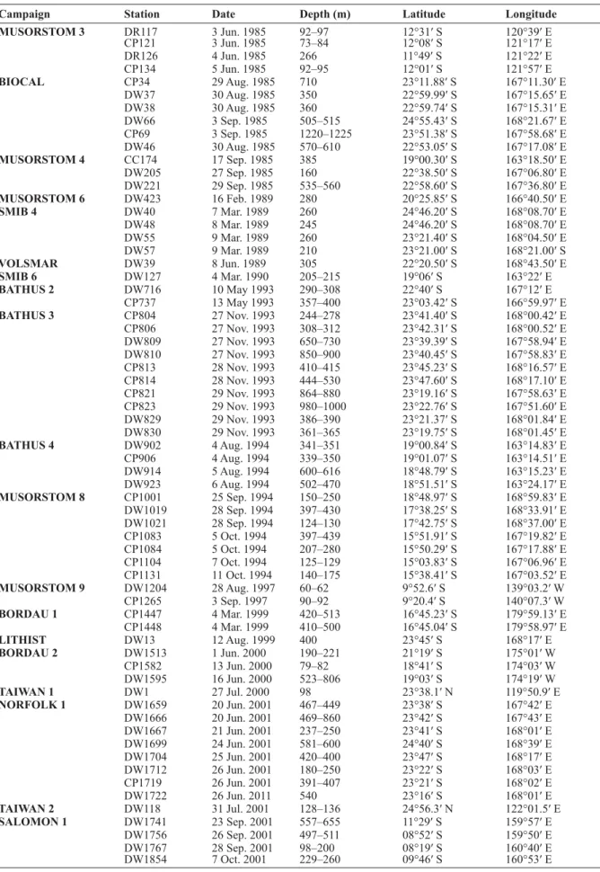
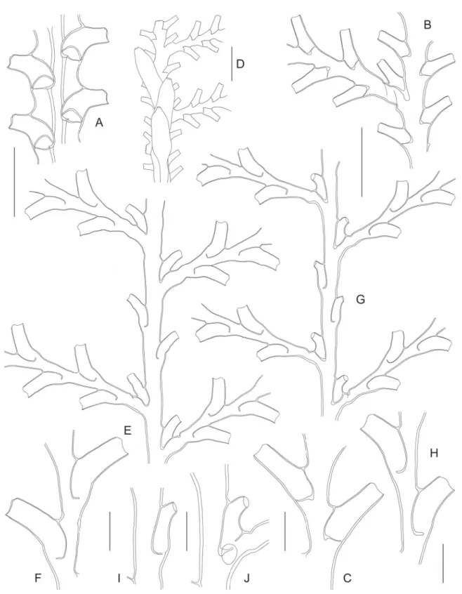
Fig. 1. ― A–B. Caminothujaria molukkana von Campenhausen, 1896 (BORDAU 2, Stn. CP1582).
A. Fragment of internode with verticil of hydrothecae. B. Gonotheca. ― C–J. Diphasia cristata Billard, 1920 (BORDAU 1, Stn. CP1448). C. Pair of hydrothecae, frontal view. D. Same, dorsal view. E. Cross section through basal part of hydrothecal pair, showing perisarcal crests. F. Female gonotheca. G. Same, apical view. H. Same, cross section. I. Male gonotheca. J. Same, apical view. ― K. Diphasia orientalis Billard, 1920 (MUSORSTOM 3, Stn. DR117), internode with pair of hydrothecae. ― L–O. Dynamena heterodonta (Jarvis, 1922) (MUSORSTOM 9). L. Pair of hydrothecae in material from Stn. DW1204.
M. Same sample, hydrothecal aperture, showing internal, submarginal cusps. N–O. Isolated (N) or stacked (O) pairs of hydrothecae in material from Stn. CP1265. ― P. Geminella ceramensis Billard, 1925a (MUSORSTOM 8, Stn. CP1084), internode with pair of hydrothecae. Scale bars: A–D, K–L, N–P = 500 μm; E = 400 μm; F–J = 1 mm; M = 100 μm.
Material examined
MUSORSTOM 3: Stn. DR117, two infertile, unbranched stems, 9 and 12 mm high, detached from substrate (MNHN-IK-2012-16515); Stn. CP121, two infertile, unbranched stems, 13 and 22 mm high, detached from substrate (MNHN-IK-2012-16516).
Remarks
For a thorough description of this species, see Billard (1925b).
Geographical distribution
Indonesia (Billard 1925b), Fiji (Gibbons & Ryland 1989), Philippines (present report).
Genus Dynamena Lamouroux, 1812 Dynamena heterodonta (Jarvis, 1922)
Fig. 1L–O Pasythea heterodonta Jarvis, 1922: 344, pl. 24, fi gs 11–12.
Dynamena heterodonta – Billard 1925b: 198, fi g. 44. ― Redier 1966: 86. ― Vervoort & Vasseur 1977:
36, fi gs 16–17.
Sertularia gracilis – Billard 1905: 334 (not Sertularia gracilis Hassall, 1848: 2223).
non Dynamena quadridentata f. heterodonta – Vannucci 1951a: 83; 1951b: 108, 112, 114 [= D. quadri- dentata (Ellis & Solander, 1786)].
Material examined
MUSORSTOM 9: Stn. DW1204, small colony composed of four infertile stems, 6–12 cm high, detached from substrate (MNHN-IK-2012-16517); Stn. DW1265, sterile colony with stems up to 2.6 cm high, epizoic on Synthecium sp. (MNHN-IK-2012-16518).
Remarks
The sterile, unbranched stems, divided here and there by oblique nodes, each of which comprises a varied number of isolated hydrothecal pairs, occasionally forming apically stacked groups of two, the lack of evidence of an abcauline caecum in retracted hydranths, the two-cusped hydrothecae with a pleated adcauline fl ap and a semicircular abcauline fl ap, and the inconstant presence of internal, submarginal cusps in the hydrothecae suggest that they belong, with little doubt, to Jarvis’ (1922) species.
Vervoort & Vasseur (1977) provided a reexamination of the type material, as well as a discussion regarding its differences with D. quadridentata (Ellis & Solander, 1786).
Geographical distribution
St. Brandon (Cargados Carajos) (Jarvis 1922), Indonesia (Billard 1925b), New Caledonia (Redier 1966), French Polynesia [Billard (1905), as Sertularia gracilis Hassall, 1848; Vervoort & Vasseur (1977);
present study].
Genus Geminella Billard, 1925b Geminella ceramensis (Billard, 1925a)
Fig. 1P Sertularella ceramensis Billard, 1925a: 649.
Geminella ceramensis – Schuchert 2003: 174, fi g. 31.
Material examined
MUSORSTOM 8: Stn. CP1084, small, sterile colony, less than 1 cm high, with tangled stems fi xed on calcareous substrate (MNHN-IK-2012-16519); Stn. CP1131, two sterile stems 0.5 cm high, detached from substrate (MNHN-IK-2012-16520).
Remarks
For a redescription of this distinctive species, see Schuchert (2003), who also provided a synonymy.
Geographical distribution
Indonesia, Philippines, New Caledonia (Schuchert 2003), and Vanuatu (present study).
Genus Gonaxia Vervoort, 1993 Gonaxia amphorifera Vervoort, 1993
Figs 2A, 3A–B Gonaxia amphorifera Vervoort, 1993: 117, fi gs 3f, 4, 5, 6a–b, 8a.
Material examined
SMIB 6: Stn. DW127, two colonies, one unbranched and 4.5 cm high, the other forked twice and 9 cm high, both bear male gonothecae (MNHN-IK-2012-16521).
Remarks
The diffi culty of distinguishing G. amphorifera from G. ampullacea Vervoort, 1993 without their female gonothecae is notorious. Since the present material bears only male gonothecae, attention was focused on the hydrothecal features, in an attempt to separate them specifi cally. Although Vervoort (1993) emphasized several distinguishing characters, the distinction was still laborious and, eventually, two features proved useful. It was found that in the present species the free, distal part of the hydrocladial hydrothecae was constantly set at about 90° with the axis of the cladia, regardless the position of the thecae (Fig. 2A) while, in the following species, that angle varied considerably (Fig. 2B) between the proximal hydrothecae (where it was of 80–90°) and the distalmost ones (usually reaching 45°). The size of the hydrothecae was comparatively smaller in the present material (Fig. 3A) than in that assigned to G. ampullacea Vervoort, 1993 (Fig. 3C). I believe that the lack of a distinct infl ation of the hydrothecae invoked by Vervoort is misleading.
Some hydrothecae exhibit incipient lateral diverticuli (Fig. 3A). Their structure is discussed under G. aff. similis Vervoort, 1993.
Geographical distribution
New Caledonia (Vervoort 1993; present study).
Gonaxia ampullacea Vervoort, 1993 Figs 2B, 3C–D
Gonaxia ampullacea Vervoort, 1993: 121, fi gs 6c, 7a–c, 9.
Material examined
BATHUS 4: DW914, a 3.5 cm high colony with unbranched stem, bearing male gonothecae (MNHN- IK-2012-16522).
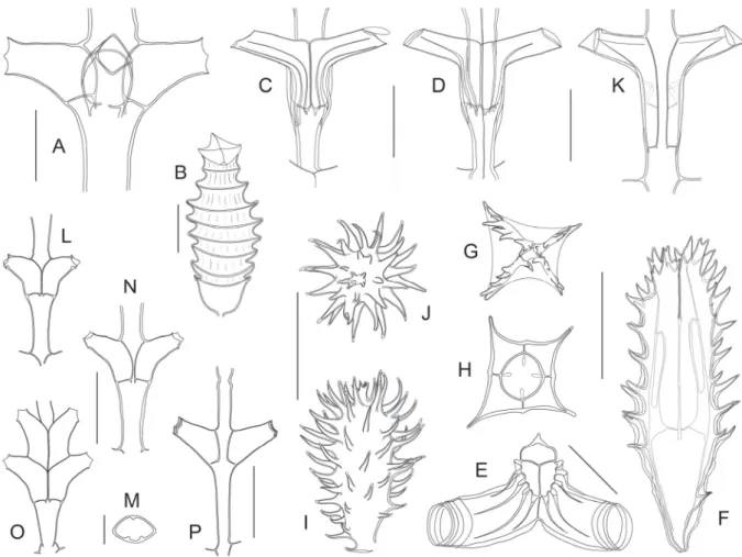
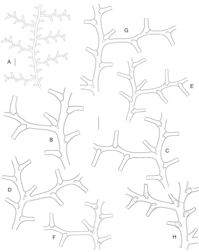
Fig. 2. ― A. Gonaxia amphorifera Vervoort, 1993 (SMIB 6, Stn. DW127), portion of stem with cladium. ― B. Gonaxia ampullacea Vervoort, 1993 (BATHUS 4, Stn. DW914), portion of stem with cladia. ― C. Gonaxia compacta Vervoort, 1993 (BATHUS 4, Stn. DW923), portion of stem with cladium. ― D. Gonaxia intermedia Vervoort, 1993 (BATHUS 4, Stn. CP906), portion of stem with cladium. ― E. Gonaxia sinuosa Vervoort, 1993 (SMIB 4, Stn. DW40), portion of stem with cladia.
Scale bar: A–E = 1 mm.
Remarks
As stated above, the larger size and the more acute angle formed by the free part of the hydrocladial hydrothecae with the axis of cladia were used to assign this material to G. ampullacea. As in G. amphorifera, some hydrothecae exhibit incipient lateral diverticuli (Fig. 3C).
Geographical distribution
New Caledonia (Vervoort 1993; present study).
Gonaxia compacta Vervoort, 1993 Figs 2C, 3E–F
Gonaxia compacta Vervoort, 1993: 135, fi gs 8c, 14a–b, 15a.
Material examined
BATHUS 4: Stn. DW923, a single, 6 cm high colony bearing an abortive gonotheca, as well as a row of incipient gonothecae, highly obscured by the accessory tubes of the stem (MNHN-IK-2012-16523).
Remarks
As already stated by Vervoort (1993), the hydrothecae of this species bear an obvious resemblance to those of G. amphorifera, G. ampullacea, and G. sinuosa, but it could be readily distinguished from them on the account of the absence of a pit formed by the free adaxial wall with the internode (Fig. 3E). Only an abortive gonotheca, arising from within the fi rst hydrotheca of a cladium (Fig. 3F), was found. Several successive, elongate masses among the accessory tubes of the stem suggest the imminent formation of normal gonothecae.
Geographical distribution
New Caledonia (Vervoort 1993; present study).
Gonaxia intermedia Vervoort, 1993 Figs 2D, 3G–K
Gonaxia intermedia Vervoort, 1993: 157, fi gs 23e, 24d, 25a–c, 26a.
Material examined
BATHUS 4: Stn. DW902, several colonies and fragments, up to 9.5 cm high, mostly fertile, bearing male gonothecae (MNHN-IK-2012-16524); Stn. CP906, a total of twelve fertile colonies (10 male and 2 female), 4–6 cm high (MNHN-IK-2012-16525).
Remarks
In some colonies, the length of cladia reaches as much as 2.8 cm, and each may bear up to 116 hydrothecae.
Aberrant secondary cladia, as those occurring in specimens from Stn. DW902, are given off from either within some cladial hydrothecae (Fig. 3J), or from short, auxiliary cladial tubes running parallel to the proximal parts of the axes of cladia.
Both male and female colonies are present in the material in hand. As stated by Vervoort (1993), the gonothecae of this species resemble those of G. amphorifera and G. ampullacea through their general shape and nearly fully adnate condition. They seem to originate from below the axillar hydrothecae. The distal part of a gonotheca overlaps the proximal part of the following one, though their bodies never fuse with each other. Their apertures (50–65 μm wide in males, and 250–290 μm wide in females)
are mounted on raised cones situated near the distal ends of their bodies. In the male colonies, the gonothecae are so obscured by the numerous auxiliary tubes running parallel to the stem, that only the tips of their neck regions protrude from the strongly fascicled mass which composes the stems. In one male specimen (from Stn. CP906) with otherwise normal gonothecae, an elongate, abortive gonotheca, with two distal apertures, is given off from the lumen of a cladial hydrotheca The occurrence of such
Fig. 3. ― A–B. Gonaxia amphorifera Vervoort, 1993 (SMIB6, Stn. DW127). A. Two successive cladial internodes with their hydrothecae. B. Portion of stem with male gonothecae. ― C–D. Gonaxia ampullacea Vervoort, 1993 (BATHUS 4, Stn. DW914). C. Two successive cladial internodes with their hydrothecae. D. Portion of stem with male gonothecae. ― E–F. Gonaxia compacta Vervoort, 1993 (BATHUS 4, Stn. DW923). E. Two successive cladial internodes with their hydrothecae. F. Abortive gonotheca (of undetermined sex) originating from within a hydrotheca. ― G–K. Gonaxia intermedia Vervoort, 1993 (BATHUS 4). G. Two successive cladial internodes with their hydrothecae (Stn. CP906).
H. Portion of stem with male gonothecae (Stn. CP906). I. Portion of stem with female gonothecae (Stn.
CP906). J–K. Abortive gonothecae arising from hydrothecal lumina (Stn. DW902). ― L–N. Gonaxia sinuosa Vervoort, 1993 (SMIB 4, DW40). L. Two successive cladial internodes with their hydrothecae.
M–N. Two female gonothecae. Scale bars: A, C, E, G, L = 300 μm; B, D, F, H–K, M–N = 1 mm.
gonothecae is considerable in the material from Stn. DW902, and specimens with one (Fig. 3K), two (Fig. 3J), or exceptionally three apertures could be seen. These so called abortive gonothecae may prove fully functional, as they are provided with apertures. However, their lumina are empty in the specimens in hand, and this assumption could not be checked at this stage.
Geographical distribution
New Caledonia (Vervoort 1993; present study).
Gonaxia sinuosa Vervoort, 1993 Figs 2E, 3L–N
Gonaxia sinuosa Vervoort, 1993: 179, fi gs 31c–e, 35b, 37a–b, 38a.
Material examined
SMIB 4: Stn. DW40, an originally 8 cm high colony with simple stem, now broken into two parts, bearing male gonothecae (MNHN-IK-2012-16526); Stn. DW48, an originally 5.5 cm high, sterile colony with simple stem, now broken into two unequal parts (MNHN-IK-2012-16527).
BATHUS 2: Stn. DW716, six colonies and fragments, 2.3–9.1 cm high, with simple or branched stems, two of which are sterile, two others bear male gonothecae, and the remaining two carry female gonothecae (MNHN-IK-2012-16528).
BATHUS 3: Stn. CP804, eleven colonies and fragments, 1.5–4.9 cm high, all with simple stems, of which four are sterile, three bear male gonothecae, and the remaining four bear female gonothecae (MNHN-IK-2012-16529); Stn. DW829, two colonies with simple stems, 3.8 and 2.4 cm high, the latter devoid of its basal part, and bearing three incipient gonothecae (MNHN-IK-2012-16530).
Remarks
The shape of the hydrothecae (Fig. 3L), as well as the condition and morphology of the gonothecae (Fig. 3M–N) are characteristic. One stem from BATHUS 2, Stn. DW716 is fully fertile (male) and bears its gonothecae in two parallel rows, each one originating from below the base of an axillar hydrotheca.
The female stems examined here bear a lesser number of gonothecae.
Geographical distribution
New Caledonia (Vervoort 1993; present study), Norfolk Ridge (Vervoort 1993).
Gonaxia crassicaulis Vervoort, 1993 Fig. 4A
Gonaxia crassicaulis Vervoort, 1993: 145, fi gs 18c–d, 19a–b.
Material examined
MUSORSTOM 4: Stn. DW205, a cladium, 2.5 cm long (MNHN-IK-2012-16531).
NORFOLK 1: Stn. DW1712, an infertile colony, 3.8 cm high (MNHN-IK-2012-16532).
Remarks
According to Vervoort (1993), G. crassicaulis is easily recognizable through the characteristic development of the perisarc, forming thick, internal, longitudinal ridges, connecting the adnate adaxial walls of the hydrothecae in each row (Fig. 4A).
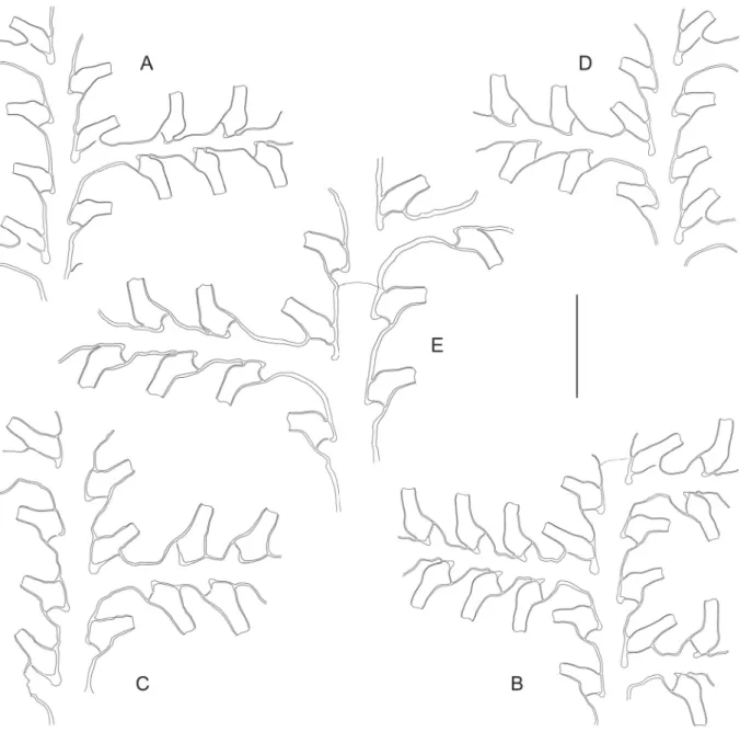
Fig. 4. ― A. Gonaxia crassicaulis Vervoort, 1993 (MUSORSTOM 4, Stn. DW205), portion of cladium. ― B–D. Gonaxia elegans Vervoort, 1993 (BATHUS 3, Stn. DW809). B. Portion of stem and basal part of a cladium. C. Two consecutive internodes with their hydrothecae. D. Portion of stem with male gonothecae. ― E–F. Gonaxia cf. errans Vervoort, 1993 (BATHUS 2, Stn. CP737). E. Distalmost portion of stem with basal parts of four hydrocladia. F. Two consecutive cladial internodes with their hydrothecae. ― G–J. Gonaxia plumularioides sp. nov. (BIOCAL, Stn. CP34). G. Distalmost part of a stem, showing the structure of internodes. H. Two consecutive cladial internodes with their hydrothecae.
I. Stem hydrotheca. J. Axillar hydrotheca. Scale bars: A–B, D–E, G = 1 mm; C, F, H–J = 300 μm.
Also characteristic is the presence of large, internal, semi-circular “thickenings” of the perisarc (Fig. 4A), arching over the basal foramina, when the hydrothecae are seen laterally. These structures, one situated in the “frontal” aspect of the hydrothecae, the other on their “dorsal” side, correspond in fact to the attachment points of the hydrothecal bases to the lateral walls of the thecae which, in this species, are raised above the level of the foramina. When the hydrothecae are seen facing their apertures, the hydrothecal bases appear U-shaped, the foramina being situated at the bottom of this concavity.
Geographical distribution
New Caledonia (present study), Norfolk Ridge (Vervoort 1993).
Gonaxia elegans Vervoort, 1993 Fig. 4B–D
Gonaxia elegans Vervoort, 1993: 153, fi gs 19c, 21b–c, 22.
Material examined
BATHUS 3: Stn. DW809, a 4.4 cm high colony devoid of hydrorhiza and bearing female gonothecae, as well as an originally 5.4 cm high colony (now broken into two parts) bearing male gonothecae and several broken cladia originating from both colonies (MNHN-IK-2012-16533).
Remarks
This species is immediately recognizable through the characteristic position and shape of its hydrothecae.
The 5.4 cm high colony is evidently male when compared to the smaller one, which bears gonothecae possessing much wider apertures. Only the female gonothecae were described by Vervoort (1993) but, upon a careful inspection of the present material, it should be stated that they do not arise from the accessory tubes on the hydrocaulus, but from the main tube itself, as this is indisputably seen in the monosiphonic part of the stem. In addition, in this colony at least, nearly all accessory tubes are found on the “dorsal” aspect of the stem, while the row of gonothecae is situated on its “front”.
The male gonothecae, observed here for the fi rst time, are similar in shape to the female ones, and also arise from the main tube of the stem, but in both the frontal and dorsal aspects of the colony. They are given off closely and in a single row (Fig. 4D), are about half adnate, and their free parts are shifted alternately left and right; their bases are obscured by the numerous accessory tubes running up the stem.
They are long (1.5–2 mm), tubular (480–520 μm wide), with small, circular apertures (100–120 μm wide) mounted on the summit of a conical, distal neck region.
Geographical distribution
New Caledonia (Vervoort 1993; present study), Norfolk Ridge (Vervoort 1993).
Gonaxia cf. errans Vervoort, 1993 Fig. 4E–F
Gonaxia errans Vervoort, 1993: 154, fi gs 23a–d, 24a–c.
Material examined
BATHUS 2: Stn. CP737, two stem fragments, 13 and 9 mm high, the former corresponding to the middle part of a colony, while the latter is likely the distalmost end of (another?) colony; both are sterile; several broken cladia are also present (MNHN-IK-2012-16534).
Remarks
The pattern of cladial arrangement on the stem in this scarce material seems to follow a rather peculiar sequence: each equivalent of internode (the nodes are inconspicuous) has a proximal apophysis with its associated axillar hydrotheca, two alternate hydrothecae above, as well as a second, distal apophysis (on the side opposite to the former) with its associated axillar hydrotheca; the same sequence is repeated on the following “internode” but, there, it is the opposite image of the preceding one. In the distalmost part of one of the available colony fragments, two successive cladia occur on the same side, and are separated on the opposite side by a single stem hydrotheca. According to Vervoort (1993), the cladial arrangement is varied, with “zero to three hydrothecae between two successive apophyses”, which is apparently the case in the present material.
The shape of the hydrothecae also fi ts the original description of G. errans, though they are comparatively shorter in the material in hand (free adaxial side 335–365 μm, adnate adaxial side 280–300 μm, abaxial side 490–510 μm), but seem to have almost the same width (maximum width 205–220 μm, diameter at aperture 165–175 μm) as in Vervoort’s samples. Consequently, the present material is provisionally assigned to G. errans.
Geographical distribution
New Caledonia (present study), Norfolk Ridge (Vervoort 1993).
Gonaxia plumularioides sp. nov.
urn:lsid:zoobank.org:act:6A07FF84-EAA6-4AE6-8755-B450B17A28EB Fig. 4G–J; Table 2
Diagnosis
Gonaxia with slender, fascicled, branched or unbranched stems; cladia monosiphonic. Equivalents of internodes composed of a proximal hydrocladial apophysis and its associated axillary hydrotheca, two alternate hydrothecae above, and a second, distal hydrocladial apophysis and its associated hydrotheca.
Hydrothecae tubular, slightly curved up- and outwards, comparatively slender and more immersed on stem than on cladia. Gonothecae unknown.
Etymology
From the Latin plūma, meaning feather, and the Greek εἶδος, meaning likeness, with reference to their plumose appearance, recalling the branching pattern displayed by some hydroids belonging to the superfamily Plumularioidea.
Material examined Holotype
BIOCAL: Stn. CP34, a 2.8 cm high, distal fragment of a branch or a stem, itself branched twice, sterile (MNHN-IK-2012-16535).
Paratype
BIOCAL: Stn. CP34, a 7.7 cm high, unbranched colony devoid of its hydrorhiza, as well as a 1 cm high fragment corresponding to the distalmost part of a colony; both are sterile (MNHN-IK-2012-16536).
Description
Colonies attaining at least 8 cm high, composed of either simple or ramifi ed stems of slender appearance, despite being fascicled; accessory tubes on both “frontal” and “dorsal” sides of stems;
the latter uniformly grading to monosiphonic distally. Side branches, when present, arising only in
“frontal” aspect of the stem or higher order branches; up to 2nd order branching observed. Division into internodes indistinct, but equivalents of internodes composed of the regular sequence: a fi rst, basal apophysis (60–90 μm long) and its associated axillar hydrotheca, two additional, alternate hydrothecae above, and a second axillar apophysis with its associated hydrotheca. Following internode with same sequence, but its proximal apophysis on side opposite to the distal apophysis of preceding internode.
Cladia leaving axis at 75–80° from upper part of stem or branches; two successive cladia on same side of stem are distant of 2200–2310 μm; strictly monosiphonic, alternate, up to 1 cm long, comprising a succession of up to 23 alternate hydrothecae. Division into internodes indistinct; equivalent of fi rst cladial internode comparatively longer (580–705 μm) than subsequent ones (480–515 μm); internodes short, accommodating a single hydrotheca distally. Hydrothecae widely spaced on stem (570–740 μm), less so on cladia (470–530 μm); tubular, slightly curving up- and outwards, free part angled at 50° to the axis of internode; stem hydrothecae slightly shifted on frontally; the two rows of cladial hydrothecae coplanar; less than half adnate on cladia, but considerably immersed into the stem internodes; free adaxial side nearly straight, adnate counterpart curved, ending basally in perisarc peg; abaxial side convex, with infl exion point in middle; aperture with three low cusps separated by shallow embayments;
opercular apparatus lost. Gonothecae unknown.
Remarks
The branching pattern of the stems of G. plumularioides sp. nov. resembles that seen in some plumularioid hydroids that build three-dimensional colonies (e.g., Macrorhynchia philippina Kirchenpauer, 1872, M. allmani (Nutting, 1900), Plumularia spiralis Billard, 1911, etc.), and is consequently different from that observed in its congeners (e.g., G. amphorifera, G. sinuosa, G. nova sp. nov., see description below), which generally build planar colonies.
The regular structure of the stem internodes is also peculiar, and is only occasionally observed in the material assigned herein to G. errans, a species known for its inconstant structure of the stem (Vervoort 1993).
The shape of the hydrocladial hydrothecae resembles that of other congeners: 1) G. complexa Vervoort, 1993, but in this species the hydrothecae have an exceedingly long part free from internode; 2) G. elegans, but its hydrothecae have a conspicuously straight axis instead of being convex (compare Fig. 4C and 4H); 3) G. errans, whose hydrothecae have, however, a much longer free part (compare Fig. 4F with Fig. 4H); 4) G. intercalata Vervoort & Watson, 2003, but its hydrothecae are huge compared to those of the new species; 5) G. stricta Vervoort, 1993, but here the hydrothecae have a much longer free part, and the cladial internodes are marked by internal, oblique perisarcal thickenings (Vervoort 1993);
6) G. tasmanica Watson & Vervoort, 2001, but its hydrothecae are comparatively larger.
Geographical distribution New Caledonia.
Table 2. Measurements (in μm) of hydrothecae of Gonaxia plumularioides sp. nov. and G. nova sp. nov.
G. plumularioides sp. nov. G. nova sp. nov.
Caulinar hydrotheca
Axillar hydrotheca
Cladial hydrotheca
Caulinar hydrotheca
Axillar hydrotheca
Cladial hydrotheca Free adaxial side 85–125 75–140 180–210 555–575 565–630 580–615 Adnate adaxial side 355–380 360–440 315–350 220–245 175–220 175–205
Abaxial side 315–345 195–245 395–440 600–630 505–575 620–665
Maximum width 110–125 125–145 195–205 260–270 270–290 250–260
Diameter at rim 80–90 80–95 160–170 160–165 150–185 165–175
Gonaxia nova sp. nov.
urn:lsid:zoobank.org:act:6C6AA611-2E5C-40B9-9502-31888388A7E9 Figs 5A–B, 6A–E; Table 2
Diagnosis
Gonaxia with either branched or unbranched, rigid, fascicled stems arising from disc-shaped hydrorhiza.
Internodes slightly geniculate, comprising a latero-distal cladial apophysis with its associated hydrotheca, and two alternate hydrothecae below. Cladia alternate, composed of moderately long, geniculate internodes. Hydrothecae arranged at a wide angle to the stem; long, tubular, slightly arcuate, tumid proximally, distally tubular, adnate for ¼ their length. Gonothecae originating on both sides of the stem from below axillar hydrothecae; adnate for about half their length; erect portion tubular, ending in rather long neck region bearing the aperture distally.
Etymology
From the Latin novus, -a, -um (and the Grek νέος), meaning new, indicating that we are dealing with a new, undescribed species.
Material examined Holotype
MUSORSTOM 8: Stn. CP1083, a 4.2 cm high, unbranched colony bearing male gonothecae (MNHN- IK-2012-16537).
Paratypes
MUSORSTOM 8: Stn. CP1083, eight colonies or fragments with branched or unbranched stems, of which four bear female gonothecae and range between 2.6 and 5.0 cm in height, while the remaining four bear male gonothecae and range between 2.8 and 3.9 cm in height (MNHN-IK-2012-16538).
Description
Colonies composed of branched or unbranched, thick, rigid, fascicled stems arising from rhizoid hydrorhiza fi rmly attached to substrate. Nodes inconspicuous, even in the monosiphonic parts, but equivalents of internodes 1580–2000 μm long, slightly geniculate, composed of a latero-distal hydrocladial apophysis with its associated hydrotheca, and two additional, alternate hydrothecae below. Hydrocladia strictly monosiphonic, arranged at approximately right angles to the stem; up to 1.2 cm long, nodes indistinct (60–70 μm wide); equivalents of internodes moderately long, geniculate, each carrying a hydrotheca distally; fi rst internode comparatively longer (1020–1240 μm) than subsequent ones (590–760 μm);
up to 20 hydrothecae per cladium. Hydrothecae long, tubular, facing outwards at nearly right angles to their corresponding internodes; slightly tumid basally, walls parallel distally; adnate for about ¼th their length; free adaxial side slightly convex proximally, then straight; abaxial wall with noticeable convexity in middle; aperture triangular in frontal view; margin with three conspicuous cusps (two latero-adaxial and one abaxial) separated by rather deep embayments; operculum composed of three triangular fl aps forming a low roof. Gonothecae on both sides of the stem, originating from below axillar hydrothecae;
about half adnate, strongly appressed proximally to the stem; distal half erect, tubular, ending in moderately long, tronconical collar, bearing the aperture distally (small in male, Fig. 6E; comparatively larger in female, Fig. 6D); perisarc of abaxial wall of fused part fi nely and densely striated (Fig. 6E–F);
individual gonothecae either solitary (towards apex of stem) or fused together through auxiliary tubes given off from one or both ends of their adnate parts; in highly polysiphonic parts of stem, adnate parts of gonothecae obscured by apposition of auxiliary tubes; length of adnate part 1250–1340 μm (♂) and 1720–1970 μm (♀), and of free part 1540–1910 μm (♂) and 1640–1720 μm (♀); width of free part
Fig. 5. ― A–B. Gonaxia nova sp. nov. (MUSORSTOM 8, Stn. CP1083). A. Distalmost, monosiphonic part of a colony. B. Portion of stem with basal part of a cladium. ― C–G. Gonaxia scalariformis Vervoort, 1993, portions of stems and basal parts of cladia from BIOCAL, Stn. DW46 (C = colony
#1; D = colony #2; E = colony #3), Stn. DW66 (F), and NORFOLK 1, Stn. DW1699 (G), showing discrete differences in shape and size. ― H. Gonaxia aff. similis Vervoort, 1993 (MUSORSTOM 6, Stn.
DW423), portion of stem with cladium. Scale bars: A = 1 mm; B–H = 500 μm.
565–590 μm (♂) and 620–690 μm (♀); length of neck region 260–365 μm (♂) and 165–245 μ (♀);
diameter at aperture 90–105 μm (♂) and 245–260 μm (♀).
Remarks
The hydrothecae of this species resemble those of several congeners: 1) G. bulbifera Vervoort, 1993, but this species has proportionally smaller hydro- and gonothecae (about two times smaller), its hydrothecal axes are straight instead of being curved, its caulinar apophyses are conspicuously infl ated, and its cladial internodes are collinear instead of being slightly geniculate; 2) G. elegans, but in this species the hydrothecae are much more closely-set, and the cladial internodes are strictly collinear (compare Fig. 5B with Fig. 4B); 3) G. perplexa Vervoort, 1993, but in this species the hydrothecae are more approximated and have straight axes, and the gonothecae are nearly fully adnate, their bodies being likely fused to each other, and their apertures are characteristically shifted outward from the stem; 4–7) G. complexa Vervoort, 1993, G. persimilis Vervoort, 1993, G. similis, and G. scalariformis, but these species are immediately distinguished through their free gonothecae, originating perpendicularly to the stems; 8) G. stricta Vervoort, 1993, but in this species the hydrothecae have straight axes and they are more approximated on cladia.
Geographical distribution Vanuatu.
Gonaxia scalariformis Vervoort, 1993 Figs 5C–G, 6F–R
Gonaxia scalariformis Vervoort, 1993: 173, fi gs 31b, 32, 33c–d, 35a.
Material examined
BIOCAL: Stn. DW46, three colonies (#1–3) slightly differing in the size of their hydrothecae: colony
#1 is 3.5 cm high, devoid of its hydrorhiza, and bears two female gonothecae (MNHN-IK-2012-16539), colony #2 is 2.8 cm high, devoid of its hydrorhiza, and bears three female gonothecae (MNHN- IK-2012-16540), and colony #3 is 2.2 cm high and broken in two parts, the basal one exhibiting remnants of hydrorhiza; both parts bear fi ve male gonothecae each (MNHN-IK-2012-16541); Stn. DW66, a 1.6 cm high colony devoid of both its hydrorhiza and apical part, with two abortive gonothecae (MNHN- IK-2012-16542); Stn. CP69, a 1.4 cm high colony devoid of its distalmost part, with a single female gonotheca (MNHN-IK-2012-16543).
NORFOLK 1: Stn. DW1699, a 1.7 cm high colony devoid of both its hydrorhiza and upper part, bearing two female gonothecae (MNHN-IK-2012-16544).
Fig. 6. (previous page) ― A–E. Gonaxia nova sp. nov. (MUSORSTOM 8, Stn. CP1083). A. Axillar hydrotheca. B. Caulinar hydrotheca. C. Cladial hydrotheca. D. Female gonothecae, lateral view. E. Male gonotheca, slightly oblique view. ― F–R. Gonaxia scalariformis Vervoort, 1993. F. Axillar hydrotheca (BIOCAL, Stn. DW46, colony #1). G. Cladial hydrotheca (BIOCAL, Stn. DW46, colony #1). H. Caulinar and axillar hydrothecae (BIOCAL, Stn. DW66). I–M. Normal gonothecae from BIOCAL, Stn. DW46 (I = colony #1; J–K = colony #2; L–M = colony #3). N–O. Abortive gonotheca from BIOCAL, Stn.
DW66. P–R. Normal gonothecae from BIOCAL, Stn. CP69 (P) and NORFOLK 1, Stn. DW1699 (Q–
R). ― S–X. Gonaxia aff. similis Vervoort, 1993 (MUSORSTOM 6, Stn. DW423). S. Cauline hydrotheca.
T. Axillar hydrotheca. U. Cladial hydrotheca. V. Lateral view of a hydrotheca, showing diverticulum.
W. Same hydrotheca viewed from above, showing minute foramen for the passage of coenosarc, and lateral diverticuli. X. Same hydrotheca in apical view, showing lateral diverticuli and basal foramen.
Scale bars: A–C, F–H, S–U = 300 μm; D–E, I–R = 1 mm; V–X = 200 μm.
Remarks
All available specimens have lightly fascicled stems basally, and none is ramifi ed. Although the arrangement and general shape of the hydrothecae perfectly match the description of the holotype, noticeable variations in size could be observed. Thus, the colony #3 from sample BIOCAL, Stn. DW46 has the slenderest hydrothecae, while those from NORFOLK 1, Stn. DW1699 are the widest ones. In the sample from BIOCAL, Stn. DW66, incipient antero-dorsal diverticuli are seen in most hydrothecae (Fig. 5F). Gonothecae of both sexes are present in the available material. Although they conform to the original description, the male gonothecae in the colony #3 from BIOCAL, Stn. DW46 are arranged on both anterior and posterior sides of the stem, and some exhibit bifi d (Fig. 6M) or trifi d apices. The female gonothecae from BIOCAL, Stn. DW66 are likely abortive, as they originate from within the hydrothecae (Fig. 6N–O); in addition, they are not fully formed, as their distal ends are missing; their sex is indeterminate. The female gonothecae from NORFOLK 1, Stn. DW1699 (Fig. 6Q–R) are the longest with respect to those present in the other samples.
Geographical distribution
New Caledonia and Norfolk Ridge (Vervoort 1993; present study).
Gonaxia aff. similis Vervoort, 1993 Figs 5H, 6S–X
Gonaxia similis Vervoort, 1993: 174, fi gs 33e–f, 34, 36a, 39a.
Material examined
MUSORSTOM 6: Stn. DW423, a 2.7 cm high, sterile colony (MNHN-IK-2012-16545).
Remarks
The present specimen is likely part of the material in RMNH-Coel. 25839 (also from MUSORSTOM 6, Stn. DW423) studied by Vervoort (1993).
According to Vervoort, his material had gonothecae adnate to the stem (p. 174, fi g. 34c), not arranged perpendicularly to it, as in the majority of samples assigned by him to G. similis. Consequently, the material in hand may belong to a separate species.
One hydrocladium in the present specimen is branched, and the secondary cladium is borne on a short, quadrangular apophysis originating from below the hydrothecal base of the fi rst internode of a cladium.
Not mentioned by Vervoort in his sample RMNH-Coel. 25839 is the constant presence of well-developed antero-posterior diverticuli within the hydrothecae (Figs 5H, 6S–V). Such variably developed diverticuli also occur in other congeners discussed herein, viz. G. amphorifera (Fig. 3A), G. ampullacea (Fig. 3C), and G. scalariformis (Fig. 5F). In the present material, their development is considerable, and extends well on each lateral side of the hydrotheca. A frontal view of the free adaxial wall of the hydrotheca (Fig. 6W), and a frontal view of the aperture (Fig. 6X) show two empty lateral “pockets” which narrow considerably the hydrothecal lumen; the foramen for the passage of the hydranth within the hydrotheca is minute and horseshoe-shaped.
Geographical distribution
New Caledonia (Vervoort 1993; present study).
Genus Idiellana Cotton & Godfrey, 1942 Idiellana pristis (Lamouroux, 1816)
Fig. 7A Idia pristis Lamouroux, 1816: 199, pl. 5, fi g. 5.
Idiellana pristis – Schuchert 2003: 175, fi g. 32.
Material examined
MUSORSTOM 3: Stn. CP121, sterile colony composed of two stems, 1.7 and 3.7 cm high (MNHN- IK-2012-16546); Stn. CP134, sterile colony composed of two stems, 1.5 and 2.7 cm high (MNHN- IK-2012-16547).
MUSORSTOM 8: Stn. DW1021, two sterile stems, 3.5 and 5 cm high (MNHN-IK-2012-16548); Stn.
CP1131, several sterile stems, 1.5–8.5 cm high (MNHN-IK-2012-16549).
TAIWAN 1: Stn. DW1, four sterile stems, 0.7–1.4 cm high, on gravel (MNHN-IK-2012-16550).
Remarks
For the latest redescription and a list of synonyms, refer to Schuchert (2003).
Geographical distribution
Circumglobal in tropical and subtropical seas (Precker & Lawn 2010).
Genus Salacia Lamouroux, 1816 Salacia macer Vervoort & Watson, 2003
Fig. 7B–G Salacia macer Vervoort & Watson, 2003: 154, fi gs 34g, 35a.
Material examined
BATHUS 3: Stn. DW809, single sterile stem, 2.4 cm high, partly damaged (several cladia missing) (MNHN-IK-2012-16551).
NORFOLK 1: Stn. DW1659, four stems 3–4.5 cm high, two of which bear female gonothecae (MNHN- IK-2012-16552).
Remarks
The slender habit of these colonies, their regularly-pinnate branching pattern and their division into internodes, each of which bears a single pair of strictly opposite hydrothecae, are diagnostic (Vervoort
& Watson 2003). The gonothecae, not previously described, are present in this material, and are female.
They are borne on either the stem or cladia, and are arranged perpendicularly from below one hydrotheca of a pair. Urn-shaped, smooth-walled, 3280–3565 μm long and 1110–1170 μm wide, tapering basally into a short pedicel; distally truncate, aperture circular, 590–615 μm wide, borne on a short, 565–575 μm wide neck region. Possibly up to 100 oocytes per gonotheca, though their number is diffi cult to ascertain.
Geographical distribution
Norfolk Ridge (Vervoort & Watson 2003; present study).
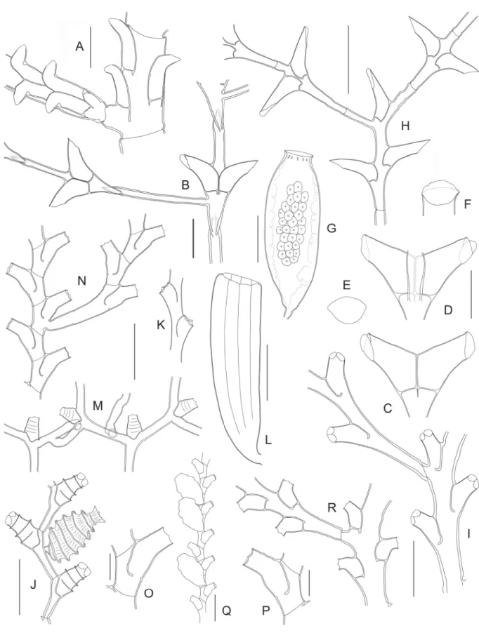
Fig. 7. (continued on next page) ― A. Idiellana pristis (Lamouroux, 1816) (TAIWAN 1, Stn. DW1), stem internode and basal part of cladium. ― B–G. Salacia macer Vervoort & Watson, 2003 (NORFOLK 1, Stn. DW1659). B. Stem internode and basal part of cladium. C. Hydrothecae, frontal view. D. Same, dorsal view. E. Hydrothecal aperture, frontal view. F. Hydrothecal aperture, oblique view, showing
Salacia sibogae Billard, 1924 Fig. 7H
Salacia sibogae Billard, 1924: 64, fi g. 1b–c.
Salacia sibogae – Schuchert 2003: 177, fi g. 34.
Material examined
MUSORSTOM 3: Stn. DR126, four small (0.4–1.1 cm high), sterile stems on axis of dead gorgonian (MNHN-IK-2012-16553).
Remarks
A recent redescription of this species and a list of synonyms are available in Schuchert (2003).
Geographical distribution
Previosuly known from Indonesia and Japan (Schuchert 2003). The present material originates from the Philippines.
Genus Sertularella Gray, 1848 Sertularella acutidentata Billard, 1919
Fig. 7I Sertularella acutidentata Billard, 1919: 20, fi gs 1e, 2.
Sertularella acutidentata acutidentata – Vervoort & Watson 2003: 154, fi g. 35b–e.
Material examined
TAIWAN 2: Stn. DW118, two infertile colonies, 7 and 8 cm high (MNHN-IK-2012-16554).
Remarks
For a redescription of this species, refer to Hirohito (1995). Its synonymy is given by Vervoort & Watson (2003).
Geographical distribution
Indonesia, Japan, Philippines, New Caledonia, and north of New Zealand (Vervoort & Watson 2003).
operculum. G. Female gonotheca. ― H. Salacia sibogae Billard, 1924 (MUSORSTOM 3, Stn. DR126), portion of stem and basal part of cladium. ― I. Sertularella acutidentata Billard, 1919 (TAIWAN 2, Stn. DW118), portion of stem with basal part of side branch. ― J. Sertularella areyi Nutting, 1904 (BATHUS 3, Stn. CP813), portion of fertile stem. ― K–L. Sertularella diaphana (Allman, 1885) (MUSORSTOM 8, Stn. CP1104). K. Two hydrothecae. L. Gonotheca. ― M. Sertularella mirabilis Jäderholm, 1896 (TAIWAN 2, Stn. DW118), portion of colony. ― N–Q. Sertularella novaecaledoniae Vervoort, 1993 (MUSORSTOM 4, Stn. DW221). N. Portion of stem and proximal part of a branch.
O–P. Hydrothecae. Q. Portion of stem with gonothecae. ― R. Sertularella quadridens (Bale, 1884) (MUSORSTOM 3, Stn. CP121), portion of stem and proximal part of a side branch. Scale bars: A–B = 500 μm; C–F = 400 μm; G–N, Q–R = 1 mm; O–P = 300 μm.
Sertularella areyi Nutting, 1904 Fig. 7J
Sertularella areyi Nutting, 1904: 83, pl. 17, fi g. 6.
Sertularella areyi – Vervoort & Watson 2003: 156, fi g. 35f–l.
Material examined
MUSORSTOM 4: Stn. CC174, several stems, up to 1.4 cm high, all but one sterile (MNHN- IK-2012-16555).
BATHUS 3: Stn. CP813, fertile colonies with stems up to 1.4 cm high on two fragments of gorgonian (MNHN-IK-2012-16556); Stn. CP814, two infertile colonies with stems up to 1.5 cm high, one on dead gorgonian, the other on sponge (MNHN-IK-2012-16557).
LITHIST: Stn. DW13, small colony composed of a few sterile stems, up to 1.2 cm high, growing on Acryptolaria sp. (MNHN-IK-2012-16558).
BORDAU 2: Stn. DW1513, small colony composed of several sterile, up to 5 mm high, stems growing on G. ceramensis (MNHN-IK-2012-16559).
NORFOLK 1: Stn. DW1704, small colony composed of a restricted number of stems, some fertile and up to 1 cm high, epizoic on Zygophylax sp., Sertularella helenae Vervoort, 1993, and tube of coronate medusa (MNHN-IK-2012-16560).
Remarks
This very distinctive species needs no additional comments on morphology. Its synonymy, as well as a redescription, are provided by Vervoort & Watson (2003).
Geographical distribution
Tropical and subtropical parts of the Atlantic and Indo-Pacifi c (Vervoort & Watson 2003).
Sertularella diaphana (Allman, 1885) Fig. 7K–L
Thuiaria diaphana Allman, 1885: 145, pl. 18, fi gs 1–3.
Sertularella diaphana – Vervoort & Watson 2003: 159, fi g. 36g–h.
Material examined
MUSORSTOM 8: Stn. CP1104, colony composed of several fertile (male gonothecae) stems, up to 6 cm high, epizoic on stem of aglaopheniid hydroid (MNHN-IK-2012-16561).
Remarks
For lists of synonyms and descriptions, see Calder (1991), Vervoort (1933) and Galea (2010).
Geographical distribution
Circumglobal in tropical and subtropical seas (Vervoort & Watson 2003).
Sertularella mirabilis Jäderholm, 1896 Fig. 7M
Sertularella mirabilis Jäderholm, 1896: 9, pl. 2, fi g. 1.
Sertularella mirabilis – Schuchert 2015: 339, fi g. 13.
Material examined
TAIWAN 2: Stn. DW118, a 3.2 cm high, sterile colony (MNHN-IK-2012-16562).
Remarks
Hirohito (1995) provided a thorough description of this peculiar species. Its synonymy is given by Schuchert (2015).
Geographical distribution
From the South China Sea to Korea and Japan (Schuchert 2015).
Sertularella novaecaledoniae Vervoort, 1993 Fig. 7N–Q
Sertularella novaecaledoniae Vervoort, 1993: 225, fi gs 50, 51aA, 52a.
Material examined
MUSORSTOM 4: Stn. DW221, a 4 cm high, fertile colony (MNHN-IK-2012-16563).
Remarks
The present material, represented by a colony with fl abellate appearance, with fascicled stem and side branches divided into very short internodes, each carrying a tubular hydrotheca adnate for about half its length, conforms to the description of the type given by Vervoort (1993). The gonothecae have undulated walls, and their apical part is fl attened (Fig. 7Q).
Geographical distribution
New Caledonia (present study), Norfolk Ridge (Vervoort 1993).
Sertularella quadridens (Bale, 1884) Fig. 7R
Thuiaria quadridens Bale, 1884: 119, pl. 7, fi gs 5–6.
Sertularella quadridens – Schuchert 2003: 185, fi g. 41.
Material examined
MUSORSTOM 3: Stn. DR117, a 18 mm high, sterile colony (MNHN-IK-2012-16564); Stn. CP121, a complete colony, 3 cm high, as well as four fragments, 1.4–3.2 cm high, all sterile (MNHN- IK-2012-16565).
Remarks
For a list of synonyms and a recent redescription, see Schuchert (2003).
Geographical distribution
Widely distributed throughout the tropical Indo-Pacifi c (Precker & Lawn 2010).
Sertularella folliformis sp. nov.
urn:lsid:zoobank.org:act:7A654AE7-6B4E-465C-9F5A-1D48528466E5 Fig. 8A–G; Table 3
Diagnosis
Sertularella species with fl abellate colonies; branching irregular; stem and branches strongly polysiphonic basally, grading to monosiphonic distally; internodes short, slightly geniculate to collinear; hydrothecae exceedingly long, tubular, curved downwards, with 8–10 broad, complete annuli and four internal, submarginal cusps. Gonothecae large, elongated-ovoid, distally truncate.
Etymology
From the Latin words follis, meaning bellows, and fōrma, meaning shape, with reference to the plicate condition of the hydrothecal wall, recalling the bellows of an accordion.
Material examined Holotype
BATHUS 3: Stn. DW809, a 4.5 × 4.5 cm sterile colony and four smaller fragments, three of which bear one gonotheca, and the fourth two gonothecae (MNHN-IK-2012-16566).
Paratype
BATHUS 3: Stn. DW809, a few colonies and fragments, largest one 7.5 × 3.5 cm, all sterile (MNHN- IK-2012-16567).
Description
Colonies fl abellate, attaining 7.5 cm high, arising from rhizoid stolons fi rmly attached to substrate.
Branching irregular, with up to 3rd order branches arranged roughly in one plane. Stems and side branches polysiphonic, main stem reaching as much as 2 mm wide basally. Both stem and branches divided into internodes by slightly-marked nodes slanting in alternate directions. Internodes short, slightly geniculate to collinear, each carrying a hydrotheca in its distal half. Side branches originate laterally below the bases of stem hydrothecae; fi rst internode comparatively longer than subsequent ones. Hydrothecae exceedingly long, tubular, slightly curved downward, adnate for ¼th or less their length, tapering imperceptibly towards aperture, walls provided with 8–10 broad, complete annuli; margin occasionally renovated; rim provided with four short cusps separated by shallow embayments; four submarginal, intrathecal projections of perisarc, two latero-adaxial and two latero-abaxial. Gonotheca originating from axil of hydrotheca, in front and/or backside of colony; elongated-ovoid, tapering abruptly below, distally truncate, wall smooth to slightly undulate.
Remarks
The hydrothecal shape is diagnostic in this species. There are few congeners which approach this condition, viz. S. catena (Allman, 1888) and S. pseudocatena sp. nov. (Fig. 9J), but both have much shorter hydrothecae, and their transverse ribs do not extend over the abaxial wall. Three other species, namely S. helenae Vervoort, 1993 (Fig. 8H), S. paucicostata Vervoort, 1993 (Fig. 8I), and S. pseudocostata Vervoort, 1993, exhibit comparatively more prominent hydrothecal ridges. The eastern Pacifi c S. exilis Fraser, 1938 has isodiametric hydrothecae “regularly curved upward and then outward”
(Fraser 1938), but their surface is reportedly smooth. In addition, it is “an especially diminutive species”
(Calder et al. 2009). Sertularella whitei Rees & Vervoort, 1987 also has exceedingly long, outwardly- curved hydrothecae, but their surface is entirely smooth (Rees & Vervoort 1987).
Geographical distribution New Caledonia.
Sertularella helenae Vervoort, 1993 Fig. 8H
Sertularella helenae Vervoort, 1993: 218, fi g. 47b–e.
Material examined
BIOCAL: Stn. DW37, several sterile stems, up to 2.6 cm high, and smaller fragments (MNHN- IK-2012-16568); Stn. DW38, single, sterile colony 3 cm high and 4.7 cm wide (MNHN-IK-2012-16569).
BATHUS 3: Stn. CP804, a 2.5 cm high and 3.8 cm wide, sterile colony (MNHN-IK-2012-16570);
Stn. CP821, a 3.4 cm high, sterile colony (MNHN-IK-2012-16571); Stn. DW830, four sterile colony fragments, 0.7–1.5 cm high (MNHN-IK-2012-16572).
NORFOLK 1: Stn. CP1719, a 4.2 cm high, sterile colony (MNHN-IK-2012-16573).
SALOMON 1: Stn. DW1741, two sterile colonies, 2.2 and 2.6 cm high (MNHN-IK-2012-16574).
Remarks
This very distinctive species needs no additional comments on morphology. For a thorough description, see Vervoort (1993). The gonothecae still remain to be discovered.
Geographical distribution
New Caledonia (Vervoort 1993; present study), Norfolk Ridge and Solomon Islands (present study).
S. folliformis sp. nov. S. plicata sp. nov.
Internodes
- length (in general) 680–865 660–890
- length (1st cladial internode) 1360–2405 1365–1450
- diameter at node 325–400 125–145
Hydrotheca
- free adaxial side 1025–1235 365–440
- adnate adaxial side 375–415 190–225
- abcauline side 1170–1295 450–495
- maximum width 320–345 255–275
- diameter at rim 290–305 170–180
Gonotheca
- length 2245–2345 –
- maximum width 780–850 –
- apical width 445–455 –
Table 3. Measurements (in μm) of Sertularella folliformis sp. nov. and S. plicata sp. nov.
Sertularella paucicostata Vervoort, 1993 Fig. 8I
Sertularella paucicostata Vervoort, 1993: 227, fi gs 51b–f, j.
Material examined
MUSORSTOM 3: Stn. DW809, several infertile stems, some branched, up to 1.3 cm high, epizoic on Sertularella folliformis sp. nov. (MNHN-IK-2012-16575).
Remarks
Surprisingly, Vervoort (1993) compared his new species with Sertularella costata Leloup, 1940, which is a much smaller and delicate hydroid (compare Fig. 8I and 8J), as illustrated by specimens of this species from the Andaman Sea, Thailand belonging to my private collection.
Sertularella costata is a rather poorly known species, with a few records around the world. Hirohito (1995) provided succinct measurements of the hydrothecae, while Calder et al. (2003) gave the fi rst description and measurements of the gonotheca. The dimensions of the hydrothecae in the Thailand material are as follows: abcauline side 315–360 μm, free adcauline side 265–280 μm, adnate adcauline side 100–110 μm, maximum width 155–170 μm, diameter at aperture 130–140 μm. The external wall is provided with 10–13 transverse ridges, and three internal, submarginal cusps are present within the hydrothecae (Fig. 8K). Through the size of its hydrothecae, the material from Thailand comes close to the Japanese specimens examined by Hirohito and, through the number of external ridges, it approaches that of the Galapagos specimens, which is apparently 10–11 (Calder et al. 2003: fi g. 14). In contrast, about 20 ridges ornament the hydrothecal walls in material from Japan (Leloup 1940; Hirohito 1995).
In my opinion, S. paucicostata most closely resembles both S. helenae and S. pseudocostata Vervoort, 1993. However, unlike S. paucicostata, S. helenae forms fascicled colonies with shorter internodes, and its hydrothecae are smaller (compare Fig. 8I and 8H) and provided with numerous perpendicular creases between the transverse ridges. According to Vervoort (1993), the hydrothecae of S. pseudocostata are comparatively larger and their outer wall has frilled ribs.
Geographical distribution
New Caledonia, Norfolk Ridge (Vervoort 1993), and the Philippines (present study).
Sertularella plicata sp. nov.
urn:lsid:zoobank.org:act:5CBEB7F6-12F7-403C-AC8D-58AE01B11AC7 Fig. 8L–O; Table 3
Diagnosis
Small Sertularella with monosiphonic stems; side branches given off irregularly in one plane. Internodes moderately long and geniculate. Hydrothecae piriform, with 8–10 conspicuous, transverse ribs; aperture on short, narrow neck facing outwards; 3 or 5 internal, submarginal, lamellar cusps. Gonothecae unknown.
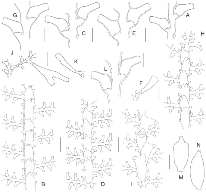
Fig. 8. (previous page) ― A–G. Sertularella folliformis sp. nov. (BATHUS 3, Stn. DW809). A. Holotype.
B. Paratype. C. Portion of stem with proximal part of a side branch. D. Successive internodes with their hydrothecae. E. Hydrothecal aperture, frontal view, showing internal cusps. F. Portion of stem with gonotheca, showing insertion site. G. Gonotheca. ― H. Sertularella helenae Vervoort, 1993 (SALOMON A, Stn. DW1714), two successive internodes with their hydrothecae. ― I. Sertularella paucicostata Vervoort, 1993 (BATHUS 3, Stn. DW809), two consecutive internodes with their hydrothecae, and comparison with Sertularella costata Leloup, 1940 (J–K). ― L–O. Sertularella plicata sp. nov. (MUSORSTOM 3, Stn. CP121). L. Portion of stem and proximal part of a side branch. M. Two consecutive internodes with their hydrothecae. N. More detailed view of the hydrotheca, to show its wrinkled surface. O. Hydrothecal aperture in frontal view, showing internal cusps. ― P–S. Sertularella aff. sinensis Jäderholm, 1896. P. Portion of stem (BATHUS 3, Stn. DW829). Q. Hydrotheca (BORDAU 2, Stn. DW1595). R. Frontal view of three hydrothecal apertures, to show variation in the number of internal cusps. S. Gonotheca (BORDAU 2, Stn. DW1595). Scale bars: A–B = 1 cm; C, F–G, L, P = 1 mm; D, H–J, M, Q, S = 500 μm; E, K, O, R = 100 μm; N = 200 μm.