ORIGINAL ARTICLE
Uncharted biodiversity in the marine benthos: the void of the smallish
with description of ten new Platyhelminth taxa from the well-studied North Sea
Werner Armonies*
Abstract
Most of our planet’s biodiversity is still unknown, particularly in the sea. Although around the island of Sylt in the North Sea, the small zoobenthos (meiofauna) has been studied intensively since the 1950s, repeating previous surveys revealed an unexpected wealth of new species in addition to the 330 species of free-living microturbellarians (non-parasitic Platyhelminthes) already known from this area. Extrapolation from well-known to less-known habitat types suggests that a total of some 520 Platyhelminth species should be expected around this island, about 670 in the North Sea ecoregion, and 830 in the ‘Northern European Sea’ ecoprovince. Assuming that the other biogeo- graphic provinces of the planet harbour a similar diversity, a total of some 20,000 marine microturbellarian species is estimated for the global shelf zones. Less than 10% of these are known by now. As a contribution to fill that gap, ten new taxa are described: Coelogynopora minuta n. sp., Coelogynopora sopottehlersae n. sp., Cirrifera paraculeata n. sp., Boreocelis fragilis n. sp., Postbursoplana noldti n. sp., Promesostoma wehrenbergi n. sp., Ptyalorhynchus oculatus n. sp., Acrorhynchides canaliculatus n. sp., Dactyloplana n. gen., and Dactyloplana tridigitata n. sp.
Keywords: Benthos, Diversity, Species richness, North Sea, Platyhelminthes
© The Author(s) 2018. This article is distributed under the terms of the Creative Commons Attribution 4.0 International License (http://creat iveco mmons .org/licen ses/by/4.0/), which permits unrestricted use, distribution, and reproduction in any medium, provided you give appropriate credit to the original author(s) and the source, provide a link to the Creative Commons license, and indicate if changes were made.
Introduction
Meiofauna is a largely neglected component of the marine benthos [1]. These are the benthic organisms small enough to pass through 1 mm meshes but large enough to be retained on a 63 µm screen. The small size makes them difficult to handle, and investigators need high-quality microscopes and expertise in morphology and taxonomy. That is why marine meiofauna is rarely studied, and if so, most studies concentrate on copep- ods or nematodes [2], which have a hard skin (the cara- pace in copepods and a cuticle in nematodes) that keeps their body shape during fixation. Therefore, they can be studied in a preserved state, which allows for short field sampling campaigns and for the evaluation of the fixed
materials over a nearly unrestricted amount of time at a distant laboratory. Other taxa, such as Platyhelminthes, have a soft skin and a body without any skeletal ele- ments. They usually shrink to a bulky mass during fixa- tion, which complicates species identification because the natural position of internal organs can no longer be seen. These soft-skinned species are best determined alive, while anatomical details may require serial sec- tions of individually fixed organisms [3]. The need for live observation requires prolonged field trips and access to a field station. Because of these complications, soft-bodied taxa are rarely studied and the studied areas are usually close to one of the few accessible field stations. Conse- quently, distributional maps show a few hot spots sur- rounding the field stations with large interspaces never studied. Outside Europe, these interspaces may include entire continents.
Open Access
*Correspondence: Werner.Armonies@awi.de
Alfred-Wegener-Institut Helmholtz-Zentrum für Polar- und Meeresforschung, Wattenmeerstation Sylt, Hafenstr. 43, 25992 List, Germany
One of the hot spots in meiofaunal research is the island of Sylt in the eastern North Sea, where field access is offered by Wadden Sea Station Sylt (now part of Alfred- Wegener Institute for Polar- and Marine Research, and formerly of Biologische Anstalt Helgoland). By area, sandy beaches are the most important habitat in the supratidal and sandy flats in the intertidal of this island.
Concentrating on a single sandy beach area next to List harbour, the interstitial micro- and meiofauna taxa have been studied one by one by several investigators since the 1960s. Altogether > 650 meiofaunal species were recorded from this beach, which exceeded the number of macrofaunal species by an order of magnitude [4].
Most meiofauna live in the system of pores left between the sand grains of the sea floor. The size of these pores depends on the granulometric sediment composition. As the sediment becomes finer, organisms need to become smaller or more slender to fit the narrow pores. There- fore, sediment composition is a key factor for meiofauna.
At the same time, sediment composition correlates with hydrographic conditions such as current velocity and wave height, which in turn affect sedimentary organic matter uptake and hence food availability for small fauna
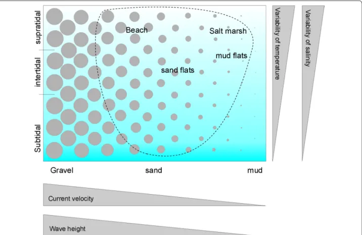
[5]. A second key factor for meiofauna is tidal level, which relates to many physical factors, including the variabil- ity of temperature and salinity (low in the subtidal and increasingly higher towards the supratidal) and sediment sorting (mostly well-sorted in the subtidal and increas- ingly heterogeneous towards the supratidal). These two key factors can be used to define habitat types in the coastal zone (Fig. 1), which may be further refined, e.g. by including habitat modifying species such as seagrasses.
The sandy beach next to List harbour mentioned above only represents the sandy inter- and supratidal section of habitats available around Sylt. Other habitat types were only studied for Platyhelminthes, including salt marshes [6, 7], mud flats [8], and sand flats with lugworm (Areni- cola marina) burrows [9, 10]. As a result of these stud- ies, the Platyhelminthes inhabiting inter- and supratidal habitats are thought to be well known on the island of Sylt. Compared to that, infra- and subtidal habitats were only marginally studied. Wehrenberg and Reise [11] gave an overview on Platyhelminth species density and abun- dance, and Noldt [12, 13] described subtidal Kalypto- rhynchia. At that time, Xenacoelomorpha (Acoela and Nemertodermatidae) were still included in the phylum
Fig. 1 Schematic representation of coastal habitat types. Circles symbolise the mean grain size and the dashed line surrounds habitats present near the island of Sylt. Further explanations in the text
Platyhelminthes, until genetic analyses revealed a distant position in the phylogenetic system [14]. By now, it seems they are most likely the sister group of Nephrozoa [15, 16]. Platyhelminth species numbers in this paper always exclude Xenacoelomorpha and therefore are not compa- rable to older references where Xenacoelomorpha were still included.
As a result of the high number of studies on a wide range of habitats, some 330 species of microturbellarians (i.e. the phylum Platyhelminthes without parasitic Neo- dermata and macrofaunal Polycladida) were recorded from the Island of Sylt. This is roughly equivalent to 20%
of the world-wide known marine microturbellarian spe- cies. No other locality of the world is known to harbour such a richness of Platyhelminth species. Nevertheless, there are still more species around this small island. Re- sampling of the beach next to List harbour 40–50 years after the first studies on meiofauna revealed 20 out of 220 Platyhelminth species previously not recorded [17]. Thus, in spite of the many studies on meiofauna in the past (summarised in [4]) species richness in this beach was still incompletely recorded. The present study gives first results from re-sampling of the subtidal sediments. In the past, the subtidal was studied in a far lower intensity than higher tidal levels. Accordingly, the percentage of new species detected was higher than in the better-studied higher tidal levels. Some of the new species are described below in the taxonomic part of the results.
As the number of species known from this small island was already high, how many more species can be expected in the Sylt area? And, as such a small area already harbours so many species, how many microtur- bellarian species might exist in the 99% of the world’s coastline that was never studied for these tiny organisms, so far? Due to a lack of knowledge, these questions pres- ently cannot be answered. But since microturbellarians in the Sylt area are exceptionally well known, we can use these data to perform a sort of mental experiment to get a rough picture on the potential total number of micro- turbellarian species world-wide in shallow coastal waters.
This is done in three steps: (1) to get an idea of local spe- cies richness around the island of Sylt, accumulative spe- cies numbers over the tidal gradient are calculated for the well-studied upper tidal levels and then extrapolated for the less-known subtidal areas. (2) Around the island of Sylt, sandy sediments predominate while very coarse sand, gravel and fine mud are lacking (Fig. 1), and spe- cies preferring these sediment types may not find suitable habitat. Thus, the species recorded around the island of Sylt are only a subset of the regional (North Sea) species pool. Therefore, the estimates for the local (Sylt) species richness are up-scaled for the North Sea ecoregion and the ‘Northern European Seas’ biogeographic province. (3)
Further up-scaling to a global level yields an idea for the world-wide species richness of marine microturbellar- ians. The estimate for microturbellarian species richness resulting from this last step suggests that we don’t even know the tip of the iceberg, today.
Materials and methods
The island of Sylt lies in the northern Wadden Sea in the eastern part of the North Sea. This is a shallow coastal region with a chain of barrier islands. The distance from the exposed sandy barrier to the sheltered mainland marshes is about 10 km. Mean tidal range is about 2 m.
In the shelter of the barrier islands, about half of the sea floor is exposed during low tides. The sea floor is sedi- mentary; natural hard substrata are limited to epibenthic mussel- and oyster beds. Sand flats prevail over muddy flats. These are dissected by tidal channels with depths down to 40 m. Of the total water volume, about half is exchanged each tide with the coastal North Sea. From the exposed sandy beaches of barrier islands and shoals of ebb deltas, depth gradually slopes down to 30 m over a distance of 80 km offshore. A detailed description of the tidal area around the island of Sylt including its biota and abiotic conditions is provided by Reise [18] and Gätje and Reise [19]. Biogeographically, Sylt belongs to the ecore- gion ‘North Sea’ within the province ‘Northern European Seas’ of the ‘Temperate North Atlantic’ realm [20].
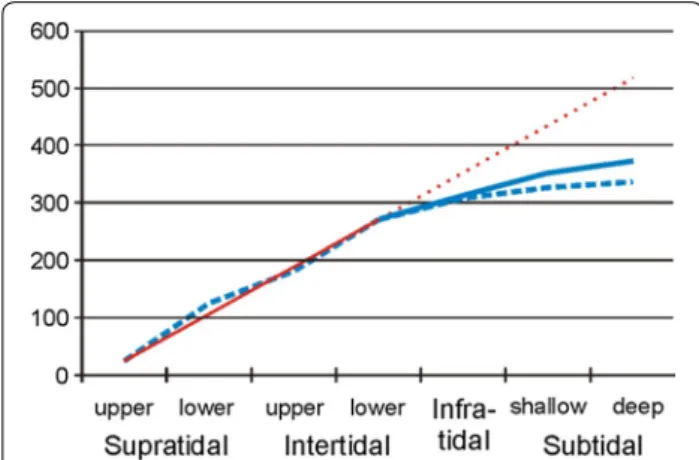
The available habitats around Sylt island range from coarse sandy beaches to muddy fine sand (Fig. 1), and most of these habitats have already been studied for microturbellarians. However, species are not restricted to habitat types delimited by man, and the width of dis- tributional ranges varies over species. Accordingly, the species composition of neighbouring habitat types may considerably overlap, and simply adding species numbers over habitats would strongly overestimate local species richness. Therefore, all available literature was screened for the position in the tidal gradient of individual Platy- helminth species using a simple scheme with 7 levels (Table 1). This scheme was constructed because tidal level of the localities was often only verbally described in older studies. The resulting assignment of species with tidal levels is given in Additional file 1. From this table, I accumulated species numbers from the upper supratidal towards the deeper subtidal, i.e. from the well-studied tidal levels to the badly-studied ones. A linear regression line was fitted to the data from the well-studied upper tidal levels. For the lower tidal levels, the deviation of cumulative species numbers from this regression line is treated as an estimate for the number of species still to be detected in the sparsely-studied subtidal around the island. In doing so, I assume that Platyhelminth species richness in the subtidal is similar to inter- and supratidal
habitats. For the subtidal down to 40 m water depth sur- rounding the Island of Sylt, this assumption is supported by previous data [21]. However, due to a lack of data from other areas, the assumption can neither be spatially gen- eralized nor be extrapolated towards larger water depths.
The reliability of the estimate for total microturbellar- ian species richness around the island of Sylt (first step of the estimates on species richness) was checked by evalu- ating the Platyhelminth species richness and composi- tion in sediment samples collected between July 2015 and April 2018 in the infra- and subtidal zones. This was done by arranging the sites studied during that period (n = 35) in chronological order and evaluating the accumula- tive numbers of species recorded (total of this study and past studies by Wehrenberg and Reise [11], and Noldt [12, 13]), of species re-discovered (i.e. species already recorded from the Sylt subtidal during the above previ- ous studies), and of new species (previously not recorded from the Sylt subtidal). ‘Sites’ were sampled in two dif- ferent ways. In the infratidal accessible from land, 6 rep- licate cores of 10 cm2 surface area were collected with a hand-held corer. Initially the same sampling scheme was applied in the sites accessible by ship only, by sub- sampling a larger box-corer with small cores of 10 cm2 surface area (one core per box-corer to avoid pseudo- replication). However in the subtidal, these small cores often contained relatively few Platyhelminth specimens and most species were represented by a single individual only. This is sufficient to record a well-known species but not adequate to study, and potentially describe, new spe- cies. Therefore larger cores were collected, i.e. a site was represented by a single box-core grab of 200 cm2 surface area (Fig. 2). The sediment depth sampled was as deep as possible, down to 30 cm in coarse sand (limited by the length of the box-corer) but only about 10 cm in fine sand (penetration depth limited by the dense package of sand grains and the weight of the box-corer). Platyhelminthes
were separated from the sediment by washing with sea- water and anaesthetization with MgCl2-solution [12].
Further details on sampling, localities, and the associ- ated platyhelminth fauna are given in Additional file 1:
Table S1.
Results
Accumulating the number of Platyhelminth species over tidal levels from the well-known supratidal towards the subtidal (Additional file 1: Table S4) showed an almost linear increase in species richness down to neap low tide level (Fig. 3). In the infratidal and below, the slope of the accumulated species numbers graph strongly flattens, as was expected from the lower study intensity.
In the past, 136 Platyhelminth species had been recorded from the Sylt subtidal (Fig. 4, ‘known from subtidal’; data in Additional file 1: Table S2). About half of these (74) were re-discovered during this study (Fig. 4,
‘re-discovered’). In addition, the 35 sites sampled dur- ing this study yielded 83 species not previously recorded from the Sylt infra- or subtidal (Fig. 4, ‘new for subtidal’).
However, many of these ‘new’ species are known to occur in the intertidal zone and therefore do not increase accumulative species numbers in the infra- and subtidal sections of Fig. 3. Elevations in the level of infra- and subtidal species richness come from 12 species recorded for the first time near Sylt and about 25 new species, some of which are described in the systematic part. In total, this study increased the number of Platyhelminth species recorded from the Sylt infra- and subtidal to 219 (Additional file 1: Table S2).
On average, each site studied revealed two new spe- cies, one not previously recorded from the infra- or subtidal zone but known from higher tidal levels, and one species not previously recorded from Sylt or unde- scribed. The subtidal accumulative species numbers increased rather steadily, with no sign of reaching a Table 1 Division of the Sylt coastal zone into tidal levels. Plant associations according to Ellenberg [22]
Tidal level Description
Upper supratidal Beach zone higher than 1 m above mean high tide (MHT) level and salt marshes grown with the plant association Armerietum mar- itimae (ungrazed) or Juncetum gerardii (grazed), also starting about 1 m above MHT. This zone is only flooded during storm tides, interstitial salinity mostly meso- to oligohaline (18–3 psu)
Lower supratidal Zone between MHT and 1 m above. In salt marshes with the plant association Puccinellietum maritimae. Interstitial salinity mostly in the polyhaline range (30–18 psu)
Upper intertidal Sand- and mudflats between mid-tide level and MHT. May not be submerged during strong offshore winds. Salinity occasionally drops to the polyhaline range
Lower intertidal Sand- and mudflats between mid-tide level and neap low tide level (NLT). Regularly submerged, salinity rarely beyond ambient level Infratidal Zone between NLT and 1 m below mean low tide (MLT); may fall dry during strong offshore winds. Earlier studies often failed to
sample this zone because the actual low water level was too high to reach it from the landward side, or too low to reach it by ship Shallow subtidal Sediments 1–10 m below MLT
Deeper subtidal Water depth > 10 m. In the vicinity of the island this level is only represented by tidal channels with medium to coarse sand
saturation level (Fig. 4). With respect to the total spe- cies richness around Sylt island, the data of the present study increase the infra- and subtidal parts of the spe- cies accumulation graph a bit (solid line in Fig. 3) but it still remains far from the level expected from the
extrapolation of the regression line calculated from the better-known tidal levels. From this regression, some 520 species are expected around Sylt Island, which means that further 150 still undetected species of Platy- helminthes may live in the subtidal around Sylt.
Fig. 2 Sites in the infra- and subtidal near the island of Sylt sampled for Platyhelminths, July 2015–April 2018. Large circles indicate large (200 cm2) cores and small circles small (10 cm2) cores. Two sites in the North Sea west of the island not indicated
Discussion
Including this study, the number of valid Platyhelminth species in the Sylt area has increased to 349 (Additional file 1: Table S3). In addition, some 50 tentatively new species (i.e., species with morphological characters that do not fit any described species) have been recorded in the past but not formally described, so far, mostly because the material was insufficient. These species only ‘exist’ in unpublished files (drawings and/or pho- tographs) left by Karsten Reise, Christian Wehrenberg, Uwe Noldt, and me. Thus, we already know of 400 spe- cies around Sylt. This is not very far from the estimate of a total of 520 species including the sparsely-stud- ied subtidal (from the extrapolation in Fig. 3). How- ever, the ‘well-known’ inter- and supratidal habitats also still harbour unknown or undescribed species, as exemplified by a previous study [17]. Strictly speaking,
the extrapolation only indicates the number of spe- cies expected until we reach a level of knowledge in the subtidal that is comparable to the present level of knowledge from higher tidal elevations. The ‘real’ num- ber of Platyhelminth species around Sylt might be even higher.
In order to get an idea on the total number of micro- turbellarian species that may exist on a world-wide scale in shallow coastal waters we may now use the above estimates from Sylt to start a mental experiment; this includes spatial extrapolations to scales that have never been studied. Therefore, the results of these extrapola- tions are merely an educated guess based on a minimum of information. For these spatial extrapolations of Platy- helminth species richness, two numbers will be used for species richness in the Sylt area: (1) a minimum of 349 species, representing the number of currently recorded species that are formally described, and (2) 520 species, representing the estimate derived from Fig. 3.
Usually larger areas include more habitat types than small ones. Currently, 457 species of microturbellarians are known from the North Sea ecoregion (i.e. about 30%
more than from the Sylt area) and 555 from the Northern European Seas province (about 60% more than from the Sylt area; only valid species excluding synonyms, numina nuda, and species dubiae; Additional file 1: Table S5).
These larger areas include habitat types such as soft mud and gravel that are absent (Fig. 1) from Sylt island.
Accordingly, the increases in species numbers by 30% and 60%, respectively, compared to the Sylt area, are assumed to represent the effects of spatial scale and the associated number of habitat types. Estimating the ‘true’ number of Platyhelminths for theses larger units also needs to include this scaling-effect. Lacking better evidence I used the above percentages derived from the number of spe- cies recorded by now. This results in estimates of a total of 676 Platyhelminth species in the North Sea and 832 in the Northern European Sea province, some 50% more than recorded by now.
The Mediterranean is another biogeographic province where Platyhelminthes have been studied reasonably well to allow for a comparison with the North Sea. The cur- rent number of microturbellarian species in the Mediter- ranean (417 and 445 including the Black Sea, Additional file 1: Table S5) is similar to the Northern European Sea.
Together both provinces harbour 862 species, with 138 joint species (roughly 30% of the total within each prov- ince). Thus, a first estimate for the global number of marine microturbellarians comes from the calculation
‘number of marine coastal ecoprovinces’ on Earth (= 62) [20] multiplied by 707 species per ecoprovince (from the estimate 832 for the ‘Northern European Seas’ minus 15% for joint occurrences of species in several provinces).
Fig. 3 Number of Platyhelminth species accumulated over the tidal gradient near Sylt island using data until 2015 (broken blue line) and including the current study (solid blue line). The red line gives the linear regression between the upper supratidal and the lower intertidal (solid) and its extrapolation towards the subtidal (dotted)
Fig. 4 Number of Platyhelminth species in the Sylt subtidal known until 2015 (blue), re-discovered during this study (green), and new records during this study (yellow)
The result of this estimate is about 44,000 marine micro- turbellarians on Earth.
However, the Mediterranean and Northern European Seas ecoprovinces are not directly neighbouring but separated by parts of the Lusitanian ecoprovince [20].
Therefore, the species overlap between neighbouring ecoprovinces is likely to be higher than the estimated 30% in the above example. In fact, we already know that many species colonise > 2 ecoregions (Additional file 1:
Table S5). Since we have no sufficient data for the per- centage of joint occurrences of Platyhelminth species in neighbouring ecoprovinces, two scenarios were calcu- lated. The first scenario assumes species occupy 2 eco- provinces, on average. The calculation of global marine microturbellarian species numbers is therefore based on half of the species number recorded from, or estimated for, the Northern European Sea ecoprovince. This results in estimates of 17,200 and 25,800 species, respectively, on a global scale. The second scenario assumes species occupy 3 ecoprovinces, on average, and calculations are based on a third of the species recorded from, or esti- mated for, the Northern European Sea. This results in estimates of 11,500 and 17,200 species of marine micro- turbellarians in shallow coastal waters on Earth.
These estimates treat species richness in the North- ern European Seas representative for all climate zones, which contradicts the general assumption of higher spe- cies richness in low than high latitudes [23, 24]. In addi- tion, quaternary glaciations covered most of the North Sea and only left a marginal part of the southern North Sea uncovered by ice [25]. After glacier retreat, the spe- cies pool available to repopulate the ‘Northern European Seas’ ecoprovince may have been exceptionally small.
Therefore the estimates for the global number marine microturbellarian species may be rather conservative.
Macrofaunal species richness is far better studied than meiofaunal. Therefore, comparing the relations between macrofaunal species richness in the North Sea and on a global scale offers an alternative way for up-scaling Plat- yhelminth species richness from the North Sea. With respect to life-style, i.e. predominantly sediment-dwell- ing species, Nemertea and Polychaeta may be closest to Platyhelminths. Assuming Platyhelminths scale like Poly- chaetes (11,757 species world-wide [26] and 326 in the North Sea [27]) we may expect 16,300–24,100 marine microturbellarian species world-wide. Assuming they scale like Nemertea (1363 species world-wide [26] and 25 in the North Sea [27]), we may expect some 25,000–
37,500 species globally.
Thus, both ways of estimating microturbellarian spe- cies number yield results in a similar order of magni- tude ranging between 11,500 and 37,500 species in shallow coastal waters. Throughout this paper, the term
‘species’ is always used for morphospecies. However, genetically different species may morphologically not be distinguishable from each other (cryptic species), morphologically different species may not be different genetically, or species limits may not be clear by either morphology or genetics in sibling species [28, 29].
Cryptic species may have the strongest effect on the estimates of global Platyhelminth diversity; they may increase the mean number of ecoprovinces occupied by single species und thus turn the estimates for global diversity down. Thus, including molecular and genetic information may yield more precise estimates in the future.
The above estimates for global microturbellarian diver- sity refer to the shallow coastal regions only because most platyhelminth studies sampled no deeper than 20 m water depth. Very few samples came from depths down to 200 m and no platyhelminth species are known from deeper waters. Currently analyses of sedimentary DNA are the only information available for deeper waters.
They indicate high platyhelminth species richness from the shelf [30] down to abyssal depth [31].
The situation exemplified here for microturbellarians in principle holds true for the smallish marine benthos in general: all knowledge comes from a few selected sites while most of the world’s oceans have never been stud- ied. Presumably, the ‘real’ numbers of species will exceed the currently known numbers by an order of magnitude for all of the small sized benthic taxa hidden in the sedi- ments. Around the island of Sylt, the relation of species richness of the small sized benthic fauna versus macrozo- obenthos currently is 2.4 [32]. With better knowledge on the smallish benthic taxa this relation will increase in the future. In the absence of better data, I hypothesise spe- cies richness of the smallish fauna in marine sediments may typically exceed macrozoobenthic species richness (by a factor of 2 or more) on a world wide scale.
Descriptions of new taxa
Eight of the 25 unknown species recorded in the subtidal sites were frequent enough for a description (Table 2).
One of them fits none of the present genera but shares morphological characters with Cheliplana paradoxa Noldt, 1989 which was only preliminarily classified with the genus Cheliplana [12]. Hence, a new genus Dactylo- plana is generated for the new species, and Cheliplana paradoxa is re-named as Dactyloplana paradoxa (Noldt, 1989). Among the new species, Acrorhynchides canalicu- latus is the only one recorded from inter- and supratidal sites but not from the subtidal, so far. All type material is deposited in the Platyhelminth collection of AWI Wad- den Sea Station Sylt.
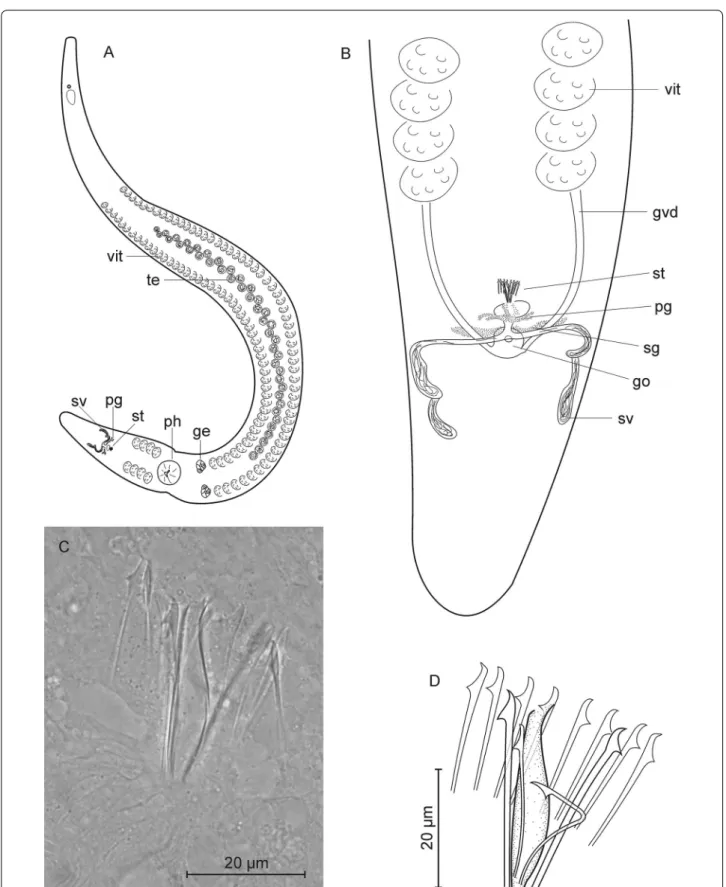
Coelogynopora minuta n. sp. (Fig. 5)
Localities (1) Type locality: Lister Ley, very fine sand, water depth 10 m (55.0225°N, 008.4586°E, 1 individual, 8 Jan 2018). (2) List, infratidal east of Oddewatt. Fine sand, water depth 0.5 m (55.0240°N, 008.4399°E, 2 individuals, 6 Oct 2016).
Material Life observations including drawings and photographs. Three whole mounts; one designated holo- type (AWI Sylt P2018-103) and two paratypes (AWI Sylt P2018-104 and AWI Sylt P2018-105).
Etymology Compared to other species in the genus the stylet spines are exceptionally small.
Diagnosis Species of Coelogynopora with a cuticular apparatus consisting of a central pipe (32 µm) associ- ated with two pairs of longer spines (one more solid and
32–34 µm long, the other very fine and 26–28 µm long) and a second group of 8 very short (21–23 µm) spines surrounding the central parts in a half circle. No acces- sory spines.
Description Very slender organisms, preserved 3–3.5 mm long and 200–250 µm in diameter. Live speci- mens may stretch to 8 mm with a diameter only slightly wider than the pharynx, i.e. about 150 µm. Body whitish, without striking epidermal glands or adhesive papillae.
The pharynx is spherical and relatively small (diameter 110 µm), in the beginning of the last fifth of the body. In specimens that are not fully stretched the body diameter is markedly narrowed besides the pharynx.
General arrangement of the reproductive system as usual in the genus: the testes follicles form a median row before the pharynx, paired germaries laterally before the pharynx, and vitellaries laterally from the Table 2 Systematic classification of the new species
Platyhelminthes Rhabditophora Proseriata
Coelogynoporidae Karling, 1966 CoelogynoporaSteinböck, 1924
Coelogynopora minutan.sp.
Coelogynopora sopottehlersaen. sp.
CirriferaSopott, 1972
Cirrifera paraculeatan. sp.
Monocelididae Hofsten, 1907 BoreocelisWestblad, 1952
Boreocelis fragilisn. sp.
Otoplanidae Hallez, 1892 Parotoplaninae Ax, 1956
PostbursoplanaAx, 1956 Postbursoplana noldtin. sp.
Rhabdocoela Dalytyphloplanida
Neotyphloplanida
Thalassotyphloplanida
Promesostomidae Hartog, 1964 PromesostomaGraff, 1882
Promesostoma wehrenbergin. sp.
Kalyptorhynchia Eukalyptorhynchia
Cicerinidae Meixner, 1928 PtyalorhynchusAx, 1951
Ptyalorhynchus oculatusn. sp.
Polycystididae Graff, 1905
Polycystidinae Schockaert & Karling, 1970 AcrorhynchidesStrand, 1928
Acrorhynchides canaliculatusn.sp.
Schizorhynchia
Cheliplanidae Schilke, 1970 Dactyloplanan. gen.
Dactyloplana paradoxa(Noldt, 1989) nom. nov.
Dactyloplana tridigitatan.sp.
Fig. 5 Coelogynopora minuta. A organization; B rear end; C, D sclerotic genital apparatus
brain to the copulatory organ, interrupted between the germaries and the caudal end of the pharynx, and cop- ulatory organs half way between the pharynx and the caudal end.
Seminal vesicles paired, laterally in the last tenth of the body. They unite to the common seminal duct that enters the copulatory bulb (prostatic vesicle) together with the distal part of prostatic glands. The ejaculatory duct pro- ceeds to the central pipe of the cuticular apparatus.
The cuticular apparatus consists of a central pipe associated with two pairs of longer spines and a second group of 8 spines surrounding the central parts in a half circle. The central pipe is 32 µm long with a small proxi- mal opening (2 µm), a bulgy (6 µm) middle part, and an obliquely cut distal end, diameter of opening about 3 µm.
The two pairs of longer spines start close to be base of the central pipe but do not seem to be fused to it. One pair of spines is more solid (about 1 µm in diameter) and 32–34 µm long, the other pair is very slender (diameter about 0.5 µm), slightly bent, and 26–28 µm long. The surrounding 8 spines are shorter than the central ones (21–23 µm). All spines have a slightly curved tip and a triangular projection 4–5 µm from the tip.
The genital opening lies ventrally of the copulatory bulb.
Shell glands enter the genital atrium laterally; the germo- vitelloducts enter laterally at the caudal side. A seminal bursa was not positively detected in live observations.
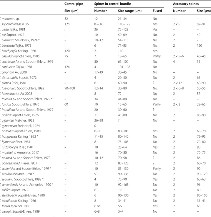
Discussion Since general morphology is highly similar over the 35 species of the genus described so far, species identity is easiest determined by the composition of the sclerotic genital apparatus. C. minuta is characterized by the small size of both the central pipe and the surrounding spines and by the absence of accessory spines (Table 3).
Coelogynopora sopottehlersae n. sp. (Fig. 6)
Locality Type locality: Lister Ley, a tidal channel in the Wadden Sea near Sylt. Medium sand, water depth 17 m (55.0322°N, 008.4892°E; 21 Feb 2018, 4 Individuals).
Material Live observation including drawings and pho- tographs. Four whole mounts, one designated holotype (AWI Sylt P2018-107), three paratypes (AWI Sylt P2018- 107 to AWI Sylt P2018-110).
Etymology This species is dedicated to Dr. Beate Sopott- Ehlers.
Diagnosis Species of Coelogynopora with a copulatory organ consisting of a central group of spines arranged in two rings that are fused at their base, and 5 pairs of acces- sory spines positioned frontally of the central spines.
Description Whitish animals, 8–10 mm long, very slen- der. Frontal end with a few tactile hairs (20–30 µm long).
Dermal glands are inconspicuous, irregularly roundish to ovoid, 6–10 µm in diameter, laterally densely packed, dor- sally and ventrally in longitudinal rows. Pharynx oriented dorso-ventrally, in the beginning of the last quarter of the body. General organization as usual, with testis follicles in a median row, paired germaries frontally of the pharynx, and lateral vitellaries reaching back to the genital opening.
Seminal vesicles paired, very slender, reaching back to the caudal end. They unite dorsally of the genital opening to a very short common duct with a weak muscular cover that enters the central group of spines in the male copula- tory organ. This common duct was not accompanied by prostatic glands and hence is not a prostatic vesicle. The male copulatory organ includes a central group of spines and 5 pairs of accessory spines. The central spines are arranged in two rings that are fused at their base, diam- eter at base 30 µm. In the inner one I counted 8 needles of 125 µm length that seem to be fused over most of their length, thus forming a slender funnel with a crown- shaped tip. The outer ring consists of 16 spines; the dor- sal ones are slightly shorter (110 µm) and slender with a tapering tip; laterally they become increasingly longer, and the ventral spines are nearly as long as the central spines (120 µm) with a broad, flattened tip. Together the outer spines are shaped as an obtuse cone surrounding the central funnel, but they may be spread outwards to give a crown-shaped appearance. The entire central scle- rotic element is enclosed in a muscular cover separating the copulatory organ from surrounding tissue; only the very tips of the spines protrude from the muscular cover.
The accessory spines form a left and a right group of 5 spines each; their longitudinal position was always fron- tal of the central spines. The accessory spines are slightly shorter than the central ones (82–91 µm) but more solid;
they are straight with a slightly curved tip. The accessory spines were surrounded by prostatic secretions, but in live observations the prostatic glands were hardly dis- cernible from the surrounding tissue. Accordingly, it is not clear where the prostatic secretions are released.
Discussion Among the 35 species of Coelogynopora described so far [34] only five have more than one pair of accessory spines and might be closer related with C. sopo- ttehlersae (Table 3). All these species differ in shape, size, and number of the sclerotic components.
The lack of a prostatic vesicle is shared with quite a number of other species, including C. axi, C. solifer, C.
gallica, C. scalpri, C. sequana, and C. solifer. In these species, prostatic secretions are either released through the accessory spines as seems to be the case in C. axi, or enter the ejaculatory duct besides the spines as in C.
scalpri. However, quite a number of species descriptions lack details on the position of prostatic glands and the existence of a prostatic vesicle. Therefore, the value of these characters for taxonomic purpose currently cannot be evaluated.
Cirrifera paraculeata n. sp. (Fig. 7)
Localities (1) Type locality: Lister Ley, medium sand, water depth 17 m (55.0322°N, 008.4892°E, 3 individuals, 21 Feb 2018). (2) List Deep, medium sand, water depth 10 m (55.0469°N, 008.4748°E, 1 individual, 26 Mar 2018).
Table 3 Sclerotic elements in the male copulatory organ of Coelogynopora species
Data based on the original descriptions and *Ax [33]
– Absent
f Central pipe fused with spines
? No or no distinct data
Central pipe Spines in central bundle Accessory spines
Size (µm) Number Size range (µm) Fused Number Size (µm)
C. minuta n. sp. 32 12 21–34 No – –
C. sopottehlersae n. sp. 125 8 + 16 110–125 Yes 2 × 5 82–91
C. alata Tajika, 1981 f 36 72–123 Yes – –
C. axi Sopott, 1972 – 10 50–69 No 2 40
C. biarmata Steinböck, 1924 * – 10–12 54–115 No 2 ?
C. birostrata Tajika, 1978 f 6 71–83 No 2 120
C. brachystyla Karling, 1966 120 2 110 Yes – –
C. cassida Sopott-Ehlers, 1985 75 7 18–65 Partly 2 × 5–6 40–45
C. cochleare Ax and Sopott-Ehlers, 1979 – 30 65–100 No 4 55
C. coniuncta Tajika, 1978 124 4 104–108 No – –
C. coronata Ax, 2008 – 17–19 30–45 No – –
C. distortofolio Sopott, 1972 – 4 20–50 No 2 65
C. erotica Riser, 1981 – 6 60–90 ? 2 × 12 60–90
C. faenofurca Sopott-Ehlers, 1992 90–100 12–14 30–80 No 2 × 6–8 50–55
C. faeroernensis Ax, 2008 – 8 72 No 2 57
C. falcaria Ax and Sopott-Ehlers, 1979 * – 34 44–88 No – –
C. forcipis Sopott-Ehlers, 1976 60 10 15–65 Partly 2 × 3 23–65
C. frondifera Ax and Sopott-Ehlers, 1979 – 20 30–60 ? – –
C. gallica Sopott-Ehlers, 1976 – 11 45–80 No 2 85–90
C. gigantea Meixner, 1938 – 26–28 ? No – –
C. gynocotyla Steinböck, 1924 – 0 – – – –
C. hamulis Sopott-Ehlers, 1980 – 8–9 80–105 Yes 2 65–70
C. hangoensis Karling, 1953 * – 11–15 80–140 No 2 75–95
C. hymanae Riser, 1981 – 8 75–105 No 2 70–80
C. juxtaforcipis Riser, 1981 – 10 25–64 Yes 2 90
C. multispina Armonies, 2017 – 16 70–80 No 2 70–75
C. nodosa Ax and Sopott-Ehlers, 1979 – 10–12 70–98 ? 2 86
C. poaceaglandis Riser, 1981 – 12 85–120 ? 2 60–70
C. scalpri Ax and Sopott-Ehlers, 1979 * – 10 85–130 Partly 2 90
C. schulzii Meixner, 1938 * – 9 90–135 No 2 90–120
C. sequana Sopott-Ehlers, 1992 * – 4 75–90 Yes 2 60–63
C. sewardensis Ax and Armonies, 1990 * – 10 92–168 No 2 96
C. solifer Sopott, 1972 – 6 110 No 2 80
C. steinboecki Sopott-Ehlers, 1980 – 6 90–130 Yes 2 80–100
C. tenuiformis Karling, 1966 – 8 34–41 No 2 31–41
C. tenuis Meixner, 1938 – 6 or 8 56 Yes 2 63
C. visurgis Sopott-Ehlers, 1989 – 6–8 5–7 No – –
Material Life observations including drawings and photographs. Two whole mounts, one designated holo- type (AWI Sylt P2018-101) and one paratype (AWI Sylt P2018-102).
Etymology The species is highly similar to Cirrifera acu- leata.
Diagnosis Cirrifera with paired seminal vesicles and a strongly muscular female atrium without spines. Cirrus Fig. 6 Coelogynopora sopottehlersae. A organization, rear end. B, E Sclerotic apparatus, gently squeezed. C Accessory spines. D Central group of spines, stronger squeezed
Fig. 7 Cirrifera paraculeata. A, B organization, rear end. C–E Sclerotic apparatus
spines robust, numerous, 7–15 µm long, without a basal plate, with a strongly curved tip.
Description Very slender organisms, adults 8–10 mm long and 200–250 µm in diameter. Live specimens may stretch even longer with a diameter only slightly wider than the pharynx, i.e. about 200 µm. Body whitish with numerous yellowish epidermal glands. Brain encapsu- lated and far frontal; statocyst frontal of the brain. The pharynx is spherical and positioned in the end of the mid- dle third of the body.
General arrangement of the reproductive system as usual in the genus: numerous testes follicles in lateral rows from the brain to some 1 mm before the pharynx, paired germaries about 0.5 mm before the pharynx, and vitellaries laterally from the brain to the copulatory organ. The common genital pore lies ventrally in the beginning of the last sixth of the body, with the copula- tory organ directly frontal of the pore. Caudally the geni- tal atrium extends to a very striking spherical muscular organ about 100 µm in diameter.
The paired seminal vesicles are very long, laterally in the last tenth of the body. They unite to the common semi- nal duct that is also very long, crosses the muscular organ dorsally and enters the copulatory bulb (prostatic vesicle) together with the distal part of prostatic glands. The pros- tatic vesicle is a slight swelling of the ejaculatory duct; from live observation it seems to be outside the copulatory bulb.
The cirrus spines are abundant (200–300) and arranged as a rather regular cylinder of 120–130 µm length and 80–90 µm diameter. Individual spines are 7–15 µm long, with smaller spines in the proximal and distal sections of the cirrus and larger ones in the centre. All spines are rather robust, lack a basal plate, and have a strongly curved tip.
Discussion The genus Cirrifera includes three species with an unpaired seminal vesicle (C. boletiformae Sopott, 1972, C. dumosa Sopott, 1972, and C. genitoductus Jouk, Martens and Schockaert, 2007) and four species with paired seminal vesicles (C. aculeata (Ax, 1951), C. cirrif- era Sopott, 1972, C. sopottehlersae Noldt and Jouk, 1988, and C. xanthoderma Riser, 1981). C. paraculeata belongs to the latter group. Among these species, a caudal exten- sion of the genital atrium with a strongly muscular organ (a female atrium according to Martens and Schockaert [35]) is only reported for C. aculeata and the new spe- cies although the ‘atrial diverticulum’ in C. sopottehlersae (Noldt and Jouk, 1988) may be equivalent to that. Thus, C. paraculeata seems to be closely related to C. acu- leata and both species co-occurred in the type locality.
Both species differ in the shape of the cirrus spines (slen- der, weakly curved, and with a basal plate in C. aculeata
against robust, without a basal plate, and with a strongly curved tip in C. paraculeata). In addition, C. paraculeata lacks the large bifid cirrus spine typical for C. aculeata, and it lacks spines in the muscular organ.
Boreocelis fragilis n. sp. (Figs. 8, 9)
Localities Subtidal medium to coarse sand of Lister Ley, the southward branch of the tidal inlet to Sylt-Rømø bight. (1) Coarse sand, 3.5 m water depth (55.0414°N, 008.4796°E, 12 Sep 2017, 1 individual). (2) Type locality:
Medium sand, 10 m water depth (55.0216°N, 008.4580°E, 20 Nov 2017, 2 individuals). (3) Medium sand, 10 m water depth (55.0225°N, 008.4586°E, 11 Dec 2017, 4 individuals).
Material Live observations on 7 individuals, including drawings and photographs; four whole mounts, one des- ignated holotype (AWI Sylt P2018-201) and three para- types (AWI Sylt P2018-202 to AWI Sylt P2018-204).
Etymology Coverslip pressure during microscopical inspection caused three out of the seven individuals stud- ied to autotomize the pre-cerebral frontal section of the body. The species name refers to this fragility.
Diagnosis Species of Boreocelis with a sclerotic appa- ratus consisting of a large clasp with paired extensions resembling a fishing-hook supporting the back of the cop- ulatory organ and two shorter poles that are connected at their tips, thus forming a bifurcated clasp. The shorter poles also bear small extensions, and each of the exten- sions of the large element proceeds to the distal end of one of the shorter poles.
Description Unpigmented specimens up to 3 mm long, but body length is extremely variable, because the tail end may be contracted to less than 1/10 of total body length—
or stretched out to become as long as the rest of the body.
Thus, body length varied between 0.8 and > 2 mm in a single individual. The pre-cerebral frontal body section is longer than usual in monocelidids and looks strongly vac- uolated. However, 3 of the 7 specimens studied alive autot- omized the pre-cerebral body end during microscopical inspection; as a result, the statocyst came extremely close to the (remaining) frontal end. Individuals like this were also found during sorting (i.e. prior to microscopy); thus the length of the pre-cerebral front section is also highly variable in this species.
Big spindle to bottle-shaped rhabdite glands (30 µm long and 6–10 µm in dimeter) occur over most of the body except in a narrow belt around the brain and in the very end of the tail. Because of the strong vacuolization, these rhabdite glands were most striking in the pre-cer- ebral section, but their number in this part of the body
varied (from none to 18) according to the state of regen- eration of the frontal end after previous autotomy. Fin- ger-shaped adhesive papillae are abundant in the caudal end.Behind the brain, there are three pairs of testis folli- cles, followed by the paired germaries still well before the pharynx, which is situated in the middle of the body. The copulatory apparatus is situated behind the pharynx, well before the tail end. It is drop-shaped, some 110 µm long and 50–60 µm wide, with a distinct layer of inner circular
and outer longitudinal muscle fibres. These muscles enclose an unpaired sclerotic element, granular secre- tions, and sperm. Outer prostatic glands and the defer- ent duct leading sperm to the seminal vesicle could not be observed.
The sclerotic apparatus consists of three elements, a large one enclosed in the copulatory organ and two shorter poles. All three elements are constructed from several tightly fitting ledges. The large element (total length 100 µm) supports the back of the copulatory organ.
Fig. 8 Boreocelis fragilis. a, b organization; a contracted, frontal end recently autotomized; b moderately stretched, with frontal end complete. c, d copulatory organ with sclerotic apparatus
It is a slightly curved half-pipe with paired extensions (21 µm long) at the distal tip of the copulatory organ, thus resembling a fishing-hook. Only from the beginning of the lateral extensions to the distal tip the half-pipe is closed (or nearly so) to form an ejaculatory duct.
The shorter (about 47 µm) poles seem to be connected at their tips by a circular muscle thus forming a bifur- cated clasp; they also bear small (7 µm) extensions at their tip. The small and large sclerotic elements are not isolated from each other. Each of the extensions of the large element proceeds to the distal end of one of the shorter poles. The nature of this connection is not clear because the sclerotic part passes into the connecting fibres without a clear demarcation. Possibly the entire element is muscular with a sclerotic coating at the ends.
Discussion The general organization of the new species agrees well with the two species of Boreocelis known up to now, viz. B. filicauda Westblad, 1952 and B. urodasyoides Ax, 1963. All three species have a similar body shape with a long pre-cerebral head section with a transparent paren- chyma and large rhabdites glands, a tail end that may be
extremely extended, and a copulatory organ with a scle- rotic apparatus consisting of three clasps, and other char- acters. But they clearly differ in the structure and size of the sclerotic apparatus. In B. filicauda the unpaired clasp is smaller (128 µm) than the paired clasps (185 µm) while in B. urodasyoides the unpaired clasp is larger (78 µm) than the semi-circular paired ones (diameter of the semi- cycles 31 µm). With respect to the dimensions of the scle- rotic elements B fragilis is closer to the latter species, but the existence of distal extensions in all 3 clasps clearly dif- fers from both known species.
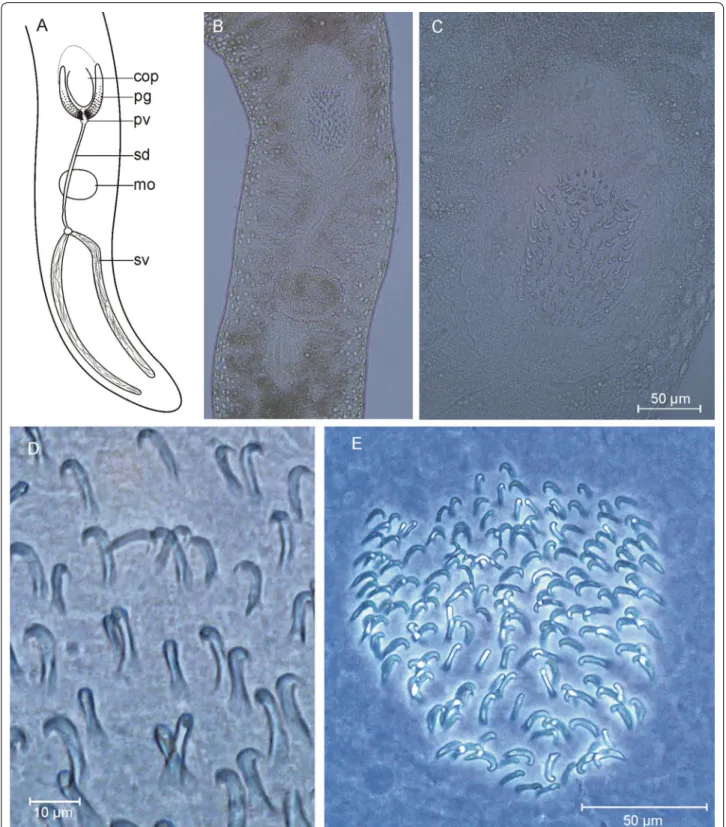
Postbursoplana noldti n. sp. (Fig. 10)
Localities Type locality: Lister Ley, medium sand, water depth 10 m (55.0216°N, 008.4580°E, 20 and 27 Nov 2017, 4 individuals). Further localities: (1) Lister Ley, medium sand, water depth 7 m (55.0445°N, 008.4767°E, 1 individ- ual on 25 Sep 2017 and 2 individuals on 22 Jan 2018). (2) List, fine sand next to the ferry landing, water depth 5 m (55.0151°N 008.4395E, 21 Aug 2017, 2 individuals). (3) Lister Ley, medium sand, water depth 6.5 m (55.0429°N, 008.4775°E, 25 Sep 2017, 5 individuals). (4) Lister Ley, Fig. 9 Boreocelis fragilis. A frontal end. B frontal and rear end. c sclerotic apparatus
Fig. 10 Postbursoplana noldti. A organization. B copulatory organ. C–E sclerotic apparatus
coarse sand, water depth 7 m (55.0458°N, 008.4760°E, Sep 2017, 2 individuals). (5) Lister Ley, medium sand, water depth 10 m (55.0225°N 008.4586°E, 12 Nov 2017, 20 indi- viduals). (6) List Deep, medium sand, water depth 13 m (55.0527°N, 008.4433°E, 19 Sep 1983, leg. Uwe Noldt).
Material Live observations on some 20 individuals, including drawings and photographs. Additional draw- ings and photographs by Uwe Noldt from the 1980s. Ten whole mounts, one designated holotype (AWI Sylt P2018- 111), nine paratypes (AWI Sylt P2018-112 to AWI Sylt P2018-120).
Etymology The species was first encountered by Uwe Noldt during his studies on Kalyptorhynchia.
Diagnosis Extraordinarily large species of Postburso- plana with a male copulatory organ armed with a cen- tral group of six smaller hooks and two larger spines that support a delicate central funnel with a bulgy stem. The copulatory organ is accompanied by two pairs of slightly curved lateral spines with the opening of secretory glands between their tips.
Description Mature animals are 3–4 mm long, flattened, with typical otoplanid shape. Head with a ciliated fur- row apically. The knob-shaped apical end bears about 50 slender sensory hairs of 30–40 µm length; single sensory hairs also occur laterally back to the brain. Ventral body ciliated from the head to the genital opening, adhesive papillae occur from the genital opening to the fan-shaped tail plate, which is densely packed with adhesive papillae.
Rhabdoids are abundant but small, apparently arranged in three longitudinal double-rows. Pharynx in the beginning of the second body half, collar-shaped, some 250 µm in diameter and oriented vertically.
Testes follicles (usually 16 pairs) in lateral rows before the pharynx, caudally followed by paired germaries and lateral rows of vitellaries that are interrupted besides the pharynx. Genital opening positioned in the beginning of the last 1/8 of the body, concealed by the male copulatory organ. Male copulatory apparatus with a longish seminal vesicle caudally, well separated from the longish granu- lar vesicle. A seminal bursa was only observed in a single individual; it was a single spherical bulb besides the semi- nal vesicle, connected to the genital atrium by a narrow duct that opens into the genital atrium at the caudal end.
Male copulatory apparatus with two pairs of lateral and a group of central spines. The lateral spines are rather long (62–70 µm) and curved inwards, the outer pair with a tapering tip, the inner with a small bifurcation 7 µm from the tip. Glands with granular secretions open between the distal tips of these lateral spines. The inner group contains eight spines of two types. Two larger (49–53 µ) spines are central in the group, with an inward bend before the distal third, giving the appearance of a slender funnel with a slightly bulgy stem. Ventrally this funnel is surrounded in a half circle by 6 shorter (28–
32 µm) spines which are slightly curved and bear a trian- gular projection some 5 µm from the tip. Phase contrast microscopy reveals that a delicate (? sclerotic) lamina stretches between the central ‘funnel’-spines forming a functional funnel dorsally but eventually the funnel is not fully closed at the ventral side.
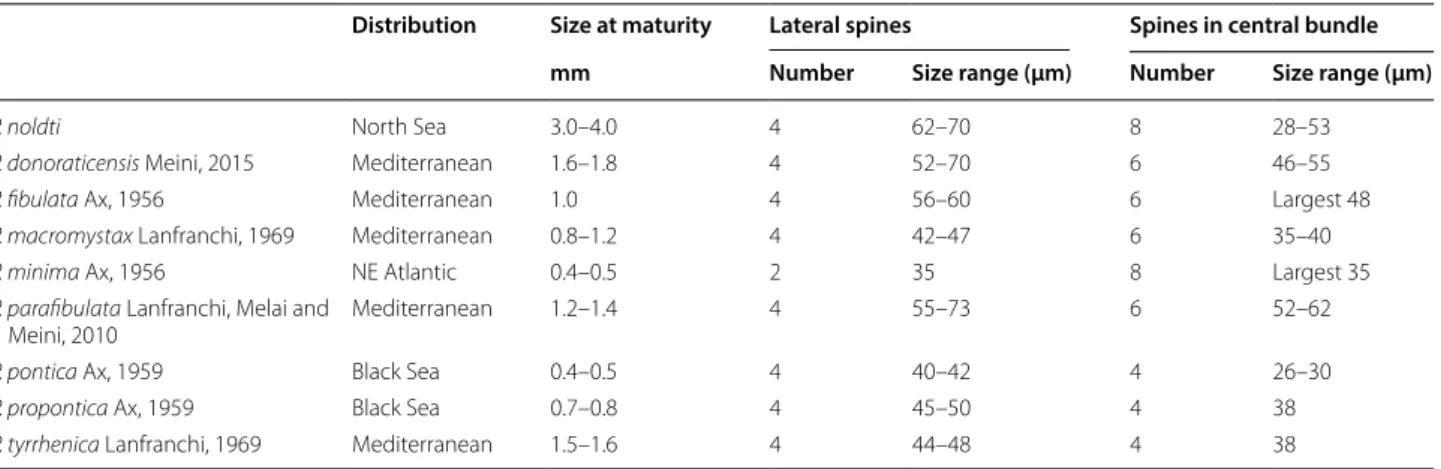
Discussion All Postbursoplana species have a male copulatory organ with a central group of 4–8 spines and (2 -) 4 lateral spines that are always slightly curved and longer than the median ones (Table 4). Species from the Mediterranean and from the Black Sea all have four or six spines in the central group while P. minima from the French Atlantic coast and P. noldti from the North Sea
Table 4 Body size and measures of the male sclerotic apparatus in Postbursoplana species
Distribution Size at maturity Lateral spines Spines in central bundle
mm Number Size range (µm) Number Size range (µm)
P. noldti North Sea 3.0–4.0 4 62–70 8 28–53
P. donoraticensis Meini, 2015 Mediterranean 1.6–1.8 4 52–70 6 46–55
P. fibulata Ax, 1956 Mediterranean 1.0 4 56–60 6 Largest 48
P. macromystax Lanfranchi, 1969 Mediterranean 0.8–1.2 4 42–47 6 35–40
P. minima Ax, 1956 NE Atlantic 0.4–0.5 2 35 8 Largest 35
P. parafibulata Lanfranchi, Melai and
Meini, 2010 Mediterranean 1.2–1.4 4 55–73 6 52–62
P. pontica Ax, 1959 Black Sea 0.4–0.5 4 40–42 4 26–30
P. propontica Ax, 1959 Black Sea 0.7–0.8 4 45–50 4 38
P. tyrrhenica Lanfranchi, 1969 Mediterranean 1.5–1.6 4 44–48 4 38
Fig. 11 Promesostoma wehrenbergi. a organization, b stylet, c tip of the stylet
both have eight spines. P. noldti is far larger than all of the other species and it is characterized by a central group of spines consisting of 6 smaller hooks and 2 larger spines that support a delicate central funnel with a bulgy stem.
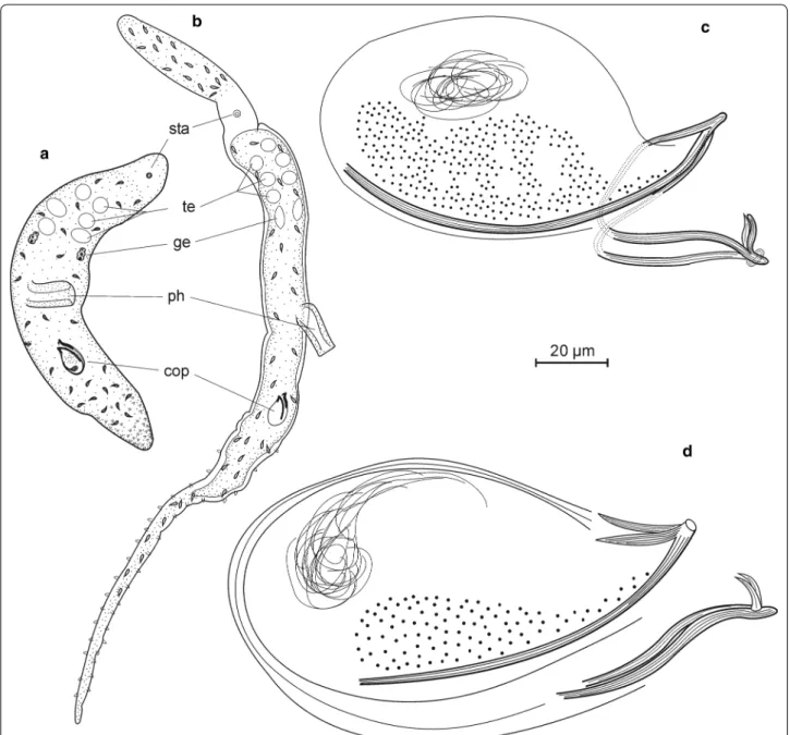
Promesostoma wehrenbergi n. sp. (Figs. 11, 12)
Localities Type locality: Lister Ley, coarse sand, water depth 7 m (55.0445°N, 8.4767°E, 25 Sep 2017, 1 individ- ual). Previously also found in the adjoining List deep by Wehrenberg but there are no detailed records of localities.
Material Live observation, including drawings and pho- tographs; further drawings by Christian Wehrenberg.
Holotype is a whole mount AWI Sylt P2018-206.
Etymology The species was first recorded by Christian Wehrenberg.
Diagnosis Species of Promesostoma characterized by the lack of an external seminal vesicle, a copulatory organ with a weak muscular cover, and the structure of the sty- let. This is a rather short (110–112 µm) branched tube with a semi-circular flap at the distal end.
Description Unpigmented animals 0.7 mm long, taper- ing at both ends of the body. With paired eyes. Pharynx in the middle of the body or shortly behind. Testes, vitellar- ies and germaries paired, position as usual in the genus.
Besides the prostatic glands the deferent ducts seem to enter the copulatory organ directly; an external seminal vesicle was not observed. The copulatory organ is pear shaped, about 60 µm long and 30 µm wide, with a mus- cular cover that is distinct but rather weak for the genus.
A long (50 µm) ejaculatory duct connects the copulatory organ with the stylet.
Fig. 12 Promesostoma wehrenbergi, stylet. A from life observation, B, C from whole mount
The stylet is a rather short (110–112 µm) branched tube with a semi-circular flap at the distal end. Proxi- mally it starts with a funnel shaped opening of 3.5 µm (up to 7 µm according to measurements by Wehren- berg). The sphincter follows at a distance of 8 µm from the proximal opening. Behind the sphincter, the sty- let is a rather straight tube of 4 µm inner diameter for about half of its length. At 45 µm from the proximal opening, the stylet branches into two parts. Only one of the branches can be followed to the distal tip, the other one is only weakly hardened and partly concealed by a semi-circular flap (about 48 µm long and 19 µm wide).
It is not clear whether this flap is a part of the stylet or part of the male genital channel.
In live material, 8–10 roundish vesicles 5–6 µm in diameter and consisting of fine granules were seen beneath the flap (Fig. 11a). After fixation these vesi- cles could no longer be seen (Fig. 11b, c). Presumably, this is the glandular organ observed in quite a number of Promesostoma species in the distal part of the male genital canal, once more challenging the nature of the semi-circular flap.
The bursa is a rather small (17 × 30 µm) bag with a striking notch in its frontal wall. It was filled with sperm to about half the stylet length.
Discussion The genus Promesostoma now comprises
> 40 species differing in stylet length and morphology.
Only 4 species have a branched stylet and potentially form a sub-group of species with P. wehrenbergi: P. balticum, P. bipartitum, P. cochleare, and P. paracochlearis. These species differ in total stylet length and position of the branch (Table 5) as well as the shape of the stylet tips. The semi-circular flap in the distal part of the stylet is unique to P. wehrenbergi in this group though a similar structure occurs in the unbranched stylet of P. digitosa Ax, 1995.
Ptyalorhynchus oculatus n. sp. (Figs. 13, 14)
Locality Type locality: North Sea, some 10 km west of the island of Sylt (55.0355°N, 008.2134°E). Fine sand, water depth 14 m.
Material Nine individuals studied alive, including draw- ings and photographs. Four whole mounts, one desig- nated holotype (AWI Sylt P2018-211), three paratypes (AWI Sylt P2018-212 to AWI Sylt P2018-214).
Etymology This is the first species in the genus equipped with eye pigmentations.
Diagnosis Species of Ptyalorhynchus with pigmented eyes, characterized by a rim-shaped cirrus with three groups of spines, small (2–3 µm) spines in the centre of the rim and larger (5–12 µm and 20–30 µm) spines in the edges.
Description Unpigmented slender specimens, free swimming up to 1.8 mm in length and 0.15 mm in diam- eter. The front end tapers from brain to tip (130 µm at brain, 50–60 µm at rostral end), the caudal end is conical.
Without obvious adhesive papillae. The pharynx (diam- eter 90 µm) is situated in the mid of the body or in the very beginning of second half. With a pair of medium sized eyes in front of the brain. Frontal gland cells behind the brain, well developed.
The proboscis is elongate (in free swimming animals about 120 µm long and 30 µm in diameter), with a very small apex and a circle of eight longish glandular vesi- cles stretching over the entire proboscis length. Probos- cis gland cells are highly developed (Fig. 13d) though the nature and function of the post-cervical complex could not be analysed from life observations.
Genital opening subterminally, copulatory organ in the last tenth of the body. The testes are paired, in free swimming animals longish (some 300 µm) in front of the pharynx or right testis in front of and left one besides the pharynx. The sperm in the testes as well as those in the seminal vesicles and bursa all have a striking pattern of very fine dots. Seminal vesicles paired, piriform.
The copulatory organ is ovoid and equipped with a weak muscle cover. Prostatic glands were seen outside the copulatory organ and prostatic vesicles inside. When the prostatic secretions are emptied, the proximal part of the copulatory organ looks strongly vacuolated. The dis- tal part of the ejaculatory duct bears a rim-shaped cirrus with numerous fine (2–3 µm) spines in the inner part of the rim. The rising edges of the rim carry larger spines, few very large ones (20–30 µm) at one side and medium sized spines (5–12 µm) at the other edge. The very large spines seem to be fused at their base.
Table 5 Stylet sizes in Promesostoma species with a branched stylet
Total stylet
length (µm) Position of stylet branch
Sources
P. balticum Luther, 1918 55/135 at 1
3 length Luther [36]
P. bipartitum Ax, 1956 80 at 1
2 length Ax [37]
P. cochleare Karling, 1935 140–155 (120–130)* at 1
4 length Karling [38], Luther [36],
*Ax [39]
P. paracochlearis Ax, 1952 210–233 at 1
4 length Ax [39]
P. wehrenbergi n. sp. 110–112 at 1
2 length This paper
Paired germaries rostrally of the copulatory organ, fused to the vitellaries that stretch forth to the pharynx or slightly below. A rostral junction of the vitellaries was
not observed. The copulatory bursa is situated between the copulatory organ and the germaries, equipped with paired cuticular mouth pieces of 12 µm total length. The Fig. 13 Ptyalorhynchus oculatus. A, B organization, C bursal mouth piece
part oriented towards the germo-vitelloduct is funnel- shaped and 7 µm long (funnel diameter at opening 6 µm) while the part reaching into the bursa (with several roots) has a length of 5 µm. The genital atrium has a weakly muscular piriform bulge besides the copulatory organ, presumably functioning as a vagina.
Discussion P. oculatus fits the genus diagnosis but differs from the existing species in the arming of the cirrus: P.
piger Brunet, 1973 bears a single group of small (4–5 µm) spines, P. coecus Meixner in Ax, 1951 a proximal group of short (6–8 µm) and a distal group of long (24–35 µm) spines while P. oculatus has a rim-shaped cirrus with three Fig. 14 Ptyalorhynchus oculatus. A, B cirrus spines, different focus. C proboscis, D proboscis with post-cervical complex