Impact of cold atmospheric pressure argon plasma on antibiotic sensitivity of methicillin-resistant Staphylococcus aureus strains in vitro
Einfluss von kaltem Atmosphärendruck-Argon-Plasma auf die
Antibiotikaempfindlichkeit von Methicillin-resistenten Staphylococcus aureus-Stämmen in vitro
Abstract
Aim: The antimicrobial activity of cold atmospheric pressure plasma (CAP), also called tissue tolerable plasma (TTP), could be a promising
Anne Lührmann
1Rutger Matthes
2option to eradicate methicillin-sensitive as well as methicillin-resistant
Axel Kramer
1Staphylococcus aureusstrains, which often colonize chronic wounds.
Currently, the influence of CAP on the susceptibility ofS. aureusto an-
tibiotics is scarcely known, but could be important for treatment of 1 Institute for Hygiene and Environmental Medicine, wounds. Therefore, the aim of this study was to investigate whether
CAP has an impact on the susceptibility of differentS. aureusstrains to different antibiotics.
University Medicine Greifswald, Greifswald, Germany
Method:For assessment, the agar diffusion test with different antibiotic
test disks (cefuroxime, gentamicin, oxacillin, vancomycin, ciprofloxacin, 2 Unit of Periodontology, Dental School, University of co-trimoxazole, clindamycin, erythromycin) was used. Test strains were
spread on agar plates and CAP treated before the antibiotic disks were Greifswald, Greifswald, Germany
placed. After 24 hours cultivation, the inhibited growth zones were measured and differences statistically evaluated.
Results:In most cases, CAP had a negligible influence on the suscept- ibility to antibiotics. For two strains, the susceptibility significantly de- creased to β-lactam antibiotics.
Conclusion:Because CAP can influence the antibiotic susceptibility of S. aureus, before conducting combined treatment with local plasma application on wounds and systemic antibiotics, their interaction must be analysedin vitroto exclude unwanted combination effects.
Keywords:antibiotics, cold atmospheric pressure argon plasma, tissue tolerable plasma, Staphylococcus aureus, sensitivity
Zusammenfassung
Zielsetzung:Die antimikrobielle Wirksamkeit von kaltem Atmosphären- druckplasma (CAP), auch als gewebeverträgliches Plasma (TTP) bezeich- net, könnte eine aussichtsreiche Option zur Eradikation von Methicillin- empfindlichen ebenso wie von Methicillin-resistentenStaphylococcus aureus-Stämmen sein, die oft chronische Wunden kolonisieren. Bisher wurde der Einfluss von CAP auf die Antibiotikaempfindlichkeit von S. aureuskaum untersucht. Da eine Veränderung der Antibiotikaemp- findlichkeit für die Wundbehandlung relevant sein könnte, sollte der Einfluss von CAP auf die Empfindlichkeit verschiedenerS. aureus-Stäm- me gegen unterschiedliche Antibiotika untersucht werden.
Methode: Im Agardiffusionstest wurden Antibiotikatestplättchen mit Cefuroxim, Gentamicin, Oxacillin, Vancomycin, Ciprofloxacin, Co-Trimoxa- zol, Clindamycin und Erythromycin eingesetzt. Die Teststämme wurden auf Agar ausplattiert und mit CAP exponiert, bevor die Testplättchen aufgelegt wurden. Nach 24 h Bebrütung wurden die Inhibitionszonen gemessen und statistisch auf Unterschiede geprüft.
Ergebnisse:In den meisten Fällen war die Einfluss von CAP auf die An- tibiotikaempfindlichkeit zu vernachlässigen. Für zwei Stämme wurde die Empfindlichkeit gegenüber β-Lactam-Antibiotika signifikant herab- gesetzt.
Schlussfolgerung:Da CAP die Antibiotikaempfindlichkeit beeinflussen kann, sollten vor beabsichtigter kombinierter lokaler CAP-Behandlung und gleichzeitiger systemischer Antibiotikagabe Interaktionenin vitro untersucht werden, um unerwünschte Kombinationseffekte auszuschlie- ßen.
Schlüsselwörter:Antibiotika, kaltes Atmosphärendruck-Argon-Plasma, gewebeverträgliches Plasma, Staphylococcus aureus, Empfindlichkeit
Introduction
Increasingly, healthcare-associated infections are caused by drug-resistant microbial pathogens [1]. Of special interest are methicillin-resistant strains ofStaphylococcus aureus(MRSA), which have a high incidence rate [2] and are mainly associated with infections on implants or catheters [3] but also on wounds [4]. The emergence and spread of MRSA in hospitals poses a challenge for infec- tion control interventions and limits treatment options [5]. Therefore, new treatment strategies against antibiotic resistance are important.
Cold physical plasma has been the focus of increasing interest as new option in medicine, especially for wound treatment [6] and cancer therapy [7]. Plasma generated at atmospheric pressure within the physiological tempera- ture range can be suitable for skin and wound treatment;
therefore, different plasma devices have been developed and used successfully [8], [9]. In wound treatment, plasma can promote the wound healing process [10] and is microbicidally active against a wide spectrum of mi- crobes [11], [12], [13], including multidrug-resistant strains [14] as well as microorganisms embedded in biofilms [15], [16]. Pharmacological effects, including microbicidal efficacy, are mainly caused by reactive oxy- gen and nitrogen species already present in the plasma flow or generated by contact of the plasma with liquids or organic substances [8].
An extensively investigated plasma source is the kinpen09®(neoplas GmbH, Greifswald, Germany) [17], which generate a tissue tolerable plasma (TTP) with defined parameters [6], [18]. TTP seems to be a hopeful new therapeutic option, for instance for chronic wound treatment or to treat MRSA carriers [6].
The antimicrobial efficacy of plasma against multidrug- resistant pathogens, including MRSA, on agar plates and embedded in biofilms have been investigated by different authors [14], [19], [20] and showed different strong ef- fects between drug-sensitive and drug-resistant Staphylococcus aureusstrains [19], [21], [22]. However, how plasma treatment of microorganisms affects antibi- otic resistance of the same is currently unknown. Maybe, plasma could reduce or increase the resistance of drug- resistant strains. Therefore, it was of interest as a pilot study to evaluate the influence of TTP application on the resistance of eight genetically differentStaphylococcus
(S.) aureus strains to different antibiotics using a disk- diffusion technique. The genetic differences of the select- ed strains were distinguished in carrying themecAand luk-Pgenes. The genemecA, encode the penicillin binding protein 2A, a protein that has an affinity to adhere to beta- lactam antibiotics and reduce hereby their biological ef- fects, and is a characteristic part of the genome of MRSA strains. The gene luk-P, an important virulence factor, because it can express the membrane toxin Panton- Valentine leukocidin (PVL). The presence ofluk-Pcould influence the activity of other vital relevance genes af- fected by cell stress. Bacterial cell stress can be caused by CAP [23] or antibiotics [24].
Material and methods
Test organisms
The genetic backgrounds of mecA and luk-P differs between theS. aureusstrains used, so that the strains carrymecAorluk-Por both genes together in their gen- ome. The SZ strains (SZ 148, SZ 179) were obtained from the nasal cavity of asymptomatic carriers from Sczcecin, Poland, and H strains (H 2966, H 3163, H 5391) from mature furuncles of furunculosis patients during the acute phase of skin infection, or during abscess incision by a surgeon as described in Masiuk et al. [25]. TheS. aureus strains numbers 05-01825 and 98-00406 were received from the Robert Koch Institute, Wernigerode, Germany, and were analysed by Strommenger et al. [26]. The S. aureusATCC 6538 strain served as reference strain, without genetic background ofmecAandluk-P. The gen- etically availability ofmecAandluk-Pis shown for each strain in the result table (Table 1).
Bacterial cultivation and preparation for treatment
TheS. aureusstrains were grown on Columbia blood agar (BD, Heidelberg, Germany) for 24 h at 37°C. The bacterial lawn was removed with 3 ml of sterile 0.9% saline (NaCl) solution and an applicator. After bacterial resuspension, a cell concentration to 108colony-forming units (CFU) was adjusted by densimetric measurement and a serial dilu- tion (108, 107, 106CFU/ml) was prepared in NaCl solution.
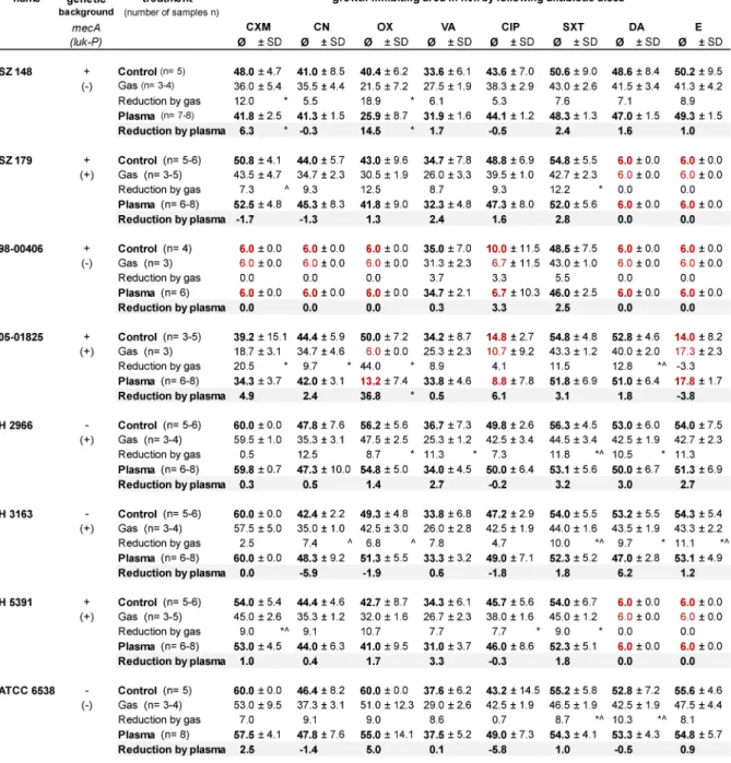
Table 1: Measured growth inhibition zone of the eight investigated clinicalStaphylococcus aureus strains under control and plasma- or gas-treatment conditions and incubated with antibiotic susceptibility test disks (CXM – cefuroxime, CN – gentamicin, OX – oxacillin, VA – vancomycin, CIP – ciprofloxacin, SXT – co-trimoxazol, DA – clindamycin, E – erythromycin) in mm (diameter), with standard deviation (SD) and number of samples (n). The reduction values show the difference from the control. Red numbers represent a growth inhibition zone less than 15 mm, which suggests resistance to the respective drug. The genetic background ofmecA and luk-P of each strain is shown, the sign “+” shows that the strain carry that gene, the sign “-” shows the absence
of that gene in their genome.
The 108CFU/ml suspension was used for argon plasma and argon gas application, and the 108, 107 and 106CFU/ml suspensions were used to inoculate the agar plates for the control (no treatment) and the antibiotic test alone. For inoculation, 100 µl of the dilutions were spread on Oxoid Iso-Sensitest™ agar plates (Oxoid, Thermo Fisher Scientific, Wesel, Germany). A note to the chosen agar: The often used Müller-Hinton agar for anti-
biotic diffusion test is not suited in combination with plasma treatment. Therefore, the Iso-Sensitest agar was preferred. Before treatment, the inoculated agar plates dried for 20 min at room temperature under laminar air flow to avoid a fluid inoculum film during treatment.
Each strain and treatment type was performed 3 times:
twice for plasma treated strains (n=6) and once for the controls and gas treatment (n=3). After treatment, the
bacteria were incubated for 24 h at 37°C. These were later statistically analyzed.
The different dilutions for the controls were used, because argon plasma reduced the bacterial load on the agar surface, and different cell densities of bacterial load on the agar surface will show different results in the antibi- otic agar diffusion test. Thus, three subsequent dilutions were chosen for the controls to obtain results which are comparable to the plasma-treated samples.
Treatment with argon plasma
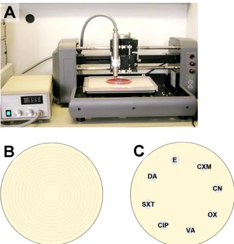
A radio frequency plasma pen (kinpen09®, neoplas GmbH, Greifswald, Germany) using argon (99.999% pure) as carrier gas served as the plasma source. The device op- erated with 1.1 MHz at 2–6 kVpp with a maximal input DC power of 3.5 W to the hand-held unit [27]. The gas flow was adjusted to 5 slm (standard liters/min) controlled by a mass flow controller (MKS Instruments, Munich, Germany). During plasma generation open to room air, the spatial afterglow plasma plume was 7 mm long. The plasma effect on the surface reaches a diameter of 3 mm around the centre of the plasma plume, which was evaluated before.
To use for treatment, the pen was fixed in a computer- controlled x/y/z stage above the prepared agar plate at a distance of 7 mm (Figure 1A) and was moved once along a spiral pattern (Figure 1B) at a speed of 10 mm/s.
The distance between the resulting concentric circles was 3 mm. This resulted in a total treatment time of 0.6 s at all treated points on the agar plate, a short treatment time to achieve a sublethal plasma dose for the most bacterial cells.
During treatment, an increase in temperature generated on the surface is not detectable, because the plasma- surface contact time is too short by moving plasma pen.
A plasma treatment on fixed place for 30 s showed a temperature of 22.6°C. After 60 s treatment, the tem- perature increased to 24.9°C. That corresponds to a temperature rise of 4.4°C and 6.7°C after 30 s and 60 s punctual treatment time. In order to measure the surface temperature, a non-contact method by using the infrared radiation emitted by the substrate was used [28] by using a pyrometer (digital infrared camera; Fluke Deutschland GmbH, Glottertal, Germany).
As plasma control, the pure argon gas without plasma ignition was applied in the same manner like the plasma application. The CAP and gas were applied as a pre- treatment, before antibiotic disks were positioned, or as sole treatment, to get respective controls.
Antibiotic susceptibility test
Within 15 min after plasma or gas treatment (time for handling), the antibiotic disks (6 mm, BBL™ Sensi-Disc™, BD, Heidelberg, Germany) for the susceptibility test were positioned on the prepared agar plates (Figure 1C) and incubated for 24 h at 37°C. Antibiotic test disks used were cefuroxime (CXM) 30 µg, gentamicin (CN) 10 µg,
oxacillin (OX) 5 µg, vancomycin (VA) 30 µg, ciprofloxacin (CIP) 5 µg, co-trimoxazol (SXT) 25 µg, clindamycin (DA) 2 µg and erythromycin (E) 15 µg. All disks were stored in their containers at –20°C until used in these experiments.
After 24 h of incubation at 37°C, the diameter of growth inhibition zone was measured. A strain was generally considered resistant to the given antibiotic if the growth inhibition zone had a diameter of less than 15 mm, as based on the Performance Standards for Antimicrobial Susceptibility Testing, Twenty-second Informational Sup- plement, 2013 [29]. A growth-inhibition-zone threshold (a set diameter) to determine bacterial resistance for each antibiotic separately was not defined, because only the differences of growth inhibition zones were of interest to decide whether the susceptibility after plasma treat- ment increased or decreased. Because ISO-Sensitive agar was used instead of Mueller-Hinton agar, the guidelines can only serve as orientation.
Statistical analyses
The diameter of the growth inhibition zone was measured in mm and was used in the statistical evaluation to com- pare plasma treatment, gas treatment, and the control (without plasma or gas treatment) in each strain. The detection limit was a radius of 1 mm from the disk’s edge, which corresponds to a limit of diameter measurement of 8 mm. In the case of detection limit, no growth inhib- ition zone was observable and the value was set 6 (diameter of the antibiotic discs). The comparison of the measured antibiotic-induced growth inhibition zones of plasma-treatedS. aureusstrains to their respective anti- biotic control was chosen depending on the result of the comparison of the plasma-treated agar plates to the dif- ferent plated dilutions (108, 107 or 106 CFU/ml) of the untreated control series (Figure 2). Control plates with analogical density of CFU after plasma treatment were used for the comparisons.
The mean diameter of the growth inhibition zone in each test run of the plasma treatment, gas treatment, and the antibiotic control (without plasma or gas treatment) of each strain was used for the statistical evaluation. Stat- istical differences were analysed with the Mann-Whitney U-test followed with Bonferroni p-value correction for multiple comparisons using statistical analysis software (Prism, GraphPad, USA).
Results
The changes of the growth inhibition zones produced by antibiotic disks with or without argon plasma or argon gas pre-treatment varied between the differentS. aureus strains examined (Table 1). Of the five strains with a genetic mecA-positive background, only the S. aureus strain 98-00406 showed resistance against the β-lactam antibiotics cefuroxime and oxacillin in the antimicrobial diffusion sensitivity disk test on agar. TheS. aureusstrain
Figure 1: Setup of the plasma application system (A), illustration of the movement pattern for the plasma or gas application (space between the circles = 3 mm) on agar plate (B), and placement of disks in on the prepared agar plates in the antibiotic sensitivity test (CXM – cefuroxime, CN – gentamicin, OX – oxacillin, VA – vancomycin, CIP – ciprofloxacin, SXT – co-trimoxazole,
DA – clindamycin, E – erythromycin) (C)
Figure 2: Illustration of the test scheme (untreated control, plasma and gas treatment) and choice of samples after plasma treatment for comparison of the growth inhibition zone caused by application of the antibiotic sensitivity test disks (AB)
98-00406 showed additional resistance to the aminoglycoside antibiotic gentamycin, the fluoroquinolone antibiotic ciprofloxacin, and the translation-inhibiting antibiotics clindamycin and erythromycin. Clindamycin and erythromycin resistances were also shown for the S. aureus strains SZ 179 and H 5391. The S. aureus strain 05-01825 showed less susceptibility to ciprofloxa- cin and erythromycin (Table 1).
The plasma treatment influenced the growth inhibition by the antibiotic test disks in some cases. A statistically significant decrease in the growth inhibition zone of 6.3 mm with cefuroxime and of 14.5 mm with oxacillin was shown for theS. aureusstrain SZ 148, as well as a decrease for strain 05-01825 of 36.8 mm by oxacillin (p<0.05) (Table 1). No test organism demonstrated a significantly increased growth inhibition zone after plasma treatment.
Interestingly, argon gas treatment without plasma ignition in some cases showed a decreased growth inhibition zone (Table 1) for co-trimoxazole – an inhibitor of the microbial tetrahydrofolate biosynthesis – which was sig- nificantly different from the growth inhibition by plasma for the strains without the genetic background ofmecA H 2966, H 3163 and ATCC 6538.
The existence of the geneluk-Pin theS. aureusgenome does not seem to influence susceptibility to antibiotics after argon plasma or argon gas treatment.
Discussion
New methods based on cold atmospheric pressure plasma are currently being developed for the treatment of chronic wounds with additional microbicidal efficacy to inactivate drug-resistant microorganisms in the wounds. Topical application of antibiotics for wound treatment is obsolete due to the increased risk of drug resistance, but systemic use of antibiotics is indicated if septic metastasis occurs [30]. For the future of wound treatment, the combination of local plasma application with systemic antibiotic therapy is a promising option. For this to be effective, knowledge of the influence of plasma treatment on microbial sensitivity to antibiotics is an es- sential precondition, because plasma can influence the sensitivity of microorganisms to antibiotics. The main ef- fect of plasma is caused by the plasma-generated reactive oxygen and nitrogen species, which interact with the cells and the surrounded media, changing the cells’ environ- ment in terms of increased oxidation processes and de- creased pH. For the used kinpen 09, in argon plasma radiating effluent, excited species of hydroxyl (OH), nitro- gen (N2), atomic oxygen (O), and argon (Ar) can generate [31], [32], which can react to further species in contact with environmental air. A study showed that induced stress and changes of environmental pH can cause an increased resistance to antibiotics [33], [34]. Thus, an increased resistance to antibiotics and decreased growth inhibition zones ofS. aureusstrains were expected in our study.
Otherwise, if plasma could lessen drug resistance of microorganisms by increasing their sensitivity to antibiot- ics, this could open new treatment options against mul- tidrug-resistant microorganisms.
S. aureus is one of the most problematic pathogens in nosocomial infections and can show different single or multiple resistances against antibiotic drugs. For this study, eight differentS. aureusstrains with or without a genetic background ofmecAand orluk-Pwere used. The tests were carried out on Iso-Sensitest agar, because it is suited for the antibiotic diffusion test and showed only a minor effect on bacterial growth if agar was plasma treated, before being inoculated with microorganisms.
So, the influences to an antimicrobial effect caused by the media was negligible. The recommend Mueller-Hinton agar for the antibiotic agar diffusion test showed clear antimicrobial effects after plasma treatment and was not suited therefore for the present study (determined in pre- tests).
The plasma treatment time used, approximately 0.6 s per treated area, was very short, considering that tissue tolerability was shown for up to 60 s of plasma exposure time [35]. Because plasma efficacy on planktonic cells on agar plates is very high, a short treatment time was necessary to maximize the number of cells remaining after plasma treatment, as the agar diffusion test requires a large number of cells. To circumvent influence of pos- sible differences of viable bacterial cells after CAP appli- cation, the diameter of the growth inhibiting zone after antibiotics of CAP-treated bacteria was compared with the suited control plate, which showed the same bacterial colony-density like the plates after CAP treatment without antibiotics (Figure 2).
The results suggest that argon plasma can increase the resistance against antibiotic drugs, but the effect was mostly very low and not significant. However, in three cases, the increased resistance was significant for the effects of β-lactam antibiotics, especially regarding oxa- cillin for the test strains SZ 148 and 05-01825. The exist- ence of the virulence factor luk-P (Panton-Valentine leukocidin) did not play a role in plasma-treatedS. aureus growth in an antibiotic environment and thus constitutes no risk for combined therapy with plasma and antibiotics.
The study of Hoon Park et al. 2015 [36] could demon- strate that an Ar plasma of a surface dielectric barrier discharged (DBD) plasma device reduced β-lactamase activity and increased therefore the susceptibility against β-lactam antibiotics. The plasma treatment time in the study of Hoon Park et al. was 300 s instead of 0.6 s of this study, resulting in different energy load, which can be one reason for the increased susceptibility against β-lactam antibiotic of plasma treated S. aureus cells.
Another reason could be the gas flow, which was 1 slm in the study of Hoon Park et al. and 5 slm in this study.
Because, the argon gas treatment also caused increased drug resistance, and showed significantly stronger effects than the plasma treatment, especially for theS. aureus strains 05-01825, H 2966 and H 3163, compared to the control or to the plasma treatment for most of the
antibiotic substances examined, which was not expected.
Argon is an inert gas and no effect on microorganisms is expected. However, studies on the proteomic and tran- scriptomic pattern of Bacillus subtilis after argon gas treatment showed an induced activity of genes known for stress responses [23]. Perhaps the genetic response to pure argon has a stronger influence on the antibiotic susceptibility ofS. aureusthan does the genetic response to argon plasma. Nevertheless, the reason why plasma showed a weaker effect than the pure argon is currently unknown. Possibly the mechanic influence of the gas flow of 5 slm cause the reduced susceptibility against antibi- otics. If plasma increases the susceptibility against β-lactam antibiotics, like reported in Hoon Park et al.
2015 [36] by reduced β-lactamase activity, the CAP maybe reduced the gas flow effect, which could explain a lower reduction of the susceptibility against antibiotics after CAP treatment than after application of pure argon.
Additionally, pure argon treatment ofS. aureuswithout subsequent antibiotic contact showed no effect (n=4 of each strain). The CAP effect without subsequent antibiotic contact can vary low between tested strains in that test setup. A previous study, with another test setup, showed also differences in correlation to the genesmecAand luk-P, which was only marginal [37].
In this study, a discrepancy between the PCR analyses – which detected the genetic code formecAgene – and the lack of resistance to oxacillin or cefuroxime after the antibiotic susceptibility test was shown. Only strains 05- 01825 and 98-00406 carryingmecAshowed a resistance against oxacillin (strain 05-01825 only after argon appli- cation), but not strains H 5391, SZ 148, and SZ 179.
Such a discrepancy between a geneticmecAbackground and lack of resistance against oxacillin in the agar-diffu- sion test does not seem unusual and is described in the literature [38], [39], [40].
This pilot study is one of the first investigations on the impact of argon plasma at low temperatures, operated using tissue tolerable parameters, on the susceptibility of differentS. aureusstrains to antibiotic drugs. One of the limits is that thein vitrostudy cannot reflect possible effectsin vivo. Infectionsin vivoare mostly associated with multispecies biofilms in a very complex wound fluid milieu. However, the results show that plasma treatment can increase the resistance ofS. aureusagainst antibiot- ics, which should be reason enough to carefully consider such effects before applying plasma to infected wounds.
A decrease of bacterial drug resistance caused by plasma influence seems not possible and could be no option to support antibiotic treatment of microbially colonized wounds. Further research should test longer plasma treatment times and include different species analysed with different methods to exclude possible plasma effects caused by interaction with the media used. The impact of pure argon on the susceptibility of microorganisms to antibiotics could be a worthwhile study.
Conclusion
For treatment of microbially infected wounds, plasma is being examined as a new potential therapy option. Our results showed that although argon plasma can influence the susceptibility of Staphylococcus aureus strains to antibiotics, the effect was mostly not significant. However, in two cases, a significant increase in resistance against β-lactam antibiotics was shown. This suggests that lower efficacy of antibiotics after plasma treatment should be considered if a combined treatment of plasma and anti- biotics is planned. Further research to reflect more real- istic treatment conditions is necessary before plasma applications on infected wounds can be recommended for combined treatment with antibiotics.
Notes
Acknowledgement
The authors thank Dr. Silva Holtfreter and Dr. Julia Kolata (Department of Immunology, Ernst-Moritz-Arndt-University, Greifswald) for providing of theStaphylococcus aureus strains.
Funding
This study was conducted within the multi-disciplinary cooperative research program “Campus PlasmaMed”, in particular within the sub-project “PlasmaCure”, and was supported by a grant from the German Ministry of Educa- tion and Research (BMBF, grant No. 13N11181).
Competing interests
The authors declare that they have no competing in- terests.
References
1. Caini S, Hajdu A, Kurcz A, Böröcz K. Hospital-acquired infections due to multidrug-resistant organisms in Hungary, 2005-2010.
Euro Surveill. 2013 Jan 10;18(2). pii: 20352.
2. Kramer A, Wagenvoort H, Ahrén C, Daniels-Haardt I, Hartemann P, Kobayashi H, Kurcz A, Picazo J, Privitera G, Assadian O.
Epidemiology of MRSA and current strategies in Europe and Japan. GMS Krankenhhyg Interdiszip. 2010;5(1):Doc01. DOI:
10.3205/dgkh000144
3. Arciola CR, Campoccia D, Speziale P, Montanaro L, Costerton JW. Biofilm formation in Staphylococcus implant infections. A review of molecular mechanisms and implications for biofilm- resistant materials. Biomaterials. 2012 Sep;33(26):5967-82.
DOI: 10.1016/j.biomaterials.2012.05.031
4. Martineau L, Davis SC, Peng HT, Hung A. Controlling methicillin resistant Staphyloccocus aureus and Pseudomonas aeruginosa wound infections with a novel biomaterial. J Invest Surg. 2007 Jul-Aug;20(4):217-27. DOI: 10.1080/10717540701481275
5. Fenner L, Widmer AF, Dangel M, Frei R. Distribution of spa types among meticillin-resistant Staphylococcus aureus isolates during a 6 year period at a low-prevalence University Hospital. J Med Microbiol. 2008 May;57(Pt 5):612-6. DOI:
10.1099/jmm.0.47757-0
6. Kramer A, Lademann J, Bender C, Sckell A, Hartmann B, Münch S, Hinz P, Ekkernkamp A, Matthes R, Koban I, Partecke I, Heidecke CD, Masur K, Reuter S, Weltmann KD, Koch S, Assadian O. Suitability of tissue tolerable plasmas (TTP) for the
management of chronic wounds. Clin Plasma Med. 2013;1(1):11- 8. DOI: 10.1016/j.cpme.2013.03.002
7. Partecke LI, Evert K, Haugk J, Doering F, Normann L, Diedrich S, Weiss FU, Evert M, Huebner NO, Guenther C, Heidecke CD, Kramer A, Bussiahn R, Weltmann KD, Pati O, Bender C, von Bernstorff W. Tissue tolerable plasma (TTP) induces apoptosis in pancreatic cancer cells in vitro and in vivo. BMC Cancer.
2012;12:473. DOI: 10.1186/1471-2407-12-473
8. von Woedtke T, Reuter S, Masur K, Weltmann KD. Plasmas for medicine. Phys Rep. 2013;530(4):291-320. DOI:
10.1016/j.physrep.2013.05.005
9. Emmert S, Brehmer F, Hänßle H, Helmke A, Mertens N, Ahmed R, Simon D, Wandke D, Maus-Friedrichs W, Däschlein G, Schön MP, Viöl W. Atmospheric pressure plasma in dermatology: Ulcus treatment and much more. Clin Plasma Med. 2013;1(1):24-9.
DOI: 10.1016/j.cpme.2012.11.002
10. Bender CP, Hübner NO, Weltmann KD, Scharf C, Kramer A. Tissue Tolerable Plasma and Polihexanide: Are Synergistic Effects Possible to Promote Healing of Chronic wounds? In Vivo and In Vitro Results. In: Machala Z, Hensel K, Akishev Y, editors. Plasma for Bio-Decontamination, Medicine and Food Security. Dordrecht:
Springer Netherlands; 2012. p. 321–34. (NATO Science for Peace and Security; Series A: Chemistry and Biology). DOI:
10.1007/978-94-007-2852-3_25
11. Daeschlein G, von Woedtke T, Kindel E, Brandenburg R, Weltmann KD, Jünger M. Antibacterial Activity of an Atmospheric Pressure Plasma Jet Against Relevant Wound Pathogens in vitro on a Simulated Wound Environment. Plasma Process Polym.
2010;7(3-4):224–30. DOI: 10.1002/ppap.200900059 12. Daeschlein G, Scholz S, von Woedtke T, Niggemeier M, Kindel
E, Brandenburg R, Weltmann KD, Junger M. In Vitro Killing of Clinical Fungal Strains by Low-Temperature Atmospheric-Pressure Plasma Jet. IEEE Trans Plasma Sci IEEE Nucl Plasma Sci Soc.
2011;39(2):815-21. DOI: 10.1109/TPS.2010.2063441 13. Daeschlein G, Scholz S, Arnold A, von Podewils S, Haase H,
Emmert S, von Woedtke T, Weltmann KD, Jünger M. In Vitro Susceptibility of Important Skin and Wound Pathogens Against Low Temperature Atmospheric Pressure Plasma Jet (APPJ) and Dielectric Barrier Discharge Plasma (DBD). Plasma Process Polym. 2012;9(4):380–9. DOI: 10.1002/ppap.201100160 14. Daeschlein G, Napp M, von Podewils S, Lutze S, Emmert S, Lange
A, Klare I, Haase H, Gümbel D, von Woedtke T, Jünger M. In Vitro Susceptibility of Multidrug Resistant Skin and Wound Pathogens Against Low Temperature Atmospheric Pressure Plasma Jet (APPJ) and Dielectric Barrier Discharge Plasma (DBD). Plasma Process Polym. 2014;11(2):175–83. DOI:
10.1002/ppap.201300070
15. Koban I, Matthes R, Hübner NO, Welk A, Meisel P, Holtfreter B, Sietmann R, Kindel E, Weltmann KD, Kramer A, Kocher T.
Treatment of Candida albicans biofilms with low-temperature plasma induced by dielectric barrier discharge and atmospheric pressure plasma jet. New J Phys. 2010;12:073039. DOI:
10.1088/1367-2630/12/7/073039
16. Matthes R, Koban I, Bender C, Masur K, Kindel E, Weltmann KD, Kocher T, Kramer A, Hübner NO. Antimicrobial Efficacy of an Atmospheric Pressure Plasma Jet Against Biofilms of Pseudomonas aeruginosa and Staphylococcus epidermidis.
Plasma Process Polym. 2013;10(2):161–6. DOI:
10.1002/ppap.201100133
17. Weltmann KD, Kindel E, Brandenburg R, Meyer C, Bussiahn R, Wilke C, von Woedtke T. Atmospheric Pressure Plasma Jet for Medical Therapy: Plasma Parameters and Risk Estimation.
Contrib Plasma Phys. 2009;49(9):631–40. DOI:
10.1002/ctpp.200910067
18. Lademann J, Ulrich C, Patzelt A, Richter H, Kluschke F, Klebes M, Lademann O, Kramer A, Weltmann KD, Lange-Asschenfeldt B. Risk assessment of the application of tissue-tolerable plasma on human skin. Clin Plasma Med. 2013;1(1):5–10. DOI:
10.1016/j.cpme.2013.01.001
19. Ermolaeva SA, Varfolomeev AF, Chernukha MY, Yurov DS, Vasiliev MM, Kaminskaya AA, Moisenovich MM, Romanova JM, Murashev AN, Selezneva II, Shimizu T, Sysolyatina EV, Shaginyan IA, Petrov OF, Mayevsky EI, Fortov VE, Morfill GE, Naroditsky BS, Gintsburg AL. Bactericidal effects of non-thermal argon plasma in vitro, in biofilms and in the animal model of infected wounds. J Med Microbiol. 2011 Jan;60(Pt 1):75-83. DOI:
10.1099/jmm.0.020263-0
20. Kvam E, Davis B, Mondello F, Garner AL. Nonthermal atmospheric plasma rapidly disinfects multidrug-resistant microbes by inducing cell surface damage. Antimicrob Agents Chemother.
2012 Apr;56(4):2028-36. DOI: 10.1128/AAC.05642-11 21. Burts ML, Alexeff I, Meek ET, McCullers JA. Use of atmospheric
non-thermal plasma as a disinfectant for objects contaminated with methicillin-resistant Staphylococcus aureus. Am J Infect Control. 2009 Nov;37(9):729-33. DOI:
10.1016/j.ajic.2009.03.010
22. Joshi SG, Paff M, Friedman G, Fridman G, Fridman A, Brooks AD.
Control of methicillin-resistant Staphylococcus aureus in planktonic form and biofilms: a biocidal efficacy study of nonthermal dielectric-barrier discharge plasma. Am J Infect Control. 2010 May;38(4):293-301. DOI:
10.1016/j.ajic.2009.11.002
23. Winter T, Bernhardt J, Winter J, Mäder U, Schlüter R, Weltmann KD, Hecker M, Kusch H. Common versus noble Bacillus subtilis differentially responds to air and argon gas plasma. Proteomics.
2013 Sep;13(17):2608-21. DOI: 10.1002/pmic.201200343 24. Kohanski MA, Dwyer DJ, Collins JJ. How antibiotics kill bacteria:
from targets to networks. Nat Rev Microbiol. 2010 Jun;8(6):423- 35. DOI: 10.1038/nrmicro2333
25. Masiuk H, Kopron K, Grumann D, Goerke C, Kolata J, Jursa- Kulesza J, Giedrys-Kalemba S, Bröker BM, Holtfreter S.
Association of recurrent furunculosis with Panton-Valentine leukocidin and the genetic background of Staphylococcus aureus.
J Clin Microbiol. 2010 May;48(5):1527-35. DOI:
10.1128/JCM.02094-09
26. Strommenger B, Kettlitz C, Weniger T, Harmsen D, Friedrich AW, Witte W. Assignment of Staphylococcus isolates to groups by spa typing, SmaI macrorestriction analysis, and multilocus sequence typing. J Clin Microbiol. 2006 Jul;44(7):2533-40. DOI:
10.1128/JCM.00420-06
27. Purevdorj D, Igura N, Ariyada O, Hayakawa I. Effect of feed gas composition of gas discharge plasmas on Bacillus pumilus spore mortality. Lett Appl Microbiol. 2003;37(1):31-4.
28. Tandian NP, Pfender E. Heat transfer in RF plasma sintering:
Modeling and experiments. Plasma Chem Plasma Process.
1997;17(3):353–70. DOI: 10.1007/BF02766840
29. Clinical and Laboratory Standards Institute (CLSI). Performance Standards for Antimicrobial Susceptibility Testing; Twenty-second Informational Supplement. Wayne, PA, USA: CLSI; 2013.
30. Dissemond J, Assadian O, Gerber V, Kingsley A, Kramer A, Leaper DJ, Mosti G, Piatkowski de Grzymala A, Riepe G, Risse A, Romanelli M, Strohal R, Traber J, Vasel-Biergans A, Wild T, Eberlein T. Classification of wounds at risk and their antimicrobial treatment with polihexanide: a practice-oriented expert recommendation. Skin Pharmacol Physiol. 2011;24(5):245-55.
DOI: 10.1159/000327210
31. Reuter S, Tresp H, Wende K, Hammer MU, Winter J, Masur K, Schmidt-Bleker A, Weltmann KD. From RONS to ROS: Tailoring Plasma Jet Treatment of Skin Cells. IEEE Trans Plasma Sci IEEE Nucl Plasma Sci Soc. 2012;40(11):2986-93. DOI:
10.1109/TPS.2012.2207130
32. Matthes R, Bekeschus S, Bender C, Koban I, Hübner NO, Kramer A. Pilot-study on the influence of carrier gas and plasma application (open resp. delimited) modifications on physical plasma and its antimicrobial effect against Pseudomonas aeruginosa and Staphylococcus aureus. GMS Krankenhaushyg Interdiszip. 2012;7(1):Doc02. DOI: 10.3205/dgkh000186 33. Albesa I, Becerra MC, Battán PC, Páez PL. Oxidative stress involved in the antibacterial action of different antibiotics.
Biochem Biophys Res Commun. 2004 Apr;317(2):605-9. DOI:
10.1016/j.bbrc.2004.03.085
34. Poole K. Bacterial stress responses as determinants of antimicrobial resistance. J Antimicrob Chemother. 2012 Sep;67(9):2069-89. DOI: 10.1093/jac/dks196
35. Hammann A, Huebner NO, Bender C, Ekkernkamp A, Hartmann B, Hinz P, Kindel E, Koban I, Koch S, Kohlmann T, Lademann J, Matthes R, Müller G, Titze R, Weltmann KD, Kramer A. Antiseptic efficacy and tolerance of tissue-tolerable plasma compared with two wound antiseptics on artificially bacterially contaminated eyes from commercially slaughtered pigs. Skin Pharmacol Physiol.
2010;23(6):328-32. DOI: 10.1159/000314724
36. Hoon Park J, Kumar N, Park DH, Yusupov M, Neyts EC, Verlackt CC, Bogaerts A, Kang MH, Uhm HS, Choi EH, Attri P. A comparative study for the inactivation of multidrug resistance bacteria using dielectric barrier discharge and nano-second pulsed plasma. Sci Rep. 2015 Sep 9;5:13849. DOI:
10.1038/srep13849
37. Matthes R, Lührman A, Holtfreter S, Kolata J, Radke D, Hübner NO, Assadian O, Kramer A. Antibacterial Activity of Cold Atmospheric Pressure Argon Plasma against 78 Genetically Different (mecA, luk-P, agr or Capsular Polysaccharide Type) Staphylococcus aureus Strains. Skin Pharmacol Physiol.
2016;29(2):83-91. DOI: 10.1159/000443210
38. Sakoulas G, Gold HS, Venkataraman L, DeGirolami PC, Eliopoulos GM, Qian Q. Methicillin-resistant Staphylococcus aureus:
comparison of susceptibility testing methods and analysis of mecA-positive susceptible strains. J Clin Microbiol. 2001 Nov;39(11):3946-51. DOI: 10.1128/JCM.39.11.3946- 3951.2001
39. Duran N, Ozer B, Duran GG, Onlen Y, Demir C. Antibiotic resistance genes & susceptibility patterns in staphylococci. Indian J Med Res. 2012 Mar;135:389-96.
40. Alipour F, Ahmadi M, Javadi S. Evaluation of different methods to detect methicillin resistance in Staphylococcus aureus (MRSA).
J Infect Public Health. 2014 May-Jun;7(3):186-91. DOI:
10.1016/j.jiph.2014.01.007
Erratum
There was a mistake in section “Material and methods”
in paragraph “Treatment with argon plasma” with sub- sequent errors in section “Discussion”. The specified treatment time of plasma at each treatment point during movement of the plasma pen at 6 s is wrong. The correct treatment time should be 0.6 s. The relevant sentences of the section “Material and methods” (“This resulted in a total treatment time of 6 s at all treated points on the agar plate, a short treatment time to achieve a sublethal plasma dose for the most bacterial cells.”) and of the section “Discussion” (“The plasma treatment time used, approximately 6 s per treated area, was very short, con- sidering that tissue tolerability was shown for up to 60 s of plasma exposure time [35].” and “The plasma treat- ment time in the study of Hoon Park et al. was 300 s in- stead of 6 s of this study, resulting in different energy load, which can be one reason for the increased suscept- ibility against β-lactam antibiotic of plasma treated S.
aureuscells.”) have been corrected.
Corresponding author:
Dr. rer. med. Rutger Matthes
Universitätsmedizin Greifswald, Zentrum für Zahn-, Mund- und Kieferheilkunde, Abt. Parodontologie, Rotgerberstr.
8, 17475 Greifswald, Germany, Phone: +49 (0)3834/
867196
rmatthes@uni-greifswald.de
Please cite as
Lührmann A, Matthes R, Kramer A. Impact of cold atmospheric pressure argon plasma on antibiotic sensitivity of methicillin-resistant Staphylococcus aureus strains in vitro. GMS Hyg Infect Control.
2016;11:Doc17.
DOI: 10.3205/dgkh000277, URN: urn:nbn:de:0183-dgkh0002779
This article is freely available from
http://www.egms.de/en/journals/dgkh/2016-11/dgkh000277.shtml
Published:2016-08-30
Published with erratum:2017-12-21
Copyright
©2016 Lührmann et al. This is an Open Access article distributed under the terms of the Creative Commons Attribution 4.0 License. See license information at http://creativecommons.org/licenses/by/4.0/.