Junge et al., Reduction of uterine blood flow 39
P«inat. Med. Acute reduction of uterine blood flow and fetal heart rate changes in pregnant sheep near term
Heinz-Dieter Junge, Wolfgang Künzel, Friedrich Karl Klöck
Universitäts-Frauenklinik Würzburg (Head: Prof. Dr. K.-H. WULF) Frauenklinik der RWTH Aachen (Head: Prof. Dr. H. JUNG)
Vigorous uterine contractions may be the cause of repetitive acute reductions of uterine blood flow (UBF) (Fig. 1) [l, 2, 6, 8, 15, 23, 33] and the cause of decelerations of the fetal heart rate (FHR).
Although this fact is of great interest for the evaluation of FHR recordings during parturition little is known about quantitative aspects. ASSALI et al. [3] reported a FHR decrease (= deceleration amplitude) of 10 bpm with a 25% reduction of UBF, a deceleration of 20 bpm with a 50% reduc- tion of UBF and a deceleration of 68 bpm with a 90% reduction of UBF (Mean values) in fetal lambs in utero. Basal UBF and basal FHR prior to UBF reduction were not taken into consideration. Yet it is known that there is a decrease in basal UBF in chronic and subchronic uteroplacental insufficiency in man [9]. So we were interested to reevaluate the quantitative relationships between basal UBF, basal FHR, the amount of acute repetitive UBF reduction and FHR changes.
l Material and methods (Fig. 2)
1.1 Five merino sheep near term were used for acute preparations. Maternal weight was 66,6 kg (Mean). Meän fetal weight was 4,2 kg. Three sheep had a single fetus and two had twin fetuses.
l .2 For recording and later evaluation of Signals a multi-channel pen recorder (Hellige) and a multi-
Curriculum vitae
HEINZ-DIETER JUNGE, M.D. Born in Kiel, 1935.
Studying Medicine at the University of Kiel. 1963- 1969 Staff member, De- partment of Obstetrics and Gynecology, University of Kiel In 1969 taking a and Computer Technique at the Bioelectronic and Com- puter Labs of the Cardi- ologic Brancht Pediatric Department, University of Kiel on a grant of the
"Stiftung Volkswagenwerk".
1970-1973 Staff member,
Department of Obstetrics and Gynecology, Medizinische Hochschule Hannover. Since 1973Lecturer (Privatdozent), Department of Obstetrics and Gynecology, University of Würzburg. Fields of interest: Technical and clinical aspects of cardiotocography, fetal cardiovascular regulation and state of arousal. Main present interest: Computer aided CTG-Monitor-Systems (CMS).
channel analog tape (Philips) were used. Calibration of heart rate meters with a function generator and of electro-manometers with a Hg-manometer was performed before the beginning and after the end of each experiment. Preliminary calibration of the flow meter was achieved by running definite amounts qf NaCl-solution per time through a catheter fitted into the flow meter cup. For definite calibration this procedure was repeated at the end
J. Perinat. Med. 5 (1977)
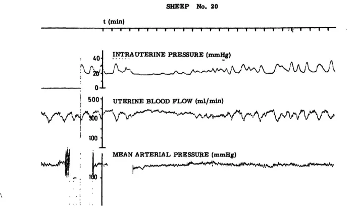
SHEEP NO. 20 t (min)
r 40 Λ Λ '
INTRA UTERINE PRESSURE (mmHg)
500
100
UTERINE BLOOD FLOW (ml/min)
MEAN ARTERIAL PRESSURE (mmHg) .^^
Fig. 1. Simultaneous recording of intrauterine pressure, uterine blood flow and mean arterial pressure in a pregnant sheep. Contraction elicited decreases in uterine blood flow can be seeh clearly.
V. urerina
A. umbilicalU XI V. umbilicaHt
flowmeter A. utcrina dextra
FEKGl Fig. 2. Diagram showing the experimental set up.
J* ^erinat. Med. 5 (1977)
Junge et al., Reduction of uterine blood flow
41 of each experiment using the ewes' own blood
running from the carotid artery.
1.3 The ewes were anaesthezised with pento- barbital (initial dose 20 mg/kg i.V., continued by infusion of 5 mg/kg/h) and Alloferin was given for relaxation. The ewe was then placed on a surgical table in left lateral position. After tracheotomia intubation was performed and Ventilation was maintained by a Starling pump. Electrodes were attached to the maternal ehest for recording maternal ECG and heart rate. Polyethylene tubes were inserted into the jugular vein for administering the anaesthetic and into the brachial artery for recording arterial pressure via a pressure transducer.
The abdominal wall was opened by a right lateral section and the uterine hörn containing the fetus was partially exposed. First of all thin catheters were inserted into branches of the umbilical artery and umbilical vein through small incisions of the uterine wall leaving the amnion intact. Fetal ECG and FHR were recorded via very thin, partially unisolated platinum wires inserted subcutaneously into the fetal back by puncturing the uterine wall and the fetal skin with a long puncture needle.
Another catheter was inserted into the uterine cavity by puncturing its wall for recording in- trauterine pressure. Both uterine arteries were freed from surrounding tissues after Infiltration with local anaesthetics to prevent spasm. Firstly the artery of the gravid hörn was inserted into the cage of the flow meter cup and blood flow was
recorded for some time. The measurement was taken valid when blood flow was stable. Then the flow meter cup was put around the artery of the other side and blood flow was measured in the same way. Both measurements were added and taken äs total uterine blood flow. At last the flow meter cup was put back around the artery of the gravid hörn again and then the abdominal wall was closed. Now the calibration scale of the blood flow channel was corrected according to the initial ratio of flows in both uterine arteries presuming that this ratio would remain constant throughout the course of the experiment.
1.4 Blood samples drawn form the catheters were kept in glass capillaries at low temperature until analyses of pÜ2, pCOa, and pH were performed.
These were done with an Eschweiler appartus.
1.5 Investigationson UBF reduction and fetal response were started at various times after initial surgical preparation depending on our program of severalitemsbeinginvestigated in each preparation.
At that time different degrees of deterioration of the ewes and hence the fetuses were seen but this was in agreement with our experimental intentions.
Repetitive and progressive acute reduction of UBF (approximately 25%, 50% and 100% from basal UBF value) was achieved three times, lasting 120 sec each and each approximatly 12 min apart, by flowmeter controlled partial or total occlusion of the aorta abdominalis of the ewe with a balloon
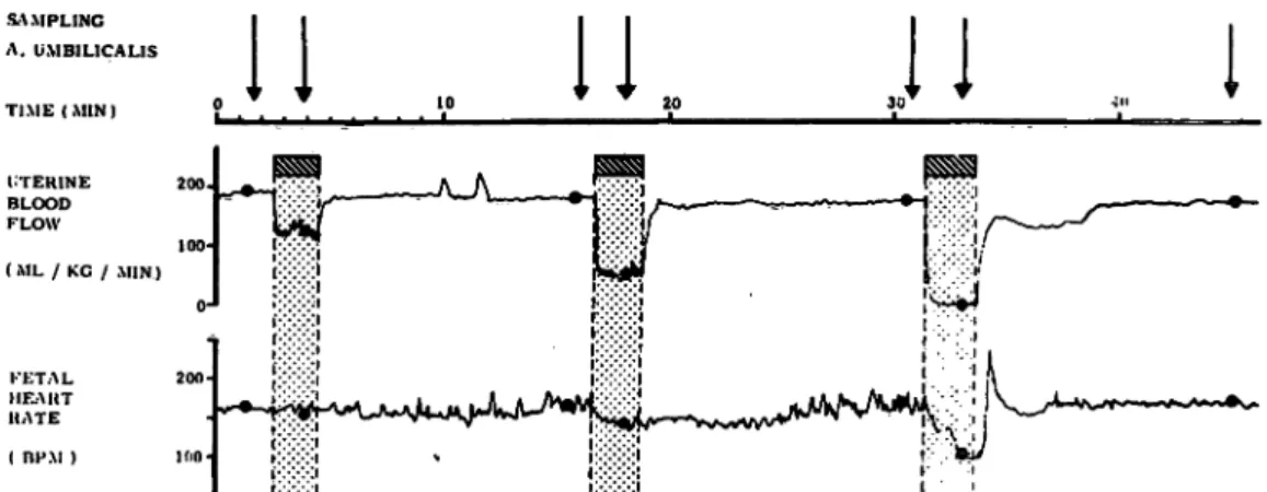
FETAL BLOOD SAMPLING A. ÜMBILICALIS
TIME ( M I N )
ITEHINE BLOOD FLOW ( ML / KG / MIN )
FETAL IIEAHT KATE
I I II .11
Fig. 3. Semischematic diagram showingdata aquisition during repetitive acute flowmeter controlled reduction of uterine blood flow.
Blood sampling was performed discontinuously (arrows). Simultaneous data of UBF and FHR were taken from contm- uous recordings (black dots).
J. Perinat. Med. 5(1977)
catheter inserted into the aorta. (In some cases the lateral abdominal section was left open and the aorta was compressed manually).
Before, 90 sec after the beginning and 10 min after the end of UBF reduction blood samples were taken from the fetal catheters (Fig. 3). In two preparations this sequence was repeated once after complete recovery of FHR.
2. Results
Data of all fetuses and all UBF reduction sequences are given in Table I and Fig. 4.
2. l Data before and after UBF reduction sequence Initial UBF values ranged from 180 to 57 ml/kg/min (Mean 104.0, SD ± 37.5 ml/kg/min). Thus situations withphysiologic avlues of UBF, borderline situations
fr -H-
7.35 7.10 7.29 7.20
FHR ibpml
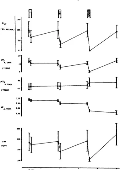
Fig. 4. Mean values of UBF, pO2, pCO2 and pH in the umbilical artery äs well äs FHR for all 7 UBF icduction sequences performed. (Arrows in upper part indicate UBF reduction).
and situations with extremely pathologic UBF values couldbe investigated, which was in agreement with our experimental intentions.
PO
2in the umbilical artery ranged from 18.5 to 6 Torr (Mean 13.3, SD ± 5.1 Torr), indicating moderate to severe fetal hypöxemia. Overall there was a combination of high UBF values with high pO
2values and vice versa, but correlation was not significant r = .42,2 < .1).
PC0
2in the umbilical artery was in the ränge of 37 to 57 Torr (Mean 44.9, SD ± 6,7 Torr). No significant correlation to UBF could be found. PH in the umbilical artery ranged from 7.43 to 7,23 pH units (Mean 7.34, SD ± .06 pH units) and there was no correlation to UBF.
FHR ranged from 130 to 235 bpm (Mean 186.4, SD ± 34 bpm). Thus a wide ränge of fetal cardio- vascular situations was under investigation too.
Except preparation No. 20 there was in inverse relationshipbetweenbasal UBF and FHR (r = - .48, 2 < .05) and an inverse relationship between, basal pO
2and FHR (r = - .46, 2 < .05). Alto- gether low basal UBF was accompanied by fetal hypöxemia and tachycardia (Figs. 5 and 6).
2.2 Ten minutes after the whole UBF reduction sequence mean values for UBF and p0
2were not altered in comparison to initial values, pC0
2was elevated by 9.7 Torr, pH was lowered by .13 pH units. FHR had increased by 48.6 to 225 bpm.
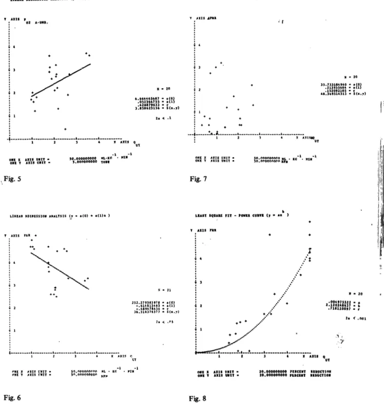
2.3 Relationship between acute reduction of of UBF (AQ
UT) and FHR decrease (AFHR = deceleration amplitude). " · The scattergram of AFHR versus AQ
UTprima vista does not show any relationship (Fig. 7). But transformation of the scattergram by taking AFHR and äs percentage of basal values gives good results (Fig. 8). (100 bpm was defined äs 0 % of basal FHR value because with 100% reduction of UBF heart rate of the fetal lamb decreased to about 100 bpm (Mean 104.2, SD ± 10.5 bpm)). In fact an alinear correlation can be seen. This fact must lead to the conchision that AFHR/AQ
UTis dependent on basal UBF and FHR levels. In order to prove this the AFHR/ A
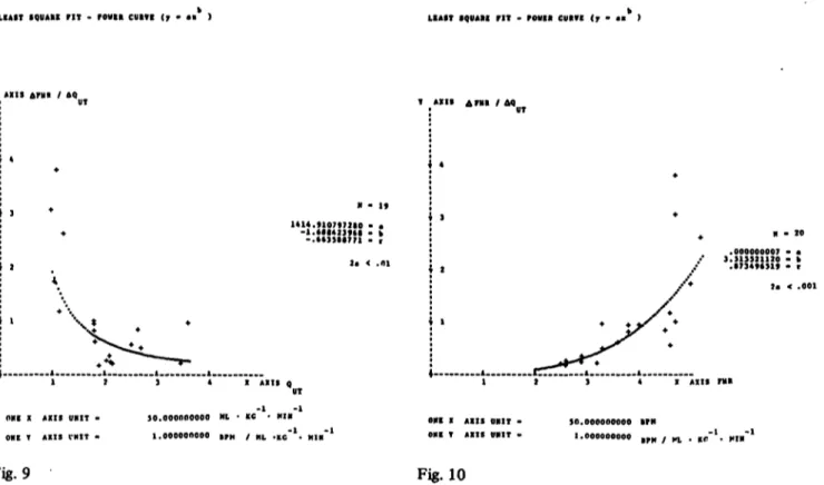
UTwere plotted versus basal UBF (Fig. 9) and basal FHR (Fig. 10).
A significant alinear correlation can be found for both. Relations are inverse because of the inverse
J. Perinat. Med. 5 (1977)
Junge et a!., Rcduction of uterine blood flow 43
a "·' -:
u: «: * * * * ·* * * *
* * ° -f y ? r « * £ 5 - 0 ω * «χ «s - P z·*;
* β*§. * o 0 χ r. IA ^0^ ,: p ^ „ „ w· 0· ^ - - - -
ΈΖ Ο · · . · . ** ΙΑ ΙΑ |Α
•Ο W V 5 C M C * £ £ β ? £ * « « - * » «Ο *Ν Ο ~ < 0 < * · ^
^ JO "" - r - r - ^ ^ ». , . . . * . * .
G S ^ ' C Ι Α Ι Α Ι Λ Ι Α
£ £ Κ £ £ 5 R 5 3 2 2 ; K"** + - ** *~ *· ·* &
ο ν >S l ^ c O ^
I A I I: '
<:
U: ^ ^ Ε? δ | » Κ ί ? ϊ ; 5 ϊ ς ^ & ς 5 κ 5? 55S κ;»
6fe
η1^.| ·|1
S«
Χ CNΟ* «χ
?1
Q« Ρ^^ >*«
αϊ S
s g·
•Γ® £§·
•S c «
Ι
^^-.S
ε ε
*c^§2 s l
5*j ·*·*OQ fli D SSc
|i 3
»-< O
Λ* ^
TS
JJ *J IA IA |A IA
«2 K ϊ\ ϊ> £ \ ^ r % «J " Φ ^ ^1 °^3C*"
1
R c M C M ^ 5 i^ K> <V (V ^ K \ | A i ^ * ° S
eo
C O > O > C M I A ^ ^ T o > f M i A o ^ r * » « · »A *· o»
.*S CM CM CM « * | A | A | Τ > | Τ % < Μ K\ |T\ r> »- »- O»
P* g r - c - t — r — r - r - c-r-r*· Γ*·Γ*·Γ»- t - t * M >
Ό
U O CM O* I * ^ O O O O ^ r C M < « - M 9 l A f\ V\ \O
^ S r - t - r - r ^ e * r - c-e-e»· r- r* e· r * c - r - JD
j.·».
d> Ί O ^ O O O I A O O I A O l A l A I A I A O - • r f i l A C M I A O » O t - l A M > t * Μ ^ Μ ί ^ Τ ^ J ^ S i
**g PCM C M C M ^ C M C M C M C M C M V W C M C M C M C M
£ C I A I A I A O O I A ο ο ο ο ο ο ° ® ί>
* C » > » tw' - M > | A » - 0 « ^ 0 M > « » 0 Γ0^ « Jj^ p CM «· ^» ^ ^ *· C M C M * · * · » · * - CM
•o
A I A O I A 0 0 0 I A O O I A O I A ^ JA » A
«2 OJ IA CM flOCJlO I A I A V O M 5 M 5 M > S j ^ S )
^ C M C M C M » - ^ « C M C M C M C M · - · · * - C M C M C M
5 U> CM C- O O M 3 C M - - I A ^ ^ ^C — —
• « • • C M l A o C & O & C D I A M » M > j T j ^ J ) l A ^ T ^ »
c F s s »
ΌS 0s ^ ° K S ° s s
e5 ? °
J i S S i 5i * S K 5 | P R K S S
§·
5 · _ .« M^ *-«A IA
5 55 R » 5
R
CV 00 t- IA CVI IA CM |A |f\ CVr* M> IA ^r IA ^r
IA |A CM IA IA CM
t* r- r» r- t* r*·
r- ω CM «* r- IA
KN K> KN |A IA |A
r- c- r- r- r- ί-
ο ΙΑ ο ΙΑ ΙΑ Ο IA CM ΙΑ ^ ^ Μ3
t 5 S « S 5
0 0 ΙΑ Ο IA IA
CM φ IA M) IA IA 0 0 0 0 0 0
«- CM O f» M> O M9 IA M> r\
ΙΑ ΟΙ Φ ΙΑ Μ> «Α
<* 0 0 0 0 0
J Perinat. Med. 5 (1977)
LINEAR REGRESSION ANALT81S (y · ·<0> * «U>· ) LINEAR REGRESSION ANALTSIS (y - «(0) * ·<!)« )
.4
ONE X AXIS UIIIT · ONE T AXXS UMIT -
50.000000000 ML-KP 5.000000000 TOBt
N - 20 6.664465687 · «(0)
.052366755 - ·<!) .428879633 · r 3.858623156 - S(x.y)
2· < .1
·--
~JT ATIS Q UT
-"*. MIN*1
• f
4
3 +
+ +
1 +
+
* + -Μ-
Ι 2 1 4 X AXISAQ UT
ΟΝΕ χ AXIS ΐ'Νΐτ · so.pnoooonfio «n. . rc~l. Mt«~l
ONE Y AXIS NIT - SO.OPOOOnocn gfa' KC MI"
35.733184940 - a(0) .213955684 · «(l) .152092105 · r 48.369514313 · S ( K . V )
, Fig. 5 Fig. 7
LIKEAX RECRESSION AHALTSIS (y - l(0) * ·(!)« ) LIAST SQOARE FIT · POVER CURTI (» · ·« >
T AXIS FHI
2 5 2 . 2 7 9 5 6 3 9 7 8 · « ( 0 ) -.6146134*3 · .(1) ..489478434 · r 36.316376377 · S ( B . V )
tT HL · Kr. · PIN-l
/t I
M · 20
718110097 Γ r 2· < .noi
-1 !.-•7
4 X AXXS o UT ONE X AXIS UNXT -
ONI T AXXS UNIT · 20.000000000 PERCEMT REDUCTION 20.000000000 PtRCIMT REDUCTXO·
Fig. 6 Fig. 8
Fig. 5. Relationship between basal pO2 in the umbilical artery and basal UBF (Qui) Pri°r to each UBF reduction.
Fig. 6. Relationship between basal FHR and basal UBF (QuiO Prior to eacl1 UBF reduction.
Fig. 7. Scattergram of values of AFHR versus ΔΟυχ. No relationship for ungrouped values.
Fig. 8. Significant alinear correlation between AFHR and ΔΟυτ» both taken s percentage of basal values.
(100 bpm was defined s 0% of basal FHR value, because with 100% UBF reduction the lowest FHR seen was about 100 bpm).
Junge et al., Reduction of uterine blood flow
Junge et al., Reduction of uterine blood flow
45
LKAfT SQUA» ΠΤ - rOWBft CUIVt (7 - ·" ) HAST S9VAKC ΠΤ · POVI« CVIVE (r · ·· )
T AXIS Am / AQ
1414.9107*7110 . «
-l.«St423fiS · k M - 20
.000000007 - ·
i....
ONE X AXIS UBIT ONE T AXIS VNIT <
X AXIS Q
3.513321120 · k .•7349*319 · t
*· < .001
-l UT
•l
X AXIS ΠΙ!
30.000000000 Nt · KC - M I M 0» X AXIS OHIT . ONE T AXIS VNIT
SO. 000000000 BM / n, · *r
Fig. 9 Fig. 10
Fig. 9. Significant alinear correlation between AFHR/AQux ratio and basal UBF (Ουτ)· Deceleration amplitude per unit of UBF decrease rises sharply when basal UBF is below 100 ml/kg/min.
Fig. 10 Significant alinear conelation between AFHR/AQuj ratio and basal FHR. Deceleration amplitude per unit of UBF decrease rises sharply when basal FHR is elevated to its maximum.
relationship between basal UBF and basal FHR (see above).
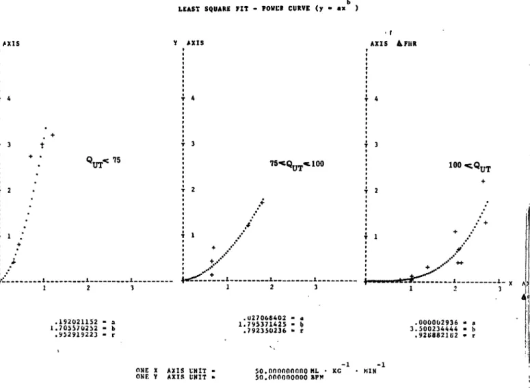
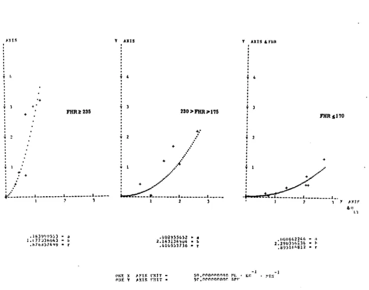
Reevaluation of the scattergram of AFHR versus AQtjx anew by grouping data according to normal, prepathologic and pathologic basal levels of QUT (Fig. 11) and FHR (Fig. 12) gives sense now. Using the least square fit power curve (y == ax
b) statisti- cally Significant alinear regression lines for the Physiologie r nge of basal UBF (Qut > 100 ml/
kg/min) and the physiologic r nge of basal FHR (FHR < 170 bpm) may be fitted. For the inter- mediate and pathologic ranges alinearity is not statistically Significant butbecause of the alinearity in the percentage regression (see Fig. 6) which includes all data, alinear regressions for the inter- mediate and pathologic ranges were calculated too.
From this the following concl sions may bevalid:
Within physiologic levels of UBF and FHR minor and moderate acute reductions of UBF do not affect FHR at all or lead to minor decelerations.
Only a substantial reduction wfll cause decelerations of larger amplitude. But with decreasing basal UBF levels (and increasing FHR levels) the same absolute amount of UBF reduction leads to increasing deceleration amplitudes.
2.4 It is a widely accepted opinion that FHR decelerations caused by acute UBF reduction are mediated by a fall in fetal p0
2via vagal Stimulation and/or direct depression of the heart. Therefore it seemed interesting to evaluate relations between UBF and pO
2alterations s well s the relations between pO
2and FHR alterations.
2.4.1 Relationship between acute reduction of UBF (AQux) and change of p0
2in the umbilical artery (Δρ0
2) during moderate to severe fetal hypoxemia.
Ninety seconds after the Start of acute UBF decrease p0
2in the umbilical artery had fallen (Range 0 to 13 Torr, Mean 6.45, SD ± 4.5 Torr). A statistically
J. Perinat. Med. 5(1977)
LEAST SQUARE FIT - POWCR CURVE (y -
Υ A X 1 S
τ 3 t
τ 2
Υ A X I S
τ Α
τ 3
τ 2
τ l
AXIS &FHR
75·<ΟΤΤ^^100
.1 1 1 1 2 3
100
1.
2
.192021152 » a 1.70557 2!>2 · b .952919223 - r
.027006402 · a 1.795371425 - b
.792350236 - r .000002936 - a
3.500234444 · b .92 8821U2 - r
ONE X
ΟΝΕ Υ A X I S UNIT
AXIS UNIT ϋο.οηοηοοηηο ML · KG
50.000000000 RPM MIN-l
Fig. 11. Relationship between absolute values of AFHR and AQuT f°r Physiologie (graph on the right), prepathologic (graph in the center) and pathologic (graph on the left) levels of basal UBF. Significant alinear correlation for AFHR versus AQuj and basal QUT > 100 ml/kg/min. Alinearity not significant for basal Qux < 100 ml/kg/min, b t the slope of regression is rising with falling basal QUT·
highly significant linear correlation exists between decrease of UBF and decrease of p0
2(r = ·
80>
2α < .001) (Fig. 13). The regression may be approximated by ΔρΟ
2= .1 ΔΟυτ, i-e. a UBF reduction of SO ml/kg/min will on an average lead to a p0
2decrease of 5 Torr.
2.4.2 Relationship between acute fall of p0
2in the umbilical artery (Δρ0
2) and FHR decrease (ΔΡΗΚ), the deceleration amplitude.
Correlation between AQ
UTand ΔρΟ
2being linear and highly significant the same relation between ΔΡΗΚ and Δρ0
2should be expected s between ΔΡΗΚ and Δ0
υτ. This holds true in general: a significant correlation between the ΔΡΗΚ/ΔρΟ
2ratio on basal FHR can be seen, but the ΔΡΗΚ/
ΔρΟ
2ratio is not well correlated to baSal pQ
f] 2and alinearity for the regression of AFHR on Δρ0
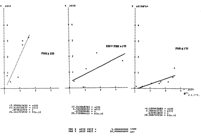
2in the normal FHR r nge could not be verified statistically. A linear regression was found signifi- cant for this r nge s well s for the r nge of severe tachycardia. For moderate tachycardia no signifi- cant correlation could be verified (Figs. 14—17).
2.5 Lag time of UBF reduction induced FHR fall A histogram of lag times between .the onset of UBF reduction and onset of FHR fall is shown in Fig. 18. It can be seen that the lag time was less than 11 sec in about 40% and less than 21 sec in about 80%. A correlation between basal FHR level and lag time could not be verified.
J. Perinat. Med. S (1977)
Junge et a)., Reduction of uterine blood flow 47
LtAST SQUARE FIT - POWER CURVE (y
Υ A X T S Υ A X I S
FHR 2 235
Υ A X I S 4 KUR
230>FHR>175
FHR $170
Α Χ Ι Γ
*<>
l . ( 7 7 J 3 f t U 4 3 - k.h/i>d574«*V « r .0029556*2 · α 2.143134*04 - l.
.610553736 · r .UGU6G2246 · ,1
2.29U3Vi.23t - h .893Η·('812 · r
ONF. X AX1S ΓΝΙΤ -
P:JF. Y AXIS ΓΜΙΤ - sn.roononoor. ML · KC SC.noopnnoor r.rr
Fig. 12. Relationship between absolute values of AFHR and
regression is rising with rising level of basal FHR. for various levels of basal FHR. The slope of
3. Discussion
All obstetricians practicing FHR monitoring are confronted with the problems of correct Inter- pretation of FHR recordings. A late deceleration pattern is commonly taken s a sign of developing fetal distress and severeness of decelerations and fetal distress is judged in part from deceleration amplitudes. But there is little Information about factors influencing deceleration amplitude. ASSALI andcoworkers [3] publisheddataon the relationship between reduction of UBF and FHR decrease. Our present data give additional knowledge concerning the influence of basal UBF and basal FHR and the role of the UBF reduction induced fall of fetal p02, which is supposed to mediate the fall of FHR.
First of all a discussion of the time courses of UBF, pO2 and pH in the umbilical artery and of FHR in connection with repetitive acute reduction of aortic flow and hence UBF is worth while (Fig. 4).
3.1 UBF dropped rapidly on reducing blood flow in the lower aorta of the ewe. On deflation of the obstructing balloon catheter UBF rose s rapidly again, but on 50% to 100% reduction this rise was sometimes followed by a slight rebound fall that gradually faded out. This phenomenon will be discussed on below.
3.2 Mean p02 in the umbilical artery - initially in the moderate to severe hypoxemic level — feil rapidly and proportionally to the amount of UBF
J. Perinat. Med. 5(1977)
LIMEAI REGRESSION ANALTS IS («'. .(0) 4- ·(!)« )
T AXIS AP 02 A.UM·.
ONE X AXIS UMIT - ONE T AXIS CNIT -
Fig. 13
LIAST SQUAR· PIT - POWIR CUR« <7 - ·« >
T AXIS ΔΡΜ* / Δ
N · 20 .1542362·« · a(O) .09932*432 · ·(!) .009«19319 · r 2.305265124 . S(*.y)
-l -l 50.000000000 ML · KC · HIN
5)000000000 TORR
02 A.UM··
.00000025» · · 3.275962721 · b .•72357*94 - r
0» X AXIS UMIT ·
ONE T AXIS UMIT - 50.000000000 RPK 10.000000000 BPH / TOtll
Fig. 15
LINEAR REGRESSION ANALTS IS (j - e(0) -*· ·<!)» ) LIAST SQDAU PIT - POWER CD·?· (y - ·« )
T AXIS F R
9.457663905 · «(0) .009333006 · ·(!) .75451344· · r 24.110100396 - S(x.y)
T AXES APUR / ΔΡ
134.225448993 · « -1.221126483 - h -.426573447 · r
20.000000000 PERCEMT REDUCTION
20.000000000 FERCENT REDUCTION ONE X AXIS UNIT ·
ONE T AXIS UMIT · 5.000000000 TOVR 10.000000000 IPH / TORR
Fig. 14 Fig. 16
Fig. 13. Significant linear correlation between absolute values of ΔρΟ2 in the umbilical artery and AQuj during modeiate to severe hypoxemia. (Basal p 2 < 20 Torr).
Fig. 14. Significant conelation between AFHR and ΔρΟ2 in the umbilical artery, both taken s percentage of basal values. (See also legend to Fig. 8).
Fig. 15. Significant correlation between ΔΡΗΚ/ΔρΟ2 ratio and basal FHR. Deceleration amplitude per unit of pO2
decrease rises with rising basal FHR.
Fig. 16. Relationship between AFHR/ApO2 ratio and basal pO2. Correlation nearly Significant. Deceleration amplitude per unit of pO2 decrease rises with falling basal pO2.
J. Perinat. Med. S (1977)
Junge et a L, Reduction of uterine blood flow 49
LINKA» EORESSIOH ANALYS1S <y · «(0) 4 .(l), )
FHR > 235
Υ ΑΧΙ5ΑΓΗ0
230>FHR>H5
.11.i
FHR* 170
A·'"
-7.V940t2b39 - a( ) 21.4:>32:»4437 · a ( l )
. 8 7 5 6 5 2 7 4 9 · r 2 b . « . 0 1 7 4 9 0 3 9 · S ( x . v )
17.71>306β783 - a< 0 ) 4 . U 2 3 2 8 423 · « ( 1 )
.073030484 - r 29.75200&513 · S ( x . y )
-4,050443880 · a ( 0 ) 3.509511731 - ·(!)
.1*32911849 · r 10.288797290 · S ( x . v )
ONF. X A X I S ΓΚΙΤ -
ΟΝΕ Υ AXIS l'NIT - s.nnnonnnnn TORR 5o.onoonnoon Hp*.
Fig. 17. Relationship between absolute values of AFHR and ΔρΟ2 for variouslevels of basal FHR. The slope of regression is rising with rising level of basal FHR.
10 20
Fig. 18. Histogram of lag times between onset of UBF reduction and onset of FHR deceleration.
reduction. Tenmin after the end of each reduction, but presumably much earlier mean p0
2was in the initial r nge again. That means changes occur with a short time constant. Publications on continuous monitoring of p0
2support this fact [14, 16, 24, 29, 30] though absolute data for the time constant are missing.
3.3 Mean pH decreased proportionally during reduction of UBF. In contrast to p0
210 min after the end of the reduction phase the fetus had not recovered in respect to pH. In fact pH remained constantiy lowered oreven a slight further decrease could be seen. Our explanation for this pheno- menon is the following:
Hypoxemia from UBF reduction leads to fetal cardiovascular reaction with redistribution of fetal
J. Perinat. Med. 5 (1977)
circulation [4, 7, 10, 11, 12, 20, 25]. The initial rapid fall of pH during UBF reduction may be caused by hypercapnia and by an immediate lactat input from regions of fetal circulation with un- changed perfusion during hypoxemic fetal cardio- vascular reaction. This component of lactat input has a short time constant and gives rise to the initial rapid pH fall. The secondary decrease of pH after cessation of UBF reduction may be the resultant of a delayed lactat input from regions of fetal circulation with diminished perfusion during hypoxemic redistribution, when these regions are reopened again on one side and overall lactat elimination on the other side.
Different time courses in the restitution from hypoxemia and acidosis after contraction elicited UBF reduction may explain well known findings taken from the dilatation phase of labors, for which a slight progressive fall of mean pH could be found despite a seemingly constant normal mean p0
2, especially when blood sampling is performed in the late phase of uterine relaxation. On the other hand, in the expulsion period a pO
2fall along with a further fall of pH is seen [5, 13, 26, 34] despite of the short p0
2time constant, because contractions become stronger and relaxation phase shortens.
3.4 Several items have to be discussed in regard to FHR changes. (It may be necessary to point out the fact again, that there was moderate to severe hypoxemia during the experimental sequences).
3.4.1 Mean FHR changes (Fig. 4) resembled the changes of UBF and pO
2, but 10 min after the end of the UBF reduction sequence mean basal FHR was slightly elevated and the explanation may be compensatory tachycardia.
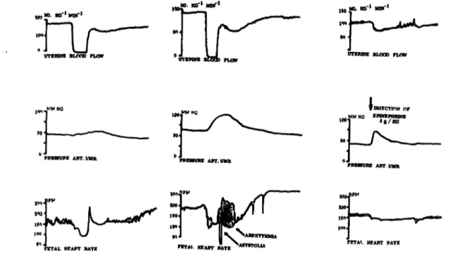
3.4.2 The slopes of FHR descents and ascents varied. In general changes were more rapid when basal FHR was high and when UBF reduction was about 50% to 100%. On several occasions after UBF reduction of larger amount a secondary deceleration could be seen together with the secondary fall of UBF mentioned above (see Figs. 3 and Figs. 19). A biphasic deceleration like this by the way quite often can be seen in clinical FHR monitoring. The reason for this phenomenon is not quite clear. It might be a simultaneous rise in fetal blood pressure because injection of
epinephrine directly into the fetal buttock elicited a blood pressure rise and UBF decrease äs well äs FHR fall (Fig. 19, right ccflumn).
3.4.3 Evaluation of relations between UBF reduction and FHR decrease (Fig. 7) was dis- appointing at first glance. The key to understanding was the fact that there is an alinear correlation between relative values only when 100 bpm (the lowestheartrateseen with complete UBF reduction) is taken äs 0 %. And this correlation of relative values points to the fact that there is a dependency from basal UBF and FHR values. This dependency can be seen clearly in Figs. 9 and 10.
According to others and own results [17, 18, 22]
the borderline to pathologic uterine blood flow near term seems to be in the ränge of about 100 ml/kg/min in various species. In fact, uterine 0
2uptake is reduced when UBf falls below 80- 100 ml/kg/min. These findings are paralleled by our results in so far äs the AFHR/AQyx ratio is constantly low and independent of basal UBF and basal FHR äs long äs basal UBF is above this borderline and that this ratio increases sharply when UBF falls well below it. The same holds true for basal FHR but in the opposite direction because of the inverse relationship between basal UBF and basal FHR: there is a definite increase of the AFHR/AQ
UTratio when basal FHR is elevated to its maximum.
Figs. 11 and 12 give further Information. Looking at the graphs for physiologic ranges of UBF and FHR (the graphs on the right sides) separately, it can be seen that in this ränge AFHR/AQux *
s afunction of AQ
UTitself: UBF reduction up 50 ml/
kg/min does not affect FHR at all. Only submaximal to complete reduction leads to minor decelerations.
On the other hand, with falling UBF levels (and increasing FHR levels) the sole functional influence of AQux is diminishing and the influence of basal UBF and basal FHR is getting into play (Figs. 11 and 12, graphs in the center and on the left).
3.4.4 Evaluation of our data verified a highly sig- niflcant correlation between p0
2decrease and UBF reduction for basal p0
2< 20 Torr (Fig. 13).
From this it is not surprising that the relationship between AFHR and 0
2to. a large extent
Js Perinat. Med. 5 (1977)
Junge et al., Reduction of uterine blood flow
51
1β. «Γ1 Huf1
UTCMHB BLOTD FLOW UTOtOfE BLOCK)
' PFB88UFE ART. UMB.
f DUECTmN (
BI/KD
' PftHSUKE ART UMB.
250 213 ISO
FETAL HEAPT RATE «TAL REART RATE FTTAT. HEART PATE
Fig. 19. Recordings of UBF (above), umbilical arterial piessure (middle) and FHR (below). Reaction of pressure and FHR on acute reduction of UBF.
A secondary fall of FHR and UBF after the end of acute UBF reduction can be seen simultaneusly with the rise in aiterial pressure. (Graphsontheleft and in the center). The latter may be the cause of secondary UBF and FHR reaction.
Rise of arterial pressure after injection of epinephrine direct into the fetal buttock is accompanied by a fall in UBF and FHR. (Graph on the right).
resembles the relationship of AFHR and
Statistics though give poorer results (Figs. 14-17).
This may be explained by the short time constant of pO
2giving rise to more overall Variation in p0
2data. On the other hand one has t take in mind that progressive pH decrease caused a shift in the oxygen dissociation graph during the course of the experiment.
3.4.5 Our present datalead to the conclusion that no single parameter being analysed is responsible for FHR alterations solely. FHR response, i.e.
deceleration amplitude is modified by basal values of UBF, fetal pO
2and FHR and by the amount and slope of UBF reduction and consecutive pO
2decrease. Thus the most extensive deceleration amplitudes were seen when all parameters were adversely altered. That the state of fetal oxygenation
and its acute changes have a major influence on the shape of FHR alterations has been shown by others [21, 27], On the other hand one may argue that the level of basal FHR modifies deceleration amplitude too haging in mind WILDER'S "Law of Initial Value" [31, 32], which in parallel has been adopted for defining heart rate regulation in the newborn[19,28].
3.4.6 The lag time between the onset of UBF reduction and the onset of FHR deceleration turned out to be less than 11 sec in about 40% and less than 21 sec in 80% (Fig. 18). Although our results are taken from fetuses of ewes ingeneralanaesthesia, they may hint to the fact that in severe hypoxemia the lag time may shorten. In fact a significant shortening of lag time with decrease of fetal arterial oxygen Saturation and oxygen tension has been reported[21].
J. Perinat. Med. 5(1977) 4*
Summary
Five merino sheep near tenn were used for acute prepara- tions to investigate the influence of basal uterine blood flow (UBF), basal fetal oxygenation and basal fetal heart rate (FHR) on FHR changes elicited by acute reduction ofUBF.
The ewes were anaesthesized with pentobarbital and Alloferin was given for relaxation. Ventilation was main- tained via a tracheal tube by a Starling pump. Maternal heart rate, arterial pressure and intrauterine pressure äs well äs fetal heart rate and umbilical artery pressure were recorded continously. Initially blood flow was recorded by cuff flow meter sequentially in both uterine arteries for a short time in order to determine total UBF and the ratio of flows. During the UBF reduction sequence flow was recorded in the artery of the gravid hörn continously and measurement was corrected according to the initial ratio of flows in both arteries, presuming that this ratio would remain constant throughout the course of the experiment.
Repetitive and progressive acute reduction of UBF (approximately 25%, 50% and 100% from basal UBF value) was achieved three times, lasting 120 sec each and each approximatly 12min apart, by flow meter controlled partial or total occlusion of the aorta abdominalis of the ewe with a balloon catheter inserted into the aorta. (In some cases the lateral abdominal section was left open and the aorta was compressed manually).
Before, 90 sec after the beginning and 10 min after the end of UBF reduction blood samples were taken from the fetal catheters (Fig. 3). In two preparations this sequence was repeated once after complete recovery of FHR.
Results
Initial UBF values ranged from 180 to 57 ml/kg/min, pO2 in the umbilical artery was 18.5 to 6 Torr, pCO2 was 37 to 57 Torr and pH 7.43 to 7.23 pH units. Basal FHR ranged from 130 to 235 bpm. Thus wide ranges of UBF äs well äs fetal cardiovascular and biochemical situations were under investigation. Altogether low basal UBF was accompanied by fetal hypoxemia and tachycardia. The time courses of UBF, pO2, pCC>2 and pH in the umbilical
artery and of FHR in connection with repetitive acute reduction of aortic flow were the following:
UBF dropped rapidly on recjucing aortic blood flow. On release of the aortic obstruciion it rose äs rapidly again, but'on 50% to 100% reduction this rise was sometimes followed by a slight rebound fall that gradually £aded out.
Mean pO2 in the umbilical artery, initially in the moderate to severe hypoxemic level, feil rapidly and proportionally to the amount of UBF reduction. Ten minutes after the end of each reduction, but presumably much earlier mean pO2 was in the initial ränge again. Mean pH decreased proportionally during reduction of UBF. In contrast to pO2 10 min after the end of the reduction phase the fetus had not recovered in respect to pH. In fact, pH remained lowered or even a slight further decrease could be seen.
Mean FHR changes resembled the changes of UBF and pO2, but 10 min after the whole UBF reduction seqüeiice mean basal FHR was slightly elevated.
As to the relationship between acute reduction of UBF (AQux) and FHR decrease (AFHR) an alinear correlation between the values, expressed äs percentage of basal level, could be verified. This fact leads to the conclusion that the AFHR/AQux ratio is dependent on basal values of UBF and FHR: statistics reveal that within Physiologie levels of UBF and FHR minor and moderate acute UBF reductions do not affect FHR at all or lead to minor deceleiations. Only substantial reductions will cause decelerations of larger amplitude. But with decreasing basal UBF levels '(and increasing FHR levels) the same absolute amount of UBF reduction leads to increasing deceleration amplitudes.
As it is widely accepted thatJFHR decelerations caused by acute UBF reduction are mediated by a fall in fetal pO2, the relations between acute UBF reduction and pO2 fall in the umbilical artery were investigated and a highly j significant linear correlation could be found. Therefore the same relations between AFHR and 2 should be expected äs between AFHR and AQuf. This holds true in general but statistics give poorer results, probably because the short time constant of pO2 gives rise to more overall Variation in pO2 data. On the other hand one has to take inmind that progressive pH decrease caused a shift in the oxygen dissociation graph during the course of the experiment. r
The lag time between oriset of UBF reduction and onset of FHR deceleration was less than 11 sec in about 40%
and less than 21 sec in
Keywords: Acidosis, animal experiments, cardiotocography, deceleration, heart rate, fetus, hypoxemia, lag time, uterine blood flow
Zusammenfassung
Akute Verminderung der Uterusdurchblutung und Ver- änderungen der fetalen Herzfrequenz beim Schaf am Ende der Tragzeit.
Es sollte der Einfluß der basalen Uterusdurchblutung, der basalen fetalen Oxygenation und der basalen fetalen Herzfrequenz auf die durch akute Drosselung der Uterus- durchblutung hervorgerufenen fetalen Herzfrequenzver- änderungen untersucht werden. Dazu wurden 5 hoch-
trächtige Merinoschafe mit Pentobarbital narkotisiert und mit Alloferin relaxiert. Die Ventilation wurde nach Tracheotomie und Intubation mit einer Starling-Pumpe aufrechterhalten. Die materne Herzfrequenz, der materne arterielle Druck, der Intrauterindruck, die fetale Herz- frequenz, der Druck in der Arteria umbilicalis wurden kontinuierlich registriert. Vor der eigentlichen Versuchs- durchführung wurde die Durchblutung mit einem Cuff- J. Perinat. Med. 5 (1977)
Junge et al, Reduction of uterine blood flow 53
flowmeter hintereinander kurzzeitig in beiden Uterin- arterien gemessen und die Gesamtuterusduichblutung sowie das Verhältnis beider bestimmt. Während des Drosselungsversuches wurde die Durchblutung auf der Seite des graviden Horns kontinuierlich registriert und die Eichskala entsprechend dem Durchblutungsverhältnis in beiden Arterien auf die Gesamtdurchblutung korrigiert.
Eine wiederholte und jeweils zunehmende Drosselung der Uterusdurchblutung (nährungsweise 25%, 50% und 100%
vom Ausgangswert der Durchblutung) von jeweils 120 sec Dauer und etwa im Abstand von 12 min wurde durch partielle oder totale Verlegung der Aorta abdominalis des Muttertieres mit einem Ballonkatheter erreicht. (In einigen Fällen blieb die Laparotomiewunde offen und die Aorta wurde manuell komprimiert). Vor, 90 sec nach Beginn und 10 min nach Ende der Durchblutungsdrosselung wurden Blutproben zur Bestimmung von PO2, PCO2 und pH aus dem in einem Ast der Arteria umbilicalis liegenden Katheter entnommen. Bei 2 Schafen wurde die Drosse- lungssequenz nach Stabilisierung der fetalen Herzfrequenz wiederholt.
Ergebnisse
Die Ausgangswerte für die Uterusdurchblutung lagen zwischen 180 und 57 ml/kg/min, der pO2 in der Arteria umbilicalis wurde mit 18,5 bis 6 Torr, der pCO2 mit 37 bis 57 Torr und pH mit 7,43 bis 7,23 pH-Einheiten be- stimmt. Das fetale basale Herzfrequenzniveau lag zwischen 130 und 235 Spm. Damit kamen also weite Bereiche basa- ler Uterusdurchblutung und fetaler kardiovaskulärer und biochemischer Situationen zur Auswertung. Insgesamt fand sich bei erniedrigter Uterusausgangsdurchblutung eine Tendenz zu fetaler Hypoxämie und Tachykardie.
Die Zeitgänge von Uterusdurchblutung, pO2, pCO2 und pH in der Arteria umbilicalis sowie der fetalen Herzfrequenz bei akuter und wiederholter Aortendurchblutungsdrosse- lung waren folgende: es kam zum steilen Abfall der Uterus- durchblutung. Nach Beendigung der Aortenkompression stieg die Uterusdurchblutung ebenso steil wieder an. Bei 50 bis 100%iger Drosselung jedoch folgte diesem Wieder- anstieg gelegentlich ein leichter Wiederabfajl, der dann langsam abklang. DerpO2-Mittelwert in der Arteria umbili- calis, schon vor der Drosselungssequenz im Bereich mittle- rer bis schwerer Hypoxämie, fiel steil und proportional zum Ausmaß der Uterusdurchblutungsdrosselung. 10 min nach Ende jeder Drosselung, wahrscheinlich aber viel
früher, fand sich der pO2-Mittelwert wieder im Ausgangs- bereich. Der pH-Mittelwert fiel während der Drosselung proportional ab. Im Gegensatz zum pO2 hatte sich der Fet 10 min nach Ende der Drosselung in Bezug auf den pH-Wert aber noch nicht erholt: der pH-Wert blieb viel- mehr erniedrigt, bzw. es wurde sogar ein weiterer pH- Abfall beobachtet. Der Mittelwert der fetalen Herzfre- quenz verhielt sich analog den Veränderungen der Uterus- durchblutung und dem pO2, allerdings fand sich 10 min nach Ende der Drosselungssequenz eine leichte Frequenz- erhöhung.
Es gelang der Nachweis einer alinearen Korrelation zwischen den prozentualen Werten der akuten Uterus- durchblutungsdrosselung und der Herzfrequenzdezele- ration. Das deutet darauf hin, daß das Verhältnis AFHR/
AQux von den Ausgangswerten der Uterusdurchblutung und der fetalen Herzfrequenz abhängig ist. Die weitere statistische Aufarbeitung zeigt, daß im Bereich physiolo- gischer Uterusdurchblutung und fetaler Herzfrequenz leichte und mittlere Drosselungen der Uterusdurchblutung die Herzfrequenz nicht beeinflussen, allenfalls minimale Dezelerationen hervorrufen und nur erhebliche Durch- blutungsdrosselungen zu Dezelerationen größerer Ampli- tude fuhren. Mit sinkender Ausgangsuterusdurchblutung (oder steigender fetaler Herzfrequenz) erzeugt die gleiche absolute Durchblutungsdrosselung jedoch Dezelerationen zunehmender Amplitude.
Weil angenommen wird, daß die durch Uterusdurch- blutungsdrosselung bedingten Dezelerationen durch einen pO2-Abfall hervorgerufen werden, wurden auch die Beziehungen zwischen der Uterusdurchblutungs- drosselung und dem pO2-Abfall in der Arteria umbilicalis untersucht. Dabei fand sich ein hoch signifikant linearer Zusammenhang. Deshalb sollten zwischen dem pO2-Ab- fall und der Dezelerationsamplitude die selben quantita- tiven Beziehungen bestehen wie zwischen der Uterus- durchblutungsdrosselung und der Dezelerationsamplitude.
Das ließ sich im vorliegenden Datenmaterial in der Tendenz auch nachweisen, doch waren die statistischen Ergebnisse nicht so eindeutig. Möglicherweise führt die kurze Zeit- konstante des pO2 zu mehr Variationen bei den pO2- Daten. Andererseits muß bedacht werden, daß der zu- nehmende pH-Abfall während der Dauer des Experiments eine Verschiebung der O2-Dissoziations-Kurve erzeugte.
Die Verzögerungszeit zwischen dem Beginn der Uterus- durchblutungsdrosselung und dem Beginn der Dezeleration lag in etwa 40% unter 11 sec und in 80% unter 21 sec.
Schlüsselwörter: Azidose, CTG, Dezeleration, Durchblutung (uterim), Fet, Herzfrequenz (fetale), Hypoxämie, Tierver- such, Verzögerungszeit.
Resume
Jugulation de la ciiculation du sang dahs l'uterus et alterations du rhythme cardiaque foetal chez les biebis enceintes approchant du terme
L'article present traite de l'influence de la circulation sanguine uterine de base, de Foxygenation et de la frequence cardiaque basales du foetus sur les alterations du rhythme cardiaque foetal resultant d'une Jugulation
aigue de la circulation uterine. A cet effet, on a administre a 5 brebis merinos en stade avance de grossesse une naicose de pentobarbital et un relaxant d'alloferine. La Ventilation a ete maintenue apres tracheotomie et intubation avec une pompe Starling. La frequence cardiaque et la pression arterielle de la mere, la pression intrauterine, la frequence cardiaque du foetus, la pression dans Tariere ombilicale J. Perinat. Med. 5 (1977)
ont ete enregistrees de fagon continue. Avant de proceder a Fexamen proprement dit, on a utilise un Cuff-flow meter pour mesurer a biefs intervaUes la circulation sanguine dans les deux älteres uterines, ce qui a permis d'observer Fensemble de la circulation uterine et le rapport entre la circulation des deux arteres. Durant toute la sequence de jugulation on a enregistre de fa^on continue la circulation dans l'artere de la corne gravide et corrige les mesures en accord avec le rapport initial entre la circulation des deux arteres, ce rapport etant suppose constant tout au long de Fexperimentation. Une jugulation aigue de la circulation sanguine dans Futerus, progressive et repetee trois fois (environ 25%, 50% et 100% de la valeur de base de la circulation uterin^), d'une duree respective de 120 sec. et a intervalles approximatifs de 12 min', a ete effectuee par occlusion partielle ou totale de l'aorte abdominale de la femelle mere a Faide d'un catheter a ballon. (Dans quelques cas, l'ouverture de la la parotomie a ete maintenue beante pour permettrela compression manuelle de Faorte). Avant la jugulation sanguine, 90 sec. apres son debut et 10 min.
apres sä fin, du sang a ete preleve dans le catheter place dans le catheter place dans une branche de Tariere ombilicale pour definir le pO2, le pCO2 et le pH. Chez 2 ,brebis, la sequence de jugulation a ete repetee apres stabilisation complete de la frequence cardiaque du foetus.
Resultats:
Les valeurs initiales de la circulation sanguine uterine se sont situees entre 180 et 57 ml/kg/min; le pO2 de l'artere ombilicale a varie entre 18,5 et 6 Torr, le pCO2 entre 37 et 57 Torr et le pH entre 7,43 et 7,23 unites pH. Le niveau initial de frequence cardiaque du foetus se situa entre 130 et 235 bpm (battement/min). C'est ainsi qu'on a pu evaluer egalement des problemes assez vastes de la circulation sanguine uterine basale et des situations caidio-vasculaires et biochimiques chez le foetus. Dans Fensemble. on observa une tendance a Fhypoxemie et tachycardie foetales dans les cas (Tun niveau bas de circulation uterine initiale.
Les temps de circulation sanguine uterine, de pO2, pCO2
et pH dans Fartere ombilicale ainsi que de la frequence cardiaque foetale en cas de jugulation aigue et repetee de 'la circulation aortique ont ete les suivants: baisse rapide de la circulation uterine qui a remonte aussi rapidement une fois cessee la compression aortique. Mais dans les jugulations de 50 a 100%, cette remontee a ete oc- casionnellement suivie d'une legere rechute qui s'attenua peu a peu. La moyenne du pO2 dans härtere ombilicale, qui se situait initialement au niveau d'hypoxemie moderee a grave, baissa brusquement et de fagon proportionnelle a
rimportance de la jugulation de la circulation uterine.
10 min apres chaque jugulation, mais probablement beaucoup plus tot, la moyenne du pO2 retrouvait a peu pres son niveau de depart.
La moyenne du pH baissa de fagon proportionnelle pendant la jugulation. En ce qui concerne le pH et au contraire du pO2, le foetus n'agait pas encore recupere 10 min apres l'arret de la jugulation: en effet, la valeur du pH resta plutot basse et subit meme occasionnellement une nouvelle et legere chute. La moyenne de la frequence cardiaque du foetus se comporta de fa$on analogue aux modifications de la circulation uterine et au pO2; mais on observa une legere hausse de la frequence initiale moyenne 10 min apres la fin de la sequence de jugulation.
On a pu verifier une correlation alineaire entre les valeurs exprimees en pourcentage du niveau initial relatives a la jugulation aigue de la circulation uterine (4QrjT) et * la
deceleration de la frequence cardiaque (AFHR). D'oü on peut conclure que le rapport AFHR/AQut depend des valeurs initiales de la circulation sanguine uterine et de la frequence cardiaque du foetus: Les evaluations statistiques montrent qu'en ce qui concerne la circulazion uterine physiologique et la frequence cardiaque foetale, les jugulations legeres et moyennes de la circulation uterine n'influencent pas la frequence cardiaque ou provoquent au plus des decelerations mineures et que seules les jugulations tresfortescausentdesdecelerationsimportantes.
Par contre, en cas de baisse de la circulation uterine initiale (et de hausise de la frequence cardiaque foetale), la meme jugulation absolue de la circulation produit des decelerations d'amplitude croissante.
Etant admis que les decelerations causees par la jugulation aigue de la ciiculation sanguine uterine sont provoquees par une chute du pO2 foetal, on a examine les rapports entre la jugulation de la circulation uterine et la chute du pO2 dans l'artere ombilicale et observe une correlation lineairetressignificative. D'oü on conclut qu'il doit exister entre la baisse du pO2 et l'amplitude de deceleration les memes rapports quantitatifs qu'entre la jugulation de la circulation uterine et l'amplitude de deceleration. Si les donnees presentes parlent en ce sens, les resultats statistiques ne sont pas aussi clairs. II est possible que la courte constante de temps du pO2 spit a l'origine d'une plus grande fluctuation dans les donnees du pO2. Par aüleurs, il faut considerer que la baisse progressive du pH pendant la duree de Fexperience a produit un deplacement de la courbe de dissociation de O2.
Le temps de retard entre le debut, d'une part, de la jugulation de la circulation sanguine uterine et, d'autre part, de la deceleration de la frequence cardiaque a ete inferieur a l l sec. dans 40% des cas et inferieur a 21 sec dans 80% des cas.
Mots-cles: Acidose, cardio-tocographie, circulation sanguine uterine, deceleration, experimentations animales, foetus, frequence cardiaque foetale, hypoxemie, temps de retard.
Bibliography
[1] AHLQUIST, R. P., R. A. WOODBURG: Influence of drugs and uterine activity upon uterine blood flow.
Fed. Proc. 6 (1947) 305
[2] ASSALI, N. S., K. DAGUPTA, A. KOLIN, L. HOLMS:
Measuiement of uterine blood flow and uterine metabolism.
J. Perinat. Med. 5 (1977)
Junge et aL, Recuction of uterine blood flow 55 V. Changes during spontaneous and induced labor in
unanaesthetized pregnant sheep and dogs. Amer. J.
Physiol. 195 (1958) 614
[3] ASSALI, NS, L. W. HOLM, N. SEHGAL: Hemo- dynamic Changes in Fetal Lamb in Utero in Response to Asphyxia, Hypoxia and Hypercapnia. Circulation Research Vol. 11 (1962) 423
[4J BEHRMANN, R. E., M. A. LEES, E. N. PETERSON, G. W. DELANNOY, A. E. SEEDS: Distribution of the circulation in the normal and asphyxiated fetal primate. Amer. J. Obstet. Gynec. 108 (1970) 956 [5) BERG, D., G. KRONENBERGER, F. KUBLI: Sta- tistische Untersuchungen zur subpartalen Azidose des Feten. Arch. Gynäk. 209 (1970) 34
[6JBORRELL, U., I. FERNSTRÖM, L. OHLSON, N. W1QU1ST: Influence of uterine contractions on the uteroplacental blood flow at term. Amer. J.
Obstet. Gynec. 93(1965)44
[7] BRINKMAN, C. R., P. WESTON, T. H. KIRSCH- BAUM, N. S. ASSALI: Effects of maternal hypoxia on fetal cardiovascular hemodynamics. Amer. J.
Obstet. Gynec. 108 (1970) 288
[8JBROTANEK, V., C. HENDRIKS, T. YOSHIDA:
Changes in uterine blood flow during uterine con- tractions. Amer. J. Obstet. Gynec. 103 (1969) 1108 [9] BROWNE, J. C. M., N. VEAL: The maternalplacental flow in normotensive and hypertensive women.
J. Obstet. Gynaec. Brit. Emp. 60 (1963) 142
[101 CAMPBELL, A. G.M., G.S. DAWES, A. P. FISHMAN, A. I. HYMAN: Pulmonary vasoconstriction during asphyxia in unmature foetal lambs. J. Physiol.
(Lond.) 181 (1965) 47
[11] DAWES, G. S.: The umbilical circulation. Amer. J.
Obstet. Gynec. 84 (1962) 1634
[12] DAWES, G. S., S. L. B. DUNCAN, B. V. LEWIS, C. L. MERLET, J. B. OWEN-THOMAS, J. T.
REEVES: Hypoxemia and aortic chemoreceptor function in the foetal lamb. J. Physiol. (Lond.).
201(1969)105
[13] FISCHER, W. M.: Untersuchungen zum Säure-Basen- Gleichgewicht im fetalen Blut vor der Geburt. Arch.
Gynäk. 200(1965)534
[14] FRANKE, R., H. BELLEE, V. SCHÖNJAHN:
Saueistoffpartialdruckmessungen am Foeten unter der Geburt. Gesundheitswesen (1971) 1693
[15JGREISS, F. C.: Effect of labor on uterine blood flow. Observation on gravid ewes. Amer. J. Obstet.
Gynec. 93 (1965) 917
[16] HUCH, A., R. HUCH: Einsatz der transkutanen PO2- Methodik zur fortlaufenden quantitativen PO2- Messung beim Kind sub partu. In: Perinatale Medizin, Band V, S. 180, Thieme, Stuttgart, 1974
[17] KÜNZEL, W., W. MOLL: Uterine O2 consumption and blood flow of the pregnant uterus. Z. Geburtsh.
Perinat. 176 (1972) 108
[18] KÜNZEL, W.,F.K. KLÖCK, H. D. JUNGE, W. MOLL:
Uterine blood flow, oxygen uptake, and vascular resistance of pregnant sheep near term. J. Perinat.
Med. 2 (1974) 101
[19] LIPTON, E. L., A. STEINSCHNEIDER, J. B. RICH- MOND: Autonomie function of the neonate:
III. Methodological Considerations. Psychosom. Med.
23 (1961)461
[20] MANN, L. L: Effects of hypoxia on umbilical cir- culation and fetal metabolism. Amer. J. Physiol. 218 (1970)1453
[21JMYERS, R. E., E. MUELLER-HEUBACH, K. ADAMSON: Predictability of the state of fetal oxygenation from a quantitative analysis of the com- ponents of late deceleration. Amer. J. Obstet. Gynec.
115(1973)1083
[22] PARER, J. T., C. W. DE LANNOY, A. S. HOVERS- LAND, J. METCALFE: Effect of decreased uterine blood flow on uterine oxygen consumption in preg- nant macaques. Amer. J. Obstet. Gynec. 100 (1968) [23] RAMSEY, E. M., G. W. CORNER, M. W. DONNER:813 Serial and cineradiographic visualisation of the maternal circulation in the primate (hemochorial) placenta. Amer. J. Obstet. Gynec. 86 (1963) 213 [24] RENOU, P., W. NEWMAN, J, LUMLEY, C. WOOD:
Fetal scalp blood changes in relation to uterine contractions. J. Obstet. Gynaec. Brit. Cwlth 75 (1968) 629
[25JRUDOLPH, A. M.: The course and distribution of the foetal circulation. In: WOLSTENHOLME, G.
E. W., Ed. Foetal autonomy. A Ciba Foundation Symposion, Churchill, London 1969
[26JSALING, E.: Das Kind im Bereich der Geburts- hilfe. Thieme, Stuttgart 1966
[27] SCHULZ, J., K. WERNICKE, D. BERG: Neue Er- kenntnisse über den Zusammenhang bestimmter Herzfrequenzalterationen von der fetalen Ausgangs- lage. In: Perinatale Medizin, Band IV, S. 233. Thieme Verlag Stuttgart 1973
[28] VALLBONA, C., M. M. DESMOND, A. J. RUDOLPH, L. F1SHER, R. M. HILL, R. R. FRANKLIN, J. B.
RUSH: Cardiodynamic studies in the newborn.
II. Regulation of the heart rate. Biol. Neonat. 5 (1963) 159
[29] WALKER, A., L. PHILLIPS, L. POWE, C. WOOD:
A new Instrument for the measurement of tissue pO2 of human fetal scalp. Amer. J. Obstet. Gynec. 100 (1968) 63
[30] WERNICKE, K., J. SCHULZ, D. BERG: Kontinuier- lich registrierter fetaler Sauerstoffdruck und seine
< Beeinflussung durch mütterliche Faktoren. In:
Perinatale Medizin, Band IV, S. 236 Thieme Verlag, Stuttgart 1973
[31] WILDER, J.: Modern psychophysiology and the law of initial value. Am. J. Psychotherapy 12 (1958) 199 [32] WILDER, J.: Basimetric approach (Law of initial value, liv) to biologic rythms. Ann. N.Y. Acad. Sei.
98(1962)1211
[33JWRIGHT, H., G. PAYLING, N. MORRIS, S. B.
OSBORN, A. HART: Effective uterine blood flow during labor. Amer. J. Obstet. Gynec. 75 (1958) 3 [34] WULF, H., W. KÜNZEL, V. LEHMANN: Verglei- chende Untersuchungen der aktuellen Blutgase und des Sälire-Basen-Status im fetalen und maternellen Z. Geburtsh. Gynäk. 167 (1967) 113
Priv. Doz. Dr. Heinz-Dieter Junge
Department of Obstetrics and Gynecology University of Würzburg
Josef-Schneider-Str. 4 D-8700 Würzburg J. Perinat. Med. 5 (1977)