Case Rep Dermatol 2018;10:145–148
DOI: 10.1159/000489163
Published online: May 24, 2018 © 2018 The Author(s) Published by S. Karger AG, Basel www.karger.com/cde
This article is licensed under the Creative Commons Attribution-NonCommercial 4.0 International License (CC BY-NC) (http://www.karger.com/Services/OpenAccessLicense).
Usage and distribution for commercial purposes requires written permission.
Stephan Schreml, MD
Department of Dermatology, University Medical Center Regensburg Franz-Josef-Strauss-Allee 11
DE–93053 Regensburg (Germany) E-Mail stephan@schreml.de
Single Case
Bullous Pemphigoid Associated with Adalimumab Therapy in a Patient with Ulcerative Colitis
Sebastian Hoffmann Mark Berneburg Stephan Schreml
Department of Dermatology, University Medical Center Regensburg, Regensburg, Germany
Keywords
Bullous pemphigoid · Adalimumab therapy · Ulcerative colitis Abstract
Bullous pemphigoid (BP) is a blistering autoimmune disease mainly observed in elderly pa- tients. Several triggers are known for this autoimmune disease and some drugs are known to be a cause of BP. However, there are only few case reports on the induction of BP under ada- limumab therapy. Other autoimmune diseases, such as lupus erythematosus, are also known to occur under TNF inhibition. Here, we report on an 81-year-old patient who received ada- limumab for ulcerative colitis and subsequently developed BP. Other causes of BP (tumors, other drugs, viral or toxoplasma infections) were excluded. We initiated a topical and systemic therapy (prednisolone 1 mg/kg/day) and stopped the adalimumab injections. The patient’s symptoms resolved quickly and we were able to taper corticosteroid therapy. This rare case highlights the importance to monitor for autoimmune events during TNF inhibition.
© 2018 The Author(s) Published by S. Karger AG, Basel
Case Rep Dermatol 2018;10:145–148
DOI: 10.1159/000489163 © 2018 The Author(s). Published by S. Karger AG, Basel www.karger.com/cde
Hoffmann et al.: Bullous Pemphigoid Associated with Adalimumab Therapy in a Patient with Ulcerative Colitis
146
Introduction
Bullous pemphigoid (BP) is a blistering autoimmune disease mainly observed in elderly patients. IgG1 and IgG4 autoantibodies against the BP180/BP230 antigen, part of hemidesmo- somes, lead to the formation of subepidermal blisters, which can form large bullae [1].
Several triggers have been discussed, such as drugs, vaccinations, radiotherapy, UV radi- ation, burns, surgical procedures, and various infections [1]. Only few cases document the on- set of BP after initiation of TNF inhibitor treatment with adalimumab. We present a patient who received adalimumab therapy for ulcerative colitis and consecutively developed BP.
Case Report
The patient (81 years old, white, 1.85 m, 105 kg) said that ulcerative colitis was diagnosed 40 years ago. After moving houses 5 years ago, the gastrointestinal symptoms aggravated.
Therefore, a therapy with adalimumab was initiated after an application with azathioprine had to be quit due to elevated liver enzymes.
Only a few days after receiving the second out of 6 injections of the second cycle, the pa- tient noticed a first blister located on his right arm. In the following days, more blisters – on both arms, on both thighs, on the abdomen, and on the back – appeared. Simultaneously, a slight pruritus emerged. Initially, therapy was started with topical betamethasone.
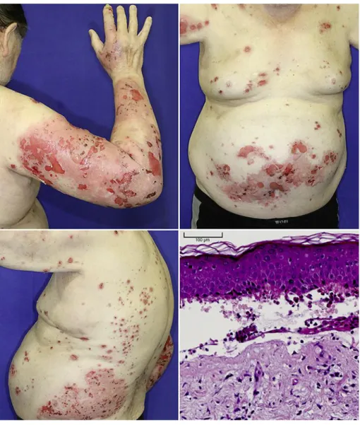
By the time the patient presented to our clinic, numerous, coalescing, hemorrhagic lesions as well as scattered tense blisters on the extremities and the abdomen were seen. Figure 1 shows the clinical appearance of the patient shortly after admission to our clinic. He had been taking irbesartan, bisoprolol, torasemide, atrovastatine, acetylsalicylic acid, mirtazapine, mesalazine, kaliumchloride, and levothyroxine for 6 years. The use of herbal medication or supplements as well as kidney dysfunction, dehydration, lately performed surgical proce- dures, strenuous physical activities, or vaccinations were denied by the patient. There was no association between the blisters’ appearance and new drugs, other than adalimumab. A skin biopsy performed shortly after admission to our hospital showed a subepidermal blister with fibrin deposits as well as eosinophils and neutrophils within the blister. Numerous granulo- cytes and eosinophils were also found in the upper corium (Fig. 1).
In addition to the already mentioned skin findings, small ulcers located at both lateral ankles were observed.
The laboratory findings showed an increased BP180 of 1:2,560 (reference range, <1:10).
Tests for HHV8, hepatitis B, hepatitis C, Helicobacter pylori, and Toxoplasma gondii were neg-
ative. We initiated a therapy with 80 mg prednisolone (1 mg/kg), which we increased up to
100 mg after 3 days as new blisters showed up. The ongoing formation of blisters during the
first 7 days of treatment led us to adjust the immune-suppressive therapy up to 120 mg pred-
nisolone for 2 days. From day 10 on, the therapy with prednisolone was tapered. Azathioprine
was not an option because of hepatic parenchyma damage (elevated liver enzymes after a
therapy with azathioprine 6 months ago). Nevertheless, we started with methotrexate 15 mg
s.c. per week on the 10th day. Although we did not presume torasemide to be the trigger for
BP, we switched this medication to hydrochlorothiazide (HCT) 12.5 mg on day 7.
Case Rep Dermatol 2018;10:145–148
DOI: 10.1159/000489163 © 2018 The Author(s). Published by S. Karger AG, Basel www.karger.com/cde
Hoffmann et al.: Bullous Pemphigoid Associated with Adalimumab Therapy in a Patient with Ulcerative Colitis
147
Discussion
To date, there are many reported cases of drug-induced BP. Furosemide has been associ- ated with the occurrence of BP before [2]. Although the dose of torasemide had been raised some weeks before the first blisters appeared, we came to the conclusion that this substance was not the trigger of the patient’s BP. In fact, he had been treated with torasemide before – for the first time in 2011. An increased dose of torasemide is very unlikely to be the cause of BP in this case.
Recently Wessman et al. [3] described a 49-year-old white man with BP after undergoing adalimumab treatment. The first time BP was mentioned as an adverse reaction to ada- limumab was by Stausbøl-Grøn et al. [4] in 2009.
TNF-α inhibitors are known to increase autoantibody production [5]. The incidence of systemic lupus erythematosus and the exacerbations of multiple sclerosis appear to be more frequent in a collective undergoing TNF-α inhibitor medication.
These findings have been explained with 2 pathomechanisms: an amount of antigens from apoptotic cells either leads to the formation of autoantibodies or there is an unbalanced cytotoxic T-cell response. Due to this disbalance, autoreactive B cells are no longer efficiently suppressed [6, 7].
These explanations are speculative, but could be taken into account for the initiation of BP under adalimumab medication. Nevertheless, there is no final answer and further work is needed in order to understand this phenomenon and to explore the pathophysiology of the whole process.
The titers of antinuclear and anti-dsDNA are checked routinely during adalimumab appli- cation in order to screen for autoimmune reactions. In line with that, Wessman et al. [3] sug- gest to establish an additional anti-BP180 measurement under therapy with TNF-α inhibitors.
This could promote an early detection and in consequence decline the outbreak of BP.
Statement of Ethics
The authors have no ethical conflicts to disclose.
Disclosure Statement
The authors have no conflicts of interest to disclose.
References
1 Lo Schiavo A, Ruocco E, Brancaccio G, Caccavale S, Ruocco V, Wolf R. Bullous pemphigoid: etiology, pathogenesis, and inducing factors: facts and controversies. Clin Dermatol. 2013 Jul-Aug;31(4):391–9.
2 Helm MF, Lin L, Santalucia P, Wilson BD, Plunkett RW, Grover R. Furosemide Induced Bullous Pemphigoid Associated with Antihistone Antibodies. N Am J Med Sci. 2014;7(2):84–6.
3 Wessman LL, Blixt EK, Wetter DA, Miest RY. Adalimumab-associated bullous pemphigoid in a patient with ulcerative colitis. JAAD Case Rep. 2017 Jul;3(4):339–41.
4 Stausbøl-Grøn B, Deleuran M, Sommer Hansen E, Kragballe K. Development of bullous pemphigoid during treatment of psoriasis with adalimumab. Clin Exp Dermatol. 2009 Oct;34(7):e285–6.
5 Mongey AB, Hess EV. Drug insight: autoimmune effects of medications-what’s new? Nat Clin Pract
Rheumatol. 2008 Mar;4(3):136–44.
Case Rep Dermatol 2018;10:145–148
DOI: 10.1159/000489163 © 2018 The Author(s). Published by S. Karger AG, Basel www.karger.com/cde
Hoffmann et al.: Bullous Pemphigoid Associated with Adalimumab Therapy in a Patient with Ulcerative Colitis
148
6 De Bandt M, Sibilia J, Le Loët X, Prouzeau S, Fautrel B, Marcelli C et al.; Club Rhumatismes et Inflammation.
Systemic lupus erythematosus induced by anti-tumour necrosis factor alpha therapy: a French national survey. Arthritis Res Ther. 2005;7(3):R545–51.
7 Ramos-Casals M, Brito-Zerón P, Soto MJ, Cuadrado MJ, Khamashta MA. Autoimmune diseases induced by TNF-targeted therapies. Best Pract Res Clin Rheumatol. 2008 Oct;22(5):847–61.
Fig. 1.