Original article:
SYNTHESIS, SPECTROSCOPIC CHARACTERIZATION, IN-VITRO ANTIBACTERIAL AND ANTIPROLIFERATIVE ACTIVITIES OF
SOME METAL(II) COMPLEXES OF
3,4-DIHYDRONAPHTHALEN-1(2 H )-ONE SCHIFF BASE Aderoju Amoke Osowole
Inorganic Chemistry Unit, Department of Chemistry, University of Ibadan, Ibadan, Nigeria E-mail: aderoju30@yahoo.com;
Phone: +2348097327529ABSTRACT
The Schiff base, 3-hydroxy-4-{[4-(methylsulfanyl)phenyl]imino}-3,4-dihydronaphthalen- 1(2H)-one, and its Mn(II), Co(II), Ni(II), Cu(II), Zn(II) and Pd(II) complexes have been syn- thesized and characterized by microanalysis, conductance,
1H NMR, infrared and electronic spectral measurements. The ligand exists in the ketoimine form in chloroform, and in the eno- limine form in the solid state, as shown by
1H NMR and IR spectroscopies. The ligand coor- dinates to the metal ions in the ratio 1:1, using NO chromophores forming complexes of the type [MLNO
3]H
2O, with the exception of the Zn(II) and Pd(II) complexes. Electronic meas- urements are indicative of a four coordinate square-planar geometry for all the complexes, except for the Cu(II) and Zn(II) complexes which assume a tetrahedral geometry. None is an electrolyte in nitromethane. The ligand and the metal complexes are air–stable, but decom- posed on heating at 120 °C and in the range 150-156 °C respectively. The antibacterial stud- ies reveal that the Co(II) and the Cu(II) complexes exhibit broad-spectrum activity against Proteus mirabilis, Escherichia coli and Staphylococcus aureus with inhibitory zones range of 14.0-22.0 and 13.0-25.0 mm respectively. The antiproliferative studies show that the Zn(II) complex has the best in-vitro anticancer activity against both HT-29 (colon) carcinoma and MCF-7 (human breast) adenocarcinoma with IC
50values of 6.46 µm and 3.19 µm, which ex- ceeds the activity of Cis-platin by 8 % and 63 % respectively.
Keywords: antibacterial and antiproliferative activities, Cis-platin, geometry, Schiff base
INTRODUCTION
Napthoquinones are very interesting molecules because of their various uses such as precursors in the syntheses of imid- azoles, phthiocols and benzophenothia- zinols (Agarwal and Mital, 1976; Efimova and Efros, 1967; Srivastava et al., 1987), and as bactericides and insecticides. Natural napthoquinones extracted from Tritonis crocosmilioro are bactericidal to Bacillus subtilis (Masuda, 1987); while acylamino napthoquinone and aminochloro napthoqui- none have larvicidal and insecticidal activi- ties against Aedes egypti and Plasmodium
falciparum (Lopes et al., 1977; Lucimi et
al., 2010). Furthermore, they are useful as
dyes in the form of dichloro napthosultam
quinines (Herzberg and Hoppe, 1922), and
as radiation modulators of induced lipid
peroxidation in Fe(II)/Fe(III) complexes of
various hydroxynapthouinone (Kumbhar et
al., 1997). In addition, 7-bromo-5-cyclo-
propyl-5H-pyridazino[4,5-b]indol-1-amine
ethanedioate derivatives and 4-anilino-5H-
pyridazino[4,5-b]indoles showed good anti-
cancer activity against human liver (Bel-
7402) and human fibrosarcoma (HT-1080)
cancer cell lines (Li et al., 2008). Thus, the
aim is to synthesize and characterize the above named Schiff base and its metal(II) complexes. Their in-vitro antibacterial and antiproliferative activities are also investi- gated with a view to assessing their suitabil- ity as active components in disinfectants, and lead compounds in drug research for breast and colon carcinomas. The ligand and its metal(II) complexes are new, being reported here for the first time as a continu- ation of my group’s research on the Synthe- sis, characterization and bioactivities of var- ious (methylsulfanyl) metal(II) Schiff base complexes (Osowole and Daramola, 2011;
Osowole, 2011).
MATERIALS AND METHODS Reagent and chemicals
Reagent grade 2-hydroxy-1,4-naptho- quinone, 4-methylthio aniline, hydrated co- balt(II) nitrate, nickel(II) nitrate, copper(II) nitrate, manganese(II)nitrate and Zinc (II) nitrate, Palladium(II) chloride were pur- chased from Aldrich and BDH chemicals, and were used as received. Solvents were purified by distillation.
Physical measurements
The electronic and infrared spectra were recorded on a Perkin-Elmer λ25 and a Thermo Nicolet FTIR 200 spectrophotome- ter respectively. The
1H NMR spectrum was recorded on a 300 MHz Brucker DRX- 400 NMR instrument in CDCl
3at 295K.
1H chemical shifts were referenced to the re- sidual signals of the protons of CDCl
3and were quoted in ppm. The elemental anal- yses for C, H and N were recorded on Thermo Quest CE Instruments flash EA1112 analyser, while manganese, cobalt, nickel, copper, palladium and zinc were de- termined titrimetrically (Bassett et al., 1978). Electrolytic conductivities in nitro- methane and melting points (uncorrected) were determined using a HANNA HI 991300 conductivity meter and a Mel-Temp electro thermal machine respectively.
Biological studies
The clinical isolates: Escherichia coli, Staphylococcus aureus, Klebsiella pneu- moniae and Proteus mirabilis were ob- tained from the Department of Medical Mi- crobiology, College of Medicine, Universi- ty of Ibadan, Ibadan, Nigeria; while MCF-7 (human breast adenocarcinoma) and HT-29 (colon carcinoma) cells were cultured at the Institute of Medicinal and Pharmaceutical Chemistry, Technical University, Braun- schweig, Germany.
Syntheses
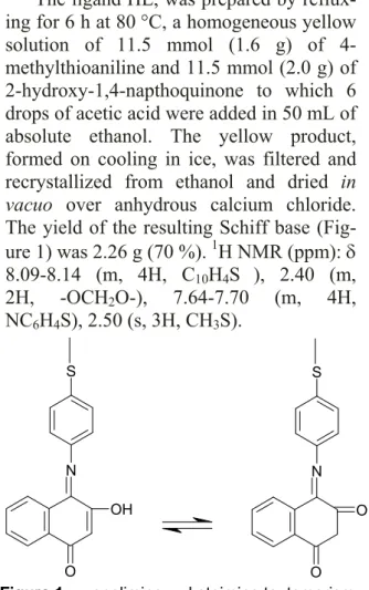
The ligand HL, was prepared by reflux- ing for 6 h at 80 °C, a homogeneous yellow solution of 11.5 mmol (1.6 g) of 4- methylthioaniline and 11.5 mmol (2.0 g) of 2-hydroxy-1,4-napthoquinone to which 6 drops of acetic acid were added in 50 mL of absolute ethanol. The yellow product, formed on cooling in ice, was filtered and recrystallized from ethanol and dried in vacuo over anhydrous calcium chloride.
The yield of the resulting Schiff base (Fig- ure 1) was 2.26 g (70 %).
1H NMR (ppm): 8.09-8.14 (m, 4H, C
10H
4S ), 2.40 (m, 2H, -OCH
2O-), 7.64-7.70 (m, 4H, NC
6H
4S), 2.50 (s, 3H, CH
3S).
S
N
O
OH
S
N
O O
Figure 1: enolimine ↔ ketoimine tautomerism
Preparation of the metal(II) complexes
The various complexes were prepared
by the addition in bits of 0.30 mmol (0.05-
0.09 g) of hydrated M(II) nitrates (M = Mn,
Co, Ni, Cu, Zn) to a stirring solution of
0.30 mmol (0.09 g) of the ligand in 30 mL
ethanol. The resulting coloured homogene-
ous solution was buffered with 0.03 mmol
(0.03 g) of triethylamine and refluxed for
6 h. The products formed on cooling to room temperature were filtered, washed with ethanol, and dried in vacuo over anhy- drous calcium chloride. The Pd(II) complex was isolated from its chloride using similar procedure.
Antimicrobial assay
The assay was carried out on the ligand and its metal(II) complexes using the Agar diffusion technique. The surface of the Muller Hinton’s agar in a Petri dish was uniformly inoculated with 0.3 mL of 18 hour old Escherichia coli, Staphylococ- cus aureus, Klebsiella pneumoniae and Proteus mirabilis cultures. A sterile cork borer was used to make 7 mm wells in the agar. A concentration of 10 mg/mL of each metal complex in DMF was then introduced into the wells and the plates allowed to stand on the bench for 30 min before incu- bation at 37 °C for 24 h. The inhibitory zones (in mm) were then taken as a measure of antimicrobial activity. The experiments were conducted in duplicates with sulfa- methoxazole as the reference drug.
Cytotoxicity assay
The MCF-7 (human breast adeno- carcinoma) and HT-29 (colon carcinoma) cells were maintained in minimum essential medium (MEM) supplemented with 10 % of fetal caw serum (FCS), and 25 mg of gentamycin at 37 °C in a humidified atmos- phere with 5 % CO
2. A concentration of 100 mL of a cell suspension in culture me- dium [7500 cells/mL for (MCF-7) and 2500 cells/mL for (HT-29)] were plated into each of 96 well plates and incubated for three days under culture conditions. After the addition of various concentrations of the test compounds, the cells were incubated for another 96 h and 72 h respectively. The medium was then removed and the cells fixed with 1 % glutardialdehyde solution and stored under phosphate buffered saline (PBS) at 4 °C. Cell biomass was deter- mined by a crystal violet staining, followed by the extraction of the bound dye with eth- anol, and a photometric measurement at 590 nm. Mean values were calculated and
the effects of the compounds were ex- pressed as % Treated/Control
corrvalues ac- cording to the following equation:
T/C
corr[ %] = ( T-C
0/C-C
0).100 where (C
0= the biomass of control cells at the tie of compound addition; C = the bio- mass of control cells at the time of the test end; T = the biomass of probes/samples at the time of the test end. The test compounds were prepared fresh as stock solutions in DMF and diluted with the cell culture me- dium to the final assay concentrations (0.1 % v/v DMF). Cis-platin was used as the reference drug. The IC
50value was de- termined as the concentration causing 50 % inhibition of cell proliferation (Scheffler et al., 2010).
RESULTS AND DISCUSSION The formation of the ligand is con- firmed by microanalyses and
1HNMR measurements. The ligand exhibits ke- toamine/enolimine tautomerism as seen in the IR and
1H NMR measurements (Figure 1). All the complexes adopt [MLNO
3]xH
2O stoichiometry, with the exception of the Zn(II) and Pd(II) complexes, which formed as [ZnL
2] and [PdLClH
2O] respectively, and are hygroscopic. The generalized equa- tion for the formation of the complexes is:
M(NO
3)
2·6H
2O + HL [MLNO
3]aH
2O + HNO
3+ bH
2O
(when M(II) = Mn/Co{a = 1, b= 5});
Ni(II)){a = 1.5, b= 4.5}); Cu(II) {a = 2.5, b= 3.5})
The analytical data, colors, percentage
yields, melting points and molar conduc-
tivities are presented in Table 1. Attempts
to isolate suitable crystals for single X-ray
structural determination have not yet been
successful.
Table 1: Analytical data for the compounds Compound
(Empirical formula) Formula
mass Color %
Yield Λm* D. T
(oC) Analysis (Calculated) %C %H %N %M
HL
(C17H14NSO2) 295.36 Dark
Purple 70 - 120 68.92 (69.07) 4.53
(4.77) 4.35
(4.74) - [MnLNO3]H2O
(MnC17H15N2SO6) 429.32 Wine 50 9.0 152 47.20 (47.56) 3.51
(3.52) 3.62
(6.53) 12.73 (12.80) [CoLNO3]H2O
(CoC17H15N2SO6) 433.31 Ma-
roon 60 12.0 150 47.52 (47.12) 3.88
(3.49) 3.94
(6.43) 13.40 (13.60) [NiLNO3]1.5H2O
(NiC17H16N2SO6.5) 442.10 Brown 60 13.0 156 46.19 (46.18) 3.80
(3.65) 3.95
(6.34) 13.27 (13.28) [CuLNO3]2.5H2O
(CuC17H18N2SO7.5) 464.93 Brown 50 18.0 150 43.33 (43.92) 3.35
(3.90) 4.41
(6.03) 13.73 (13.67)
# [ZnL2]7H2O
(ZnC34H40N2S2O11) 779.86 Red 50 15.0 150 52.25 (52.37) 3.81
(5.17) 3.60
(3.59) 8.40 (8.34)
# [PdLClH2O]5H2O
(CuC17H18N2SO7.5) 544.38 Brown 40 22.0 150 37.15 (37.51) 2.72
(4.63) 3.10
(2.57) 19.52 (19.55) D.T = decomposition temperature; # = hygroscopic; *Ω-1 cm2 mol-1
Conductance measurements
The molar conductivities of the com- plexes in nitromethane, are in the range 9.0-22.0 ohm
−1cm
2mol
−1, showing that they are non-electrolytes in the solvent. A value of 94.0-105.0 ohm
−1cm
2mol
−1is ex- pected for a 1:1 electrolyte (Geary, 1971).
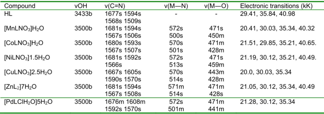
Infrared and electronic spectra
The relevant infrared data are presented in Table 2. The band at 3433 cm
-1in the ligand is assigned as νOH and its broadness is attributed to intramolecular hydrogen bonding (Derebe et al., 2002). The absence of this band in the complexes indicates the involvement of the napthaquinol O in bond- ing to the metal ions. The new broad band
at 3500 cm
–1in the complexes is assigned to (OH) of crystallization water. The un- coordinated C=N vibrations in the ligand are observed as four bands in the range 1677-1509 cm
–1. These bands are observed as three to four bands in the complexes, and are hypsochromic/bathochromic shifted to 1681-1506 cm
–1; confirming the involve- ment of the imine N atom in coordination to the metal(II) ion. The appearance of the bands due to ν(M―O) and ν(M―N) in the complexes, at 471-428 and 572-534 cm
–1is further evidence of coordination. These bands are absent in the ligand spectrum (Abd El-Wahab, 2008).
Table 2: Relevant infrared and electronic spectral data of the complexes
Compound νOH ν(C=N) ν(M―N) ν(M―O) Electronic transitions (kK)
HL 3433b 1677s 1594s
1568s 1509s
- - 29.41, 35.84, 40.98 [MnLNO3]H2O 3500b 1681s 1594s
1567s 1506s
572s 500s
471s 450m
20.41, 30.03, 35.34, 40.32 [CoLNO3]H2O 3500b 1680s 1593s
1567s 1507s
570s 501s
471m 428m
21.51, 29.85, 35.21, 40.65.
[NiLNO3]1.5H2O 3500b 1681s 1592s 1566s
572s 513s
471s 459m
21.19, 30.12, 35.21, 40.49.
[CuLNO3]2.5H2O 3500b 1667s 1605s
1590s 1570s 570s
514s 443m
428m 20.0, 30.03, 35.34 [ZnL2]7H2O 3500b 1681s 1594s
1567s 1508s 571m
514s 471m
428s 21.05, 30.12, 35.34, 40.49 [PdLClH2O]5H2O 3500b 1676m 1608m
1592s 1570s 572s
501m 471m
441m 21.28, 30.12, 35.34 Key: s = strong, m = medium, b = broad; 1 kK = 1000 cm-1
The electronic spectra are presented in Table 2. The Mn(II) and Co(II) complexes both exhibit a single band each at 20.41 kK and 21.51 kK respectively, suggestive of a 4-coordinate, square-planar geometry (Osowole et al., 2009). Similarly, the Ni(II) and Pd(II) complexes have an absorption band each at 21.19 kK and 21.28 kK, indic- ative of a 4- coordinate square-planar ge- ometry; and are assigned to
1A
1g→
1E
1gtransition (Sonmez and Haciyusufoglu, 2006). The Cu(II) complex has a single band at 20.0 kK, suggestive of a distorted tetrahedral geometry with the assignment
2
T
2→
2E (Gaber et al., 2001). As expected, the Zn(II) complex has no d-d band. How- ever, the presence of a M→L CT band at 21.05kK is indicative of a tetrahedral ge- ometry. The ligand bands are observed at 29.41, 35.84 and 40.98 kK, and are as- signed to n→π*, π→π* and CT transitions.
These bands are mostly bathochromic shifted in the complexes to 29.85-30.12, 35.21-35.34 and 40.32-40.65 kK due to co- ordination (Pandya and Shah, 2009).
1
H NMR spectra
The phenyl napthoquinone protons are observed as a multiplet at 8.09-8.14 ppm, while the CH
2protons are observed as a singlet at 2.40 ppm. The four phenyl pro- tons in NC
6H
4S moiety resonate as a multi- plet at 7.64-7.70 ppm, while the methyl group in the CH
3S moiety is seen as a sin- glet at 2.51 ppm.
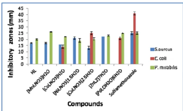
Antibacterial activity
The results of antibacterial activities are shown in Figure 2 and presented in Table 3.
The ligand is active against Staphyloccous aureus and Proteus mirabilis with inhibito- ry zones range of 17.0 and 20.0 mm but it is inactive against Klebsiella pneumoniae and E.coli. As expected, the complexes are more susceptible to Proteus mirabilis, a gram negative bacterium, due to its thin peptidoglycan layer, which makes it more permeable to the complexes with inhibitory zones range of 19.0-26.0 mm (Thangadurai and Natarajan, 2001). Furthermore, none of the complex is active against Klebsiella
pneumoniae. All the metal complexes are active against S. aureus with inhibitory zones range of 13.0-25.0 mm, except for the Pd(II) complex, which has no activity.
Figure 2: Histogram of the antibacterial activi- ties of the ligand and its complexes
The resistance of Klebsiella pneumo-
niae to the ligand and the metal complexes,
and the insensitivity of E. coli to the Ni(II),
Mn(II) and Zn(II) complexes is attributed
to the production of extended-spectrum be-
ta-lactamases (ESBL) by these bacteria,
which inactivates the compounds (Kama-
lakannan and Venkappayya, 2002; Vit-
kauskiene et al., 2006). Surprisingly E. coli
is sensitive to Co(II), Pd(II) and Cu(II)
complexes with inhibitory zones range of
14.0-25.0 mm. As expected, the complexes
are mostly more active than the ligand
against P. mirabilis and E. coli. This is due
to the partial sharing of their positive
charge with donor groups of the ligand and
possible π-electron delocalisation on the
aromatic rings which in turn increased the
lipophilic character, favouring its permea-
tion into the bacterial membrane, thus caus-
ing the death of the organisms (Mostafa
and Badria, 2008). The ligand has the same
activity of 17.0 and 20.0 mm as the Mn(II)
and Pd(II) complexes against S. aureus and
P. mirabilis. This observation could not be
explained. The lower activities of the
Co(II) and Cu(II), and Ni(II) complexes
relative to the ligand against S. aureus and
P. mirabilis may be attributed to low de-
gree of permeability of the cells of the bac-
teria, or the difference in the bacteria ribo-
some (Rafique et al., 2010).
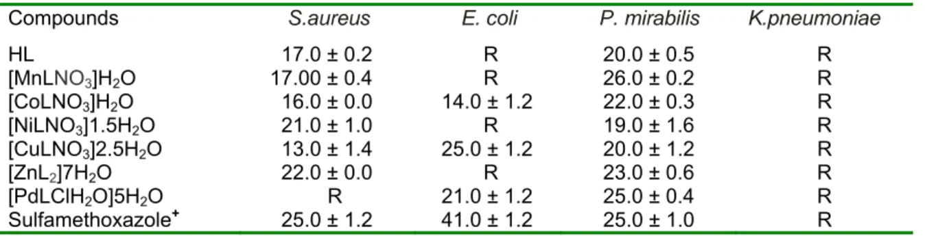
Table 3: Zones of inhibition (mm) of the compounds against various bacteria isolates
Compounds S.aureus E. coli P. mirabilis K.pneumoniae
HL 17.0 ± 0.2 R 20.0 ± 0.5 R
[MnLNO3]H2O 17.00 ± 0.4 R 26.0 ± 0.2 R [CoLNO3]H2O 16.0 ± 0.0 14.0 ± 1.2 22.0 ± 0.3 R [NiLNO3]1.5H2O 21.0 ± 1.0 R 19.0 ± 1.6 R [CuLNO3]2.5H2O 13.0 ± 1.4 25.0 ± 1.2 20.0 ± 1.2 R
[ZnL2]7H2O 22.0 ± 0.0 R 23.0 ± 0.6 R
[PdLClH2O]5H2O R 21.0 ± 1.2 25.0 ± 0.4 R Sulfamethoxazole+ 25.0 ± 1.2 41.0 ± 1.2 25.0 ± 1.0 R Key: R = Resistance; + = positive standard
The activities of sulfamethoxazole (25.0-41.0 mm) against the various bacteri- al isolates, when compared to the metal complexes (13.0-26.0 mm), indicate that most of the metal complexes have lower activity. However, the Zn(II) complex shows optimum activity of about 90 % that of sulfamethoxazole against S. aureus and P. mirabilis. It is note worthy that the Pd(II) complex has the same activity of 25.0 mm as the antibiotic against P. mirabi- lis; while the Mn(II) complex’s activity (26.0 mm) against P. mirabilis, is marginal- ly higher than that of the antibiotic (25.0 mm). Thus, the Co(II) and Cu(II) complexes exhibit broad-spectrum antibac- terial activity against Staphyloccous aureus, Escherichia coli and Proteus mirabilis, with inhibitory zones range of 14.0-22.0 and 13.0-25.0 mm respectively;
proving their usefulness as potential broad- spectrum antibacterial agents.
Anticancer activity
The results of the anticancer activities are presented in Table 4. The metal com- plexes are more sensitive to the HT-29 (co- lon carcinoma), than the MCF-7 (human breast adenocarcinoma) cells. The activities of the Zn(II), Cu(II) and Pd(II) complexes are 63 %, 58 % and 44 % Cis-platin activity respectively, against MCF-7 cells; while the ligand has an activity of 29 % that of Cis-platin. On the contrary, the Zn(II) com- plex has the best activity against HT29 cells, exceeding that of Cis-platin by 8 %.
The Cu(II) complex has about the same ac- tivity as Cis-platin, while the activities of the Pd(II) complex and the ligand are 65 % and 56 % of Cis-platin activity respective- ly. Thus, in both cases chelation enhances the antiproliferative activity of the com- plexes.
Table 4: IC50 values of the ligand and its Co(II), Cu(II), Pd(II) complexes against MCF-7 and HT-29 cells
Compounds MCF-7 (human breast
adenocarcinoma) [µM] HT-29(colon carcinoma cells) [µM]
CDDP 2.0 7.0
HL 6.92 ± 0.1 12.53 ± 0.1
[CuL2]7H2O 3.46 ± 0.1 7.27 ± 0.1
[ZnL2]3H2O 3.19 ± 0.2 6.46 ± 0.2
[PdL2]3H2O 4.53 ± 0.1 10.81 ± 0.1
Results are expressed as means (± error) of at least two independent experiments