ALTEX preprint published October 21, 2020
doi:10.14573/altex.2003211
Research Article
Galleria mellonella as an Alternative In Vivo Model to Study Bacterial Biofilms on Stainless Steel and
Titanium Implants *
Gopala K. Mannala
1, Markus Rupp
1, Francisca Alagboso
1, Maximilian Kerschbaum
1, Christian Pfeifer
1, Ursula Sommer
2, Marian Kampschulte
3, Eugen Domann
4and Volker Alt
11Department of Trauma Surgery, University Hospital Regensburg, Regensburg, Germany; 2Experimental Trauma Surgery, Justus Liebig University Giessen, Giessen, Germany; 3Departmentof Radiology, University Hospital Giessen-Marburg, Campus, Giessen, Giessen, Germany; 4Institute for Medical Microbiology, Biomedizinisches Forschungzentrum Seltersberg (BFS), Giessen, Germany
Abstract
The purpose of this study was to establish an infection model of Galleria mellonella larvae as an alternative in vivo model for biofilm-associated infections on stainless steel and titanium implants. First, the model was established with bacteria-free implants to evaluate the biocompatibility of implants in the larvae. Titanium or stainless steel implants were implanted without any adverse effects over the entire observation period of 5 days compared to controls. Then, stainless steel and titanium implants pre-incubated with Staphylococcus aureus were implanted into the larvae to mimic biofilm-associated infection. For both materials, pre-incubation of the implant with S. aureus led to significantly reduced survival of the larvae compared to bacteria-free implants.
Survival rates of the larvae could not be improved in this biofilm infection situation by the addition of gentamicin, whereas gentamicin could significantly improve the survival of the larvae in case of planktonic infection of the larvae with S. aureus without an implant, confirming the typical characteristics of reduced antibiotic susceptibility of biofilm infections. Additionally, biofilm formation and various stages of biofilm maturation were confirmed by surface electron microscopy and by measuring gene expression of biofilm-related genes with the pre-incubated implant, which showed strong biofilm formation and upregulation of autolysin (atl) and sarA genes. In conclusion, G. mellonella can be used as an alternative in vivo model to study biofilm-associated infections on stainless steel and titanium implants, which may help to reduce animal infection experiments with vertebrates in the future.
1 Introduction
Progress in medical research led to implementation of a multitude of implantable medical devices. Their extensive use in the clinical routine as metal or ceramic prostheses, catheters or heart valves, only to mention a few, is strongly connected to the success story of modern medicine (Bechert et al., 2000; Waldvogel, 2000). However, colonization of medical devices with pathogens finally forming biofilm on implant surfaces is a critical problem in clinical routine. After formation of a biofilm, medical antibiotic or antimycotic treatment in general fails due to manifold bacterial defense mechanisms (Zimmerli et al., 2004; Ribeiro et al., 2012). Hence, biofilms established on medical devices often require removal of the entire implant to achieve infect eradication. The impact of such surgical procedures for removal and often reimplantation after infect eradication is high, not only for the individual patient but also with respect to costs (Zimmerli et al., 2004).
Several in vitro and in vivo models have been developed to uncover the process of biofilm formation (Lebeaux et al., 2013). In translational research, in vivo models such as rat, rabbit, dog and sheep are widely used to check the compatibility of orthopedic implants as well as to screen antimicrobial coatings and compounds against biofilm formation from various microorganisms.
In general, vertebrate models are restricted to use due to animal welfare and ethical reasons. This is particularly true for infection experiments as inoculation of bacteria or other microorganisms often results in a high burden of the disease in the animals (Moriarty et al., 2019). Therefore, ethical approval is usually restricted. To provide best possible protection of research animals, each research project should follow the 3R (replacement, reduction, refinement) principles introduced for animal welfare by Russel and Burch in 1959 (Russell and Burch, 1959).
In recent decades, invertebrates such as Drosophila, Caenorhabditis elegans, Zebrafish and G. mellonella have been widely used as infection models to study host-pathogen interactions as well as virulence of bacterial and fungal pathogens (Mukherjee et al., 2010; Mannala et al., 2017a; Mannala et al., 2018). In addition, those models enabled testing of
*
Received March 21, 2020; Accepted October 15, 2020;
Epub October 21, 2020; © The Authors, 2021.
ALTEX 38(#), ###-###. doi:10.14573/altex.2003211
Correspondence: Prof. Dr. med. Dr. biol. hom. Volker Alt Department of Trauma Surgery, University Hospital Regensburg, Franz-Josef-Strauß-Allee 11, 93053 Regensburg, Germany (volker.alt@ukr.de)
ALTEX preprint published October 21, 2020
doi:10.14573/altex.2003211
antimicrobial activity and drugs. These models are economical, ethically legitimate and easy to handle. Among those, the larva of the greater wax moth, G. mellonella, has been extensively used to test virulence of bacterial pathogens. Recently our group evaluated virulence levels of various clinical relevant Staphylococcus aureus strains isolated from implant-associated infections using this model (Mannala et al., 2018). Earlier, G. mellonella model was used to assess accumulation of bacterial pathogens on different tooth brush bristles to mimic biofilm infections on a foreign body. After piercing of tooth bristles in the larvae proleg, infection signs observation, bacterial enumeration and biofilm analysis by SEM have been done subsequently in this model (Benthall et al., 2015; Campos-Silva et al., 2019).
However, to our best knowledge, orthopedic metallic implants have not been yet tested in G. mellonella to study implant associated infections and bacterial biofilm formation. Therefore, the aim of the current study was the establishment of this insect infection model for biofilm-associated infections for stainless steel and titanium implants in order to mimic biofilm-infections in orthopedic surgery. For this purpose, the compatibility of the injected implants without bacterial loading as well as effects of pre-incubated implants with S. aureus and the additional use of gentamicin, including survival analysis, SEM and gene expression analysis, were evaluated.
2 Materials and Methods
The study was carried out in a 2-stage manner. First, establishment of the model with bacteria-free implants was performed to evaluate the biocompatibility of the inoculation of the implants into the larvae. After successful establishment of the model, the second stage of the work was conducted with inoculation of pre-incubated stainless steel and titanium implants with S. aureus into the larvae to mimic biofilm-associated infection on the implants.
2.1 G. mellonella, bacterial strain, growth conditions and preparation of stainless steel and titanium implants
G. mellonella larvae were ordered from Fauna Topics GmbH (Rielingshausen, Germany) and maintained on artificial diet in an incubator at 30°C. For each experiment, ten larvae weighing around 200-250 mg and present in the last instar stage were used. After infection, G. mellonella were maintained at 37°C.
In this study, Staphylococcus aureus EDCC 5055 strain was used. This strain is known for its high biofilm formation capacity and its whole genome sequence is available (Mannala et al., 2017b; Mannala et al., 2018). Brain-Heart- Infusion (BHI) broth was used to maintain S. aureus aerobically at 37°C by constant shaking at 180 rpm. An overnight culture was diluted to 1:50 and grown to mid-exponential phase containing an optical density of 1.0 at 600nm. This bacteria culture was then washed twice with 0.9% NaCl. Thereafter, the bacterial number was measured with the help of optical density, adjusted to required cell number and used for the experimental purpose.
Sterile stainless steel and titanium K-wires with a diameter of 0.8mm (Synthes, Zuchwil, Switzerland) were used as implant materials. Small pieces with a length of 4-5mm were cut with a cable cutter and one edge of each K-wire was sharpened with help of a sharpener (Fig.1).
Fig. 1: Preparation of larvae, implants and implantation process
(A) For the implantation, larvae with weight between 200-250 mg were purchased. Stainless steel or titanium implants with a length of 4-5mm and 0.8mm diameter with one side sharp edged were used. (B) The implants were inoculated inside the larvae with the help of a forceps. The larvae were held by one person and another person pierced the cuticle of the larvae segmented region (rear part of the larvae) with the sharp edged part and pushed it completely into the body of the larvae. Further details can be seen in the supplementary video1.
2.2 Establishment of the G. mellonella implant model
For the implantation of implant materials inside the G. mellonella, the larvae were held by one person and another person performed the implantation with the help of a metal tweezer. The implants were placed in the rear part of the larvae through piercing the cuticle of the larvae with the sharp edge of implant material and was pushed inside the larvae simultaneously.
For easy piercing and implantation, it is recommended to implant the implant at the segment region due to less cuticle thickness (see supplementary video1). After implantation, the larvae were placed in petri dishes containing G. mellonella artificial diet at 30° C and observed for their survival for 5 days. Further, the observation of the implanted larvae and control
1 doi:10.14573/altex.2003211s1
ALTEX preprint published October 21, 2020
doi:10.14573/altex.2003211
larvae was performed until the pupae and subsequently moth stages. Presence of any metal toxicity leads to sickness and death of the larvae. Observation of the larvae activeness and survival were considered as parameters of metal toxicity.
Wound healing and melanization at the implantation site were measured based on the leaking of hemolymph or pus generation and color of the skin changes. Hence, we tested the biocompatibility of the implantation procedure and toxicity of stainless steel and titanium implants in G. mellonella with this setting.
2.3 Micro-CT analysis
Cross-sectional imaging was performed using a SkyScan 1173 micro-CT system (Bruker microCT, Kontich, Belgium). The system is equipped with a high voltage tube for imaging of dense materials. Larvae were wrapped in parafilm and mounted on a rotational stage for ex-vivo scanning. Cross sectional images were reconstructed by filtered back projection using the NRecon software (Version: 1.7.1.0, Bruker microCT, Kontich, Belgium). Image acquisition and reconstruction parameters were described in Table S12.
2.4 Infection experiments with pre-incubated stainless steel and titanium implants in bacterial suspension
After confirmation of the biocompatibility of the metallic implants inside the G. mellonella, this model was evaluated to study implant-associated biofilm infections. For the infection process, implants were pre-incubated in specified bacterial growth culture with the above mentioned S. aureus EDCC 5055 at 1x10⁶ CFU/ml for 30 min at 150 rpm shaking conditions.
Later, these implants were washed with 10ml PBS and implanted in the larvae as mentioned before. For the control group, the same process was applied but without bacterial contamination of the implants.
To determine the number of adherent bacteria before implantation, implants were sonicated in PBS at 40 kHz, 0.1- 1 W/cm2 in sonication bath for one minute and followed by the vortex of the samples for one minute. Later, the suspension was plated out on the LB agar plates. The bacterial colonies were counted after incubation of plates at 37°C for 16 hours.
After implantation, the larvae were maintained at 37° C for five days and observed for the survival each day.
Comparison of larval survival with infective implants (stainless steel, titanium), biofilm formation, biofilm maturation stages and gene expression analysis were done after the infection process. We used ten larvae for each group of the experimental setup and each experiment was repeated for a least three times.
2.5 Planktonic infection experiments without implants and effects of gentamicin
In order to determine the impact of the biofilm formed by S. aureus on the pre-incubated implants and to distinguish this effect from planktonic infection with S. aureus without an implant, larvae were injected with 4,000 CFUs of S. aureus without an implant. This bacterial load corresponded to the one of the adhering bacteria on the metallic surface of the stainless steel and titanium implants in the biofilm infection model. Survival rates of this experiment were compared to survival rates from the biofilm infection model with inoculation of pre-incubated stainless steel implants with S. aureus as described above.
In order to determine the effect of gentamicin on the survival in the biofilm infection model on stainless steel implants and in the planktonic infection model, the larvae were injected on day 1 with gentamicin (120mg/kg) after infection with the S. aureus preincubated implant in the biofilm model and after planktonic infection in the planktonic infection model as described before. All experiments were run for three times.
2.6 Scanning electron microscopy
For the observation of biofilm on day 2 and its maturation from day 1-4, scanning electron microscopy (SEM) was used. For the SEM analysis of the implants, the larvae were dissected at specified time points, implants were taken out and placed in PBS. These implants were washed twice with PBS to remove planktonic cells and then fixed with 1% sucrose and 2.5%
glutaraldehyde at 4°C for 24 hours. Thereafter, the fixative reagents 1% sucrose and 2.5% glutaraldehyde were removed by washing six times with PBS and dehydrated with lower to higher ethanol concentrations (30%, 50%, 70%, 80%, and 96%) for 15 min, then finally three times with 100% ethanol for 30 min. Samples were dried in a critical point dryer (Leica EM CPD300) auto and sputter coated with gold and palladium (Polaron Sputter Coater SC7640). SEM analysis was performed with a LEO1530 at 15kV. The biofilm formation was analyzed on both stainless steel and titanium implants. The biofilm maturation was analyzed on the stainless steel implant on day 1-4. For each setting, four samples were analyzed for SEM analysis.
2.7 RNA isolation and qRT-PCR analysis
For RNA extraction from S. aureus grown extracellularly in BHI, we applied aliquots of 0.5 ml from the same S. aureus culture grown until mid-exponential phase used to infect G. mellonella. The bacterial cells were treated with 1.0 ml RNA protect (Qiagen) for 5 min and were collected by centrifugation for 10 min (8000 g). The bacterial pellets were stored at
−80°C until use. For RNA extraction from the biofilms formed on implants in G. mellonella, the implants were taken out of the larvae on day 2, sonicated, pelleted down by centrifugation and stored at -80oC. Total RNA was isolated using miRNeasy kit (Qiagen, Hilden, Germany) with some modifications. The collected bacterial pellets were washed with SET buffer (50mM NaCl, 5mM EDTA and 30mM Tris-HCl (pH 7.0) by centrifugation at 16000g for 3 min. After wash steps, pellets were resuspended into 0.1ml Tris-HCl (pH 6.5) containing 50 mg/ml lysozyme (Sigma), 25U of mutanolysin (Sigma), 40U of SUPERase (Ambion), 0.2mg of proteinase K (Ambion). The suspension was incubated for 30min on a thermomixer at 37°C and with shaking (350 rpm). Lysis of the cell suspension with performed with addition of QIAzol (Qiagen) followed by mixing and incubation for 3min at room temperature. To the suspension, 0.2 volumes of chloroform was added, mixed well and centrifuged at 16000g at 4°C for 15 min. From the vial, upper aqueous phase containing RNA was collected into a
2 doi:10.14573/altex.2003211s2
ALTEX preprint published October 21, 2020
doi:10.14573/altex.2003211
new collection tube and 1.5 volumes of 100% ethanol was added and mixed thoroughly. The samples containing RNA were transferred into columns of miRNeasy Kit (Qiagen, Hilden, Germany) and treated on-column DNase digestion (RNase-Free DNase, Qiagen). RNA was eluted with RNase-free water and stored at −20°C until needed. The quantity of the RNA was determined by absorbance at 260nm and 280nm and the quality was assessed using Nano-chips on Agilent 2100 Bioanalyzer.
Reverse transcription was performed by SuperScript II Reverse Transcriptase (Invitrogen) using 1μg RNA. The samples were processed to quantitative real-time PCR in a final volume of 25μl using QuantiTect SYBR Green PCR kit (Qiagen) as stated in the manufacturers instruction. A standard curve was generated for the all used primer pairs (see Tab.
S22) using different copy numbers of genomic DNA from S. aureus EDCC 5055. For each primer pair a negative control (water) and RNA sample without reverse transcription reaction (for genomic DNA contamination) were included as controls during cDNA quantification. After real-time PCR, all samples were tested on a 1.5% agarose gel to check that only a single band was produced. The expression level of each gene was measured by normalizing its mRNA quantity to the quantity of the mRNA of gyrB for the same sample using a formula for relative quantification in real-time PCR published by Pfaffl (2001).
2.8 Statistical analysis
Statistical analysis of the data was performed using sigma plot 10.0. For the analysis of qRT-PCR Student’s t-test was applied. For the survival analysis two-way ANOVA was performed. The data was represented from means ± standard deviation from three independent experiments. The data was considered significant if the p-value was < 0.05.
3 Results
3.1 Implantation of G. mellonella with implants
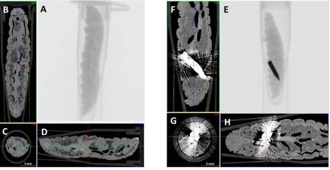
To determine the biocompatibility of the inoculation procedure and of the stainless steel and titanium implants in this model, the larvae were implanted with each type of the implant. The inoculation procedure could successfully be established and micro-CT demonstrated intra-body location of the implantation inside the larvae in all cases (Fig. 2). The implanted larvae were active and similar to the control group, which were injected with 0.9% NaCl. The survival rates of the larvae with the tested implants were above 90% from the observation for five days after the implantation and almost as high as the 100%
survival rate of the NaCl control (Fig. 3). The implantation of stainless steel as well as titanium implants did not show any adverse effects such as metal toxicity, wound healing disturbances or melanization at the site of implantation. There were no effects detected on the life stages of G. mellonella larvae as the implanted metals could be detected in pupae and moth stages (Fig.S12).
Overall, the model can be deemed biocompatible for the implantation of stainless steel and titanium K-wires.
3.2 G. mellonella as implant-associated biofilm infection model with S. aureus on titanium and stainless steel K-wires
Determination of the total adherent bacteria on the implant before implantation into the larvae revealed comparable bacterial adherence between stainless steel (4,000 ± 700 CFUs) and titanium (3,800 ± 500 CFUs) implants. The number of adherent bacteria per area on the stainless steel and titanium were calculated as 137/mm2 and 130/mm2, respectively.
Fig. 2: Micro-CT cross-sectional imaging of larvae
The control larva that was not inoculated with an implant shows typical G. mellonella anatomy in all scans (A-D). The implant treated larvae shows a completely inoculated implant in the rear part of body of the larvae (E-H).
ALTEX preprint published October 21, 2020
doi:10.14573/altex.2003211
Fig. 3: Survival analysis of G. mellonella with inoculated titanium and stainless steel without bacteria
No significant differences between the survival rates of NaCl- treated larvae and larvae treated with stainless steel or titanium implants. Experiments were conducted with 10 larvae per group and repeated three times. The data is represented as means ± standard deviation from three independent experiments.
Fig. 4: Survival analysis of G. mellonella with pre- incubated titanium and stainless steel with S.
aureus
The larvae treated with S. aureus contaminated implants showed significant reduced survival rates compared to larvae with bacteria-free control implants, both for titanium and for stainless steel implants (***p<0.001). The data are presented as means ± standard deviation from three independent experiments.
Fig. 5: Comparison between biofilm infection and planktonic infection and effects of gentamicin.
The larvae that were infected with S. aureus only without an implant (planktonic infection model) showed a different and higher survival rate, particularly in the first 4 days, compared to the biofilm infection model with the pre- incubated stainless steel K-wire implants.
Gentamicin had no effects on the survival of the larvae with implants, whereas gentamicin could significantly improve survival rates in the planktonic infection model (***p<0.001).
Fig. 4 shows the survival curves of the larvae with infection of S. aureus with stainless steel and titanium implants after implantation process with the pre-incubated implants. The larvae showed significant reduction of survival rates 5 days after the implantation of S. aureus contaminated stainless steel (survival rate: 27±3.5%) and titanium implants (survival rate:
47±3.5%) compared to controls (steel: 90±0.0%, titanium: 95±7.0%) (p<0.001).
3.3. Comparison between biofilm infections on the metallic K-wires and planktonic infections and the effects of gentamicin
The larvae with the planktonic infection that were infected with S. aureus only without an implant showed a different survival behavior compared to the biofilm infection model with the preincubated stainless steel K-wire implants. Planktonic infection resulted in a higher survival rate of the larvae, particularly in the first 4 days, compared to the biofilm infection situation.
There was a clear difference in the susceptibility against gentamicin in the larvae between the two infection models. Gentamicin had no effects on the survival of the larvae with implants mimicking a biofilm infection, whereas gentamicin could significantly improve survival rates in the planktonic infection model (p<0.001) (Fig. 5).
3.4 RNA isolation and qRT-PCR analysis
To study the biofilm related gene expression, the RNA was isolated from the planktonic cells as well as the biofilm formed on implants inside the larvae on day 2. Among these genes atl, sarA, and icaA genes are upregulated whereas fib, fnbB and fnbA genes are down regulated. No changes in the gene expression levels of clfB and agrA genes were observed. Among the upregulated genes, autolysin (atl) showed 6 fold upregulation followed by sarA 2 fold and icaA gene one fold upregulation (Fig. 6).
3.5 Biofilm visualization on implants with SEM analysis
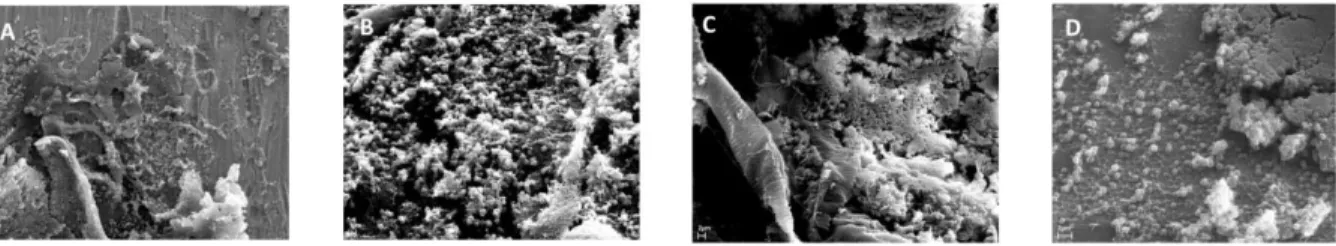
Fig. 7 shows clearly the biofilm formation on the surface of both the titanium and stainless steel implants with S. aureus bacteria cell to cell adherence and clump formation features. Further to visualize the biofilm maturation over time, SEM analysis was performed on stainless steel implants that were explanted from the larvae on each day till fourth day. Our results revealed attachment of bacteria (Fig. 8A), accumulation (Fig. 8B), maturation (Fig. 8C) and detachment or reduction of biofilm mass due to effect immune response on the biofilm (Fig. 8D) or by leaving with empty lacunae on the surface of the implant.
ALTEX preprint published October 21, 2020
doi:10.14573/altex.2003211
Fig. 6: Biofilm related gene expression analysis
To investigate key genes involved in biofilm formation, we have isolated RNA from planktonic bacteria and biofilms and qRT-PCR analysis was performed. Among these autolysin (atl), sarA and icaA were significantly upregulated (`*´p <0.05) and fibrin (fib) and fibronectin binding proteins (fnbA and fnbB) were significantly down regulated ( `*´p <0.05; `**´p <0.01). No changes in the gene expression levels were observed with clfB and agrA genes. The data is represented from means
± standard deviation from three independent experiments.
Fig. 7: Biofilm visualization on implants with SEM analysis
After 2 days of infection, the larvae were dissected and the implant was explanted and fixed for the SEM analysis.
Representative SEM images of biofilm formed on stainless steel (A+B) and on Titanium (C+D) implants with bacterial cell to cell attachments.
The SEM images A and C were taken at magnification of 2000X ( scale bar: 200µm &
scale bar: 100µm)) and B and D were taken at magnification of 10,000X (scale bar:
1µm). For each setting four samples were analyzed for SEM analysis.
4 Discussion
To the best of our knowledge G. mellonella was already used with different tooth brush bristles to evaluate biofilm-related infections with a foreign material (Benthall et al., 2015; Campos-Silva et al., 2019). However, metallic implants were not studied in this insect model before. Therefore, the main aim of the current work was to establish and to evaluate a G.
mellonella infection model with implantation of stainless steel and titanium implants to mimic biofilm infections for orthopedic purposes.
Our results of the presented G. mellonella model demonstrate that this is a suitable model to study biofilm infections with stainless and titanium implants as it shows major characteristics of a biofilm infection on metallic surfaces.
First, biocompatibility of the implantation procedure and of the metallic implants was shown. The implantation could be performed easily by piercing the cuticle without any adverse effects to the larvae, such as hemolymph bleeding, toxic effects, wound or melanization formation. In addition, the larvae well tolerated the implanted metallic implants over the entire observation period with only a slight reduction of survival rate compared to NaCl treated larvae. The implants even kept integrated in the course of all stages of G. mellonella life cycle, including pupae and moth. All these results indicate that the metallic implants are well tolerated in the larvae system.
Second, the infection part of the study with S. aureus pre-incubated implants with S. aureus revealed significant reduction of survival rates of the infected larvae compared to uninfected controls. Further, this model exhibited typical
ALTEX preprint published October 21, 2020
doi:10.14573/altex.2003211
Fig. 8: Visualization of biofilm maturation stages with SEM.
The stainless steel implants were explanted from day 1 through day 4, fixed and processed for SEM analysis. The figure shows representative SEM images of biofilm maturation with attachment of bacteria to surface on day 1 (8A) (10,000X; scale bar:
1µm) , accumulation of bacteria on day 2 (8B) (10,000X; scale bar: 3µm) , maturation of biofilm on day 3 (8C) (10,000X; scale bar: 2µm) and dispersal or removal of biofilm of biofilm on day 4 by leaving with empty lacunae on the implant surface of implant (8D) (10,000X; scale bar: 2µm) For each setting, four samples were analyzed for SEM analysis.
characteristics of a biofilm infection, which is in general associated with a reduced susceptibility against antibiotics of the bacteria embedded in the biofilm compared to a planktonic infection (Benthall et al., 2015). We could clearly demonstrate this phenomenon in our experiments as gentamicin did not show any effect on the survival rates in the biofilm-infection model with the pre-incubated metallic implants, whereas gentamicin was highly effective in the planktonic model with a significant improvement of survival rates.
Furthermore, both titanium and stainless steel contaminated implants were shown to exhibit characteristic biofilm formation on their surface that could be evaluated by SEM analysis, which we have evaluated on day 1 - 4 in our model. All typical stages of biofilm formation with attachment, accumulation, maturation and dispersal of bacteria from biofilm were found as evident in previous studies (Joo and Otto, 2012; Nishitani et al., 2015; Khatoon et al., 2018). When comparing these results with biofilm study using a mouse model, it should be considered that biofilm was dispersed on the 14th day post infection in mouse model, whereas in our model the dispersal of biofilm was achieved on the 4th day, which might be due to the short life cycle of the larvae. Besides, reduced biofilm mass might be also due to the impact of the larvae innate immune system on the biofilm (Nishitani et al., 2015).
S. aureus biofilm formation is majorly regulated by two genetic loci namely sarA (staphylococcal accessory regulator) and quorum sensing system (agr) (Balamurugan et al., 2017). Several in vitro and in vivo studies revealed that sarA gene is involved in biofilm formation with regulation of several biofilm related genes (Yarwood et al., 2004; Valle et al., 2007). In addition, sarA mutants are known to result in defect of biofilm formation (Beenken et al., 2003; Tsang et al., 2008). The agr quorum sensing system (QS) functions with sensing of S. aureus growth through extracellular levels of autoinducing peptides (AIPs) (Le and Otto, 2015) and is known for its role and regulation in initiation of biofilm formation and the dispersal of the biofilms (Paharik and Horswill, 2016). In the current study, we used this new model further to analyze the gene expression in biofilm formation. Autolysin (atl), a key gene in the context of bacterial attachment to foreign body surfaces (Dai et al., 2012; Khatoon et al., 2018), was shown to be relatively highly expressed. Biofilm regulatory molecules sarA and intracellular adhesion protein (icaA) were upregulated as well, which is in line with similar findings in the literature with in vivo biofilm formation (Khatoon et al., 2018; Moriarty et al., 2019). The quorum sensing system gene agrA did not show any changes in gene expression levels, which is dependent on the various stages of biofilm formation.
Several studies demonstrated that clumping factor B (clfB) fibrin coding gene (fib) and fibronectin binding protein coding genes (fnbA and fnbB) are upregulated in S. aureus biofilms (Kot et al., 2018; Azmi et al., 2019). Interestingly, fibrin coding gene (fib) and fibronectin binding protein coding genes (fnbA and fnbB) were down regulated, whereas clumping factor B (clfB) was not down regulated in the presented model. This might be due to lack of fibrin and fibronectin proteins in G.
mellonella and a limitation of the model.
The model is of relevance due to its low costs and its potential for high throughput analysis, e.g. for screening of antimicrobial materials, such as coatings or other anti-infective treatment strategies. Furthermore, application and decision processes with animal welfare committees, which are sometimes complicated and time-consuming, can be avoided by the use of the presented model. Also, different materials than titanium and stainless steel can be inoculated, which broaden the interest of material for other medical disciplines.
Despite these positive aspects, there are several further limitations to the model and this study. The major drawbacks of this models are the lack of an adaptive immune system of Galleria and its short life cycle that does not allow to study chronic infections. The absence of a skeletal system prevents typical bone-associated reactions and limits the conclusion of these results for orthopedic implants. Furthermore, although the use of animals can be reduced by the current alternative, this model also relies on a living organism, which remains questionable from an ethical point of view. The study itself has only used S. aureus strain and must be confirmed by other causative agents. Other relevant materials in orthopedics, such as polyethylene (PE) or polymethylmethacrylate (PMMA) have not been tested. In a next step, a direct comparison between this model and a clinically relevant orthopedic infection model, e.g. for infected non-unions in rats (Alt et al., 2011) or others (Moriarty et al., 2017) for the orthopedic setting, should be conducted.
5 Conclusion
In conclusion, our results show that Galleria mellonella can be used as an alternative in vivo model to study biofilm- associated infections on stainless steel and titanium implants and potential new treatment methods, which may help to reduce animal infection experiments with vertebrates in the future.
ALTEX preprint published October 21, 2020
doi:10.14573/altex.2003211
References
Alt, V., Lips, K. S., Henkenbehrens, C. et al. (2011). A new animal model for implant-related infected non-unions after intramedullary fixation of the tibia in rats with fluorescent in situ hybridization of bacteria in bone infection. Bone.
doi:10.1016/j.bone.2011.01.018
Azmi, K., Qrei, W. and Abdeen, Z. (2019). Screening of genes encoding adhesion factors and biofilm production in methicillin resistant strains of Staphylococcus aureus isolated from Palestinian patients. BMC Genomics.
doi:10.1186/s12864-019-5929-1
Balamurugan, P., Praveen Krishna, V., Bharath, D. et al. (2017). Staphylococcus aureus quorum regulator SarA targeted compound, 2-[(Methylamino)methyl]phenol inhibits biofilm and down-regulates virulence genes. Front Microbiol.
doi:10.3389/fmicb.2017.01290
Bechert, T., Steinrücke, P. and Guggenbichler, J. P. (2000). A new method for screening anti-infective biomaterials. Nat Med. doi:10.1038/79568
Beenken, K. E., Blevins, J. S. and Smeltzer, M. S. (2003). Mutation of sarA in Staphylococcus aureus limits biofilm formation. Infect Immun. doi:10.1128/IAI.71.7.4206-4211.2003
Benthall, G., Touzel, R. E., Hind, C. K. et al. (2015). Evaluation of antibiotic efficacy against infections caused by planktonic or biofilm cultures of Pseudomonas aeruginosa and Klebsiella pneumoniae in Galleria mellonella. Int J Antimicrob Agents. doi:10.1016/j.ijantimicag.2015.07.014
Campos-Silva, R., Brust, F. R., Trentin, D. S. et al. (2019). Alternative method in Galleria mellonella larvae to study biofilm infection and treatment. Microb Pathog 137, 103756. doi:10.1016/j.micpath.2019.103756
Dai, L., Yang, L., Parsons, C. et al. (2012). Staphylococcus epidermidis recovered from indwelling catheters exhibit enhanced biofilm dispersal and self-renewal through downregulation of agr. BMC Microbiol. doi:10.1186/1471-2180- 12-102
Joo, H. S. and Otto, M. (2012). Molecular basis of in vivo biofilm formation by bacterial pathogens. Chem Biol.
doi:10.1016/j.chembiol.2012.10.022
Khatoon, Z., McTiernan, C. D., Suuronen, E. J. et al. (2018). Bacterial biofilm formation on implantable devices and approaches to its treatment and prevention. Heliyon 4, e01067. doi:10.1016/j.heliyon.2018.e01067
Kot, B., Sytykiewicz, H. and Sprawka, I. (2018). Expression of the biofilm-associated genes in methicillin-resistant Staphylococcus aureus in biofilm and planktonic conditions. Int J Mol Sci. doi:10.3390/ijms19113487 Le, K. Y. and Otto, M. (2015). Quorum-sensing regulation in staphylococci—an overview. Front Microbiol 6, 1174.
doi:10.3389/fmicb.2015.01174
Lebeaux, D., Chauhan, A., Rendueles, O. et al. (2013). From in vitro to in vivo models of bacterial biofilm-related infections. Pathogens. doi:10.3390/pathogens2020288
Mannala, G. K., Izar, B., Rupp, O. et al. (2017a). Listeria monocytogenes Induces a Virulence-Dependent microRNA Signature That Regulates the Immune Response in Galleria mellonella. Listeria monocytogenes Induces a Virulence- Dependent microRNA Signature That Regulates the Immune Response in Galleria mello. Front Microbiol 8, 1–12.
doi:10.3389/fmicb.2017.02463
Mannala, G. K., Hain, T., Spröer, C. et al. (2017b). Complete Genome and Plasmid Sequences of Staphylococcus aureus EDCC 5055 (DSM 28763), Used To Study Implant-Associated Infections. genome Announc 5, 9–10.
doi:10.1128/genomeA.01698-16
Mannala, G. K., Koettnitz, J., Mohamed, W. et al. (2018). Whole-genome comparison of high and low virulent Staphylococcus aureus isolates inducing implant-associated bone infections. Int J Med Microbiol.
doi:10.1016/j.ijmm.2018.04.005
Moriarty, T. F., Harris, L. G., Mooney, R. A. et al. (2019). Recommendations for design and conduct of preclinical in vivo studies of orthopedic device-related infection. J Orthop Res. doi:10.1002/jor.24230
Moriarty, T. F., Schmid, T., Post, V. et al. (2017). A large animal model for a failed two-stage revision of intramedullary nail-related infection by methicillin-resistant staphylococcus aureus. Eur Cells Mater. doi:10.22203/eCM.v034a06 Mukherjee, K., Altincicek, B., Hain, T. et al. (2010). Galleria mellonella as a model system for studying Listeria
pathogenesis. Appl Environ Microbiol 76, 310–317. doi:10.1128/AEM.01301-09
Nishitani, K., Sutipornpalangkul, W., De Mesy Bentley, K. L. et al. (2015). Quantifying the natural history of biofilm formation in vivo during the establishment of chronic implant-associated Staphylococcus aureus osteomyelitis in mice to identify critical pathogen and host factors. J Orthop Res. doi:10.1002/jor.22907
Paharik, A. E. and Horswill, A. R. (2016). The Staphylococcal Biofilm: Adhesins, Regulation, and Host Response.
Microbiol Spectr. doi:10.1128/microbiolspec.vmbf-0022-2015
Pfaffl, M. W. (2001). A new mathematical model for relative quantification in real-time RT-PCR. Nucleic Acids Res 29, e45.
doi:10.1093/nar/29.9.e45
Ribeiro, M., Monteiro, F. J. and Ferraz, M. P. (2012). Infection of orthopedic implants with emphasis on bacterial adhesion process and techniques used in studying bacterial-material interactions. Biomatter 2, 176–194.
doi:10.4161/biom.22905
Russell, W. M. S. and Burch, R. L. (1959). The Principles of Humane Experimental Technique.
https://caat.jhsph.edu/principles/the-principles-of-humane-experimental-technique
Tsang, L. H., Cassat, J. E., Shaw, L. N. et al. (2008). Factors contributing to the biofilm-deficient phenotype of Staphylococcus aureus sarA mutants. PLoS One. doi:10.1371/journal.pone.0003361
Valle, J., Vergara-Irigaray, M., Merino, N. et al. (2007). sigmaB regulates IS256-mediated Staphylococcus aureus biofilm phenotypic variation. J Bacteriol 189, 2886–96. doi:10.1128/JB.01767-06
Waldvogel, F. A., Bisno, A. L. (2000). Infections Associated with Indwelling Medical Devices, Third Edition. F. A.
Waldvogel and A. L. Bisno (eds.),. American Society of Microbiology. doi:10.1128/9781555818067
Yarwood, J. M., Bartels, D. J., Volper, E. M. et al. (2004). Quorum Sensing in Staphylococcus aureus Biofilms. J Bacteriol.
ALTEX preprint published October 21, 2020
doi:10.14573/altex.2003211
doi:10.1128/JB.186.6.1838-1850.2004
Zimmerli, W., Trampuz, A. and Ochsner, P. E. (2004). Prosthetic-Joint Infections. N Engl J Med 351, 1645–54.
doi:10.1056/NEJMra040181
Conflict of interest
The authors declare no conflicts of interest.
Acknowledgments
The authors thanks Silke Zechel and Steffan Olejniczak for their technical assistance.