Development of a novel in-vitro vascular model for determination of physiological and pathophysiological
mechanobiology
I n a u g u r a l D i s s e r t a t i o n zur
Erlangung des Doktorgrades Dr. nat. med.
der Medizinischen Fakultät und
der Mathematisch-Naturwissenschaftlichen Fakultät der Universität zu Köln
vorgelegt von Robin Bayer
aus Neuss
Prost Druck GmbH, Jülich
2021
I Betreuer Prof. Dr. Jürgen Hescheler
Prof. Dr. Dr. Aysegül T. Artmann
Referenten Prof. Dr. Stephan Baldus Prof. Dr. Günter Schwarz
Datum der mündlichen Prüfung : 15.01.2021
II Table of Contents
1. Abstract ... 1
2. Introduction ... 5
3. Working Hypothesis ... 7
4. Background ... 8
4.1. Vascular Wall ... 8
4.2. Blood pressure regulation ... 9
4.3. Hypertension ... 12
4.4. Mechanical Cellular Stimulation ... 14
4.5. Biomechanics ... 14
4.5.1. Cellular Force Measurement ... 15
4.6. In-vitro Model ... 16
4.6.1. CellDrum Technology... 17
5. Material & Methods ... 18
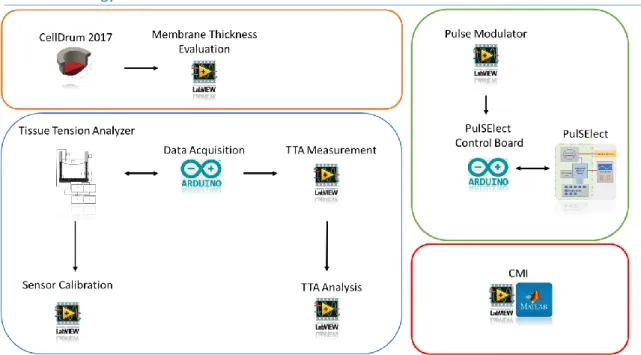
5.1. Technology Overview ... 20
5.2. CellDrum ... 20
5.2.1. Membrane Characterization... 21
5.2.2. Membrane Functionalization ... 21
5.3. Tissue Tension Analyzer ... 22
5.3.1. Development ... 22
5.3.1.1. Mechanical Construction ... 23
5.3.1.2. Electronic Components ... 23
5.3.1.3. Proximity Measuring... 25
5.3.1.4. Calibration ... 25
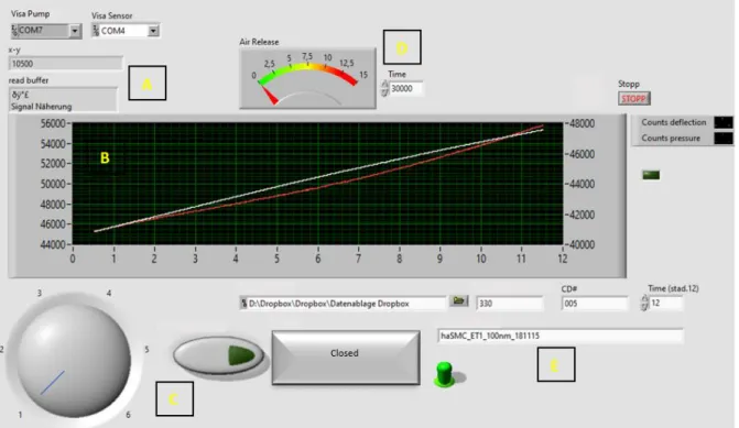
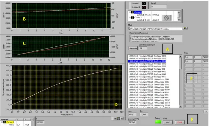
5.3.1.5. Programming ... 26
5.4. Physical Model ... 28
5.5. General Biomechanics ... 29
5.6. Cell Culture ... 29
5.6.1. Human Aortic Smooth Muscle Cells ... 29
5.6.2. Human Aortic Endothelial Cells ... 30
5.6.3. Co-Cultures ... 30
5.6.3.1. Conditioned Co-Culture ... 31
5.6.3.2. Direct Co-Culture ... 31
5.6.3.3. 3D Co-Culture ... 32
5.6.4. Vitality Test ... 33
5.6.5. Microscopic Analysis ... 33
III
5.7. Pharmacological Testing ... 34
5.8. Blood Sera Screening ... 38
5.8.1. Blood Collection and Sera Extraction ... 38
5.8.2. Blood Sera Analysis ... 38
5.9. PulSElect ... 38
5.10. Stimulation Protocol ... 40
5.11. Cell Morphology Index (CMI) ... 41
5.12. Gene Analysis ... 42
5.12.1. Microarray Analysis ... 42
5.12.1.1. Microarray Analysis Evaluation ... 42
5.12.2. Quantitative PCR... 42
5.12.2.1. RNA Isolation ... 44
5.12.2.2. Reverse Transcription ... 44
5.12.2.3. qPCR Analysis... 45
5.12.3. Endothelin-1 ELISA ... 45
6. Results ... 46
6.1. Biomechanics – Proof of Principle ... 46
6.2. Pharmacology ... 47
6.2.1. Ca² Channel Modulators ... 47
6.2.2. K+ Channel Modulators ... 48
6.2.3. NO ... 50
6.2.4. Vasoactive Mediators ... 51
6.2.5. PDE5 Inhibitor ... 52
6.2.6. Stimulants & Toxins ... 53
6.2.7. Dose-Response Model ... 55
6.3. Mechanical Stimulation ... 56
6.3.1. Microarray Analysis ... 60
6.3.2. qPCR ... 63
6.4. Co-Culture ... 65
6.5. Blood Sera ... 68
7. Discussion ... 69
7.1. Mechanobiological Vascular in-vitro Model ... 69
7.2. Hypertension Disease Model Induced by Mechanical Stimulation ... 74
7.3. Co-Cultivation and Arrangement of haSMC and haEC... 80
7.4. Application for Medical Laboratory Screening ... 82
7.5. Methodical Discussion ... 83
8. Conclusion ... 85
IV
9. References ... 87
10. Appendix ... 102
10.1. List of Abbreviations ... 102
10.2. List of Figures ... 103
10.3. List of Tables ... 109
10.4. Constructions ... 110
10.5. Electronics ... 113
10.6. Coding ... 114
1 1. Abstract
Background/Aims
The aim of the study was to develop a biological and technological method to investigate in-vitro the physiology and pathophysiology biomechanical phenomena of a vascular wall. In particular, cellular contraction and relaxation as a biomechanical response to vasoactive substances and different mechanical stimulation intervals were studied to provide data for basic research and pharmacological developments in cardiovascular diseases such as arterial hypertension.
Methods:
Methodologically, the study is based on CellDrum technology, which is a method to determine cellular stress changes of a few kilo Pascal(kPa). Especially for this study, a new approach was developed to characterize the cell stresses of monolayers and multilayer tissue equivalents in a standardized way.
A monolayer model consisting of human aortic smooth muscle cells (haSMC) was primarily developed for CellDrum as a vascular in-vitro cell culture model. Also, a model of human aortic endothelial cells (haEC) was established, and an approach for a 3D co-culture model of both cell types was developed.
Vasoactive substances with different mechanisms of action and concentrations were tested to represent the physiological properties of the model. For the first time, the biomechanical influence of blood sera was analyzed on the CellDrum models to test the potential possibility of a laboratory screening procedure.
The PulSElect system was developed, which exposes the CellDrum models to a defined, cyclical, mechanical stress by stretching, to simulate the symptoms of mechanically induced hypertension. The influence of the mechanical stress was observed by cytoskeletal alignment quantification, transcriptome analysis, gene expression of mechanosensitive as well as biomechanically relevant genes and biomechanical stress evaluation to elucidate cellular stiffening and cellular stress management.
Results
The haSMC cell models showed significant physiological and biomechanical changes in cell tone after application of the vasoactive substances, sera and conditioned media (~-6-10% relative to initial tension).
Mechanical stimulation of the cells allowed quantification of both mechanical and transcriptomic changes as well as morphological adaptation. Furthermore, it was possible to present the obtained results in a time-dependent manner. Also, mechanical stimulation has been shown to induce the development of the contractile phenotype of haSMC and improve its cellular integrity, resulting in increased basal tension and overall contractility.
As an extension of a well-established haSMC CellDrum model, an approach for direct co-cultivation of human aortic smooth muscle cells and endothelial cells was elaborated.
Conclusion
Different CellDrum models have been established to replicate biomechanical processes of the vascular system. The study showed that the CellDrum technology is a suitable method to analyze biomechanical stress changes caused by different stimuli using haSMC. The analysis of blood sera using CellDrums allows for possible future use as a screening method for pharmacological and medical laboratory research.
Since the CellDrum technology is not limited to the use of monolayers, it is possible to think about an extension of cell models with additional cell types and cell layers. Although we have already been able
2
to show partial co-cultivation of smooth muscle cells and endothelial cells, further research is needed to establish this sufficiently.
Increased expression levels of mechanosensitive genes have been shown to correlate with literature data on the pathogenesis of hypertension, using microarray analysis (Affymetrix) and qPCR.
Nevertheless, it remains a speculative reflection of the cellular changes due to induced hypertension.
The data and findings obtained to provide the promising potential supporting research and development of personalized medication, sports medicine, cell biology and stem cell research using CellDrum technology.
3 Hintergrund /Ziele:
Das Ziel der Studie ist es, ein biologisches und technologisches Verfahren zu entwickeln, um in-vitro Physiologie und Pathophysiologie biomechanischer Phänomene einer vaskulären Gefäßwand zu untersuchen. Insbesondere wird die zelluläre Kontraktion und Relaxation als biomechanische Antwort auf vasoaktive Substanzen und verschiedener Intervalle mechanischer Stimulationen untersucht, um Daten für die Grundlagenforschung und pharmakologische Entwicklungen kardiovaskuläre Erkrankungen wie Bluthochdruck zu erhalten.
Methoden:
Methodisch basiert die Studie auf der CellDrum-Technologie, welche ein Verfahren ist, um zelluläre Spannungsänderungen von wenigen Kilopascal (kPa) zu erfassen. Speziell für diese Studie wurde ein neues Analyseverfaren entwickelt, um die Zellspannungen von Zellmonolayern und mehrschichtigen Gewebeäquivalenten standardisiert zu charakterisieren und zu analysieren.
Ein Monolayer-Modell, bestehend aus glatten Muskelzellen der menschlichen Aorta (haSMC), wurde primär für CellDrum als vaskuläres in-vitro-Zellkulturmodell entwickelt. Außerdem wurde ein Modell aus humanen Aortenendothelzellen (haEC) etabliert und ein Ansatz für ein 3D-Co-Kulturmodell beider Zelltypen entwickelt.
Um die physiologischen Eigenschaften des Modells darzustellen, wurden Substanzen mit unterschiedlichen Wirkmechanismen und Konzentrationen getestet. Erstmals wurde der biomechanische Einfluss von Blutseren an den CellDrum-Modellen untersucht, um die potentielle Verwendung der CellDrum, als eines labortechnischen Screeningsverfahrens zu testen.
Das PulSElect-System wurde entwickelt, um die CellDrum-Modelle durch Dehnung einer definierten, zyklischen, mechanischen Belastung auszusetzen. Dies soll die Symptome einer mechanisch induzierten Hypertonie simulieren. Der Einfluss des mechanischen Stresses wurde mittels Quantifizierung der zytoskelettalen Ausrichtung, Transkriptomanalyse, Genexpression mechanosensitiver sowie biomechanisch relevanter Gene und biomechanischer Analyse ausgewertet, um die zelluläre Verfestigung und das zelluläre Stressmanagement zu verdeutlichen.
Ergebnisse:
Die haSMC Zellmodelle zeigen nach Zugabe der vasoaktiven Substanzen, Seren und konditionierten Medien signifikante physiologisch-biomechanische Änderung der Zellspannung(~-6-10% relativ zur mechanischen Grundspannung).
Die mechanische Stimulation der Zellen ließ sich, sowohl mechanisch als auch durch Änderungen des Transkriptoms sowie anhand morphologischer Anpassung quantifizieren. Darüber hinaus war es möglich die gewonnenen Ergebnisse zeitabhängig zueinander darzustellen. Außerdem hat sich gezeigt, dass die mechanische Stimulation die Ausprägungen des kontraktilen Phänotyps der haSMC verstärkt und deren zelluläre Integrität verbessert, was zu einer Erhöhung der Basalspannung und der Gesamtkontraktilität geführt hat.
Als Erweiterung eines etablierten haSMC CellDrum-Modells wurde ein Ansatz zu einer direkten Co- Kultivierung von humanen glatten Aortenmuskelzellen und Endothelzellen ausgearbeitet.
Zusammenfassung:
Verschiedene CellDrum-Zellmodelle konnten etabliert werden, um biomechanische Prozesse des vaskulären Systems nachzubilden. Die Studie zeigt, dass die CellDrum Technologie eine geeignete Methode ist, um biomechanische Spannungsänderungen durch verschiedene Stimuli anhand von haSMC zu analysieren. Die Auswertung der Blutseren mittels CellDrums lässt über eine zukünftige Nutzung als Screeningverfahren für pharmakologische und labortechnische Forschung spekulieren.
4
Da die CellDrum nicht auf die Nutzung von Monolayern limitiert ist kann man über eine Erweiterung des Zellmodels, mit weiteren Zellentypen und Zelllagen, nachdenken. Obwohl wir bereits teilweise Co- Kultivierung von glatten Muskelzellen und Endothelzellen zeigen konnten, bedarf es weiterer Forschungsarbeit, um diese ausreichend zu etablieren.
Mittels Transkriptom-Analyse und qPCR konnten erhöhte Expressionslevel von mechanosensitiven Genen gezeigt werden, welche mit Literaturdaten der Pathogenese von Bluthochdruckerkrankungen korrelieren. Nichtsdestotrotz bleibt es stets spekulativ, die erhobenen Daten auf eine mechanisch induzierte Hypertonie zu projizieren.
Die gewonnenen Daten und Erkenntnisse liefern vielversprechendes Potential, um die Forschung und Entwicklung personalisierter Medikation, Sportmedizin, zellbiologischer Forschung und Stammzellforschung mittels CellDrum Technologie zu unterstützen.
5 2. Introduction
Hypertension describes the chronic and pathological increase of blood pressure, which is a major cause of premature death worldwide. According to estimations of the WHO, 1,13 billion people suffer from hypertension worldwide.
Arterial hypertension is called a “silent killer”, as there are usually no recognizable symptoms, especially in the early stages of pathogenesis. In severe cases, hypertension can cause fatigue, nausea, vomiting, confusion, anxiety, chest pain and muscle tremors. Even though medical progress has been made, there is still a leak of knowledge to fully understand the mechanism of hypertension pathogenesis and early recognition of the diagnosis.
Global epidemiological statistics from 2015 (WHO), visualize increased prevalence and grade of severity in low or middle income. Increased degree of seriousness in low and middle-income countries is still a development issue and is explainable by missing awareness, diagnostics and effective health care services.
In total, only approximately 75% of affected people are aware of their diagnosis from which less than one of five patients suffering from hypertension have the problem under control. A fatal consequence of uncontrolled hypertension can be the development of a cardiovascular disease (CVD), which is globally the number one cause of death. According to the World Health Organization(WHO), 17,9 million people, representing 31% of all global deaths, died from CVD in 2016[CVD – WHO 17.5.2017 / checked 22.07.2019]. CVDs sum up a group of vessel and heart disorders, leading to insufficient sustenance of organs, cardiac and brain tissue, consequently having fatal consequences like heart attacks and strokes.
The increase of blood pressure is most commonly associated with the increase in vascular wall stiffness caused by increased cellular tone, vascular injury or mechanical cardiac overload. Vascular smooth muscle cell tone is the primary regulation factor of the blood pressure and can vary to regulate temporary or long-lasting blood pressure imbalances. The cellular structure can adapt to none physiological conditions, causing pathophysiological developments and cardiovascular diseases.
Due to the steady increase of medical findings, the development of medications needs methods that permit a fast, precise and personalized evaluation system for the development and testing of hypertensive regulating agents. Additionally, new combinations of applied medication need to be determined for their appropriate personalized dosages and side effects.
Due to modern medicine and nowadays pharmacology, a great variety of suitable medications and treatments are available to decrease blood pressure. The pharmacological adjustment for each patient takes a lot of effort and expenditure to find an appropriate remedy, dosage and or even multi- medication.
For drug development and validation, animal testing is an essential matter, as appropriate in-vitro models to avoid animal testing are currently non-existent. Current trends try to minimize the usage of animal testing. Therefore, new approaches are required to analyze biological and medical relevant characteristics, which display comparable physiological conditions.
Generally, in-vitro models are not capable to re-give the complexity of a living organism, including metabolism and real-live tissue. Nevertheless, in-vitro models offer ideal conditions to isolate cellular or tissue mechanisms, as they are easy to standardize, reproducible and ethically reasonable. Cell culture technique and tissue engineering enable to model living samples, which can be used for a broad field of experiments. Even though such models neglect great parts of the metabolic system, the specimen usually allows an excellent derivation of the cellular physiology and metabolism. Being aware
6
of this, this reduction of complexity allows an even better understanding of single pathway mechanisms.
As the general physiology and biochemistry of vasoconstriction and -dilation, as well as stiffening of blood vessels, is largely understood. The possibilities for mechanical evaluation of single cells and cell clusters are always limited since the challenge is usually to enable direct force transmission without a preferred mechanical direction. Also, many methods only allow for one-time measurements due to complex sample handling and a sample damaging measurement procedure. In means of the research and development of new and existing pharmacological agents, measurement methods that can evaluate the biomechanics of agent combinations and their time-dependent characteristics are highly interesting.
On the physiological level, there are also still open questions, especially regarding the adaptation to extrinsic and intrinsic changes. Thus, changes are known at both the macroscopic and transcriptomic level, but cannot be measured.
The working hypothesis of this thesis is to create a vascular model that can be used for mechanobiological and pharmacological research. Investigation of cellular adaptation caused by mechanical stimulation might be similar to mechanically induced hypertension.
For this purpose, a CellDrum approach is presented, which allows the measurement of functional cellular force generation, taking into account the cellular arrangement and composition of the extracellular matrix (ECM) as well as the anchoring of the cells within the surrounding matrix. In contrast or even better, complementary to standard in-vitro technologies such as microelectrode arrays (MEA), patch-clamp and high-resolution imaging. This approach is intended to provide answers to the question of the extent to which cellular and biochemical mechanisms turn into cellular force generation.
For mechanical stimulation, a system specially adapted to the CellDrum was developed, which exposes the samples to a defined cyclical mechanical stimulus. The combination of the PulSElect system and the CellDrum technology enabled a rhythmic specimen stretching due to pressure pulses. This cellular mechanical stimulation provides insights to track changes in cellular tone increase and mechanically induced stress management of the cells.
The herein presented work describes the biological and technical development of a systematical approach to elucidate the cellular effects of vasoactive substances under varying mechanical conditions, from scratch to potential medical lab applications.
7 3. Working Hypothesis
1. With the in-vitro preparation of human arterial smooth muscle cells, the biomechanical effects of vasoactive and pharmacological agents can be demonstrated physiologically and functionally.
2. Development of an in-vitro hypertension model. Due to cyclic mechanical stimulation of human arterial smooth muscle cells, physiological and pathological cellular adaptions on morphological, genetic and biomechanical levels can be identified and correlated.
3. The direct co-cultivation of arterial smooth muscle cells and endothelial cells allows an advanced mechanobiological investigation, including NO-mediated vasoactivity.
4. An established vascular CellDrum model can be used for clinical or laboratory examination of patient material.
8 4. Background
4.1. Vascular Wall
The circulatory system describes the vasculature network across the body, transporting nutrients, oxygen, carbon dioxide, hormones and blood cells throughout the organism.
There are two categories of blood vessels depending on the direction of the blood transport, whether from (veins) or towards the heart (arteries). Veins transport deoxygenated blood from the tissue back to the heart. The structure of the two types is basically the same, only the composition of the individual layers are adapted to the requirements of the respective area[1]. In comparison to arteria, venous vascular walls are most commonly thinner, especially the intima-media, as the internal pressure is usually lower[2].
Arteries transport oxygenated blood from the heart towards the tissue. In direct comparison to veins, arteria walls are more robust due to their thickness and elasticity, but also have improved contractile abilities. Great lumen vessels close to the heart have high flexibility to turn pulsatile blood flow into a laminar bloodstream. The so-called Windkessel-function stores the high energetic bloodstream pulses from the cardiac contraction to a vascular extension, which can release it in a laminar stream.
Additionally, this effect decreases the mechanical load of the heart muscle by storing energy during the vascular expansion [3,4].
To have an evenly distributed blood flow and appropriate tissue supply throughout the organism, the vascular wall thickness and lumen cross-section differ from adapting the variations in blood flow, depending on the body region. These differences are classified in further subgroups, elastic, contractile and a mixed type artery.
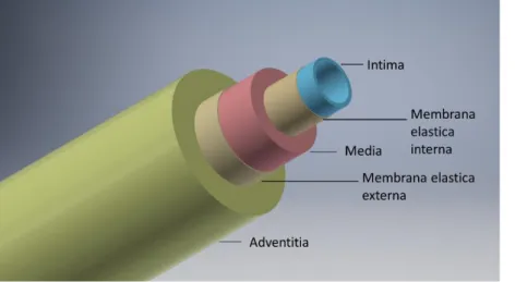
Having a closer look at the arterial vessel wall, they consist out of three layers, the tunica intima, media and adventitia, which are composed of endothelial cells, smooth muscle cells and connective tissue[Figure 1].
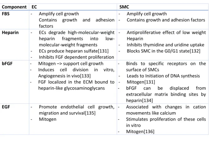
The inner layer is called Intima and consists of a single endothelial cell layer, which is located on an internal elastic membrane of collagen type IV. These two layers build a direct barrier to the lumen and bloodstream. Like in other hollow organs, the lumen is lined by a single layer of endothelial cells, which primary task is to enable oxygen diffusion and prevent blood from clotting[5]. Also, endothelial cells have blood flow sensory properties to regulate blood flow and play a major role in vascular wall healing and angiogenesis [6,7]. Besides, regulation of the lumen diameter due to permanent secretion of vasoactive substances[8], the endothelium also regulates the proliferation of vascular muscles by producing a heparin-like substance to prevent lumen rejuvenation through cellular over-proliferation [9,10].
The outer layer is called tunica adventitia, which consists mainly of connective tissue and provides vascular anchorage to adjacent tissue and protects the inner layers. This protective and shaping structure accounts for a large part of the mechanical stability of the vascular wall. Under healthy physiological conditions, it is not involved in blood pressure generation.[11]. However, in the pathological case, the strength of the connective tissue structures can be linked to the development of certain hypertensive diseases[12,13].
In between, the tunica intima is situated, which mainly consists of smooth muscle cells and connective tissue. This layer provides regulation and stabilization of the blood flow by cellular contraction or relaxation. The key player for this feature is the smooth muscle cell, which mechanisms will be discussed in more detail during the next chapters.
9
Figure 1 Schematic structure of an artery with the designation of the individual functional layers (Adventitia, Media & Intima) and the related connecting layers.
The ratio of connective tissue and cell amount varies depending on the body region and blood flow condition. Hence, arteria close to the heart is usually classified as elastic arteries, having a large lumen diameter consisting of a comparable low cell density, which is even less aligned. In contrast, peripherical arteries situated at the limbs contain higher smooth muscle cell density with higher cellular alignment, offering better contraction abilities and greater variations of the vessel volume.
These features keep muscular tissue oxygenated during physical stress and provides stabilization of the body's core temperature.
4.2. Blood pressure regulation
Blood pressure regulation is a crucial player in keeping body homeostasis. Material transport, tissue nutrition, pH and body core temperature are depending on permanent and sufficient blood circulation.
The mean arterial blood pressure consists of the resulting cardiac output and the peripheral arterial flow resistance and can be temporarily adjusted. The arterial blood pressure is measured by pressoreceptors situated in the aortic arch and the carotid sinus. Sensed blood pressure deviations are transmitted to the cardiovascular center of the vegetative nervous system, which is located in the medulla oblongata and nearby areas. For bilateral blood pressure regulation, there are two vasomotor centers, the excitatory, to increase and the inhibitory, to decrease the blood pressure. An additional blood pressure regulator is the hypothalamic center, adjusting blood pressure to the overall vegetative status[14].
Moreove, blood pressure can be regulated locally due to mechanical stimuli[15], drop of CO2 or O2
partial pressure [16,17] or changes in pH-value. Local, as well as global blood pressure regulation, causes the vascular system to modulate the inner lumen diameter to adapt to current conditions.
The endothelium of the intima permanently secretes vasoactive substances that contribute to the homeostasis of blood flow. Thus, blood pressure is regulated by the endothelium mostly via secreted messengers such as endothelin-1 (ET1), angiotensin-2 (AT2) and endothelial nitric oxide (eNO). Since the vascular endothelium is in direct contact with the bloodstream, it can react to biochemical changes in the blood and also mechanosensory to changes in blood flow. Studies have shown that the regulatory mechanisms of the endothelial cells can be affected and amplified by mechanical stimuli, especially shear and stretch forces[18–21].
Vascular constriction and dilation are provided by the contractile properties of smooth muscle cells.
With the help of the Actin-myosin apparatus, the smooth muscle cells can vary the vascular wall tone and lumen cross-section, by cellular shortening and/or elongation[22,23]. The contractile apparatus
10
consists of alpha and beta Actin as well as Myosin light chains (MLC) and Myosin heavy chains (MHC), which are netlike distributed throughout the cellular body. Dense bodies within the sarcolemma are used as the anchoring points for (intermediate) actin filaments [Figure 2]. Further intermediary filaments like desmin, vimentin and filamin transduce the generated force of the myosin filaments to the sarcolemma. The contracting of smooth muscle cells can be mediated in several ways, but all seem to work through trimeric G-protein receptors [24].
Like all other muscle cells, the contraction of smooth muscle cells is Ca2+ dependent. As soon as the intracellular Ca2+ level increases, especially for smooth muscle cells of great lumen vessels, additional Ca2+ is released via the IP3 receptor, which is initiated by the endo-plasmatic reticulum. In its resting phase, the head domain of Myosin is fixed to an actin filament, while the head domain of the heavy myosin chain and light myosin chain is angled 90°. The cross-bridge cycle (CBC) of smooth muscle cells (SMC) is the same as the CBC of straightened muscle cells. However, the initiation is not triggered by Troponin C. Instead, Ca2+ ions bind to Calmodulin, resulting in a Ca2+-Calmodulin Complex, which activates the Myosin Light Chain Kinase (MLCK). MLCK phosphorylates the MLC, leading to activation of Myosin ATPase, resulting in cleavage of the myosin head transforming ATP to ADP by releasing a phosphate, triggering the CBC. Due to the biochemical binding in the CBC, the actin-myosin apparatus mechanically moved forward by changing the myosin head angle from 90° to 45°, resulting in smooth muscle contraction.
Figure 2 Schematic
representation of the net-like distribution of dense bodies and intermediate filaments that cross the sarcoplasm. The contraction of the actin-myosin complex causes the dense bodies and intermediate filaments to shrink, resulting in cellular deformation and shortening[1].
The sensitivity of LCM phosphorylation is increased due to RhoGTP. A parallel signaling-pathway activates the Rho-kinase, which partially inhibits myosin phosphatase, consequently increasing the number of phosphorylated myosin cross bridges and force generation at any given Ca2+
concentration[25].
For cellular relaxation, the myosin needs to be removed from the actin, requiring another ATP molecule. The phosphorylation of ATP causes the myosin heads to reach back to the initial resting state. Additionally, intracellular Ca2+ needs to be decreased, which is promoted by cyclic Adenosine Monophosphate(cAMP) and cyclic Guanosine Monophosphate (cGMP)[26–28].
11
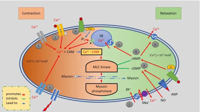
Figure 3 Contraction and relaxation mechanism of a smooth muscle cell as a schematic overview. The left half of the picture describes the contraction by electrical tension (1), receptor (2) and strain (3) dependent Ca2+ channels as well as by receptor- controlled (4) release of CA2+ ions from the sarcoplasmic reticulum (SR). Muscle relaxing mechanisms are shown in the right half of the figure. Decrease of the cytosolic CA2+ nucleotide by CA2+-ATPase(5,6)and 3Na+/Ca2+ exchange (7) as well as the effect of the cyclic nucleotide cAMP (8) and cGMP (9). Molecular contraction mechanism in the center of the image, CAM calmodulin, PLC phospholipase C, AC adenylyl cyclase, G G protein, IP3 inositol triphosphate, ANP atriopeptin, GC guanylylcyclase[1] (figure adapted).
The Ca2+ exchange is mainly ensured by voltage-dependent Ca2+ ion channels, including the L-type and T-type Ca2+ channels. Both channels are primarily responsible for the membrane potential of the cell and are activated and opened by depolarization so that Ca2+ ions can reach into the cell compartment.
The L-Type Ca2+ channels describe a group of ion channels with long-lasting activation time and are essential for the contraction of smooth muscles. T type Ca2+ channels, on the other hand, have a lower depolarization limit than the L type Ca2+ channels and have a shorter activation time. This fast activation behavior enables the cell to react to quick and rhythmically arriving action potentials.
The group of strain-dependent Ca2+ ions reacts to mechanical stimuli and allows depolarization and influx of Ca2+ by mechanical stretching of the cell membrane[26,29–31].
Another group of Ca2+ ion channels is the stored operated channels, which open after the intracellular Ca2+ stores of the cell are depleted and to refill the cellular Ca2+ storage. The exact physiological function and meaning of these channels are not yet clear at this stage. Furthermore, other ion channels can be summarized, which have a physiological and pathophysiological influence on intracellular Ca2+
levels and regulate the myogenic tone. These group of channels include, voltage-controlled K+ channels, Ca2+-activated K+ channels with high conductivity, ATP-sensitive K+ channels, ryanodine receptors, inositol 1,4,5-trisphosphate receptors (IP3Rs), and a multitude of channels of transient receptor[31,32].
12 4.3. Hypertension
A continuous pathological increase in arterial blood pressure is called arterial hypertension. The subjective perception of increased blood pressure varies from imperceptible to severe impairment.
Therefore, hypertension is classified according to various ranges of blood pressure. As soon as the general blood pressure at rest overcomes 140-159mmHg and or systolic 90-99mmHg diastolic blood pressure, a person is diagnosed with hypertension.
Table 1 High Hypertension categorization according to the level of blood pressure [1]
Classification Systolic mmHg Diastolic mmHg
Optimum <120 <80
Normal 120-129 80-84
High-Normal 130-139 85-84
Mild Hypertension (1.) 140-159 90-99
Moderate Hypertension (2.) 160-179 100-109
Severe Hypertension (3.) >= 180 >= 110
Isolated systolic hypertension >=140 <90
Hypertension can be divided into two subgroups, primary (or essential) and secondary hypertension, whereas the pathogenesis of primary hypertension cannot be defined in 90-95% of all cases. Secondary hypertension is caused by another primary disease like renal, cardiac and nervous dysfunction, or endocrine imbalance. Both instances can be pathologically subdivided to increase peripheral resistance and increased heart time volume, leading to increased systolic blood pressure, as the diastolic pressure stays normal.Due to the increased load on the peripheral system, structural changes and adaptations of the vascular tissue can occur, which can manifest themselves in the compression of the connective tissue and ECM as well as cellular stiffening. In most cases, hypertension is often a hybrid of described subgroups.
The pathogenesis of primary hypertension is mostly unknown, but various factors were identified, inducing hypertensive pathogenesis.
Genetic deposits could be a decisive factor in the development of hypertension [33]. The most common are polygenic mutations that are responsible for a pathological increase in blood pressure due to malfunctioning of the genomic material, which has been demonstrated in various twin studies.
In contrast, Liddle Syndrome is a rare example of monogenetic defects, which mutates the tubulin sodium channel of the distal kidney epithelia, increasing the resorption of water and hypertonic saline[34,35].
Psychosocial factors, in which acute racket and stress might amplify the activation of sympathoadrenal systems, causing increased vascular contraction through neurotransmitters. In addition, excessive sodium intake, obesity and regular alcohol consumption promote pathogenetic development. The coherence of excessive NaCl intake and hypertension was proved by only a partial group of global hypertension patients. It is assumed that the high consumption of NaCl increases cytosolic Na+, which deranges the Na+/ Ca2+ exchange, resulting in an increased intracellular Ca2+ level and a steady increased cellular tone[36].
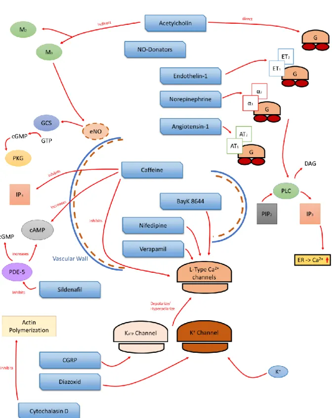
Moreover, a disbalance of the body´s natural vasoactive substances like eNO, ET1, AT2[37–39] or imbalanced of the endocrine system affect the blood regulation significantly [Figure 4]. Patients with diagnosed hypertension showing an increased level of catecholamines and norepinephrine in the blood sera, leading to increased vascular tone[40,41]. In the case of pregnancy hypertension, elevated ET1 levels were often found in the blood of affected women.
13
To understand the multiple pathogenesis and their connections to the genomic level, multidirectional pathways can be described using big data analysis and so-called signature genes can be identified, which may be responsible for the development of hypertension[42–44].
Figure 4Overview and progress of different causes of high blood pressure[1] (figure adapted)
The development and problems of high blood pressure can also be traced back to changes in the vascular cells. Many of the already mentioned causes lead to uncontrolled cell proliferation or calcification at the macroscopic level, which in turn leads to narrowing of the vascular lumen[45]. As the number of cells increases, the elasticity of the vessel wall is also impaired.
Genetic defects, mechanical overload and increased cell mass, can lead to severe changes in the connective tissue of the vessel wall[12,46]. In particular, the additional production and uncontrolled restructuring of the extracellular matrix can lead to reduced elasticity, strength or defective structures such as fibrosis[47].
Likewise, long-term biochemical and biomechanical changes cause adjustments of the ion channels and receptors located in the cell membrane[48,49]. Such adaptations may shift the resting or activation potential, making the cell more difficult to depolarize, or in the opposite, keep the cell depolarized all the time [50].
Till now, there is no pharmaceutical cure for hypertension. Only a timely adjustment of blood pressure during the pharmacological treatment can be achieved. In severe cases, surgical interventions can diminish hypertension long-dated. There are various drugs to relieve the symptoms of high blood pressure, but due to the multiple sources of hypertension, patients are usually medicated by several compounds at once. The most common anti-hypertensive regulating agents are ACE-inhibitors, AT1- antagonists, Potassium inhibitor, beta-antagonists and thiazides. Aside from their desired effects, the drug administration goes along with side effects, prolonging the pharmaceutic adjustment and additional ailments.
14
4.4. Mechanical Cellular Stimulation
Mammalian cells need mechanical tension to survive. The mechanical, environmental conditions determine the orientation, growth behavior, force development and essential functions of the cells.
Cells in the human body are permanently exposed to mechanical stress caused by movement, digestion, respiration and in the vascular system by the pulsatile ejection of the heart.
Since the mechanical stimulus of the cell plays such a crucial role in tissue development[51] and also in regenerative behavior [52], a wide variety of methods for mechanical cell stimulation have been developed over the last decades[53–55].
Mechanical stimulation at the cellular level can be subdivided according to physiological stimuli so that methodologies can be found in the literature that exposes cells to compression, shear stress and mechanical strain. Besides, the stimulation protocols differ in static and dynamic mechanical load.
Static stimuli describe a one-time change in the stress situation to which the cells are exposed. Thus, some studies observe the growth behavior of cells on growth surfaces with varying degrees of solidity.
Especially in stem cell research, these protocols gain great attention since the maturation of cardiomyocytes, in particular, is strongly influenced by the growth condition mechanics[56,57].
For the field of vascular research, cyclically dynamic mechanical stimulation methods of shear stresses and strain stresses are of particular interest. Thereby, cells are exposed to different, rhythmic and recurrent stimuli. The rheology of blood vessels has an essential effect on the cellular characteristics of the vascular endothelium. Thus, it has already been possible to show the correlation of mechanical stress and metabolites/messenger secretion[6].
Due to the permanent cyclical blood ejection of the heart, the cells of the vascular system experience mechanical stimulation throughout the organism. To understand the cellular response to this mechanical stimulation and also changes, methods for cellular stretching are of particular interest.
To date, studies have shown that cyclical mechanical stimulation of vascular smooth muscle cells has a fundamental influence on their phenotype[58][59], biomechanics, cell connection, signaling[60][61]
and restructuring of both intra- and extra-cellular structures[18,62,63]. All these findings serve for a detailed physiological understanding to better understand pathological developments and regenerative medicine in medical applications as well as biological and biomechanical research[19,64].
4.5. Biomechanics
Biomechanics is an interdisciplinary discipline that applies the laws of mechanics and material sciences to biological organisms, biomaterials and tissue. The resulting data are used in research and medicine to explain mechanobiological phenomena based on experimental and mathematical models.
Over the last decades, mechanical tissue properties have been studied in detail and their fields of application are as diverse as the methods used to determine them[46]. Thus, classical mechanical material parameters serve the necessary physiological characterization and the definition of material requirements for medical and tissue-engineered implants[65,66]. Due to the experimental research work by G.A.Holzapfel mechanical properties of vessels could be measured by dissections and tissue equivalents. This enables to determine both, the tensile strength and the deformation properties of biological material consisting of ECM and cells[67,68].
In extension, pathological or aging-related findings can also be correlated with biomechanical models, which can be reconstructed and improve fundamental understanding and prognosis[69,70].
In classical physiology, biomechanics is mostly applied to the study of movement and tissue consolidation or contraction and relaxation. At the beginning of the last century, the contractile force
15
of an isolated and perfused heart could be measured using the so-called Langendorff heart, which is still used in physiology today, especially for pharmacological investigations[71].
The measurement of force variations due to muscle shortening can be projected onto all other muscular structures, including vascular structures. Especially this requirement is still used today in so- called aortic rings, whose cross-sectional deformation provides detailed information about mechanobiological processes[72–75]
4.5.1. Cellular Force Measurement
In addition to the macroscopic level, the biomechanical laws can also be applied to the microscopic and subcellular levels. Taking biophysical laws into account, complex and dynamic mechanical properties of individual cells and cell organelles can be measured[76–78].
The state of the art currently offers various methods to measure force changes of monolayers and single cells, both directly and indirectly. Due to different hypotheses of force distribution and stress development of the cells[79,80], diverse force measurement methods are considered, which can measure uniaxial, monoaxial or bi-axial stresses[81–83]. The high sensitivity of the methods is essential to resolve cellular forces in the pN-µN range.
One of the most common methods for indirect force or stress measurement is atomic force microscopy and traction force microscopy[84–86]. Both techniques allow deformations of the growth substrate to be measured by induced cell forces. In addition, the atomic force microscopy method allows us to assess the strength of single cells. Due to material stiffness analysis, it is possible to infer cell contraction or relaxation indirectly[87].
Probably the most common method for measuring cell forces is tensile testing of cell cultures on defined growth substrates. Usually, biocompatible polymers or matrices in rectangular shape are prepared from which the induced forces of the cells are determined by means of using load cells.
However, the geometry and fixation of these preparations usually generate different stress fields on the carrier material by the cells. Thus, in most cases, only an averaged and summed force generation of the cells can be shown. In order to counteract this problem, the group around Kit Parker has developed a procedure that enables the contraction of the cells through the deformation of eyelash structures. By deforming the "lash", a mechanical model can be used to determine the force of shrinking from the eyelash bending radius[88]. Micro-structuring of the carrier material across methods can help to direct the generated strength to a particular preferred direction, which makes the evaluation more precise and sensitive[89].
The bi-axial stress measurement neglects the directional stress distribution, as the stress distribution is the same in all directions in biaxial systems. For bi-axial stress measurement, methods that also serve for mechanical cell stimulation are usually used. Deviations of the parameters for precise adjustment of the strain can thus be indirectly inferred from changes in cell tension[60,90]. The best known and most widely used method for bi-axial stimulation is the FlexCell system, which transfers the principle of the simple tensile test in a round geometry[91,92].
The choice of methodology is therefore extremely versatile and can be made with the scientific question. While most in-vitro cell force methods currently serve for basic research and laboratory development, clinical approaches continue to pursue the use of tensile tests and deformation analyses of real tissues in-vitro.
16 4.6. In-vitro Model
Developments in cell culture technology and tissue engineering make it gradually easier to produce tissue equivalents from cell cultures. Besides the less ethically questionable handling towards living tissue, isolated cells benefit the reduction of mechanically influencing factors. So that mechanical cell force alterations can be characterized more precisely without the passive contractive influence connective tissue.
Cell culture technics and tissue engineering enables the cultivation of living tissue or tissue equivalents under in-vitro condition. Such models are idealized cellular systems for biological analyses. Due to the substantial simplification of a living organism, these models enable to multitudinous aspect elimination compared to in-vivo samples.
From an ethical point of view, such in-vitro models are much more favorable than animal testing or experiments with living tissue dissections, as the specimen material can be cultured under laboratory conditions and reproduced to a certain extent. According to the 3Rs principle, the development of in vitro models makes it possible to reduce and replace animal experiments and to make the experimental set-ups more reproducible and ethically acceptable[93]. Nevertheless, for functional testing, animal testing is still an important matter as the functional testing of pharmaceutics to complete organisms cannot be simulated entirely[94].
Current studies show that there is an urgent need for disease models and functional physiological assays to support and improve medical findings and diagnostics in order to understand sophisticated mechano-biological phenomena of physiology, pathophysiology and aging in-vitro[95,96].
The scientific application and implementation of arterial in-vitro models are versatile and depend on the scientific problem and the phenomena to be observed, ranging from single-cell models and monolayers to three-dimensional tissue-equivalent or organoids. The knowledge gained from these models is used to analyze functional physiological processes concerning biochemical, electrophysiological and biomechanical phenomena. Beyond that, they are also used to answer more complex questions that arise at the molecular level or in regenerative medicine, in which processes such as angiogenesis or wound healing are central[56,97,98].
A great variety of model designs depend on the scientific question and observing phenomena and ranging from single-cell model and monolayer to three-dimensional tissue-equivalent or organoids.
The most fundamental way to set up a cell culture model is the cultivation of a single monolayer of one cell type. Due to the isolation of cells, it is easy to characterize and interpret single-cell physiology and experimental findings. In terms of studying rheological effects within the vasculature, flow chambers and organ on a chip approaches became quite famous in recent years[99–103].
Especially while studying vascular walls, the cross-communication between smooth muscle cells and endothelial cells is a highly interesting topic, with high medical impact. To get inside to this topic, it requires a co-cultivation of both cell types, allowing intercellular signaling. According to literature, there are three main subgroups of co-cultures: Conditioned co-culture, direct co-culture and three- dimensional co-culture.
A conditioned co-culture offers no direct contact between the cells. Both cell types are physically separated from each other. Such systems can be realized by so-called inlet membranes, which separates two different culture compartments, but allows cell signaling and molecule transfusion throughout the separation layer. Another approach to achieve a conditioned cell-culture is to exchange or mix the media of two distinct cell cultures. Hence, the effect of metabolites and signaling molecules can be evaluated by employing a mono-culture[104,105].
17
In contrast to conditioned co-cultures, direct co-cultures describes the cultivation of two different cell types in the same cell culture compartment. Respecting cellular communications, such systems are closer to physiological conditions, but also exacerbate the interpretation and analysis of differentiated results[106]. Concerning the physiology and experimental setup, it is possible to arrange the cell in a physiological pattern by gel structures or decellularized tissue[107–109]. Such a setup benefits the functionality of an in-vitro model as the arrangement of cells can be simulated equally to an in-vivo system[110].
Working with vascular three-dimensional cell models requires a well-defined matrix equivalent. In most cases, three-dimensional structures are generated by gel-like structures made of collages, matrices or similar[111–113]. The use of such systems usually has a relatively compact and stable structure due to the matrix components and is, therefore, mostly applied in biochemical and molecular biological research.
A recent trend that is closely related to stem cell research is the application of organoids, which commonly suitable for the study at hollow organs. Organoids are spherically modeled organs made of primary or stem cells[56], promising to have great physiological properties and having similar in-vivo arrangement.
4.6.1. CellDrum Technology
The CellDrum technology was firstly described by Jürgen Tzerwiki, Institute for Bioengineering FH Aachen, in 2001 [114–116]. The CellDrum was developed to estimate the biomechanical properties of standardized cellular monolayer and 3D tissue equivalents[117]. The CellDrum is methodologically an important technology that, in contrast to known indirect electrophysiological methods, quantifies direct lateral bi-axial generated cellular forces in micronewton (µN). Previous studies have shown that the CellDrum is a useful tool to elucidate the mechano-pharmacological characterization of human- induced pluripotent cardiomyocytes[118,119]. This has been shown in the experimental work by M.Goßmann, which has also demonstrated that a three-dimensional arrangement of different types of cells is possible[120]. Besides the possibility to biomechanically analyze cells, the softness of the membranes results in a unique growth surface, which provides mechanically seen an almost physiological growth environment.
Over the past decades, the CellDrum went through various optimizations and changes, leading to the current state of the art, which will be presented in this work. Especially the description of standard operating protocols and automatization are described in detail in a recent publication by R.Bayer[121,122].
The recent developments of the CellDrum technology showed the versatile application of the standard CellDrum. In addition to mechanobiological characterization, the system can be used to expose the cell to mechanical stress. Combining both techniques could be an excellent foundation for biophysical and biomechanical basic research[122,123].
18 5. Material & Methods
Table 2 Chemicals
Material Producer Vendor Cat.No.
Cell culture
M200 Gibco ThermoFisher M200500
M231 Gibco ThermoFisher M231500
DMEM-GlutaMAX Gibco ThermoFisher 31966021
DMEM + F12 Gibco ThermoFisher 11330057
M199 Gibco ThermoFisher 12340030
DPBS Gibco ThermoFisher 14190250
Trypsin/EDTA (0,05%) Gibco ThermoFisher 25300054
FBS Gibco ThermoFisher 16140071
SMGS Gibco ThermoFisher S00725
SMDS Gibco ThermoFisher S0085
LSGS-Kit Gibco ThermoFisher S003K
Collagen Type I Merck Merck 9007-34-5
Human Collagen Type IV Merck Merck CC076
PureCol EZCollagen Type I Cellsystem Cellsystem 5074-35ml
Fibronectin Bovine Plasma Merck Merck F1141-2MG
DMSO Roth Roth 0728.1
Trypan Blue Solution 0,4% Gibco ThermoFisher 15250061
LIVE/DEAD®
Viability/Cytotoxicity Kit
Invitrogen ThermoFisher L3224
LDH-KIT Invitrogen ThermoFisher -
ET1-ELISA Kit Invitrogen ThermoFisher EIAET1
Staining
Alexa Fluor® 488 – Phalloidin
Invitrogen ThermoFisher A12379
CellTracker™ Deep Red Invitrogen ThermoFisher C34565 CellTracker™ Green CMFDA Invitrogen ThermoFisher C7025 Anti-Actin, α-Smooth
Muscle - Cy3™ antibody
Sigma-Aldrich Merck C6198-100UL
Antibody CD31 Abcam Abcam Ab28364
Gen Analysis
SYBR Green Sigma Aldrich Merck 163795-75-3
RNeasy Mini Kit Qiagen Qiagen 74106
QIAshredder Qiagen Qiagen 79656
miRNA Kit Qiagen Qiagen 217604
QuantiNova Reverse Transcription Kit
Qiagen Qiagen 205411
SsoAdvanced Universal SYBR Green Supermix
BioRad BioRad 1000076382
Ultra Pure Water PCR grade Invitrogen ThermoFisher AM9935
ROTIPURAN®Ultra Carl-Roth Carl-Roth HN68.3
RNAseZAP Sigma Sigma R2020-250ML
![Table 1 High Hypertension categorization according to the level of blood pressure [1]](https://thumb-eu.123doks.com/thumbv2/1library_info/3712398.1506518/17.892.112.789.276.447/table-high-hypertension-categorization-according-level-blood-pressure.webp)
![Figure 4 Overview and progress of different causes of high blood pressure[1] (figure adapted)](https://thumb-eu.123doks.com/thumbv2/1library_info/3712398.1506518/18.892.111.761.189.618/figure-overview-progress-different-causes-pressure-figure-adapted.webp)