Effect of reduced expression of sorcin on Ca handling, 2+
in vivo heart function and transcriptional regulation
Inaugural-Dissertation zur Erlangung des Doktorgrades
der Mathematisch-Naturwissenschaftlichen Fakultät der Universität zu Köln
vorgelegt von Malik Alock Suchitra aus Muzaffar Nagar, India
im Dezember 2005
Berichterstatter: Herr Prof. Dr. Jens Brüning Frau Prof. Dr. A.A. Noegel
Herr Prof. Dr. Robert H. G. Schwinger
Tag der mündlichen Prüfung: 10 February 2006
To My Parents
TABLE OF CONTENTS
Abbreviations Abstract
Zusammenfassung 1 Introduction
1.1 Calcium cycling in the normal heart 1
1.2 Altered Ca
2+cycling in the hypertrophied and failing heart 3 1.3 Role of Ca cycling proteins in the altered calcium cycling
2+3
1.3.1 Ryanodine receptors-A macromolecular complex 4 1.3.2 Proteins interacting with ryanodine receptors 4 1.3.3 Sarcoplasmic reticulum Ca ATPase
2+6
1.3.4 Phospholamban 6
1.4 Sorcin and its role in calcium cycling 8
1.4.1 Structure of sorcin 8
1.4.2 Expression and subcellular localization of sorcin 8 1.4.3 Interaction of sorcin with other calcium binding proteins 10 1.4.4 Adenoviral mediated gene transfer and transgenic mouse model 10
1.5 Aims of the thesis 12
2 Materials and methods
2.1 Materials 13
2.1.1 Animals 13
2.1.2 Materials (reagents, buffers, cell culture medium, 13 enzymes and antibodies)
2.2 Methods 19
2.2.1 Generation of adenoviral antisense sorcin GFP 19 2.2.2 Isolation of adult rat cardiomyocytes 22 2.2.3 Transfection of cardiomyocytes 22
2.2.4 Protein preparation from the transfected cardiomyocytes 23
2.2.5 Extraction of transcription factors from transfected 24
cardiomyocytes
2.2.6 Quantitative immunoblotting 24
2.2.7 Immunofluorescence in transfected cardiomyocytes 26 2.2.8 Contractility measurement in transfected cardiomyocytes 26 2.2.9 Reverse transcriptase polymerase chain reaction 27 2.2.10 Calcineurin activity measurement in transfected cardiomyocytes 30 2.2.11 Isolation and purification of recombinant sorcin protein 30 from pGEX2TK vector
2+
uptake 31
2.2.12 Sarcoplasmic reticulum Ca
2.2.13 Calcium transient measurement in transfected cardomyocytes 31 2.2.14 Catheter based adenoviral gene delivery into rat hearts, 32 2.2.15 Echocardiography and morphological assessment 32
2.2.16 Statistical analysis 32
3 Results
3.1 Generation and amplification of adenoviral vectors 33 3.2 Optimization of transfection with adenoviral vectors 34
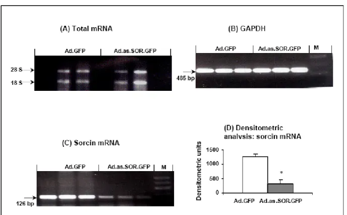
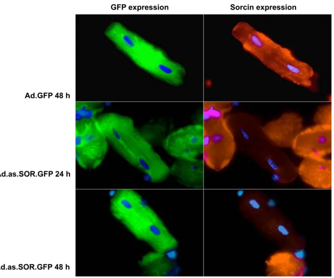
3.3 Downregulation of sorcin by Ad.as.SOR.GFP 34 3.3.1 Downregulation of sorcin mRNA 35 3.3.2 Downregulation of sorcin protein 35 3.3.2.1 Immunofluorescence staining of transfected cardiomyocytes 36 3.3.2.2 Immunoblotting of transfected cardiomyocytes 36 3.4.1 Measurement of contractility in transfected cardiomyocytes 37
2+
transient measurement in cardiomyocytes 39 3.4.2 Intracellular Ca
3.4.3 Force-frequency relationship in transfected cardiomyocytes 41 3.4.4 Pharmacological interventions in transfected cardiomyocytes 42 3.5 Expression and purification of recombinant GST-sorcin 43 3.6 Sarcoplasmic reticulum Ca
2+uptake 43 3.7 Effect of down regulation of sorcin on expression 45
of calcium handling proteins
3.7.1 Ryanodine receptor 45
2+
ATPase 45
3.7.2 Sarcoplasmic reticulum Ca
3.7.3 Phospholamban 47
3.7.4 FK binding protein 12.6 50
3.7.5 Triadin 51 3.8 In vivo adenoviral based sorcin gene delivery in adult rat hearts 52 3.9 Calcineurin signaling in the transfected cardiomyocytes 56
3.9.1 Calcineurin expression 57
3.9.2 Calcineurin enzymatic activity 57 3.9.3 Expression and nuclear translocation of NF-AT in the 59 transfected cardiomyocytes
3.9.4 GATA-4 expression in the transfected cardiomyocytes 61 3.9.5 Expression of cardiomyopathic marker genes in 62
the transfected cardiomyocytes
4 Discussion
4.1 Depressed levels of sorcin and excitation contraction coupling 65
4.2 Sorcin and SR Ca
2+content 66
4.3 Decreased expression of sorcin: Effect on force-frequency 67 relationship and β-adrenergic stimulation 4.4 Decreased expression of sorcin and functional remodeling 69
of the heart
4.5 Calcineurin-NF-AT signaling 70
4.6 Conclusion 73
5 References 74
6 Erklärung
7 Acknowledgements 8 Publication and abstracts 9 Lebenslauf
10 Appendix
Abbreviations
(m/m) Mass/Mass
°C Degree Celsius A, mA Ampere, Milliampere
Ad Adenovirus
Ad.as.Sor.GFP Adenovirus antisense sorcin vector expressing Green fluoresecent protein Ad.GFP Adenoviral vector expressing Green fluorescent protein
Amp Ampicillin
ANF Atrial natriuretic factor
AP Action potential
ATP Adenosine triphosphate ARC Adult rat cardiomyocyte β-AR β-Adrenergic receptor
β-MHC β-Myosin heavy chain
BNP B-type natriuretic peptide BSA Bovine serum albumin
Ca
2+Calcium ions
cAMP cyclic Adenosine monophosphate
cDNA complementary DNA
cGMP cyclic Guanosine monophosphate CICR Calcium induced calcium release
Ci Curie
CMV Cytomegalovirus
cpm Counts per minute
CSQ Calsequestrin
DAPI 4',6-Diamidino-2-phenylindole dihydrochloride DCM Dilated cardiomyopathy
DEPC Diethylpyrocarbonate DHPR Dihydropyridine receptor DMSO Dimethylsulfoxide
dNTP Deoxyribonucleotide triphosphate DTT 1,4-dithiothreitol
DU Densitometric units
E-C coupling Excitation contraction coupling
ECL Enhanced chemiluminiscence reaction
EDTA Ethylenedinitrilotetraaceticacid disodiumdihydrate EGTA Ethylene glycol-bis (β-Aminoethyl ether) tetracetic acid FKBP12.6 FK binding protein 12.6
g, mg, µg Gram, Milligram, Mikrogram
GAPDH Glyseraldehyde-3-phosphate dehydrogenase GATA4 GATA binding protein 4
GFP Green fluorescent protein h Hour
HEPES 2-[4-(2-Hydroxyethyl)-1-piperazinyl]-ethane-sulfonic acid HRP Horse radish peroxidase
Hz Hertz [1/s]
IgG Immunoglobulin G
kb Kilobase
K
DDissociation constant kDa Kilodalton
l, ml, µl Liter, Milliliter, Microliter LB Luria bertani medium LTCC L-Type calcium channel mm, µm, nm Millimeter, Micrometer, Nanometer
mM, µM, nM Millimol(ar), Micromol(ar), Nanomol(ar) Mean Arithmatic mean
MG Molecular weight in g/mol
MOI Moieties of infection or Multiplicity of infection (Virus/Cell) MOPS 3-(N-Morpholino)propanesulfonic acid
mRNA Messenger ribonucleic acid n Number
+ 2+
NCX Na Ca -exchanger
NF-AT Nuclear factor of activated T cells NFU Normalised fluorescent units NGS Normal goat serum
OD Optical density
PAGE Polyacrylamide gel electrophoresis
PBS Phosphate buffered saline
pCa Negative logarithm of the calcium ion concentration pfu Plaque forming units
pH Negative log of the H
+concentration
PKA Proteinkinase A
PLB Phospholamban
PMSF Phenylmethylsulphonylfluoride PNPP p-Nitrophenyl phosphate PVDF Polyvinyldifluoride
RT Room temperature
RyR2 Ryanodine Receptor s, ms Second, Millisecond SDS Sodium dodecyl sulphate SEM Standard Error Mean
SERCA 2a Sarcoplasmic reticulum Ca -ATPase Isoform 2a
2+SR Sarcoplasmic reticulum TAE Tris acetate EDTA
TBS Tris buffered NaCl TBST Tris buffered NaCl with tween
TEMED N,N,N`,N`Tetramethylendiamine Tris Tris(hydroxymethyl)-aminomethane TTCC T-type calcium channel
T to peak Time to peak
U Enzymatic activity
U/min Units per minute V, mV Volt, Millivolt
vs. versus
Abstract
An adequate and efficient Ca
2+handling is the essential condition for effective functioning of the heart. Decreased peak systolic Ca
2+with prolongation of the duration of Ca transient, slower
2+rates of SR Ca
2+uptake and various other alterations in Ca
2+efflux leading to elevation in diastolic Ca
2+are the key features of the failing heart. These key alterations in heart failure necessitate further evaluation of proteins involved in these regulatory mechanisms. Sorcin, a penta E-F hand family protein associates with cardiac ryanodine receptors, L-type Ca
2+channel as well as SR Ca
2+ATPase and modulates excitation-contraction coupling in the heart. The present thesis aims at understanding the role of sorcin in calcium handling, the effect of decreased level of sorcin on remodeling of the heart and subsequent transcriptional regulation, by using the adenoviral antisense RNA approach.
A decrease in the endogenous sorcin expression (75% on the mRNA level and 53% on the protein level) was obtained in the adult rat cardiomyocytes. The decreased amount of sorcin resulted in reduced cell contractility and significantly depressed Ca
2+transient amplitude accompanied with the decreased rate of relaxation in the transfected cardiomyocytes. The increase in stimulation frequency was associated with a negative force-frequency relationship in cardiomyocytes with depressed sorcin, mimicking the behavior exhibited by the failing human cardiomyocytes. However, the β-adrenergic stimulation was unaltered. An oxalate facilitated Ca
2+uptake assay indicated decreased Ca
2+uptake by the sarcoplasmic reticulum in the cardiomyocytes with decreased expression of sorcin. Moreover, incubating the sarcoplasmic reticulum vesicle preparations with recombinant sorcin (1 µM) enhanced the SR Ca
2+uptake and brought it back to the control level. The depressed expression of SR Ca
2+ATPase on the mRNA as well as protein level was also observed. The expression of ryanodine receptors, triadin and phospholamban was unaltered, while a mild increase in the expression of FKBP12.6 was observed. In vivo downregulation of sorcin, achieved by the catheter based cardiac specific gene delivery resulted in severe chamber dilation. Echocardiography revealed ventricular enlargement, decreased heart rate and increased chamber dimension indicating dilated cardiomyopathy in the hearts with depleted levels of sorcin. After 14 days the animals were sacrificed and an increase in the heart weight was observed. In addition, upregulation of calcineurin expression and higher phosphatase activity was observed which was accompanied with increased dephosphorylation of NF-AT
C3. GATA4 expression was significantly upregulated in the antisense sorcin transfected cardiomyocytes. The mRNA expression of hypertrophic marker gene β-myosin heavy chain was upregulated, while expression of atrial natriuretic factor and B-type natriuretic peptide was unaltered.
In conclusion, using the antisense mRNA approach for sorcin it can be concluded that
downregulation of sorcin diminishes the cardiac contractile performance and leads to the
ventricular remodeling of the heart. This process is at least partially due to the activation of
calcineurin-NFAT signaling pathway. The antisense approach proves to be a valuable tool to
identify targets that modulate cardiac contractility and may open new avenues for the treatment
of myocardial diseases with diminished cardiac output such as heart failure.
Zusammenfassung
Für eine effektive Herzfunktion ist eine effiziente Ca
2+-Homöostase wesentlich. Vermindert systolisch freigesetztes Ca
2+, eine Verlängerung des Ca
2+-Transienten und eine reduzierte Aufnahmerate des sarkoplasmatischen Retikulums (SR) sind wesentliche Veränderungen einer Herzinsuffizienz. Diese Störungen machen eine weitere Untersuchung von Proteinen notwendig, die die myokardiale Ca
2+-Homöostase regulieren. Sorcin ist ein EF-Hand Protein, dass mit dem kardialen Ryanodin-Rezeptor, dem L-Typ Ca
2+-Kanal sowie mit der SR Ca -ATPase assoziiert
2+ist und die elektromechanische Kopplung am Herzen mitreguliert. Die vorliegende Doktorarbeit hatte zum Ziel, die Rolle von Sorcin für die Ca
2+-Homöostase, die Bedeutung einer verminderten Expression von Sorcin auf die Herzentwicklung und die transkriptionale Regulation mit Hilfe von adenoviralem Antisense Sorcin zu untersuchen.
Eine Reduktion der endogenen Sorcin Expression (74% der mRNA Expression und 53% der Proteinexpression) konnte mit Hilfe dieses adenoviralen antisense Ansatzes in isolierten Rattenkardiomyozten erzielt werden. Die verminderte Sorcin Expression führte zu einer reduzierten Zellkontraktion, einer verminderten Amplitude des Ca
2+-Transienten und einer Verlängerung der Relaxationsrate. Erhöhung der Stimulationsfrequenz zeigte eine negative Kraft-Frequenz-Beziehung (KFB) in Kardiomyozyten mit verminderter Sorcin Expression, die mit der KFB an insuffizienten, menschlichen Kardiomyozyten vergleichbar ist. Die β-adrenerge Stimulation blieb erhalten. Der Ca
2+-Uptake mit Oxalat zeigte verminderte Aufnahmeraten des SR an Kardiomyozyten mit verminderter Sorcin Expression, Inkubation mit rekombinantem Sorcin (1µM) führten zu einer erhöhten SR Ca
2+-Aufnahmerate. Zudem wurde eine reduzierte Expression der SR Ca
2+-ATPase nachgewiesen. Die Expression des Ryanodin Rezeptors, von Triadin und Phospholamban blieb unverändert, während die Proteinexpressin von FKBP 12,6 erhöht war. Reduktion der Expression von Sorcin mit Hilfe eines katheterbasierten, intrakoronaren Applikationsweges in vivo führte zu einer schweren Herzdilatation.
Echokardiographie der transfizierten Herzen zeigte eine ventrikuläre Herzvergrösserung, eine verminderte Herzfrequenz und einen erhöhten end-diastolische Durchmesser. Nach 14 Tagen Transfektion wurde morphologisch eine Zunahme des Herzgewichtes gesehen. Zudem wurden an Antisense Sorcin transfizierten Kardiomyozyten eine erhöhte Calcineurin Aktivität und Expression, verbunden mit einer höheren NF-AT
C3Dephosphorylierung, und eine erhöhte Expression der GATA4 Expression gefunden. Das Hypertrophiemarker-Gene „β- Myosin Heavy Chain“ war erhöht, während die Expression des atrialen, natiuretischen (ANP) und gehirnspezifischen, natiuretischen Peptiden (BNP) unverändert blieb.
Zusammenfassend werden durch eine Verminderung der Sorcin Expression mit Hilfe eines
adenoviralen Anti-sense Ansatzes eine reduzierte kardiale Kontraktionskopplung und eine
dilatative Kardiomypathie ausgelöst. Dieser Prozess beinhaltet Störungen der Ca
2+-Homöostase
und Aktivierung des Calcineurin- NFAT Signaltransduktionsweges. Die adenovirale anti-sense
Strategie kann dazu verwandt werden, um Zielproteine bei der Entstehung einer Herzinsuffizienz
zu identifizieren, die die kardialen Kontraktilität und Leistung insbesondere bei verminderten
kardialen Pumpleistung verbessern können. Dieser Ansatz kann in der Entwicklung neuer
Therapieformen bei der menschlichen Herzinsuffizienz verwandt werden.
1. Introduction
1.1 Calcium cycling in the normal heart
Calcium (Ca2+) ions function as ubiquitous intracellular messengers that regulate many different cellular processes in cardiac myocytes (Frank et al., 2003, Bers, 2005). Ca2+ plays a requisite role not only in the electromechanical and contractile activity in the excitation contraction coupling (E-C coupling) (Bers, 2002), but also in modulating E-C coupling by activation of kinases and phosphatases which can modulate gene expression responsible for hypertrophic signaling and heart failure (Chien et al., 2003).
The process ensuring contraction and relaxation through a series of events from electrical excitation of the myocyte to the contraction of the heart is termed as excitation contraction coupling. Depolarization of the plasma membrane during the cardiac action potential activates voltage gated L-type Ca2+ channels (LTCC or dihydropyridine receptors) leading to a mild Ca2+ influx into the cytosol which in turn triggers a massive release of Ca2+ from the sarcoplasmic reticulum through ryanodine receptors (RYR2), this phenomenon is termed as calcium induced calcium release (CICR) (Fabiato, 1985). The tremendous increase in cytoplasmic Ca2+ concentration (almost 10 fold) leads to the actin-myosin cross bridge formation, which is activated by Ca2+ binding to troponin C and ultimately results in the contraction of myocyte (Solaro et al., 2002, Brixius et al., 2002).
The increase in cytosolic Ca2+ concentration during the contraction is immediately followed by Ca2+ removal, which results in deactivation of contractile machinery and myocardial relaxation during diastole. Cytosolic Ca2+ is pumped back into the sarcoplasmic reticulum (SR) by sarcoplasmic reticulum Ca2+ ATPase (SERCA2a) (Bers, 2002, Frank et al., 2003). Activity of SERCA2a is under the control of phospholamban (PLB), which in the nonphosphorylated form inhibits SERCA2a activity while phosphorylation of PLB reverses this inhibition (Koss et al., 1996, Frank et al., 2000).
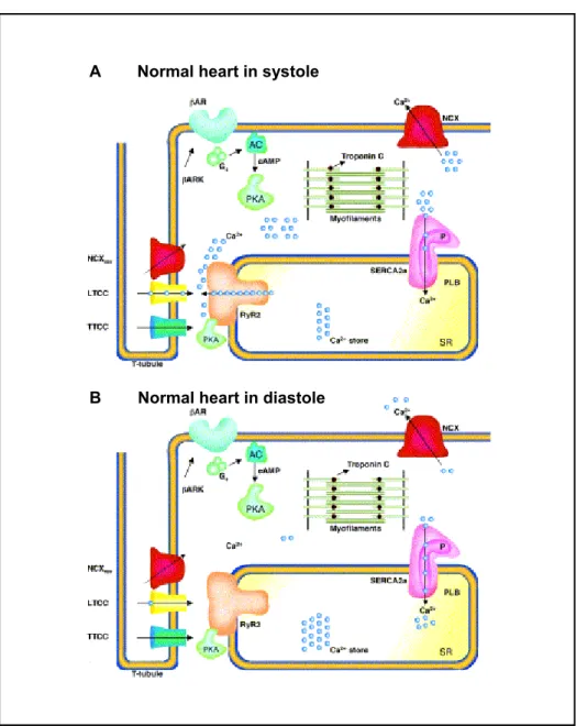
A
BB Normal heart in diastole A Normal heart in systole
Fig. 1.1 Excitation contraction (E-C) coupling in normal heart
(A) Depolarization due to action potential activates voltage gated L-type Ca2+ channels in the transverse tubule (yellow) resulting in a mild Ca2+ influx (blue circles) into the cytosol, Ca2+ influx can also occur through T-type Ca2+
channels (blue) or reverse mode Na+/Ca exchanger (NCX+ rev, red). The Ca2+ influx from LTCC triggers a profound Ca2+ release from sarcoplasmic reticulum (SR) via RYR2 (orange). During systole, there is a ten-fold increase in the intracellular Ca2+ reaching a concentration up to 1 μM. At this high concentration, free Ca2+ binds to troponin C (brown), inducing a conformational change that results in cross bridge formation in actin–myosin myofilaments (green) and muscle contraction. The β-adrenergic signaling pathway (green) is capable of increasing the E-C coupling gain. Agonist activation of β-adrenoceptors (β-AR) allows activation of adenylate cyclase (AC) by production of cyclic AMP (cAMP), which is responsible for the activation of protein kinase A (PKA, green). β-AR kinases regulate the β-AR agonist activation. (B) During diastole intracellular Ca2+ is pumped back from the cytosol to the SR via SR Ca2+ ATPase (SERCA2a, pink) and relaxation of the myocyte is achieved. SERCA2a is regulated by phospholamban (PLB, purple). Phosphorylated PLB (with ‘P’) does not inhibit SERCA2a under baseline conditions. A part of Ca2+ is extruded from the cytosol by sarcolemmal Na / Ca exchanger mode (NCX, red). + + Modified from: Wehrens and Marks (2003).
1.2 Altered Ca cycling in the hypertrophied and failing heart
2+Abnormal and altered intracellular Ca2+ handling plays a crucial role in the development of cardiac hypertrophy, heart failure and fatal ventricular arrhythmias (Wehrens et al., 2003). Depressed systolic intracellular Ca2+ transients, increased diastolic intracellular Ca2+ concentration and the slow rate of diastolic decay of intracellular Ca2+ concentration are the key features of the failing human heart (Beuckelmann et al., 1992, Schwinger et al., 1995). Reduction in the SR Ca2+ content (Lindner et al., 2002) and a decreased E-C coupling gain have also been reported in the failing human myocardium (Gomez et al., 1997).
Chronic hyperactivity of the sympathetic nervous system is one of the main characteristic of congestive heart failure (Chidsey, 1962). The constantly elevated high adrenergic activity leads to maladaptive changes in the β- adrenergic signaling pathway and results in the decreased expression and coupling of the β-adrenoceptors (Bristow et al., 1984), reduction in the coupling of the β2-adrenoceptors (Daaka et al., 1997), an increase in the expression of the inhibiting G protein Gi, an increase in the expression of the β-adrenoceptor kinases (Neumann et al., 1988, Ungerer et al., 1993) and depressed expression and function of adenylyl cyclases (Rockmann et al., 2002). Altered β-adrenergic signaling and depressed phosphatase activity in heart failure is responsible for hyperphosphorylation of L-type Ca2+ channels (Chen et al., 2002), NCX (Wie et al., 2003) and cardiac RYR2 (Reiken et al., 2003, Marx et al., 2002).
1.3 Role of Ca cycling proteins in the altered calcium cycling
2+Periodic change in Ca2+ concentration in cardiomyocytes is the prerequisite for cardiac contraction and relaxation. Various proteins associated with the SR, integrally regulate the vital periodic change in intracellular Ca2+ concentration ensuring the smooth functioning of the myocardium. Abnormal cardiac E-C coupling is the result of altered function of Ca2+ proteins responsible for intracellular Ca homeostasis. Altered expression 2+
and activity of SERCA2a (Schwinger et al., 1995,) phosphorylation of PLB (Dash et al., 2001), RYR2 (Marks et al., 2002), Na+/Ca+ exchanger and Na+ K+ ATPase (Schwinger et al., 1999) have been reportedly associated with various features of contractile dysfunction (Arai et al., 1993, Go et al., 1995, Hasenfuss 1994).
1.3.1 Ryanodine receptors – A macromolecular complex
Ryanodine receptors (RYR2), a 565 kDa homotetramer consisting of four monomeric subunits is the main SR Ca2+ release channel and plays a predominant role in E-C coupling (Marx et al., 2002). The subunit contains a high conductance Ca2+ selective pore, Ca2+ activation and inactivation sites, several phosphorylation sites and multiple binding sites for an array of endogenous regulators, which include ATP, Mg+ and calmodulin. Knock out of RYR2 gene in mice proved to be fatal and the mice died at embryonic day 10 with morphological abnormalities. Prior to death the knock out cardiomyocytes showed structurally abnormal mitochondria, highly vacuolated SR with elevated concentrations of Ca2+, suggesting that RYR2 is absolutely required for cellular Ca2+ homeostasis as a major Ca2+ release channel (Takeshima et al., 1998). RYR2 also functions as a scaffolding protein for various other proteins like FK binding proteins, Calmodulin (CaM), calsequestrin, triadin, junction, and several other proteins forming a macromolecular complex channel protein system (Bers, 2004) and alterations in these interacting proteins have been associated with the pathogenesis in the heart.
1.3.2 Proteins interacting with ryanodine receptors (a) FK binding proteins
FKBP 12.6, a member of immunophilin family of proteins associates with the cytosolic domain of cardiac RYR2 with high affinity (Marx et al., 2002). Disruption of the FKBP12.6 gene in mice resulted in cardiac hypertrophy in male mice, but not in female mice. Female mice hearts were normal, despite the fact that male and female knockout mice displayed similar dysregulation of Ca2+ release, the amplitude, duration of Calcium sparks and calcium-induced calcium release gain (Xin et al., 2002). Another study reported consistent display of exercise-induced cardiac ventricular arrhythmias that cause sudden cardiac death due to the ablation of FKBP 12.6 (Wehrens et al., 2003). FKBP 12.6 stabilizes RYR2 channel activity and inhibits the aberrant activation of RYR2 during diastole, the protein kinase A based phosphorylation of FKBP 12.6 causes dissociation of FKBP 12.6 from the RYR2 resulting in unstable RYR2 channel which can induce life threatening arrhythmias (Wehrens et al., 2004).
(b) Calmodulin
Calmodulin (CaM is a 16.6 kDa Ca2+ binding protein with four E-F hands existing as pairs on the bi-globular structure (Strynadka et al., 1989). At high [Ca]i levels, CaM activates its targeting partners by binding to them with high affinity. It is suggested that under physiological conditions CaM bound to RYR2 may inhibit open basal probability and alter the Ca2+ dependent activation of the RYR (Yamaguchi et al., 2003).
(C) Protein Kinase A, Calmodulin dependent protein kinase II and phosphatases
Protein Kinase A (PKA) is anchored to cardiac RYR via protein kinase A anchoring protein (AKAP) in the cytosolic domain of RYR2. PKA dependent RYR2 phosphorylation alters RYR2 gating and reportedly speeds up the time course of release without affecting the amount of released Ca2+ (Valdiva et al., 2000). In the complex physiological system PKA activation also increases ICa trigger and SR calcium load in such a way that the amount of SR Ca2+ release during E-C coupling is greatly enhanced.
Calmodulin dependent protein kinase II (CaMKII) is mainly expressed as the δ isoform in the heart and is also known to phosphorylate RYR2 and alter RYR2 activity (Wichter et al., 1991). Endogenous CaMKII increases the amount of SR Ca2+ release for a given SR Ca2+ content and ICa trigger in intact voltage clamped ventricular cardiomyocytes (Li et al., 1997). Transgenic mice overexpressing CaMKII δ exhibited increased diastolic calcium spark frequency and fractional SR Ca2+ release during E-C coupling despite a reduced SR calcium content and diastolic [Ca] (Maier et al., 2003). i
In heart failure, despite the increased phosphatase expression there is less phosphatase associated with RYR2 that could possibly lead to enhanced RYR2 phosphorylation in heart failure resulting in increased diastolic calcium leak (Bers, 2003).
(d) Calsequestrin, junctin, triadin and histidine rich Ca binding
2+protein
Cardiac calsequestrin is a 45.2 kDa high capacity moderate affinity Ca2+ binding protein localized at lumen of
the junctional SR in cardiac muscle (Scott et al., 1988; Frank et al., 2001). Calsequestrin (CSQ) undergoes a structural transformation after binding to calcium. Transgenic mice overexpressing more than 10-fold CSQ developed cardiac hypertrophy and the cardiomyocytes showed increased amount of SR Ca2+ content (Jones et al., 1998, Sato et al., 1998) but the twitch Ca2+ transients and contractions were depressed. Adenoviral mediated moderate overexpression of CSQ (2-4 fold) resulted in enhanced Ca2+ transient amplitude and increased SR Ca2+ load (Terentyev et al., 2003).
Triadin and junctin are present at the junctional SR and they interact with each other, CSQ and RYR2. The interaction between triadin, junctin and CSQ is suggested to influence indirectly RYR2 gating and Ca2+ release (Gyoerke et al., 2004). A histidine rich Ca2+ binding protein (170 kDa) is also present in the SR lumen and has been reported to bind to triadin (Sacchetto et al., 2001) and may also influence Ca2+ handling (Fan et al., 2004).
1.3.3 Sarcoplasmic reticulum Ca ATPase (SERCA2a)
2+SR Ca ATPase is the prime regulator of the rate of Ca
2+ 2+reuptake during relaxation in the heart (Periasamy et al., 2001). Five distinct Ca
2+ATPase isoforms are found in skeletal, smooth and cardiac muscles and are encoded by three different mammalian genes (SERCA1, SERCA2 and SERCA3) (Arai et al., 1994, Loukianov et al., 1998). SERCA2a is the main isoform expressed in cardiac muscle. Transgenic mice overexpressing SERCA2a exhibited increased myocardial contractility and relaxation by increasing SR Ca
2+transport (He et al., 1997, Baker et al., 1998).
Ablation of SERCA2a and SERCA2b resulted in the embryonic lethality (Periasamy et al., 1999). Heterozygous mutant hearts expressing 65% of the protein levels of SERCA2 as compared to the wild type, exhibited impaired cardiac contractility and relaxation (Ji et al., 2000). Ablation of SERCA2a specifically, resulted in increased incidence of neonatal mortality and structural malformations accompanied with mild cardiac hypertrophy with impaired cardiac contractility and relaxation (Van der Heyen et al., 2001).
1.3.4 Phospholamban
The calcium pumping activity of SERCA2a is primarily regulated by phospholamban (PLB), a low molecular weight (52 amino acid) integral SR membrane phosphoprotein that inhibits SERCA2a in its unphosphorylated state (Frank et al., 2000). Phospholamban is highly conserved and present in single copy in the genome of mammalian and avian species (Chu et al., 1998). Oligomeric phospholamban is a pentamer of five identical subunits (Luo and Kranias, 1998). Phosphorylation of PLB by cyclic-AMP dependent or calmodulin dependent protein kinases (PKA or CaMKII) relieves this inhibition, allowing faster twitch relaxation and decline of [Ca]i
(Munch et al., 2000). Transgenic mice overexpressing two fold higher PLB in the heart displayed impaired cardiac contractility and relaxation but exhibited normal growth (Kadambi et al., 1996). However, 4 fold transgenic overexpression of PLB in mice resulted in development of overt heart failure and a premature mortality with aging (Dash et al., 2001). In contrast ablation of PLB was followed by increased force of
contraction, attenuated β-adrenergic responsiveness, increased relaxation rates and enhanced cardiac in vivo performance (Luo et al., 1994, Chu et al., 1996).
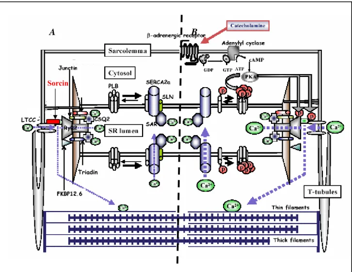
A B
Sorcin
LTCC
PLB
Triadin
Fig 1.2 Regulation of intracellular calcium homeostasis by Ca2+ cycling proteins.
(A) During the action potential a small amount of extracellular Ca2+ enters the cardiomyocyte through the sarcolemmal L-type Ca2+ channels (LTCC), which in turn triggers the release of a larger amount of Ca2+ from the sarcoplasmic reticulum (SR) via ryanodine receptors (RYR2) into the cytosol. Phosphorylated PLB relieves its inhibition on SERCA2a activity, resulting in an increase in Ca2+ uptake into the SR. The RyR2 is also associated with cardiac calsequestrin (CSQ), triadin and junctin. Sorcin is a penta-EF hand Ca2+ binding protein that binds directly to both RyR2 and the LTCC. (B) β-adrenergic stimulation causes enhanced Ca2+ release and uptake by via cAMP-dependent PKA signal pathway. PKA phosphorylates L-type Ca2+ channels (LTCC), the cardiac ryanodine receptor (RyR2) and phospholamban (PLB). The phosphorylated RyR2 is dissociated from FKBP12.6, resulting in pronounced channel open probability. Phosphorylated PLB relieves its inhibition on SERCA2a activity, resulting in an increase in Ca2+ uptake into the SR. The Ca2+ movement is indicated with blue broken lines. Modified from:
Minamisawa et al. (2004).
1.4 Sorcin and its role in calcium cycling
Sorcin (soluble resistance-related calcium-binding protein) is an evolutionary conserved, 21.6 kDa Ca2+
binding protein. Sorcin was originally isolated from multidrug resistant cells in which it was overexpressed as a result of amplification of the sorcin gene (Meyers et al, 1991). In recent years sorcin has emerged as a potential calcium cycling protein and growing literature has emphasised its role in the regulation of intracellular homeostasis and modulation of E-C coupling. However, the ambiguity associated to its function demands further research to fully understand the role played by sorcin in the regulation of calcium cycling in the heart.
1.4.1 Structure of sorcin
Sorcin belongs to the newly found family of penta E-F hand proteins, which contains five E-F hand motifs that associate with membranes in a calcium dependent manner. Binding of Ca2+ to the high affinity sites E-F1 and E-F2 induces translocation of sorcin to membranes where it interacts with specific targets (Zamparelli et al., 1997). Sorcin is capable of binding two calcium ions per monomer and exist as dimers in the absence of calcium (Zamparelli et al., 2000). Calcium free human sorcin forms a homodimer, in which each monomer adapts the penta E-F hand motif. Sorcin molecule has a globular shape apart from the extended N-terminal portion. The C-terminal domain is predominantly α-helical containing eight α-helices and connecting loops incorporating five E-F hands. The E-F hands associate into pairs in a way that E-F 1 pairs with E-F2 and E-F3 pairs with E-F4, The left E-F 5 motif is unpaired in the monomer but pairs with the E-F5 of the second monomer forming a dimer. Dimeric sorcin exhibits an asymmetrical structure (Xie et al., 2001).
1.4.2 Expression and subcellular localization of sorcin
Sorcin is widely expressed in most tissues including heart and skeletal muscle (Van der Bleik, et al., 1986).
Sorcin also associates with presenilin-2 in brain and annexin-7 in adrenal medulla (Pack-Chung et al., 2000, Salzer et al., 2002). At subcellular level sorcin localizes to T-tubule junctions of cardiac SR (Meyers et al., 1995) and co-localizes with RYR at the Z-lines in the normal heart whereas the degree of co-localization is noticeably disrupted in myopathic heart (Pickel et al., 1997).
A
B
Fig. 1.3 Ribbon structure presentation of dimeric sorcin.
(A) Ribbon structure as viewed along the symmetry axis of the dimmer. Each monomer contains helices and loops that form five E-F hands. These E-F hands associate into pairs; E-F1 pairs with E-F2 whereas E-F3 pairs with EF4, and EF5 pairs with its counterpart from the other monomer to form part of the dimer interface. One of the monomers is shown in monochrome (gray) whereas the other is color-coded to show the sub-domain structures: E-F1 (green); E-F2 (blue); E-F3 (magenta); E-F4 (cyan); E-F5 (red); LE-F12, the linker connecting the adjacent E-F-hands within the first pair E-F1–E-F2 (yellow); LE-F34, the linker connecting the adjacent E-F hands within the second pair E-F3–EF-4 (gray); and the short N-terminal fragment (dark orange). For each E-F hand, the individual helices are denoted as E (N-terminal helix) and F (C-terminal helix) with an appropriate number indicating to which E-F hand it belongs, whereas the loops flanked by the E and F helices are denoted as L with the same corresponding number. A sorcin monomer contains eight helices that are numbered from _1 to _8. (B) Ribbon structure of sorcin as viewed perpendicular to the symmetry axis. Xie et al. (2001).
1.4.3 Interaction of sorcin with other calcium binding proteins
Sorcin associates with the cytoplasmic side of RYR in skeletal and cardiac muscle and participates in channel gating (Meyers et al., 1995). Co-immunoprecipitation of metabolically labelled cardiac myocyte proteins and several other in vitro binding studies indicated that sorcin inhibits open cardiac RYR2 from being incorporated into planar lipid bilayers with high affinity. This inhibitory effect of sorcin on ryanodine receptor was relieved by phosphorylation of sorcin with the catalytic subunit of PKA (Farell et al., 2003).
In cardiac myocytes, sorcin significantly inhibits both the spontaneous activity of RYR in quiescent cells (visualized as calcium sparks) and the inward calcium current triggered activity that gives rise to intracellular transients. Sorcin decreases spark efficiency and amplitude, and the dynamic interaction with RYR2 occurs at a rate that would allow for modulation of the channel by sorcin on a beat-to-beat basis (Farell et al., 2003). The rapid and reversible effect of sorcin on RYR2 closure is kinetically capable of playing a role in terminating the positive feedback loop of calcium induced calcium release (Seidler et al., 2003). Calcium bound sorcin binds to the cytoplasmically oriented C-terminal domain of the cardiac L-type calcium channel’s pore-forming alpha 1-C subunit, either at or near the I-Q calmodulin binding region, thus participating in channel inactivation. The functional effects of sorcin on RyR2 and the L-type calcium channel suggest that sorcin plays a role in interchannel communication (Meyers et al., 1998).
Sorcin interacts with SERCA in sorcin overexpressing adult rat cardiac myocytes (Matsumoto et al., 2005) and increases NCX activity in sorcin overexpressing rabbit cardiac myocytes (Seidler et al., 2003). Other binding partners for sorcin include presenilin-2, which in complex with sorcin, may be involved in intracellular calcium modulation in neuronal cells (Pack-Chung et al., 2000) and annexin-7, as sorcin has been shown to inhibit annexin-7 mediated aggregation of chromaffin granules isolated from adrenal medullary tissue (Verzili et al., 2000).
1.4.4 Adenoviral mediated gene transfer and transgenic mouse model
A transgenic mice model overexpressing sorcin by mouse α-myosin heavy chain (α-MHC) promoter was developed. The transgenic mice developed normally with no change in expression of other calcium regulatory proteins. However, contractile abnormalities were observed in isolated adult transgenic myocytes along with significant depression of Ca2+ transient amplitudes. The rate of inactivation of transgenic myocyte L-type Ca2+
channels was accelerated by 15%, as compared to wildtype, while the whole cell calcium density and time course of channel activation were found to be normal (Meyers et al., 2003).
On contrary, the adenoviral mediated overexpression of sorcin in either normal or diabetic mice and in adult rat cardiomyocytes using an adenoviral gene transfer approach showed an increase in cardiac contractility of the normal heart and dramatically rescued the abnormal contractile function of the diabetic heart. This study advocated that viral vector mediated delivery of sorcin to cardiac myocytes is beneficial resulting in improved contractile function in diabetic cardiomyopathy (Suarez et al., 2004).
One study proclaimed that a point missense mutation in sorcin gene resulting in phenylalanine to leucine (F112L) was associated with an uncommon apical form of familial hypertrophic cardiomyopathy and hypertension. The prevalence of this mutation was 2 out of 200 patients with hypertrophic cardiomyopathy.
The study reported that sorcin F112L lacks the inhibitory action of sorcin in RYR mediated Ca2+release
(Mohiddin et al., 2002).
Another study indicated that a mild over expression of sorcin (1.7 fold) exhibits a significantly higher peak force of contraction. Echocardiography of in vivo transfected rat hearts with the adenoviral based overexpression showed enhanced fractional shortening and decreased end-systolic diameters indicating increased cardiac contractility though the gross morphology did not show any remarkable difference in the sorcin over expressed hearts (Frank et al., 2005).
1.5 Aims of the thesis
Despite the growing literature, advocating the role of sorcin in calcium cycling and intracellular calcium homeostasis, the role of sorcin in heart remains controversial. The present study was aimed at apprehending the role of sorcin in Ca2+ signalling pathway by using antisense RNA strategy directed against de novo synthesis of sorcin. The adenoviral antisense sorcin vector was utilized to transfect isolated adult rat cardiomyocytes to investigate functional, biochemical and molecular consequences of adenoviral mediated downregulation of sorcin. In addition, the antisense sorcin vector was used for the adenoviral mediated in vivo sorcin gene delivery in rat hearts to study the functional consequences of depleted sorcin in the whole heart.
The aims of the thesis were to specifically address the following questions:
1. Does the downregulation of sorcin affects the cardiac Ca2+ handling and excitation- contraction coupling?
2. Does the downregulation or ablation of sorcin induce any compensatory mechanism in the expression or activity of the Ca2+ binding proteins and alters the contractility in the E-C coupling?
3. Does lowered expression of sorcin affect the SR Ca2+ storage and SR Ca release? 2+
4. Does reduced expression of sorcin result in functional remodelling of the heart in vivo?
5. Does the ablation of sorcin affect the transcriptional regulation in the heart?
2. Materials and Methods
2.1 Materials 2.1.1 Animals
Male wistar rats, 220–250 g were used for the isolation of adult rat cardiomyocytes (ARC). For the catheter based in vivo gene transfer in hearts, male wistar rats 240-350 g were used. All the animals used were obtained from Charles River, Germany. The rats were housed in plastic cages in a room with a controlled humidity of 40 % and temperature of 22 °C. A controlled environmental 12 hour light-dark cycle was maintained. The experimental design was carried out according to German animal care legislation.
2.1.2 Materials (reagents, buffers, cell culture medium, enzymes and antibodies)
(a) Antisense adenoviral expression vector
AdEasyTM adenoviral vector system, Stratagene Shuttle vector pAdTrack-CMV, Stratagene PCR 2.1 cloning vector, Invitrogen
Restriction endonucleases, New England Biolabs Alkaline phosphatase, New England Biolabs Polyfect transfection kit, Stratagene
PCR purification kit, Qiagen Electroporation cuvettes, Biorad Cell electroporator, Biorad AD293 cells, Stratagene
Growth medium for AD293 cells: DMEM (4.5 g/L glucose, 110 mg/L sodium pyruvate and 2 mM L- glutamine) supplemented with 10% v/v heat inactivated foetal bovine serum.
Sea plaque® agarose, Stratagene
(b) Isolation of rat cardiomyocytes
Cell culture medium: Medium M199 500 ml, Gibco, MEM-Vitamin 10 ml, Gibco, non essential amino acids 5 ml, Gibco; Penicillin/Streptomycin 100 IU/ml 5 ml, Sigma, Insulin H 40 IE/ml, Höchst.
Diethylether, Roth
Powell-Medium (in mM): NaCl 110, KCl 2.5, KH2PO 1.17, NaHCO4 3 25, Glucose 2 g/l.
Carbogen (95% O2 + 5% CO ), Linde 2
Collagenase Typ II, Biochrom Calcium chloride, Sigma Laminin natural mouse, Sigma Bovine serum albumin (BSA), Sigma
(c) Protein preparation from transfected rat cardiomyocytes
Cell-lysis buffer (in mM): Tris-HCl (pH 7.5) 5, EDTA (pH 8.0) 5 Complete mini EDTA free protease inhibitor cocktail tablet, Sigma Freezing buffer (in mM): Saccharose 400, HEPES 5, TRIS 5, pH 7.2.
(d) Quantitative immunoblotting Acrylamide-Bis 30%/0.8 % v/v, Roth Coomassie brilliant blue, Serva Dodecyl sodium sulphate (SDS), Serva
Enhanced Chemiluminiscence (ECL) Kit, Amersham Life Science Acetic acid, Merck
Glycerine, Merck Glycine, Merck
Kaleidoscope prestained protein standards (≅ 7 –205 kDa), BioRad 2-βMercaptoethanol 98% v/v, Sigma
N, N, N´, N´-tetramethylendiamine (TEMED), Serva
TBS (in mM): NaCl 150, Tris-HCl 10; pH 7.5
TBST (in mM): NaCl 150, Tris-HCl 10, 0.05% v/v TWEEN 20, pH 7.5 TWEEN 20, Merck
Polyvinyldienfluoride (PVDF) membrane, BioRad Ponceau S, Serva
Loading buffer (2x): 100 mM TRIS/HCl (pH 6.8), 2% v/v
2-βMercaptoethanol, 4% SDS w/v, 0.2% Bromphenolblue 20% v/v X-ray film, Amersham
(e) Antibodies for western blot
Monoclonal (Mouse) Anti-sorcin antibody, (1:1000), Zymed, Polyclonal (Rabbit) Anti-sorcin antibody, (1:1000), self produced
Monoclonal (Mouse) Anti-ryanodine receptor isoform 2 antibody, (1:1000), Affinity BioReagents Monoclonal (Mouse) Anti-calsequestrin antibody, (1:1000), Biomol
Polyclonal (Rabbit) Anti-calcineurin-A antibody, (1:1000), Santa Cruz Biotechnology Polyclonal (Rabbit) Anti-FKBP12.6 antibody, (1:1000), Biomol
Monoclonal (Mouse) Anti-SERCA2 ATPase antibody, (1:1000), Affinity Bioreagents Monoclonal (Mouse) Anti-triadin antibody, (1:1000), Affinity Bioreagents
Monoclonal (Mouse) Anti-phospholamban antibody, (1:1000), Upstate Biotechnology
Ployclonal (Rabbit) Anti-GATA-4(H112) antibody, (1:1000), SantaCruz Biotechnology Polyclonal (Goat) Anti-NF-ATC3 antibody, (1:1000), Santa Cruz Biotechnology Peroxidase-conjugated Anti goat IgG antibody, (1:2000), Sigma
Peroxidase-conjugated Anti-mouse IgG antibody, (1:2000), Sigma Peroxidase-conjugated Anti-rabbit IgG antibody, (1:2000), Sigma
(f) Immunofluoroscence measurement in transfected cardiomyocytes
Triton X-100, Sigma,
Polyclonal (Rabbit) Anti-sorcin antibody (1:500), self produced Alexa fluorTM 546 Anti-rabbit IgG, Molecular probes
DAKO mounting medium, Molecular probes
PBS (in mM): NaCl 137, KCl 2.6, K2HPO 1.8, Na4 2HPO 10 (pH 7.4) 4 Paraformaldehyde (4% w/v in PBS), Sigma
(g) Contractility measurement of the transfected cardiomyocytes
Tyrode buffer (in mM): NaCl 132, KCl 4.8, MgCl2 1.2, CaCl2 1.8, Glucose 10, HEPES 10, pH 7.3, Sigma Forskolin, Calbiochem
(h) RNA isolation and reverse transcription
RNase free water, Invitrogen Ethanol, Merck
Ist strand buffer, Gibco
0.1M dithiothreitol (DTT), Gibco
Random primer, RNase inhibitor, Promega M-MLV reverse transcriptase, Gibco
Deoxynucleotide triphosphate set, PCR grade, Roche Diagnostics Chloroform, Merck
2-Propanol, Roth
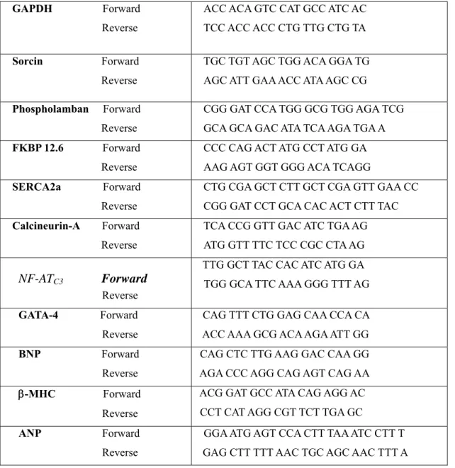
Table 2.1 List of primers
GAPDH Forward ACC ACA GTC CAT GCC ATC AC Reverse TCC ACC ACC CTG TTG CTG TA
Sorcin Forward TGC TGT AGC TGG ACA GGA TG Reverse AGC ATT GAA ACC ATA AGC CG
Phospholamban Forward CGG GAT CCA TGG GCG TGG AGA TCG Reverse GCA GCA GAC ATA TCA AGA TGA A FKBP 12.6 Forward CCC CAG ACT ATG CCT ATG GA Reverse AAG AGT GGT GGG ACA TCAGG
SERCA2a Forward CTG CGA GCT CTT GCT CGA GTT GAA CC Reverse CGG GAT CCT GCA CAC ACT CTT TAC Calcineurin-A Forward TCA CCG GTT GAC ATC TGA AG Reverse ATG GTT TTC TCC CGC CTA AG
TTG GCT TAC CAC ATC ATG GA
Forward
NF-AT
C3 TGG GCA TTC AAA GGG TTT AGReverse
GATA-4 Forward CAG TTT CTG GAG CAA CCA CA Reverse ACC AAA GCG ACA AGA ATT GG BNP Forward CAG CTC TTG AAG GAC CAA GG Reverse AGA CCC AGG CAG AGT CAG AA ACG GAT GCC ATA CAG AGG AC β-MHC Forward
CCT CAT AGG CGT TCT TGA GC Reverse
ANP Forward GGA ATG AGT CCA CTT TAA ATC CTT T Reverse GAG CTT TTT AAC TGC AGC AAC TTT A
For each primer pair the annealing temperature (TA) was calculated according to the following formula:
T = 2( A + T ) + 4( G + C) – 5 A
(i) Calcineurin activity measurement in transfected cardiomyocytes
Tris, Roth
Acetylated BSA (Bovine serum albumin), Promega Nickel chloride, Sigma
Calmodulin (phosphodiesterase 3’-5 –cyclic nucleotide activator), Sigma ’
PNPP (p-Nitrophenyl phosphate), Sigma
ELISA reader MRX Revealation , DYNEX Technologies
(j) Measurement of calcium transient in transfected cardiomyocytes
X- rhod-1 AM, Molecular Probes
Tyrode sloution (in mM): NaCl 132, KCl 4.8, MgCl 1.2, CaCl2 2 1.8, Glucose 10, HEPES 10, pH 7.3 aCl2 1.8
Stimulator
Confocal laser scanning microscope LSM 510 Meta, Zeiss (k) Expression and purification of recombinant sorcin
Luria Bertani (LB) medium (1l) Bacto-trypton 10 g, Yeast extract 5 g, NaCl 5 g, ddH2O to 1l (pH 7.3) Ampicillin 100 mg/ml, Sigma
IPTG, Sigma
Elution buffer: Reduced glutathione 50 mM Tris-HCl (pH 8.0), Sigma PBS (in mM): NaCl 137, KCl 2.6, K2HPO 1.8, Na4 2HPO 10 (pH 7.4). 4
Lysozyme, Sigma EDTA, Roth
TritonX-100, Sigma
Glutathione agarose beads, Amersham
(l) Sarcoplasmic reticulum Ca uptake
2+Potassium dihydrogen phosphate, Merck Sodium fluoride, Merck
Sucrose, Roth
Phenylmethanesulfonylfluoride (PMSF), Sigma Dithiothreitol, Merck
Imidazole, Merck
Potassium chloride, Merck Sodium azide, Merck
Magnesium chloride hexahydrate, Merck
’ ’
Ethylene glycol bis (β-Aminoethyl ether)-N, N, N , N , tetra acetic acid, Sigma Potassium oxalate monohydrate, Merck
Calcium chloride dihydrate, Merck Ruthenium red, Sigma
Tris, Roth
Adenosine-5’-triphosphate, Roche
Whatman glass microfibre filters 2.5 cm, poresize 0.45 μm ACS II scintillation cocktail, Amersham
Liquid scintillation analyzer TR-1600, Packard
(m) Catheter based adenoviral injection in rat hearts and Echocardiography
Acetylcholine, Sigma Isoflurane, Sigma Ketamine, Sigma Diazepam, Sigma Epinephrine, Hoechst Heparin 100 IU, Sigma Sodium bicarbonate, Braun
3F balloon catheter, 2 F lumen balloon catheter, Edwards life science Pressure transducer, Stratham P23 XL
Four chamber power lab system, AD instruments
2.2 Methods
2.2.1 Generation of adenoviral antisense sorcin GFP (Ad.as.Sor.GFP)
Adenovirus serotype 5 with antisense sorcin GFP (Ad.as.SOR.GFP) was generated using the AdEasyTM Adenoviral vector system from stratagene (Hajjar et al., 1997).
(a) Cloning sorcin cDNA and insertion in transfer vector
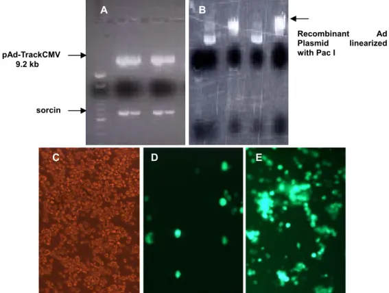
Sorcin human cDNA fragment was cloned in PCR 2.1 vector and confirmed by restriction digestions and sequencing. The cloned cDNA fragment was subsequently inserted in the multiple cloning site of shuttle vector pAD-TrackCMV (9.2kb) in reverse orientation using the endonuclease restriction enzyme sites Hind III and Xho I. The ligation was performed by using T4 DNA ligase (Molar ratio from Insert: Vector = 5:1) The orientation of the fragment was confirmed by restriction digestion and sequence analysis.
(b) Production of recombinant Ad plasmid by homologous recombination
The shuttle plasmid was linearized by Pme I, confirmed the complete digestion by agarose gel electrophoresis and purified by Qiagen purification kit to remove any salts, which can interfere in the subsequent electroporation. The purified linearized plasmid was dephosphorylated with alkaline phosphatase for 1 h at 37 °C. The BJ5183 cells were cotransformed with the linearized shuttle vector containing sorcin cDNA and the pAdEasy vector as per the manufacturer’s instructions, which resulted in the recombination event inside the BJ5183 cells. In short 1 μg of linearized, dephosphorylated shuttle vector and 1 μl of pAdEasy supercoiled vector (100 ng/ μl) were mixed with BJ5183 gently and transferred to prechilled electroporation cuvettes. A pulse of 200 Ω, 2.5 kV, 25 μF was used for electroporation. The cuvette was removed immediately; 1 ml of sterile LB broth was added and gently mixed by pipetting up and down to resuspend the cells. The transformants were incubated at 37 °C for 1 h while shaking at 225-250 rpm. The recombinant reaction mixture was plated on kanamycin plates and incubated overnight at 37 °C. The recombinant Ad plasmid was purified from the clone and confirmed by restriction digestion with Pac I enzyme.
(c) Production of recombinant Ad virus
The positive recombinant Ad plasmid was used to transform XL10-GOLD ultracompetent
cells to amplify the recombinant adenovirus plasmid DNA by using the heat shock method
following the manufacturer’s protocol. Briefly 50 ng of plasmid DNA was added to the pre
aliquoted 100μl XL10-Gold ultra competent cells and incubated for 30 min on ice followed
by heat pulsing the reaction mixture at 42
°C for 30 s. The reaction tube was incubated on
ice for 2 min, 0.9 ml of pre warmed (42
°C) NZY
+broth was added and incubated at 37 C
°for 1 hour at 225-250 rpm. The transformant reaction was plated on kanamycin plates and incubated overnight at 37
°C. Positive colonies were checked by restriction digestion with Pac I. A maxiprep was done and purified by standard caesium chloride density gradient centrifugation to get highly pure DNA. The plasmid DNA was subjected to Pac I digestion, purified the reaction mixture with Qiagen PCR purification kit and resuspended in PCR grade water. AD293 cells produce the adenovirus E1 gene in trans allowing the production of infectious virus particles when the cells are transfected with E1 deleted pAdEasy vector.
7-8 x 10
5AD293 cells were plated in 60 mm tissue culture dish containing growth medium.
After 24 hours the transfection was done by using Polyfect
TMtransfection kit following the
manufacturer’s instructions. The progress of the transfection was monitored by the
fluorescence microscopy (by detecting GFP expression). Adenovirus producing AD293 cells
were washed with PBS and harvested by scraping the cells in 0.5 ml of PBS. The cell
suspension was subjected to four rounds of freeze/thaw by alternating the tubes between dry
ice-methanol bath and 37
°C water bath. The cellular debris were collected by
microcentrifugation at 12000 g for 10 min at room temperature. The supernatant obtained
was used as the primary virus stock. The titer (pfu/ml) was determined by plaque assay
using agarose overlay. Similarly a control expressing GFP (Ad.GFP) was generated with the
shuttle vector. Fig. 2.1 represents the schematic presentation of the generation of the
adenoviral vectors.
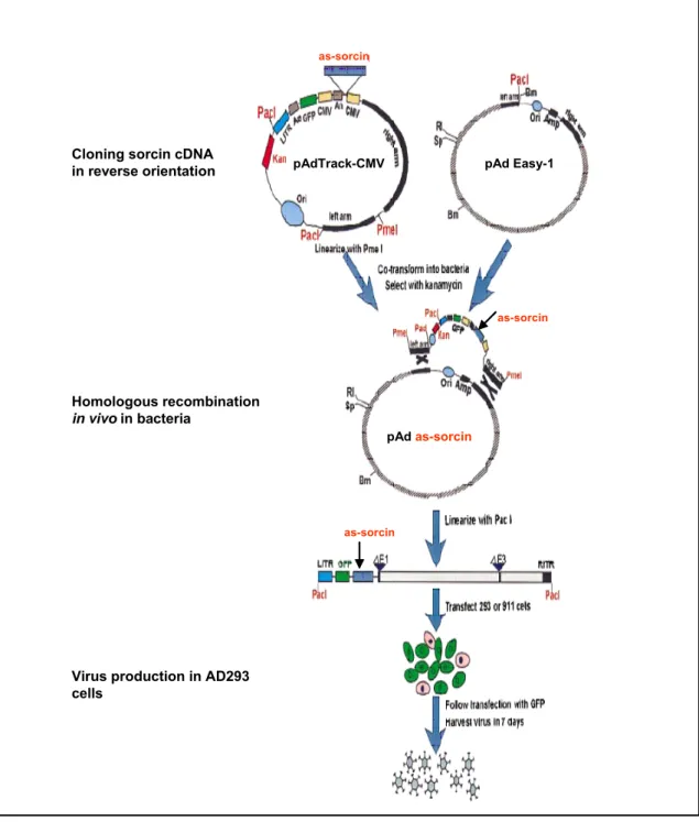
pAd
as-sorcin
Fig. 2.1 Schematic outline of the generation of adenoviral vector using AdEasy system
The gene of interest (sorcin) was first cloned in reverse direction into shuttle vector pAdTrack-CMV. The resultant plasmid was linearized by digesting with Pme I and subsequently cotransformed into E.coli BJ5183 cells with an adenoviral backbone plasmid pAdEasy-1. Recombinants were selected for kanamycin resistance and recombination was confirmed by multiple restriction endonuclease analyses. Finally the linearized recombinant plasmid was transfected into adenovirus packaging cell line (AD293 cells). The “left arm” and
“right arm” represents the regions mediating homologous recombination between the shuttle vector and adenoviral backbone vector. Modified from Tong-Chuan He et al. (1997).
- as-sorcin
as-sorcin
as-sorcin as-sorcin
pAd
Cloning Sorcin cDNAin reverse orientation
Homologous recombination in vivo inbacteria
Virus production in A293 cells
- as-sorcin
as-sorcin
as-sorcin
as-sorcin
as-sorcin Cloning sorcin cDNA
in reverse orientation
Homologous recombination in vivoin bacteria
Virus production in AD293 cells
pAdTrack-CMV pAd Easy-1
pAd as-sorcin
as-sorcin
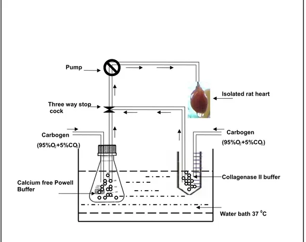
2.2.2 Isolation of adult rat cardiomyocytes (ARC)
The isolation of cardiomyocytes was done by the enzymatic digestion technique. Male wistar rats (age 10-12 weeks, 220 – 250 g) were anaesthetised by diethyl ether followed by cervical dislocation. Hearts were quickly excised, aorta cannulated and perfused with the calcium free oxygenated (95% O2 + 5%
CO2) powell buffer on the Langendorff perfusion apparatus for 5 minutes at 37 °C at a constant flow rate of 8 ml/min. The heart was retroperfused with collagenase buffer (collagenase II, Biochrome), 26-30 mg (210 U/mg) in 40 ml oxygenated powell buffer with 31.25 μM CaCl at a constant temperature of 372 o C for 15 to 20 min taking care that the buffer was oxygenated constantly. The heart was taken from the cannula and gently pipetted up and down in collagenase buffer with a blunt ended 10 ml pipette till all the cardiomyocytes were loosened, followed by centrifugation at 300 g for 3 min at room temperature, the supernatant was aspirated and the pellet (cardiomyocytes) was filtered through a nylon filter (200 μm) and adjusted to a volume of 10 ml with 4% BSA. The cardiomyocytes were subjected to increasing Ca2+
concentration in order to get calcium stable cardiomyocytes. Pre calculated volume of 100 mM CaCl2 was added in four gradual steps at an interval of 3 min each to achieve the final Ca2+ concentration 1.8 μM, followed by final centrifugation step 300 g for 3 min at room temperature. Supernatant was aspirated and the cardiomyocyte pellet was resuspended in 10 ml of culture medium. Cardiomyocyte count was calculated with neubaeur chamber. The cardiomyoctes were plated on 10.5 cm laminin precoated culture plates for protein preparation and on the laminin precoated 18 mm and 12 mm glass coverslips for contractility measurement and immunocytochemistry, respectively. Fig. 2.2 depicts the scheme of isolation of the cardiomyocytes .
2.2.3 Transfection of cardiomyocytes
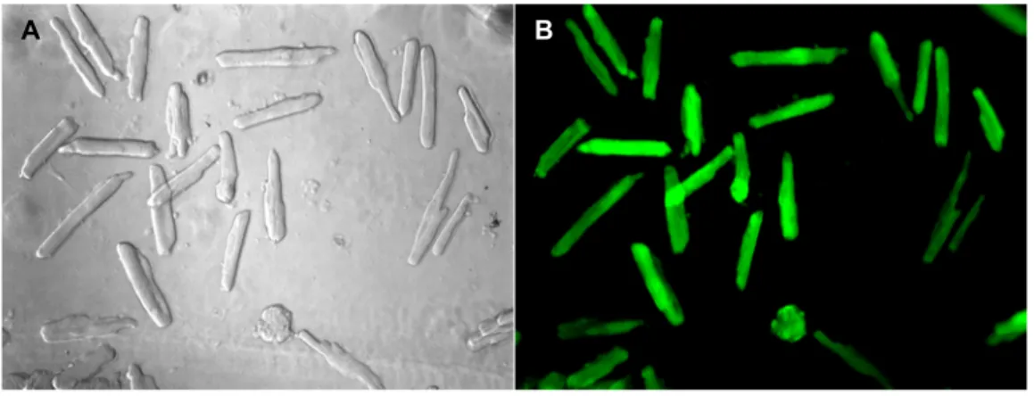
The isolated cardiomyocytes were transfected with the Ad.as.SOR.GFP with 100 moieties of infection (MOI) and Ad.GFP as control (MOI 100) after three hours of isolation and allowing the cells to adhere to the laminin coated plates. After incubation with the virus for 12-20 h the media was changed. Successful transfection of cardiomyocytes was determined by monitoring the culture plates for green fluorescence using Axiovert microscope (Zeiss) with an excitation wavelength 473 nm by a fluorescence filter.
Expression of green fluorescence protein marker by the cardiomyocytes verified the successful transfection. The cardiomyocytes were cultured for 48 h before protein preparation and other functional studies.
Carbogen (95%O 2+5%CO 2)
Pump
Isolated rat heart
Calcium free Powell Buffer
Carbogen (95%O 2+5%CO 2)
Water bath 37 0C Collagenase II buffer Three way stop
cock
Fig. 2.2 Schematic presentation of the isolation of adult rat cardiomyotes.
Fig. 2.2 Schematic presentation of the isolation of adult rat cardiomyotes.
The aorta from the excised rat heart was cannulated and perfused with the Ca2+ free Powell buffer for 5 min followed by retroperfusion of collagenase II buffer for 15 to 20 min. All the buffers and solutions were constantly oxygenated (95% O2 + 5% CO2) and kept at 37 °C in the water bath.
The aorta from the excised rat heart was cannulated and perfused with the Ca2+
free Powell buffer for 5 min followed by retroperfusion of collagenase II buffer for 15 to 20 min. All the buffers and solutions were constantly oxygenated (95% O2 + 5% CO2) and kept at 37 °C in the water bath.
2.2.4 Protein preparation from the transfected cardiomyocytes 2.2.4 Protein preparation from the transfected cardiomyocytes
Cardiomyocytes were cultured on laminin coated plates for 48 h post transfection with Ad.as.SOR.GFP or Ad.GFP. Culture medium was changed and the cells were washed with PBS, 1 ml of lysis buffer (with complete mini EDTA free tablet / 10 ml of lysis buffer) was added to the culture plate and allowed to stand at room temperature for 5 min, the cells were scraped with the cell scraper and homogenised by using the ultrasonic homogeniser with two pulses for the whole cell homogenates. The cardiomyocyte homogenates were pooled from different preparations to scale up the amount of protein. The amount of protein was quantified by Bradford assay and stored in aliquots at –80
°C.
2.2.5 Extraction of transcription factors from transfected cardiomyocytes
Isolation of nuclear extract and transcription factor was performed based on the protocol described earlier (Andrews et al., 1991). The procedure utilizes the hypotonic lysis followed by high salt extraction of nuclei. Briefly, 48 h post transfection, cardiomyocytes were scraped into 1.5 ml of cold PBS. Cells were pelleted and resuspended in 400 µl cold buffer A (10 mM HEPES-KOH pH 7.9 at 4 0C, 1.5 mM MgCl2, 10 mM KCl, 0.5 mM dithithreitol, 0.2 mM PMSF). Cells were allowed to swell on ice for 10 min and then vortexed for 10 seconds. Samples were centrifuged and the supernatant discarded, the pellet was resuspended in 20-100 µl (according to starting number of cells) of cold buffer B (20 mM HEPES-KOH pH 7.9 at 4 0C, 25% v/v glycerol, 420 mM NaCl, 1.5 mM MgCl2, 0.2 mM EDTA, 0.5 mM dithiothreitol, 0.2 mM PMSF) and incubated on ice for 20 min for high-salt extraction. Cellular debris were removed by centrifugation for 2 min at 4 °C and the supernatant fraction (containing the DNA binding proteins) was stored at –70 °C for later use.
2.2.6 Quantitative immunoblotting
(a) SDS PAGE
SDS-polyacrylamide gel electrophoresis (SDS-PAGE) was performed using the discontinuous buffer system (Laemmli, 1970). Discontinuous polyacrylamide gel was prepared using glass plates of 10 cm x 7.5 cm dimensions and spacers of 0.5 mm thickness. The composition of resolving and stacking gels is given in the table 2.2.
Table 2.2 Composition of stacking and separating gels for SDS PAGE
component stacking gel 4% separating gel 8%
AA/Bis (30 %/0.8 %) 4.1 ml 7.2 ml
1.5 M Tris pH 8.8 - 10.2 ml
0.5 M Tris pH 6.8 3.7 ml -
H O 2 22.3 ml 9.2 ml
300 µl 300 µl
10% SDS
300 µl 95 µl
10% APS
41 µl 24 µl
TEMED
Samples were mixed with suitable volumes of loading buffer, denatured by heating at 95 ºC for 5 min and loaded into the wells of the stacking gel. A prestained molecular weight marker (kaleidoscope) was run simultaneously on adjacent lane as a standard to establish the apparent molecular weights of proteins resolved on SDS-PAGE. Electrophoresis was performed in 1x gel-running buffer at a constant voltage of 100-150 V until the bromophenol blue dye front had reached the bottom edge of the gel or had just run out of the gel.