THE EFFECTS OF LACTATE ON SKELETAL MUSCLE
Inaugural Dissertation
zur
Erlangung des Doktorgrades Dr. nat. med.
der Medizinischen Fakultät und
der Mathematisch-Naturwissenschaftlichen Fakultät der Universität zu Köln
vorgelegt von
Lena Willkomm aus Hildesheim
Hundt Druck, Köln
2014
II Berichterstatter/-in: Prof. Dr. Rudolf Wiesner
Prof. Dr. Aleksandra Trifunovic
Tag der letzten mündlichen Prüfung: 25.03.2014
III
Table of Contents T
ABLE OFC
ONTENTSList of Figures VI
List of Tables VII
Abbreviations VIII
Abstract 1
Zusammenfassung 2
1 Introduction 4
1.1 Exercise and physical activity and associated health benefits 4 1.2 Satellite Cells and skeletal muscle adaptation 6 1.3 Myogenesis and its regulatory epigenetic modifications 7 1.4 The role of p38 MAPK in skeletal muscle adaptation 11 1.5 Exercise and physical activity and histone modifications 13 1.6 Lactate as a metabolic signal of gene expression 14
1.7 Aims 16
2 Materials & Methods 17
2.1 Materials 17
2.1.1 Lab equipment and disposables 17
2.1.2 Chemical and solutions 18
2.1.3 Buffers and cell culture media 19
2.1.4 Kits 22
2.1.5 Software 22
2.2 Methods 22
2.2.1 Cell Culture 22
2.2.1.1 Thawing Cells 22
2.2.1.2 Freezing cells 22
2.2.1.3 Splitting Cells 23
2.2.1.4 Seeding cells 23
2.2.1.5 Experimental Design 23
2.2.1.6 La and pH measurement 25
2.2.1.7 Bromdeoxyuridin(BrdU)-Assay 25
2.2.1.8 Gene expression analysis 25
2.2.1.8.1 RNA isolation 25
2.2.1.8.2 RNA concentration determination and quality assessment 26
IV 2.2.1.8.3 Complimentary DNA (cDNA) synthesis 26
2.2.1.8.4 Primer Design 26
2.2.1.8.5 Quantitative real-time reverse transcription polymerase chain reaction (RT-PCR) 27
2.2.2 Human Muscle Biopsies 28
2.2.2.1 Subjects 28
2.2.2.2 Experimental design and resistance training protocols 28
2.2.2.3 Muscle biopsy 30
2.2.3 Protein analysis 30
2.2.3.1 Western Blot (WB) Analysis 30
2.2.3.1.1 Cell lysis 30
2.2.3.1.2 Homogenisation of human muscle biopsies 30 2.2.3.1.3 Protein concentration determination and sample preparation 31
2.2.3.1.4 SDS-PAGE and Western Blot 31
2.2.3.2 Immunofluorescent staining (IF) 33
2.2.3.2.1 Fixation and permeabilisation of cells 33 2.2.3.2.2 Fixation of human tissue sections 33
2.2.3.2.3 Immunological detection 33
2.2.3.2.4 Microscopy and analysis 34
2.2.3.2.5 Quantification of fluorescence 34
2.2.3.3 Immunocytochemistry (ICC) 34
2.2.3.3.1 Fixation, permeabilisation and inactivation of endogenous peroxidase of cells 34 2.2.3.3.2 Immunological detection and DAB staining 35
2.2.3.3.3 Microscopy and densitometry 35
2.2.4 Statistical analysis 35
3 Results 36
3.1 Changes in La concentration and pH in incubation media 36
3.2 La reduced proliferation 36
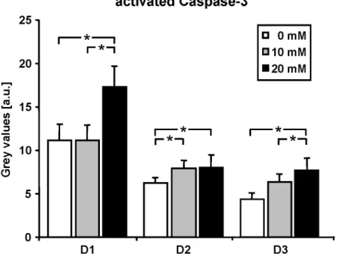
3.3 La increased activation of Caspase-3 37
3.4 La timely delayed late differentiation in a dose-dependent manner 39
3.5 Lactate increased oxidative stress 43
3.6 Involvement of p38 MAPK and histone modifications in vitro 48 3.7 p38 MAPK and histone modifications in vivo 52
4 Discussion 55
5 Conclusion 65
6 References 67
V
7 Appendix 93
8 Erklärung 99
9 Curriculum Vitae 100
VI List of Figures L
IST OFF
IGURESFigure 1.1: Satellite cell development and markers of myogenesis 7
Figure 1.2: Histone modifications H3K4me3 and H3K27me3 during SC proliferation and differentiation 10
Figure 1.3: A model for the activity of p38 MAPK in myogenesis 12
Figure 3.1: Changes in La concentration and pH in incubation media 36
Figure 3.2: La suppressed proliferation of C2C12 cells 37
Figure 3.3: La induced cellular stress in C2C12 cells 38
Figure 3.4: La delayed the differentiation process of C2C12 cells in a dose-dependent manner 39
Figure 3.5: La delayed myogenin synthesis in C2C12 cells 40
Figure 3.6: La delayed MHC synthesis in C2C12 cells in a dose-dependent manner 42
Figure 3.7: La increased oxidative stress 43
Figure 3.8: La-induced cell cycle withdrawal was reversible by different antioxidants 44 Figure 3.9: La altered gene expression of cell cycle control genes Cdkn1a and Trp53 45 Figure 3.10: La influenced gene expression of Pax7, Myf5, myogenin, MHC1, and MHC2 46 Figure 3.11: La-induced delayed differentiation in C2C12 cells was reversible by the use of antioxidants 47 Figure 3.12: La has acute effects on p38 MAPK phosphorylation and H3K4 and H3K27 trimethylation 48 Figure 3.13: La-induced delayed late differentiation in C2C12 cells is similar to p38 MAPK-inhibition in cells
treated with 0 mM La DM + SB203580 49
Figure 3.14: La did not affect H3K27me3, but decreased H3K4me3 during differentiation 51 Figure 3.15: HIT dephosphorylates p38 MAPK and diminishes H3K4me3 and H3K27me3 compared to STD 53 Figure 5.1: La induces cell cycle arrest and inhibits differentiation progression 65
VII List of Tables L
IST OFT
ABLESTable 2.1: Primers used in real-time RT-PCR 27
Table 2.2: Cycling conditions for real-time RT-PCR 27
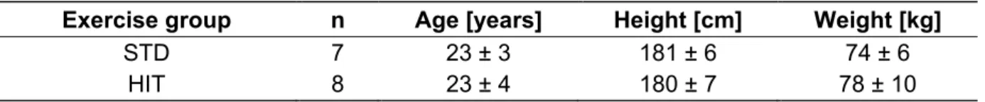
Table 2.3: Anthropological data of subjects 28
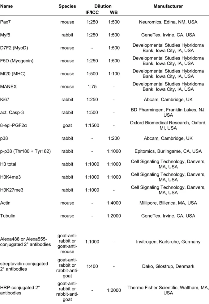
Table 2.4: Antibodies used for IF/ICC and WB 32
Table A.1: La delayed myogenin synthesis in C2C12 cells 93
Table A.2: La delayed MHC synthesis in C2C12 cells in a dose-dependent manner 93 Table A.3: La altered gene expression of cell cycle control genes Cdkn1a and Trp53 94 Table A.4: La influenced gene expression of Pax7, Myf5, myogenin, MHC1, and MHC2 95 Table A.5: La-induced delayed differentiation in C2C12 cells was reversible by the use of antioxidants 97 Table A.6: La-induced delayed differentiation in C2C12 cells is comparable to p38 MAPK-inhibition 98
VIII Abbreviations A
BBREVIATIONS8-epi-PGF2α 8-epi-prostaglandin F2α a.u. arbitrary units (grey values) AA ascorbic acid (vitamin C) act. Casp-3 activated Caspase-3
AMPK adenosine monophosphate-activated protein kinase Ash2L absent, small, or homeotic-like, variant 2
ASK1 apoptosis-stimulating kinase 1 ATF activating transcription factor ATP adenosine triphosphate BCP 1’-bromo-3’chloropropane bHLH basic helix-loop-helix
bp base pair
BrdU bromodeoxyuridine BSA bovine serum albumine
CaMK calcium and calmodulin-acitvated protein kinase
Cat Catalase
Cdkn1a cyclin-dependent kinase inhibtor 1a
cDNA complimentary deoxyribonucleic acid Ct cycle threshold
CTL cytotoxic t-cell
D7F2 myogenic determination protein (MyoD) antibody (trade name)
DAB diaminobezindine
DAPI 4’,6-diamidino-2-phenylindole DM differentiation medium
DMEM Dulbecco’s modified Eagle medium DMSO dimethyl sulfoxide
DNA deoxyribonucleic acid
DPBS Dulbecco’s phosphate buffered saline EDTA ethylenediaminetetraacetic acid ERK extracellular signal-regulated kinase Ezh ehancer of zeste homolog
F5D myogenin antibody (trade name) FCS fetal calf serum
F
maxmaximum force
G1 gap 1
GLUT glucose transporter GPx glutathione peroxidase H
2O
2hydrogen peroxide
H histone 3
IX H3K4me3 trimethylated histone 3 lysine 4
H3K9me3 trimethylated histone 3 lysine 9 H3K27me3 trimethylated histone 3 lysine 27 H4K20me3 trimethylated histone 4 lysine 20 HDAC histone deacetylase complex HIT high intensity resistance training HRP horse radish peroxidase
HS horse serum
ICC immunocytochemical staining IF immunofluorescent staining JMJD Jumonji domain-containing protein JNK c-Jun N-terminal kinase
KDM lysine demethylase KMT lysine methyltransferase LA linolenic acid
La lactate
mTOR mammalian target of rapamycin
MAPK mitogen-activated protein kinase MCT monocarboxylate transporter Mef myocyte enhancer factor
Mf20 myosin heavy chain antibody (trade name) MHC myosin heavy chain
MKP mitogen-activated protein kinase phosphatase
mM mmol·L
-1MOPS 3-(N-morpholino)propanesulfonic acid MP dry milk powder
MRF muscle regulatory factor
MRF4 muscle-specific regulatory factor 4, also known as Myf6 mRNA messenger ribonucleic acid
Myf5 myogenic factor 5
MyoD myogenic determination protein NAc N-Acetyl-L-cysteine
p- phosphorylated
p21 Cyclin-dependent kinase inhibitor 1a p53 transformation related protein 53
PAGE polyacrylamid gel electrophoresis Pax7 paired box transcription factor 7
PBS phosphate buffered saline
PcG polycomb group protein
PCR polymerase chain reaction
Pen/Strep penicillin/streptomycin
X
PFA paraformaldehyde
PGC1α peroxisome proliferator activated-receptor
γ coactivator-1αPKB protein kinase B
PM proliferation medium pRb retinoblastoma protein PRC2 polycomb repressor complex 2 PVDF polyvinylidene fluoride RNA ribonucleic acid ROM range of motion
ROS reactive oxygen species
RT room temperature
RT-PCR reverse-transcription polymerase chain reaction SC satellite cell
SDS sodium dodecyl sulphate S.E.M. standard error of the mean
Ser serine
SOD superoxide dismutase STD standard resistance training SWI/SNF switch/sucrose non-fermentable TAE Tris acetate ethylenediaminetetraacetic acid TBS Tris buffered saline
TBST Tris buffered saline containing Tween20
®Thr threonine
T
meltmelting temperature
Tris Tris(hydroxymethyl)aminomethane Trp53 transformation related protein 53
TSC2 tuberous sclerosis 2 Tx100 TritonX-100
TrxG trithorax group protein
Tyr tyrosine
UTX transcribed tetratricopeptide repeat, X chromosome
WB Western blot
Wnt wingless-type MMTV integration site
YY Ying Yang
Commonly used abbreviations and SI units are not listed separately.
1 Abstract A
BSTRACTRegular exercise and physical activity are cornerstones in the prevention and treatment of
numerous chronic conditions, such as type 2 diabetes, coronary heart disease, and age-
related sarcopenia. The associated health benefits arise from a number of tissues but due to
its high plasticity skeletal muscle plays a pivotal role. The resident stem cells of skeletal
muscle tissue, so called Satellite cells (SCs), contribute significantly to skeletal muscle
adaptation and hence, maintenance of healthy tissue. The specific stimuli regulating SC
development, i.e. activation, proliferation, an differentiation, depend on the form of exercise
and consist of hormonal, mechanical, and metabolic signals. While hormonal and mechanical
factors have been well documented, the importance of metabolic stimuli such as Lactate (La)
remains less clear. La is produced continuously under aerobic conditions, but elevated levels
occur during exercise when glycolysis is increased. La is able to induce muscle adaptation,
but the underlying molecular mechanisms are not yet understood. Therefore, one aim of this
study was to identify the phenotypical effects of high La levels as observed during resistance
or high intensity endurance training on the proliferation and differentiation in a model of
activated SCs, C2C12 cells. Furthermore, possible signalling targets for La, such as p38
mitogen-activated protein kinase (p38 MAPK), and subsequent histone modifications were
investigated. Lastly, to confirm the observed mechanisms in vivo, a human intervention study
was conducted. Treatment with La (10 mM, 20 mM) increased the serum deprivation-induced
withdrawal from the cell cycle and initiated early differentiation in C2C12 cells as analysis of
gene expression and protein patterns of cell cycle (Ki67, Trp53, and Cdkn1a) and
differentiation markers (Pax7, Myf5, myogenin, and myosin heavy chain) revealed. However,
La delays late differentiation in a dose-dependent manner. La-induced production of ROS,
marked by high 8-epi-PGF2α levels, might at least be partly responsible as the effects
induced by La were reversible by the addition of the antioxidants ascorbic acid, N-
acetylcysteine, or linolenic acid. Observed downregulation of p38 MAPK activation and its
downstream modifications histone 3 lysine 4 (H3K4) and histone 3 lysine 27 (H3K27)
trimethylation suggests that La inhibits late differentiation progress by this mechanism which
is crucial for muscle specific gene transcription. Experiments using the p38 specific inhibitor
SB203580 add further evidence for this hypothesis. Additionally, it was demonstrated that
diminished p38 MAPK activation and subsequent histone modifications are conserved in
differentiated muscle tissue in vivo. Conclusively, the reported data confirm that La modifies
skeletal muscle adaptation via a ROS-sensitive signalling network by delaying late
differentiation of SCs, an important mechanism of skeletal muscle adaptation. This
conclusion implies reassessment of traditional views on training design and periodisation in
order to accelerate skeletal muscle adaptation.
2 Zusammenfassung Z
USAMMENFASSUNGRegelmäßige körperliche Aktivität ist in der Prävention und Therapie vieler chronischer Krankheiten, wie zum Beispiel Typ 2 Diabetes, koronare Herzkrankheiten und altersbedingte Sarkopenie von großer Bedeutung. Die positive Wirkung von körperlicher Aktivität sind u. a.
auf die Adaptation der Skelettmuskulatur zurückzuführen, welche eine hohe Plastizität, d.h.
eine sehr hohe Anpassungsfähigkeit auf wiederkehrende Reize, besitzt. Die
gewebsspezifischen Stammzellen der Skelettmuskulatur, sogenannte Satellitenzellen (SCs),
tragen wesentlich zur Skelettmuskelanpassung und damit zur Gesunderhaltung des
Gewebes bei. Die Satellitenzellentwicklung, d.h. die Aktivierung, Proliferation und
Differenzierung von SCs, wird durch verschiedene Stimuli reguliert, welche von der Art des
Trainings abhängen. Diese können hormonellen, mechanischen oder metabolischen
Ursprungs sein. Während hormonelle und mechanische Effekte gut beschrieben sind, bleibt
die Rolle der metabolischen Reize, wie zum Beispiel Laktat (La), unklar. La wird ständig
produziert, vor allem aber bei körperlicher Aktivität und der damit verbundenen erhöhten
Glycolyserate. Kürzlich wurde gezeigt, dass La die Genexpression beeinflussen und damit
die Muskelanpassung fördern kann. Die zugrunde liegenden molekularen Mechanismen sind
aber unbekannt. Daher waren die Ziele dieser Arbeit zum einen, die Aufklärung der
phänotypischen Effekte erhöhter Laktatkonzentrationen, wie sie bei Kraft- oder
hochintensivem Ausdauertraining auftreten, auf die Proliferation und Differenzierung in
einem Modell aktivierter SCs (C2C12-Zellen). Desweiteren sollten Signalmoleküle, die
potentiell durch La modifiziert werden könnten, wie z.B. p38 mitogen-activated protein kinase
(p38 MAPK) und darauf folgende Histonmodifikationen untersucht werden. Außerdem sollte
eine humane Trainingsstudie zeigen, ob die aus der Zellkultur gewonnenen Erkenntnisse
auch in vivo zutreffen. Genexpressions- und Proteinanalysen von Zellzyklus- (Ki67, Trp53,
Cdkn1a) und Differenzierungsmarkern (Pax7, Myf5, Myogenin, Myosinschwerekette) an
C2C12-Zellen, die mit physiologisch relevanten La-Konzentrationen (10, 20 mM) inkubiert
wurden, zeigten, dass La den durch den Serumentzug verursachten Zellzyklusaustritt
verstärkt und die frühe Differenzierung einleitet. Die späte Differenzierung wird jedoch durch
La zeitlich und dosisabhängig verzögert. Die Produktion von ROS scheint bei der Vermittlung
des La-Effekts eine wichtige Rolle zu spielen. Dieses beruht auf der erhöhten 8-epi-PGF2α-
Bildung und der Umkehrbarkeit des Laktateffekts durch Zugabe der antioxidativen
Substanzen Ascorbinsäure, N-Acetylcystein oder Linolensäure. Desweiteren konnte eine
Reduktion der p38 MAPK-Aktivierung und der Trimethylierung des Histons 3 an den Lysinen
4 und 27 (H3K4me3 und H3K27me3) durch La festgestellt werden. Auch Versuche unter
Einsatz des p38-spezifischen Inhibitors SB203580 bestätigen, dass die
Differenzierungsverzögerung durch eine Hemmung von p38 MAPK und der damit
3
zusammenhängenden, für die muskelspezifische Genexpression notwendige H3K4me3 und
H3K27me3 zustande kommt. Zudem konnte die Konservierung dieses Mechanismus auch
im humanen Modell in vivo gezeigt werden. Zusammenfassend hat La einen modifizierenden
Effekt auf die Skelettmuskeladaptation. Dieser besteht in der Verzögerung der späten
Differenzierung von SCs, welche ROS-vermittelt auftritt. Dieser Schluss impliziert ein
Überdenken und Modifikation der bisherigen Trainingsperiodisierung, um die
Skelettmuskelanpassung zu beschleunigen.
4 1 Introduction I
NTRODUCTION1.1 Exercise and physical activity and associated health benefits
Regular exercise and physical activity are cornerstones in the prevention, management, and treatment of numerous chronic conditions, such as hypertension, coronary heart disease, obesity, type 2 diabetes mellitus, and age-related sarcopenia (1, 2). Exercise and physical activity loweres the risk of all cause mortality, coronary heart disease, stroke, bone-related diseases, colon and breast cancer, and depression (3, 4). The associated health benefits arise from a number of tissues. However, skeletal muscle plays a pivotal role as detailed below. It makes up ~40% of body mass and accounts for ~30% of the basal metabolic rate (5). It is the predominant (~80%) disposal site of glucose under insulin-stimulated conditions (6) and a major regulator of glycemic control and metabolic homeostasis (7). One striking physiological characteristic of skeletal muscle tissue is its high plasticity, i.e. it has a high ability to adapt to sustained stimuli. Within the tissue, exercise and physical activity lead to activation of signalling pathways which induce acutely altered gene expression and/or protein synthesis. This is subsequently followed by chronic functional and structural adaptations.
Additionally, muscular adaptations not only occur through changes within the existing muscle fibre but also by altering extracellular components. Extracellular matrix remodelling, capillarisation, and especially the addition of skeletal muscle stem cells, so called Satellite cells (SCs), to myofibres contribute further to functional adaptation and remodelling, and hence maintenance of healthy skeletal muscle tissue.
To date it is difficult to recommend specific exercises and activities to achieve a specific outcome as the molecular mechanisms of muscular adaptation in response to different forms of exercise are at large poorly understood. Generally, three forms of exercise can be distinguished: endurance, resistance, and patterned movement exercise (8). While the latter deals with a motor program in the central nervous system resulting in only small biochemical changes within the muscle, the former two have a large impact on muscle phenotype (8).
Resistance training which is usually short in duration and high in intensity increases muscle
wet mass (9), fibre cross-sectional area (10), RNA and protein content (11), and force
generation capacity (12, 13), i.e. hypertrophy. This kind of training elicits health benefits for
pathological conditions where muscle weakness comprises function such as sarcopenia and
neuromuscular disorders, or following immobilisation, prolonged bed rest or injury (14). In
contrast, endurance training which is comprised of low to moderate intensities and high
volume induces adaptational changes that differ immensely. Here the main effects observed
within skeletal muscle tissue are increased capillarisation (15) and mitochondrial mass (16),
decreased glycolytic (17) and increased oxidative enzymes (18), increased slow contractile
and regulatory proteins (17, 19), and a decrease in fast fibre area (19). Hence, this kind of
5 training is recommended in the prevention and treatment of cardiovascular diseases and metabolic abnormalities which involve coronary heart disease or obesity risk factors, such as elevated blood lipids.
The question arising with these findings of distinct adaptations is how skeletal muscle is able to respond differentially to what appears to be the same set of signals. Both forms of exercise elicit changes in intracellular calcium, deplete nutrients, and lead to energy stress (20). Nevertheless, the duration and intensity of those signals vary and might deliver the explanation for exercise form-specific adaptations. A lot of research in this field investigated the distinct signalling pathways activated by each form of exercise. The main conclusions are that [1] resistance training induces adaptation mainly by increasing protein synthesis via the protein kinase B (PKB) and mammalian target of rapamycin (mTOR) pathways; and [2] that in response to endurance training adenosine monophosphate (AMP)-activated protein kinase (AMPK) and calcium and calmodulin-activated protein kinase (CaMKII) are thought to be the key signals inducing gene expression and hence, adaptation (20). AMPK has moreover been shown to activate TSC2, which deactivates mTOR and hence mTOR-initiated protein synthesis (21). This could explain the observation that endurance training impairs hypertrophic effects induced by concurrent resistance training (22). In contrast, strength training improves endurance performance, possibly by improved neuromuscular characteristics and running economy (23). However, underlying molecular mechanisms remain unknown. Regarding health benefits, it is now agreed upon that a combination of both forms of training elicits a favourable additive effect (24), reflected by recent exercise guidelines for health (2). This notion is further supported by the success of training that combines aspects of both classical forms of training, such as high intensity endurance training which uses modes of non-weight bearing endurance exercise (e.g. cycling) combined with high intensities and low volumes usually related to resistance training. This form of training is less time consuming but research shows that is as efficient as traditional endurance-based training regarding physiological adaptations (25). As little as 15 min all out cycle exercise spread out over 2 weeks are sufficient to increase muscular oxidative capacity reflected by protein content and/or maximal activity of mitochondrial enzymes (26, 27).
Several weeks of low-volume, high intensity cycling are furthermore associated with increased resting glycogen content (26), increased capacity of whole-body and skeletal muscle lipid oxidation (28), and enhanced peripheral vascular structure and function (29).
The underlying molecular mechanisms are only partly understood. However, HIT activates
peroxisome proliferator activated-receptor γ coactivator-1α (PGC1α) which is considered the
master regulator of mitochondrial biogenesis in skeletal muscle (30). As evidence suggests
that the intensity of exercise is the key factor regulating PGC1α activation in human muscle
(31) this result is not surprising. Further investigations identified AMPK and p38 mitogen-
6 activated protein kinase (p38 MAPK) as upstream activators of PGC1α following high intensity endurance training (32), possibly via increased ROS generation (33). Although the beneficial effects of this kind of training have been established, metabolic signals initiating adaptational processes remain to be identified.
1.2 Satellite Cells and skeletal muscle adaptation
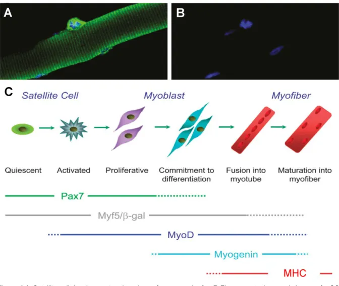
Next to altered gene expression and/or protein synthesis in skeletal muscle fibres, another important aspect of skeletal muscle adaptation or regeneration is the activation, proliferation, and differentiation of SCs, i.e. myogenesis. SCs lie quiescent between the basal membrane and the sarcolemma and were first discovered by Alexander Mauro who named them SCs for their localisation surrounding a muscle fibre (34). They originate from the somites and make up the necessary muscle specific stem cell pool needed for embryonic and adult myogenesis. In postnatal myogenesis, as it occurs in response to training or muscle injury, SCs are activated by multiple stimuli to become myoblasts. Myoblasts proliferate and increase in number, but at some point exit the cell cycle to enable differentiation and eventually fuse with existing myofibres to enlarge, to replace or to repair muscle tissue (Figure 1.1).
The role of SCs in muscle regeneration has been well characterised. However, their function
in mediating exercise-induced adaptations is lively debated (35). Most emphasis has been
placed upon elucidating the contribution of SCs in skeletal muscle adaptation in response to
resistance training, where muscle fibre hypertrophy is the main adaptation. It is discussed
whether SCs are necessary for hypertrophic events (36). However, the current literature
suggests that after an initial phase where muscle hypertrophy mainly occurs through
increased protein synthesis by myonuclei within the muscle fibre, the addition of new
myonuclei to existing myofibres is required (14) for the maintenance of myonuclear domains,
i.e. a theoretical cytoplasmic area associated with a myonucleus, to allow for sufficient supply
of genetic machinery and protein synthesis without changing the kinetics of protein
production for each nucleus (7). Only a few studies have looked at the role of SCs in
response to training regimens that do not induce hypertrophy, e.g. high intensity endurance
training as mentioned above. Results from an animal study (37) and two human studies (38,
39) have implied that endurance-based training enhances the SC pool. Furthermore, the
literature suggests that this enhancement is influenced by rather the intensity than the
duration of exercise (40). This would also explain the results of Snijders et al. who did not
find a change the satellite cell content in diabetes type 2 patients following 6 month of a
continuous, endurance-type exercise programme (41). Their workload corresponded to about
75% VO
2max, whereas the other studies used intermittent protocols with intensities around 75-
7
Figure 1.1: Satellite cell development and markers of myogenesis. A + B Fluorescent microscopic image of a SC on a mouse muscle fibre (yellow fluorescent protein – SC; blue – DNA). Modified from (52). C SC myogenesis scheme and markers typical for each stage. SCs lie quiescent underneath the basal membrane but can be activated by training. Activated SCs are called myoblasts that proliferate and increase in number. At some point they exit the cell cycle and start differentiating and fusing to form myotubes. Pax7 is expressed in quiescent, proliferating and early differentiating cells. Myf5 and MyoD expression increases during the proliferative and early differentiation phase, whereas myogenin marks the commitment to differentiation. During late differentiation, i.e.
that of sarcomeric assembly, MHC expression is initiated. SC – Satellite cell; Pax7 – paired box transcription factor 7; Myf5 – myogenic factor 5; MyoD – myogenic determination protein; MHC – myosin heavy chain. Modified from (167).
95% VO
2peak(38) or 75-95% HR
max(39) i.e. intensities that were considerably higher.
Moreover, one study provides evidence that SCs are not only activated by high intensity endurance training and lead to increase SC availability but also have a role in functional adaptations following non-hypertrophic training such as skeletal muscle fibre remodelling (42). Taken together, the current literature suggests that in response to resistance and high intensity endurance training SCs are activated and play a crucial role in hypertrophic and even possibly non-hypertrophic skeletal muscle adaptations.
1.3 Myogenesis and its regulatory epigenetic modifications
During myogenesis, SCs and myoblasts underlie a strict sequential expression pattern of
different transcription factors and structural muscle proteins (43). These can be used as
8 markers to study the proliferation and differentiation behaviour of this cell population in vitro.
One of these proteins is the paired-box transcription factor 7 (Pax7). It is expressed during quiescence, activation, and proliferation as well as early differentiation and it is the most commonly used marker to identify SCs as such (44–46). To briefly describe the expression pattern during differentiation which is extensively reviewed in (47), a reduction of Pax7 expression coincides with the withdrawal from the cell cycle and transition from the proliferative to the differentiation phase. This state is also accompanied with the onset of the expression of several myogenic regulatory factors (MRFs). This group of muscle specific transcription factors of the basic helix-loop-helix (bLHL) class of DNA binding proteins consists of four members: myogenic factor 5 (Myf5), myogenic determination protein (MyoD), muscle-specific regulatory factor 4 (MRF4), and myogenin. Whereas MyoD and Myf5 are soon expressed following activation and early differentiation of SCs, myogenin is not expressed until a later stage marking the commitment to differentiation to which it is essential in adult myogenesis (48, 49). The exact expression pattern of MRF4 is less clear. It is expressed before as well as after myogenin expression (50, 51). At an even later stage of differentiation, i.e. that of sarcomeric assembly, the expression and synthesis of myosin heavy chain (MHC) indicates end-terminal differentiation of myotubes and myofibres. A number of stimuli that activate SCs were identified and are comprehensively reviewed in (52) and (53). Among these are transforming growth factor β, myostatin, insulin-like growth factor 1, fibroblast growth factor, hepatocyte growth factor, and the wingless-type MMTV integration site (Wnt) family of proteins. Although a number of SC activators were identified, their mechanisms have not been completely understood. Furthermore, some factors possibly might have not even been identified as yet.
The gene expression pattern during myogenesis is regulated by epigenetic modifications (54, 55), especially histone modifications. Generally, 147 bp of double stranded DNA are wrapped around a histone octamer consisting of two copies of each core histone, namely 2A, 2B, 3, and 4, forming so called nucleosomes which make up the basic units of chromatin.
This ensures compactness of the genome to fit into the nuclei but also results in limited
accessibility for the transcription machinery and hence hindered gene expression. This
structure can however be reversibly modified to allow gene expression if indicated. Histone
modifications include acetylation, methylation, phosphorylation, and ubiquitination and
regulate gene expression differently. Especially histones 3 and 4 (H3 and H4) are prone to
such alterations (56). Histone acetylations, the most common post-translational histone
modification, lead to enhanced transcriptional activity. Histone acetyltransferases (HAT) and
histone deacetylases (HDAC) add or remove, respectively, acetyl groups from histone lysine
residues, converting the charge of the residue and alter DNA-histone binding as well as
nucleosome associations (57, 58). Histone methylation however, is more complex as it can
9 either increase or repress gene expression. It occurs most commonly at lysine and arginine residues where up to three or two methyl groups can be added, respectively (59, 60).
Methylated residues provide binding sites for chromodomains (61) and PHD domains (62–
64) which are both related to chromatin remodeling. Trimethylation of H3 at lysine 4 (H3K4me3) occurs in as many as 75% of all gene promoters in some cells (65, 66) and it is positively correlated with active gene transcription (67, 68) marking its critical role in gene expression. In contrast, trimethylation of H3 at lysine 9 and 27 (H3K9me3 and H3K27me3) as well as H4 at lysine 20 (H4K20me3) are linked to gene repression. Adding to the complexity compared to histone acetylation is the finding that methylation of different residues appears to be catalysed by different lysine methyltransferases (KMT). They are two forms of KMT: polycomb group (PcG) and trithorax group (TrxG) proteins that work antagonistically. For example, H3K4 methylation is mediated by KMT2, a TrxG member that acts as the subunit of the histone methyltransferase complex absent, small, or homeotic-like, variant 2 (Ash2L), whereas H3K27 is methylated by PcG proteins from the KMT6 family, namely enhancer of zeste homolog 1 and 2 (Ezh1 and Ezh2) that act as the active subunit of the polycomb repressor complex 2 (PRC2), another histone methyltransferase complex (69).
The H3K27me3 mark is dominant to H3K4me3 (67). So, in order for gene transcription to
occur at gene loci marked with both modifications, H3K27me3 has to be demethylated. This
is achieved by the lysine demethylase family 6 (KDM6) members ubiquitously transcribed
tetratricopeptide repeat, X chromosome (UTX) and Jumonji domain-containing protein 3
(JMJD3) (70, 71, 71).
10
Figure 1.2: Histone modifications H3K4me3 and H3K27me3 during SC proliferation and differentiation. A During proliferation and cell cycle progression Pax7 and Myf5 genes are expressed. These genes are known to be marked with H3K4me3. Myf5 is important for the progress from the proliferative to the differentiation stage. The H3K4me mark is established through the recruitment of TrxG proteins by Pax7. B In terminally differentiating SCs Pax7 and cell cycle progression genes are silenced. This repression involves PcG mediated H3K27me3 at the promoter regions of the respective genes. At this time point MyoD collaborates with Mef2d and Six4 to establish the H3K4me3 mark at the myogenin promoter, initiating its expression. H3Kxme3 – trimethylated histone 3 at lysine x; SC – Satellite cell; Pax7 – paired box transcription factor 7; Myf5 – myogenic factor 5; TrxG – trithorax group protein; PcG – polycomb group protein; MyoD – myogenic determination protein; Mef2d – myocyte enhancer factor 2d; Six4 – Six homeobox 4. Modified from (69).
11 During myogenesis (Figure 1.2), Pax7 expression is switched off in order for differentiation to proceed. p38 MAPK promotes recruitment of PRC2 to the Pax7 promoter via interaction with Ezh2, which is the direct substrate for phosphorylation-mediated association with Ying Yang 1 (YY1). This leads to the enrichment in chromatin occupancy by Ezh2/YY1 which mediates Pax7 repression (72, 73). During proliferation cell cycle genes are enriched with the permissive H3K4me3 mark (74). However, when differentiation is initiated, the repressive H3K27me3 conformation accumulates (75) by a mechanism involving E2F family members and the retinoblastoma protein (pRb) (76). Furthermore, during the proliferative state, Myf5 expression is induced via a Pax7-dependent recruitment of a KMT2 family member and subsequent H3K4me3 (77). Myogenin expression marks the commitment to differentiation in myoblasts. Here, the repressive H3K27me3 is removed during differentiation (78, 79).
Furthermore, Rampalli et al. showed that Ash2L is recruited by Mef2d and targets the H3K4me3 mark to specific promoters, such as myogenin (80). The same group further demonstrated that this recruitment is regulated through the p38 MAPK phosphorylation of Mef2d, linking the p38 MAPK signalling pathway with the activation of muscle-specific genes in myogenesis (79, 80). The histone modification H3K9me3 is controversially discussed in the literature. Although it has been thought to repress gene expression (81, 82), it was recently shown to also be able to induce the same (83). The H4K20me3 is another histone methylation known to play a role in myogenesis, but so far it was only shown to affect myogenesis by the repression of myogenin with the further interpretation being elusive (84).
Taken collectively, the current literature suggests that epigenetic events, in particular histone trimethylation of H3K4 and H3K27, are fundamental to the regulation of myogenesis.
However, apart from histone methylations, other epigenetic modifications might be important during myogenesis.
1.4 The role of p38 MAPK in skeletal muscle adaptation
Interestingly, a number of the above mentioned events are regulated via the p38 MAPK.
MAPKs are a family of protein kinases, i.e. proteins that covalently bind a phosphate to a
threonine (Thr), serine (Ser), or tyrosine (Tyr) side chain of other proteins and thereby alter
their targets activity or interactions with other molecules. This leads to changes in cell cycle
machinery, gene expression, protein synthesis, differentiation, metabolism, migration, and
cell death (85, 86). 3 subfamilies have been well characterised in the literature: extracellular
signal-regulated kinases (ERK1/2), the c-Jun NH2 terminal kinases (JNK1/2/3), and p38
MAPK (p38α, p38β, p38γ, p38δ). During myogenesis, p38 MAPK has been identified as a
major regulator (87, 88). Upon differentiation cues, p38 MAPK is phosphorylated by the
upstream MAPK kinases 3 and 6 (MKK3 and MKK6) at threonine 180 and tyrosine 182 (p-
p38), and activated. The isoforms p38α and p38β were shown to be involved during
12 myoblasts differentiation (89–91). The role of p38γ in myogenesis is so far not fully understood. Although it is expressed in skeletal muscle and its expression is upregulated during myogenesis (90, 92–94), a differentiation regulatory effect could not be confirmed.
p38δ appears to play no important role in skeletal muscle (reviewed in (95)).
Additionally to the histone methylations described above, p38 MAPK has additional pro- myogenic effects (Figure 1.3). Upon differentiation signals, activated p38 MAPK directs switch/sucrose non-fermentable (SWI/SNF) and Ash2L-KMT containing complexes to regulatory muscle genes, promoting their transcription (96–98). Furthermore, p38 is directed to the chromatin of muscle specific genes through interactions with E47 (99), Mef2d (80) and BAF60 (96). CAM-downregulated by oncogenes (CDO) is a transmembrane protein of the Ig- fibronectin III repeat subfamily. It can also activate p38 MAPK which in turn leads to the formation of MyoD-E47 heterodimers that bind to E-box sequences on the regulatory regions of muscle-specific genes (100, 101). It is moreover involved in cell cycle exit. p38 MAPK decreases cyclin D1 expression and pRb phosphorylation, therefore promoting cell cycle withdrawal and differentiation progression (102).
Figure 1.3: A model for the activity of p38 MAPK in myogenesis. p38 MAPK is activated by MKK3/6 in muscle cells and subsequently phosphorylates various substrates contributing to myogenesis. It induces the transcriptional activity of Mef2 transcription factors and promotes the heterodimerisation of E47 with MyoD.
Additionally, it induces Myf5 expression. p38 MAPK recruits the SWI/SNF complexes to muscle promoter regions, thereby affecting the epigenetic landscape. p38 MAPK – p38 mitogen-activated protein kinase; MKK3/6 – MAPK kinase 3/6; Mef2 – myocyte enhancer factor 2; MyoD – myogenic determination protein; Myf5 – myogenic factor 5; SWI/SNF - switch/sucrose non-fermentable. Modified from (87).
13 In addition to its regulatory role during the differentiation of SCs, p38 MAPK has also been shown to possess the ability to induce skeletal muscle adaptation within differentiated muscle tissue in response to exercise or physical activity. The level of phosphorylation and hence activation depends on the exercise status, i.e. p38 MAPK is more phosphorylated in untrained subjects compared to highly trained athletes (103, 104). This in turn leads to the stimulation of activating transcription factor 2 (ATF2) and Mef2 (105) which are upstream transcription factors of the PGC1α gene and induce its expression following exercise (31, 105, 106). These data implies a superior role of p38 MAPK during SC differentiation and regulating gene expression in response to exercise and physical activity making it a key molecule for skeletal muscle adaptation.
1.5 Exercise and physical activity and histone modifications
Epigenetic events are not only important during myogenesis, but also regulate skeletal muscle fibre adaptations in response to exercise and physical activity through exercise- induced alterations of the metabolic state (107–109). This implies that epigenetic responses should also elicit an intensity-dependent effect. This concept was supported by the finding of decreased whole genome methylation in muscle biopsies from sedentary subjects after an acute bout of exercise. Exercise induced dose-dependent expression of PGC1α and Mef2a mRNA accompanied by a pronounced hypomethylation on both promoters, a state associated with gene induction (110). Proposed metabolic stimuli to regulate epigenetic events following exercise are AMPK and CaMKII. Both have a pronounced regulatory effect on class II HDACs. This class consists of HDAC 4, 5, 7, and 9 and are enriched in striated muscle (111). They exert their function by association with the Mef2 family of transcription factors to repress Mef2-dependent transcription (112). Following a single bout of exercise, HDAC5 was found to be less abundant within the nucleus and less HDAC5 was associated with Mef2 (113). These changes were accompanied by increased Mef2-binding activity to the DNA (114) and elevated glucose transporter 4 (GLUT4) mRNA levels (113). The concept that HDAC5 nuclear abundance is reduced following exercise could also be confirmed for HDAC4 (115). AMPK and CaMKII can both phosphorylate HDAC4 and 5, mediate their nuclear export and hence render these enzymes to suppress histone acetylation (115, 116).
Although this direct relationship was shown for GLUT4, other oxidative genes are also
thought to be regulated by this mechanism (31, 117). The literature also suggests that
epigenetic modifications are involved in the differential expression of the MHC genes and
therefore determination of muscle phenotype (118). Changes in acetylation and methylation
of H3 led to differential expression of MHC1, MHC2x, and MHC2b fibres in response to
muscle unloading in skeletal muscle tissue of mice (119, 120). A knockout model of HDAC5
in mice furthermore suggested a regulatory role for slow fibre formation. Following HDAC5
14 loss, MHC1 fibre type increased (121). Collectively, epigenetic modifications in response to exercise provide evidence for the role of metabolic control of gene expression and the concept that skeletal muscle adaptation via epigenetic mechanisms is therefore exercise- intensity dependent. However, knowledge of epigenetic alterations, especially histone modifications is scarce. Understanding the exercise and physical activity-dependent alterations in skeletal muscle gene expression by altering the epigenetic landscape will add new implications to exercise and physical activity prescriptions in prevention and therapy.
1.6 Lactate as a metabolic signal of gene expression
Muscle phenotype alterations only occur after repeated bouts of exercise. Over time, skeletal muscle demonstrates remarkable plasticity in response to contractile activity by functional remodelling (122, 123). Repeated stimuli triggered in each single session at a sufficient frequency are necessary to induce a new steady state within the muscle (8) including the activation and regulation of the development of SCs necessary for adaptation. These signals include hormonal factors (124), mechanical stimuli (125), and alterations of the metabolic state (126). While the importance of hormonal and mechanical factors has been well established (127, 128), the importance of metabolic influences especially in resistance and high intensity endurance training is indicated (129) but remains elusive.
The metabolic responses to resistance and high intensity endurance exercise are thought to be initiators of skeletal muscle adaptation by influencing motor unit activation, hormones, and muscle damage (130). The challenge arising with this idea is to identify the intracellular signalling molecules that occur at different concentrations depending on the exercise intensity. One possible candidate is La. All muscle contractions require energy in form of adenosine triphosphate (ATP). ATP stores are limited within the muscle fibre and therefore ATP has to be resynthesised at its rate of use during muscular activity in order to maintain contractions. During high intensity exercise the main source of ATP is glycolysis that produces pyruvate. Pyruvate is transferred into mitochondria for further oxidative phosphorylation and ATP resynthesis. However, if exercise intensity and the ATP breakdown rate exceed the rate of oxidative phosphorylation, pyruvate accumulates in the cytosol and is converted to lactic acid by the lactate dehydrogenase enzyme. Lactic acid is almost fully dissociated to La and protons at physiological pH, resulting in La accumulation within the working muscle. La has long been considered a metabolic waste product and its accumulation was thought to be the cause of the decrease in muscle pH and hence, muscle fatigue. This idea has changed massively in the past. La is now recognised as a metabolic stimulus able to induce skeletal muscle adaptation. For example, La has been shown to increase norepinephrine and growth hormone levels following resistance exercise (131–133).
Furthermore, it increases recruitment of motor units at a given load (131, 134). La was also
15 shown to correlate with muscle damage (135). These events lead to enhanced skeletal muscle adaptation and therefore underline the role of La as a metabolic stimulus for altering muscle phenotype.
The aforementioned La effects are indirect and have no immediate impact on muscle gene expression or protein synthesis. However, recently Hashimoto et al. demonstrated that La increases monocarboxylate transporter-1 (MCT-1) and PGC1α mRNA content indicating the induction of mitochondrial biogenesis in L6 rat myoblasts (136). La was termed “Lactormone”
as it possesses signalling properties inducing gene expression necessary for skeletal muscle adaptation (137).
With the identification of La as a gene expression-altering molecule in skeletal muscle, the question arises which signalling pathways are involved in the mediation. It was previously shown that in macrophages La concentrations correlated with the induction of angiogenic factor gene expression (138). Later on this effect was shown to be mediated by H
2O
2(139).
H
2O
2is a member of the reactive oxygen species (ROS) which also include the superoxide
anion (·O
2-) and hydroxyl radicals (·OH). Although H
2O
2is not a free radical and a weaker
oxidising agent, it can be oxidised into the extremely active and toxic ·OH (140) and with
other ROS can affect signalling cascades that affect cell growth, proliferation, migration, and
apoptosis (141). ROS are produced at various sites in skeletal muscle. Main sources are
Complex I and III of the electron transport chain of mitochondria (142). Furthermore, NADPH
oxidases (143) and xanthine oxidase (144) contribute to ROS formation within the cytosol of
skeletal muscle. ROS-induced redox signalling regulates numerous transcription factors and
alter gene expression and hence skeletal muscle phenotype (145). One known target of ROS
are MAPKs (146). Applying the above mentioned mechanism suggested in macrophages to
muscle tissue, investigations confirmed that La increases intracellular H
2O
2and therefore
ROS, and subsequently promoted binding of ROS-sensitive transcription factors to DNA
(136). In the same study it was furthermore speculated that ROS led to the downstream
activation of MAPK (136). However, no data on La and its effect on the MAPKs are available
for skeletal muscle and hence the role of these key regulators in La-signal mediation remains
to be investigated. Taken together, La is an important signalling molecule for skeletal muscle
adaptations. Although a La-activated ROS-sensitive signalling network has been suggested,
conclusive data is missing if the La-effects depend on ROS-formation. Furthermore,
molecular insights in the mediation of the La effect are not yet provided.
16 1.7 Aims
1. To identify phenotypical effects of a simulated training microcycle of resistance or high intensity endurance training by intermittent incubation with a potent metabolic signalling molecule, i.e. La, on the proliferation and differentiation in a model for skeletal muscle stem cells.
2. To investigate the role of ROS formation and oxidative stress induced by La and whether these could be responsible for the observed effects.
3. To elucidate whether elevated La levels influence key signalling molecules of skeletal muscle adaptation, i.e. p38 MAPK, and its downstream epigenetic targets H3K4me3 and H3K27me3 to further establish the important role of La as a metabolic signal of gene expression.
4. To confirm that these mechanisms in muscle stem cells are conserved during
differentiation and remain active in differentiated muscle tissue exposed to similar La
concentrations evoked by exercise in vivo.
17 2 Materials & Methods M
ATERIALS& M
ETHODS2.1 Materials
2.1.1 Lab equipment and disposables
Name Company A-Plan 10x/0,25 Ph1 objective Carl Zeiss AG, Jena Germany
A-Plan 20x/0,4 Korr Ph2 objective (200M) Carl Zeiss AG, Jena Germany ApoChromat 63x/1,4 Oil DIC objective Carl Zeiss AG, Jena Germany
ApoTome Carl Zeiss AG, Jena Germany
Axiovert 200M microscope Carl Zeiss AG, Jena Germany AxioCam MRm Carl Zeiss AG, Jena, Germany
Biofuge 13 Heraeus, Hanau, Germany
BioMax Exposure Cassette Eastman Kodak Co, Rochester, NY, USA BIOSEN S-Line Lactate analyser EKF-Diagnostic GmbH, Barleben, Germany Bürker hemocytometer Karl Hecht, Sondheim, Germany
CCD camera MC-3255 Sony, Tokio, Japan
Cell Culture Flasks and Consumables BD Biosciences, San Jose, CA, USA CO
2Incubator C150 Binder, Tuttlingen, Germany
Criterion
TMelectrophoresis chamber Bio-Rad Laboratories, Hercules, CA, USA Criterion
TMXT 4-12% Bis-Tris gels Bio-Rad Laboratories, Hercules, CA, USA Filter Set 10 (exc. 450-490 nm; em. 515-565 nm) Carl Zeiss AG, Jena Germany
Filter Set 14 (exc. 510-560 nm; em. 590 nm) Carl Zeiss AG, Jena Germany Filter Set 49 (exc. 365 nm; em. 545-450 nm) Carl Zeiss AG, Jena Germany Flexcylcer
2thermocycler AnalytikJena AG, Jena, Germany
Glass cover slips VWR International, Langenfeld, Germany Hypodermic Needle Henry Schein, Melville, NY, USA
ISOMED 2000 isokinetic equipment D & R Ferstl GmbH, Hemau, Germany Kinetic Microplate Reader MWG Biotech, Ebersberg, Germany KS300 microscope Carl Zeiss AG, Jena Germany Lamina Air Flow HS12 Heraeus, Hanau, Germany
Leica CM1900 Cryostat Leica Microsystems, Wetzlar, Germany Leitz Orthoplan Leica Microsystems, Wetzlar, Germany Lode Excalibur cycle ergometer Lode, Groningen, Netherlands
Micro-dismembrator Braun, Melsungen, Germany
Microscope slides VWR International, Langenfeld, Germany Nalgene
TMCryo 1°C Freezing Container Thermo Fisher Scientific, Waltham, USA pH-mV-meter Mettler Toledo, Gießen, Germany Plan Neofluar 20x/0.5 objective (KS300) Carl Zeiss AG, Jena Germany
Poly-L-lysine microscope slides VWR International, Langenfeld, Germany PowerPac
TMBasic power supply Bio-Rad Laboratories, Hercules, CA, USA PVDF membrane Pall Corporation, Port Washington, NY, USA Scaltec SBA53 weighing scale Scaltec Instruments, Heiligenstadt, Germany Table centrifuge 5417R Eppendorf, Hamburg, Germany
Thermocycler Mx3005P Agilent Technologies, Santa Clara, CA, USA
Thermomixer 5436 Eppendorf, Hamburg, Germany
18 Titramax 101 shaker Heidolph Instruments, Schwabach, Germany TransBlot Turbo Bio-Rad Laboratories, Hercules, CA, USA Vortex Genie2 Scientific Industries, Bohemia, NY, USA X-OMAT x-ray films Eastman Kodak Co, Rochester, NY, USA 2.1.2 Chemical and solutions
Name Company 100 bp DNA ladder Invitrogen, Karlsruhe, Germany
Acetic Acid Merck, Darmstadt, Germany
Agarose Biozym Scientific, Hessisch Oldendorf, Germany Aqua-Poly/Mount Polysciences, Warrington, PA, USA
Ammonium chloride Merck, Darmstadt, Germany
β-mercaptoethanolInvitrogen, Karlsruhe, Germany Bovine Serum Albumine PAA, Pasching, Austria
BCP Molecular Research Center, Cincinnati, OH, USA bromphenol blue Merck, Darmstadt, Germany
Cell lysis buffer (# 9803) Cell Signaling Technology, Danvers, MA, USA Complete Mini
®Protease Inhibitor Cocktail Roche, Mannheim, Germany
D-Glucose Roth Chemicals, Karlsruge, Germany DAB Sigma-Aldrich, Steinheim, Germany
DAPI Invitrogen, Karlsruhe, Germany
DMSO Merck, Darmstadt, Germany
DMEM Invitrogen, Karlsruhe, Germany
DPBS Invitrogen, Karlsruhe, Germany
Dry milk powder Bio-Rad Laboratories, Hercules, CA, USA EDTA Sigma-Aldrich, Steinheim, Germany
Ethanol Merck, Darmstadt, Germany
Entellan
®Merck, Darmstadt, Germany
FBS PAA, Pasching, Austria
Fresubin
®protein energy drink Fresenius Kabi, Bad Homburg, Germany Gelatine Sigma-Aldrich, Steinheim, Germany Glucoseoxidase Sigma-Aldrich, Steinheim, Germany Glutamine Invitrogen, Karlsruhe, Germany Glycerol Merck, Darmstadt, Germany
Glycine Merck, Darmstadt, Germany
H
2O
2Merck, Darmstadt, Germany
HALT Phosphatase Inhibitor Cocktail Pierce Biotechnology, Rockford, IL, USA HD Green IntasScience Imaging Instruments, Göttingen,
Germany
Hemolyzing solution (ready-to-use) EKF Diagnostic GmbH, Barleben, Germany Horse Radish Peroxidase Dako, Glostrup, Denmark
Isopropanol Merck, Darmstadt, Germany KH
2PO
4Merck, Darmstadt, Germany L-Ascorbic Acid Sigma-Aldrich, Steinheim, Germany Linolenic Acid Sigma-Aldrich, Steinheim, Germany Methanol Merck, Darmstadt, Germany
N-Acetyl-L-cysteine Sigma-Aldrich, Steinheim, Germany
19 Na
2HPO
4Merck, Darmstadt, Germany
NiSO4 Merck, Darmstadt, Germany
Paraformaldehyde Roth Chemicals, Karlsruge, Germany Penicillin/Streptomycin Invitrogen, Karlsruhe, Germany Precision Plus Protein Standard Dual
Color Bio-Rad Laboratories, Hercules, CA, USA Recovery
TMCell Culture Freezing Medium Invitrogen, Karlsruhe, Germany
Restore
TMWestern Blot Stripping Buffer Thermo Fisher Scientific, Waltham, MA, USA SB203580 Cell Signaling Technology, Danvers, MA, USA Sodium bicarboante Invitrogen, Karlsruhe, Germany
Sodium chloride Merck, Darmstadt, Germany Sodium dodecyl sulfate Amresco, Karlsfeld, Germany Sodium-L-Lactate Sigma-Aldrich, Steinheim, Germany Sodium pyruvate Invitrogen, Karlsruhe, Germany
SuperSignal West Dura Luminol/Enhancer Thermo Fisher Scientific, Waltham, MA, USA TissueTek
®O.C.T.
TMCompound Sakura Finetek, Zoeterwoude, Netherlands
Tris Merck, Darmstadt, Germany
TritonX-100 Sigma-Aldrich, Steinheim, Germany
TriReagent
®Molecular Research Center, Cincinnati, OH, USA Trypsin-EDTA Invitrogen, Karlsruhe, Germany
Tween20 Sigma-Aldrich, Steinheim, Germany XT MOPS running buffer Bio-Rad Laboratories, Hercules, CA, USA Xylol Alfred Quadflieg, Gelsenkrichen, Germany All compounds/used chemicals were adjusted for purity.
2.1.3 Buffers and cell culture media
0.1% gelatine 0.1% gelatine in DPBS
PM 20% FCS
1% pen/strep 4 mM glutamine
0.15% sodium bicarboante
1 mM sodium pyruvate in DMEM
(0 mM La) DM 4% HS 1% pen/strep 4 mM glutamine
0.15% sodium bicarboante
1 mM sodium pyruvate in DMEM
5 mM La DM 4% HS
1% pen/strep 4 mM glutamine
0.15% sodium bicarboante 1 mM sodium pyruvate
5 mM sodium lactate in DMEM
20
10 mM La DM 4% HS
1% pen/strep 4 mM glutamine
0.15% sodium bicarboante 1 mM sodium pyruvate
10 mM sodium lactate in DMEM
20 mM La DM 4% HS
1% pen/strep 4 mM glutamine
0.15% sodium bicarboante 1 mM sodium pyruvate
20 mM sodium lactate in DMEM
DM + DMSO 4% HS
1% pen/strep 4 mM glutamine
0.15% sodium bicarboante 1 mM sodium pyruvate
0.1% DMSO in DMEM
DM + SB 203580 4% HS 1% pen/strep 4 mM glutamine
0.15% sodium bicarboante 1 mM sodium pyruvate
0.1% DMSO
10 µM SB203580 in DMEM 20 mM La DM + AA 4% HS
1% pen/strep 4 mM glutamine
0.15% sodium bicarboante 1 mM sodium pyruvate 20 mM sodium lactate
100 µm ascorbic acid in DMEM
20 mM La DM + NAc 4% HS 1% pen/strep 4 mM glutamine
0.15% sodium bicarboante 1 mM sodium pyruvate 20 mM sodium lactate
5 mM N-acetylcysteine in DMEM
20 mM La DM + LA 4% HS 1% pen/strep 4 mM glutamine
0.15% sodium bicarboante 1 mM sodium pyruvate 20 mM sodium lactate
5 µM linolenic acid in DMEM
21
TAE 40 mM Tris
20 mM acetic acid
1 mM EDTA pH = 8.0
PBS 137 mM NaCl
10 mM Na
2HPO
41.8 mM KH
2PO
4pH = 7.4
TBS 50 mM Tris
150 mM NaCl pH = 7.6
4% PFA 4% PFA in PBS pH = 7.4
Permeabilisation
solution 500 mM NH
4Cl
0.25% Tx-100 in PBS DAPI solution 300 nM DAPI in PBS PB 80 mM Na
2HPO
4·2 H
2O
20 mM NaH
2PO
4·H
2O pH = 7.4
DAB 2 mM DAB
7.5 mM NH
4Cl 1.5 mM NiSO4
10 mM D-glucose
0.0004% glucose oxidase in PB
Lysis buffer 1% Tx100 in TBS pH = 8.0
Laemmli buffer 2x 125 mM Tris 4% SDS 20% glycerine
10%
β-mercaptoethanol0.004% bromphenol blue pH = 6.8
TBST 150 mM NaCl
50 mM Tris
0.1% Tween20
®pH = 7.4
Transfer buffer 25 mM Tris 150 mM glycine
10% methanol
22 2.1.4 Kits
Name Company Cell Proliferation ELISA BrdU (colorimetric) Roche Diagnostics, Mannheim, Germany
RC DCTM