Diversity of Voltage Activated Calcium Currents in Identified Olfactory Interneurons
I n a u g u r a l – D i s s e r t a t i o n
zur Erlangung des Doktorgrades
der Mathematisch‐Naturwissenschaftlichen Fakultät der Universität zu Köln
vorgelegt von
Andreas Husch
aus Prüm
Köln 2007
Berichterstatter: Prof. Dr. P. Kloppenburg PD Dr. J. Schmidt
Tag der mündlichen Prüfung: 28.11.2007
Contents
Abbreviations...6
Zusammenfassung...7
Abstract...9
1 Introduction...11
1.1 The insect olfactory system...11
1.2 Variety of voltage gated calcium channels...13
1.3 Objectives of this thesis...15
2 Materials & Methods...16
2.1 Animals and materials...16
2.2 Cell culture...17
2.2.1 Cell culture dissection...17
2.2.2 Dissociation of olfactory interneurons...17
2.3 Intact brain preparation...18
2.4 Electrophysiological recordings...20
2.4.1 Whole cell patch clamp recordings...20
2.4.2 Data aquisition...21
2.4.3 Data analysis...21
2.5 Odor stimulation...23
2.6 Histology...25
2.6.1 Whole mount preparation of biocytin filled neurons...25
2.6.2 Slice preparation of the stained neurons...25
2.6.3 Confocal laser scanning microscopy...26
3 Results...27
3.1 Voltage activated calcium currents in vitro and in situ...28
3.1.1 Voltage activated inward currents...28
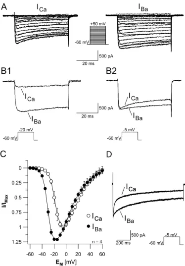
3.1.2 Charge carrier...31
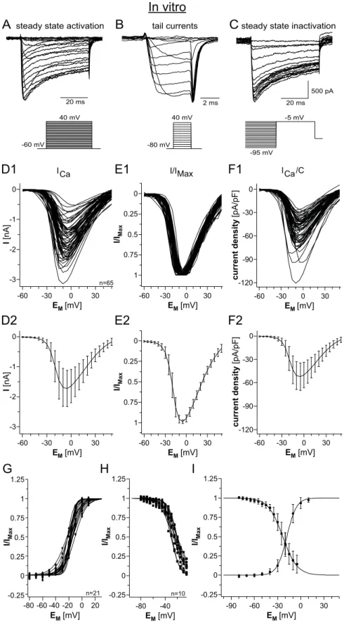
3.1.3 Calcium currents in vitro...33
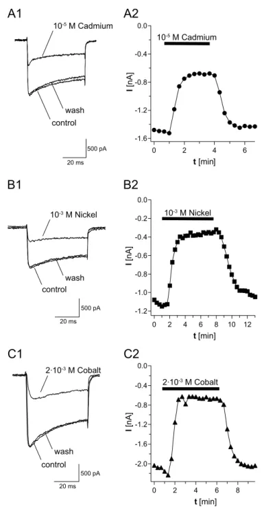
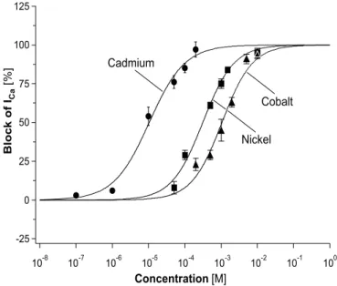
3.1.4 Inorganic ions as calcium channel blockers...35
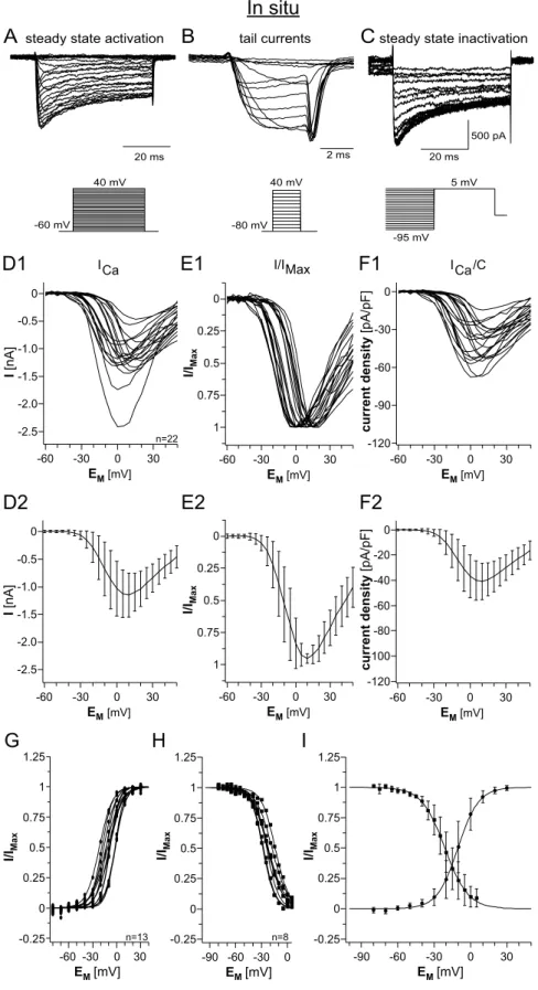
3.1.5 Calcium currents in situ...38
3.2 Identification of different olfactory interneuron types...41
3.2.1 Odor responses ...43
3.2.2 Uniglomerular projection neurons (uPNs)...45
3.2.3 Type I local interneurons (type I LNs)...47
3.2.4 Type II local interneurons (type II LNs)...49
3.2.5 Comparison between uPNs, type I LNs and type II LNs...50
3.3 Voltage activated calcium currents in identified neuron groups...53
3.3.1 I/V‐relationship of calcium current activation...53
3.3.2 Tail current analysis...57
3.3.3 Steady state inactivation...59
3.3.4 Characterization of calcium currents in type II LN subpopulations...62
4 Discussion...71
4.1 Voltage activated calcium currents in vitro and in situ...71
4.1.1 General parameters of voltage activated calcium currents...72
4.1.2 Rundown during whole cell voltage‐clamp recordings ...75
4.2 Identification of olfactory interneurons...76
4.3 Diversity of voltage activated calcium currents in identified neuron types...78
4.3.1 Analysis of calcium currents in identified neurons...79
4.3.2 Functional significance of voltage activated calcium currents...80
4.4 Diversity of other voltage activated ion currents...82
4.5 Conclusions...83
List of Figures...85
References...87
Acknowledgments...96
Erklärung...98
Teilpublikationen...99
Abbreviations
4‐AP 4‐aminopyridine
AL(s) antennal lobe(s) AP(s) action potential(s) HVA high voltage activated IA transient potassium current
IBa barium current
ICa calcium current
Ih h‐current; activated by hyperpolarization IK(V) sustained potassium current
IK(Ca) calcium dependent potassium current
Imax maximum current
INa sodium current
LN(s) local interneuron(s) LVA low‐voltage activated M‐LVA mid/low‐voltage activated MWRS Mann‐Whitney rank sum ORN(s) olfactory receptor neuron(s) PN(s) projection neuron(s)
s slope factor
SD standard deviation
TEA tetraethylammonium chlorid
TTX tetrodotoxin
uPN(s) uniglomerular projection neuron(s)
V0.5(act) potential at half‐maximal activation
V0.5(inact) potential at half‐maximal inactivation
VGCC voltage‐gated calcium channel
Zusammenfassung
Innerhalb des Antennallobus (AL) der Insekten projeziert jede Rezeptorzelle zu einem Glomerulus und viele Rezeptorneuronen konvergieren innerhalb jedes Glomerulus, wo sie lokalen Interneuronen (LNs) und (Ausgangs‐) Projektionsneuronen (PNs) synaptische Eingänge liefern. Die Verzweigungen der LNs beschränken sich auf den AL. Die Projektionsneurone hingegen projizieren ihre Axone in höherrangige Neuropile des Protocerebrums, einschließlich der Pilzkörper und dem lateralen Lobus des Protocerebrum. Insbesondere Projektionsneurone waren im Fokus intensiver Studien, da sie zahlreiche Funktionen erfüllen und eine Schlüsselrolle im zentralen olfaktorischen Verarbeitungsweg spielen. Im Gegensatz dazu sind die Rolle und funktionellen Eigenschaften der LNs noch unklar. Zum besseren Verständnis der biophysikalischen Parameter, die die olfaktorische Informationsverarbeitung auf zellulärer Ebene vermitteln, wurden spannungsaktivierte Kalziumströme (ICa) in olfaktorischen Interneuronen des Antennallobus von adulten Schaben wie folgt analysiert: 1) in akut dissoziierten Zellen (in vitro), 2) in einer intakten Hirnpräparation (in situ) und 3) in eindeutig identifizierten und intakten Neuronen.
Die Kombination von whole cell patch‐clamp Ableitungen in Strom‐ und Spannungsklemme mit Einzelzellmarkierungen ermöglichte es, uniglomeruläre Pns (uPNs) und LNs verschiedener Untergruppen zu studieren. Anhand ihrer
physiologischen und morphologischen Eigenschaften konnten zwei Hauptgruppen von LNs identifiziert werden, die im Folgenden als Typ I LNs und Typ II LNs bezeichnet werden. Duftstimulation und depolarisierende Strominjektionen lösten in Typ I LNs TTX‐sensitive Aktionspotentiale aus. Ableitungen in der Spannungsklemme lösten spannungsaktivierte Natriumströme, transiente und gleichrichtende Kaliumströme und Kalziumströme aus. Im Gegensatz dazu lösten Duftstimulationen und Strominjektionen in Typ II LNs zwar Membrandepolarisationen, jedoch keine TTX‐sensitiven Aktionspotentiale aus. In wenigen Ableitungen saßen kleine (2 mV) „spikelets“ auf den duftevozierten Depolarisationen auf. Spannungsaktivierte Natriumströme waren in Typ II LNs nicht messbar. Die spannungsaktivierten Kalziumströme hingegen waren signifikant größer als in Typ I LNs und die Eigenschaften der Aktivierung waren signifikant unterschiedlich. In Typ I LNs lag die Spannung der halbmaximalen Aktivierung (V0.5(act)) des ICa bei ‐11.1 ± 6.5 mV, was im Bereich der V0.5(act) von uPNs (V0.5(act) = ‐10.6 ± 3.4 mV) lag. Im Gegensatz dazu war V0.5(act) in Typ II LNs zu signifikant negativeren Potentialen verschoben (V0.5(act) = ‐19.4 ± 4.7 mV). Diese Ergebnisse deuten darauf hin, dass die durch Duftstimulation hervorgerufenen Depolarisationen in Typ II LNs vor allem durch Kalzium Ströme getragen werden könnten und dass die spannungsaktivierten Kalzium Ströme eine Rolle in graduierter synaptischer Übertragung zwischen Typ II LNs und anderen olfactorischen Interneuronen spielen könnten.
Abstract
In the insect antennal lobe (AL) each olfactory receptor cell projects to one glomerulus and many receptor axons converge in each glomerulus, where they provide synaptic input to local interneurons (LNs) and projection (output) neurons (PNs). The arborizations of LNs are confined to the AL. In contrast, the PNs extend axons to higher order neuropiles of the protocerebrum, including the mushroom bodies and the lateral lobus of the protocerebrum. In particular PNs have been in the focus of intensive studies, because they serve multiple functions and play a key role in the central olfactory pathway. However, the role and functional properties of LNs are less well understood. Towards the goal to better understand the biophysical parameters that mediate olfactory information processing on the cellular level, voltage activated calcium currents (ICa) in olfactory interneurons of the AL from adult cockroach were analyzed: 1) in acutely dissociated cells (in vitro), 2) in an intact brain preparation (in situ), and 3) in unequivocally identified and intact neurons.
Using whole cell patch‐clamp recordings in current and voltage‐clamp mode in combination with single cell staining, uniglomerular PNs (uPNs) and two major types of LNs could be identified by their physiological and morphological properties, which in the following are referred to as type I LNs and type II LNs. In type I LNs odor stimulation and depolarizing current injection elicited overshooting TTX‐
sensitive action potentials. Voltage‐clamp recordings revealed a voltage‐activated
sodium current, a transient and a sustained potassium current and a calcium current.
In contrast, in type II LNs odor stimulation and current injections induced membrane depolarization, but no TTX‐sensitive action potentials. In some recordings small (~ 2 mV) ʹspikeletsʹ were riding on the odor evoked depolarization. In type II LNs voltage activated sodium currents were not detectable. However, ICa was significantly larger compared to type I LNs and the activation characteristics were significantly different.
For type I LNs the voltage for half maximal activation (V0.5(act)) of ICa was ‐11.1 ± 6.5 mV, which is in the range of V0.5(act) for uPNs (V0.5(act) = ‐10.6 ± 3.4 mV). In contrast to this, in type II LNs the V0.5(act) of ICa is significantly shifted to more negative potentials (V0.5(act) = ‐19.4 ± 4.7 mV). These results suggest that the odor‐evoked depolarization in type II LNs might be carried largely by ICa and that ICa might play a role in graded synaptic release between type II LNs and other olfactory interneurons.
1 Introduction
1 Introduction
Odor discrimination is crucial for the survival of most animals. The first‐order synaptic relay in olfactory systems of vertebrate and invertebrate animals have striking similarities in physiology and neuronal organization, suggesting that olfactory information is processed through similar mechanisms in these evolutionary remote animals (Eisthen 2002; Hildebrand and Shepherd 1997; Strausfeld and Hildebrand 1999; Wilson and Mainen 2006). One experimental system that has served very successfully as a model to understand olfactory information processing is the first order olfactory relay or antennal lobe (AL) of insects (Keene and Waddell 2007; Laurent 1999; Vosshall and Stocker 2007). As an important step towards the
long term goal to better understand the cellular mechanisms that mediate olfactory
information processing I characterized the biophysical properties of voltage activated Ca2+ currents in identified olfactory interneurons from the ALs of adult Periplaneta americana.
1.1 The insect olfactory system
Inside the insect antennal lobe each olfactory receptor cell projects to one glomerulus and many receptor axons converge in each glomerulus, where they provide cholinergic synaptic input to local interneurons (LNs) and projection neurons (PNs).
The arborizations of local interneurons are confined to the antennal lobe, whereas the
1 Introduction
projection neurons extend their axons to higher order neuropiles of the protocerebrum, including the mushroom bodies and the lateral lobe of the protocerebrum. Studies in the fruit fly, Drosophila melanogaster, using genetically encoded activity sensors (Ng et al. 2002; Wang et al. 2003), proposed that olfactory information proceeds along a straight and simple path (labeled‐line), with minimal interaction among receptor‐specific tracks: receptor → glomerulus → output neuron
→ higher brain regions. For some odors of particular biological significance such as CO2 straight processing tracks may exist (Suh et al. 2004), but recent work shows that the general picture is more complex (for review see Stopfer 2005). With in vivo patch‐
clamp recordings in the antennal lobe of the fruitfly, Wilson et al. (2004) have shown, that a given PN can respond to odors of many different chemical classes. The authors compared explicitly the odor sensitivities of olfactory receptors and their immediate postsynaptic PNs. Surprisingly the electrophysiological results indicated that PNs are more broadly tuned than their presynaptic ORNs (Wilson et al. 2004). They concluded that PNs not only receive direct input from ORNs, but also input from neighboring glomeruli, possibly mediated by local interneurons. As already described in other insects, especially in the american cockroach, many antennal lobe LNs are GABAergic (Boeckh and Tolbert 1993; Distler 1989; Distler 1990; Malun 1991b), providing inhibitory input to the PNs (Christensen et al. 1993; MacLeod and Laurent 1996; Waldrop et al. 1987). As expected, also in Drosophila many LNs contain
the inhibitory transmitter GABA. Blocking GABA‐mediated inhibition reduced the
temporal variability in odor evoked PN firing patterns (Wilson and Laurent 2005), demonstrating that olfactory information is dramatically restructured within the antennal lobe. However, inhibitory local interneurons alone can not generate the complex response patterns, observed in PNs. Recent work from Shang et al. (2007) described that some of the apparent complexity in the antennal lobes output rises from a previously unidentified population of excitatory cholinergic local
1 Introduction
interneurons. These data indicate the presence of at least two or even more completely different physiological and morphological types of local interneuron populations. In the bee antennal lobe at least two morphologically different types of local interneurons have been described (Fonta et al. 1993). Immunohistochemical data from insect antennal lobe suggest an even more complex role of local interneurons. Some LNs are histaminergic (Gebhardt and Homberg 2004; Loesel and Homberg 1999; Nassel 1999) and other subpopulations are immunoreactive for other neuropeptides, amines or nitric oxide (Berg et al. 2007). However, even basic questions regarding the physiology of different types of LNs are not completely understood. For example in the locust (Locusta migratoria) olfactory system only non‐
spiking local interneurons are reported (MacLeod and Laurent 1996), whereas in the honey bee (Apis melifera) and in the moth (Manduca sexta) there are exclusively data available that describe spiking local interneurons (Christensen et al. 1993; Sun et al.
1993). The cellular mechanisms that underlie these important intrinsic properties remain obscure, as relatively little is known about the physiological properties of antennal lobe interneurons. It has been shown that the properties of neurons are largely determined by the types of ion channel and the rate of channel expression for the different channel types (Baro et al. 1997). As a first step toward addressing this issue, in this study I have explored the electrophysiological properties of voltage activated calcium currents in antennal lobe interneurons in the brain of Periplaneta americana.
1.2 Variety of voltage gated calcium channels
Due to their different functions and tasks antennal lobe neurons do not only differ in their morphology and arborization patterns, but also in their basic physiological and neuronal properties. In this context calcium handling plays a critical role in the
1 Introduction
control of a variety of neural processes such as synaptic release, membrane excitability, enzyme activation and activity dependent gene activation (Berridge 1998;
Augustine et al. 2003). The spatial and temporal dynamics of these signals are determined by a variety of cellular parameters including: calcium influx, calcium buffering, calcium extrusion, geometry of the cell and locally changing diffusion coefficients (Neher and Augustine 1992; Helmchen 2005). A main source of cytoplasmic Ca2+ that contributes significantly to the dynamics of intracellular Ca2+
signals are voltage gated Ca2+ channels (VGCC). Multiple types of voltage‐gated Ca2+
channels, characterized by different functional properties, are usually differentially distributed in functionally specialized subcellular compartments of the neuron and contribute to its whole cell Ca2+ current. Characterized by their physiological and biophysical phenotypes the following voltage gated Ca2+ channel types can be presently distinguished: Low‐voltage activated (LVA) channels (T‐type, Cav3.1 ‐ Cav3.3) and high‐voltage activated (HVA) channels (L‐, N‐, P/Q‐, R‐type, Cav1.1 ‐ Cav1.4, Cav2.1 ‐ Cav2.3).
In important structural aspects insect VGCCs seem to resemble the vertebrate VGCCs. For instance, the Drosophila genome contains genes, which are homologous to mammalian α2‐ subunits that underlie T‐, L‐ and N‐type currents as well as genes encoding β‐ and α2β ‐subunits (Littleton and Ganetzky 2000). Despite the similarities of Ca2+ channel sequences in invertebrates and vertebrates, invertebrate channels greatly differ in their pharmacological profile. For instance, one of the characteristics of L‐type channels in vertebrates is their sensitivity to 1,4‐dihydropyridines (e.g.
nifedipine), whereas most invertebrate channels with homologous ‘L‐type‐like’
sequences lack this feature (for reviews see Jeziorski et al. 2000; Wicher et al. 2001).
In the insect central nervous system, VGCCs can be separated electrophysiologically into low‐voltage activated (LVA) or mid‐low‐voltage activated (M‐LVA) and high‐
voltage‐activated (HVA) calcium channels (Grolleau and Lapied 1996; Wicher and
1 Introduction
Penzlin 1997). It has been demonstrated that the LVA current in dorsal unpaired median (DUM) neurons of P. americana could be further bisected into two components according to their sensitivity to Ni2+ ions: a transient (tLVA) and a sustained LVA current (mLVA; (Grolleau and Lapied 1996)). In DUM neurons, LVA currents start to activate at ‐80 mV, M‐LVA at ‐50 mV and HVA currents at ‐40 mV. M‐
LVA and HVA currents were further characterized by their differential sensitivity to inorganic ions (Ni2+, Cd2+) and peptide toxins (conotoxins and agatoxins; Wicher and Penzlin 1997). Differential sensitivity of HVA currents in embryonic brain neurons
from P. americana to peptide toxins suggests 2 current components resembling the
vertebrate P/Q‐ and R‐type channels (Benquet et al. 1999).
Because of the obvious importance of voltage activated calcium currents in olfactory information processing, the objective of this thesis was to characterize in detail ICa in identified interneurons of the insect antennal lobe.
1.3 Objectives of this thesis
The specific goals of this thesis were:
1. To characterize physiological and biophysical properties of voltage activated calcium currents in olfactory interneurons from the ALs of adult Periplaneta americana under controlled in vitro conditions.
2. To establish an in situ recording method that allows unequivocal identification of the recorded neurons and their stimulation with biologically relevant odors.
3. To characterize voltage activated calcium currents in identified olfactory interneuron types such as uniglomerular projection neurons and different types of local interneurons in an intact brain preparation.
2 Materials & Methods
2 Materials & Methods
2.1 Animals and materials
P. americana were reared in crowded colonies at 27 °C under a 13:11 h light/dark photoperiod regimen and reared on a diet of dry rodent food, oatmeal and water. All experiments were performed with adult animals, in situ preparations were done with adult males. Before dissection the animals were anesthetized by CO2 or cooling (4 °C) for several minutes. For cell culture, they were then adhered in plastic tubes with adhesive tape and the heads were immobilized using dental modeling wax (S‐U Modellierwachs, Schuler‐Dental, Ulm, Germany) with a low solidification point (57 ° C). For in situ experiments, the animals were placed in a custom built holder,
and the caput was immobilized with tape (tesa ExtraPower Gewebeband, tesa AG,
Hamburg, Germany). The antennae were immobilized on a removable plastic ring that was later used to transfer the brain with antennae to the recording chamber (for details see Figure 2.1).
All chemicals, unless stated otherwise, were obtained from Applichem (Darmstadt, Germany) or Sigma‐Aldrich (Taufkirchen, Germany) with a purity grade of p.a. (per analysis).
2 Materials & Methods
2.2 Cell culture
2.2.1 Cell culture dissection
To examine the electrophysiological properties of isolated antennal lobe neurons, cells were dissociated and cultured using modified protocols reported previously (Grolleau and Lapied 1996; Hayashi and Hildebrand 1990; Kirchhof and Mercer 1997). The head capsule was opened with razor blade pieces fixed in a knife holder
and the antennal lobes were dissected with fine forceps. Typically, ALs from eight
animals were pooled in sterile ’culture’ saline (kept on ice) containing (in mM): 185 NaCl, 4 KCl, 6 CaCl2, 2 MgCl2, 35 D‐glucose, 10 HEPES, 5% fetal bovine serum (S‐10, c.c.pro, Neustadt, Germany), adjusted to pH 7.2 (with NaOH), which resulted in an osmolarity of 420 mOsm.
2.2.2 Dissociation of olfactory interneurons
For dissociation the ALs were transferred for 2 min at 37 °C into 500 μl Hanks’ Ca2+
and Mg2+ free buffered salt solution (14170, GIBCO, Invitrogen, Karlsruhe, Germany) containing (in mM): 10 HEPES, 130 sucrose, 8 units ml‐1 collagenase (LS004194, Worthington, Lakewood, New Jersey, USA) and 0.7 units ml‐1 dispase (LS02100, Worthington), adjusted to pH 7.2 (with NaOH) and to 450 mOsm (with sucrose).
Dissociation of neurons was aided by careful titruation with a fire‐polished Pasteur pipette for 3 ‐ 5 min. Enzyme treatment was terminated by cooling and centrifuging the cells twice through 6 ml of culture medium (4 °C, 480 g, 5 min). The culture medium consisted of 5 parts Schneider’s Drosophila medium (21720, GIBCO) and 4 parts Minimum Essential Medium (21575, GIBCO) to which was added (in mM): 10 HEPES, 15 glucose, 10 fructose, 60 sucrose, 5% fetal bovine serum adjusted to pH 7.5 (with NaOH) and 430 mOsm (with sucrose). After centrifugation, the cells were
2 Materials & Methods
resuspended in a small volume of culture medium (100 μl per dish, 6 dishes), and allowed to settle for 2 h to adhere to the surface of the culture dishes coated with concanavalin A (C‐2010, Sigma, 0.7 mg ml‐1 dissolved in H2O). The cultures were placed in an incubator at 26 °C, and used for electrophysiological experiments on the same day. For recordings the cells were visualized with an inverted microscope (IX71, Olympus, Hamburg, Germany) using a 40x objective (U‐Apo/340, 40x/1.15, Olympus) and phase contrast optics.
2.3 Intact brain preparation
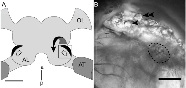
The intact brain preparation was based on an approach described by (Kloppenburg et al. 1999a; Kloppenburg et al. 1999b), in which the central olfactory network was left intact. Shortly before the experiment, the holder was filled with ‘normal’ saline (see below). The head capsule of the anesthetized animal was opened by cutting a window between the two compound eyes and the bases of the antennae. The brain with antennal nerves and antennae attached was dissected from the head capsule and transferred to the recording chamber using the plastic ring for stabilization. The brain was pinned with fine wire in a Sylgard‐coated (Dow Corning Corp., Midland, Michigan, USA) recording chamber containing ʹnormalʹ saline (see below). To gain better access to the recording site and facilitate the penetration of pharmacological agents into the tissue, the brain was enzyme treated (papain, P4762, Sigma, 0.3 mg ml‐1 and L‐cysteine, 30090, Fluka/Sigma, 1 mg ml‐1 dissolved in ‘normalʹ saline) for ~ 3 minutes at room temperature before the AL was desheathed using fine forceps. The AL neurons were visualized with a fixed stage upright microscope (BX51WI, Olympus) using a 40x water‐immersion objective and IR‐DIC optics (Dodt and Zieglgänsberger 1994).
2 Materials & Methods
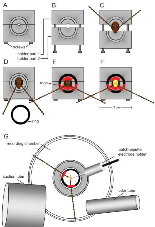
Figure 2.1. Schematic representation of the intact brain preparation
A and B: For the intact brain preparation a custom build holder was used, that could be closed (A) and opened (B) via two metal screws. C: The animal was placed inside the opened holder. D: To stabilize the animal and to avoid loss of saline during dissection, the two parts were pushed together tightly and sealed with vaseline. E: The caput was immobilized with tape stripes and the antennae were mounted on a plastic ring. F: The head capsule was opened and the brain was dissected with antennae and antennal nerves attached. G: The ring including the intact brain preparation was transferred to the recording chamber. It was possible to record from neurons under visual control and stimulate the antennae with physiologically relevant odors.
A B C
D E F
G
screws holder part 1 holder part 2
ring tape
odor tube suction tube
patch-pipette + electrode holder recording chamber
3 cm
2 Materials & Methods
2.4 Electrophysiological recordings
2.4.1 Whole cell patch clamp recordings
Whole cell recordings were performed at 24 °C following the methods described by Hamill et al. (Hamill et al. 1981). Electrodes (tip resistance between 3‐5 MΩ) were fashioned from borosilicate glass (GB150‐8P, 0.86 x 1.5 x 80 mm, Science Products, Hofheim, Germany) with a temperature controlled pipette puller (PIP5, HEKA‐
Elektronik, Lambrecht, Germany) and filled with a solution containing (in mM): 190 K‐Aspartate, 10 NaCl, 1 CaCl2, 2 MgCl2, 10 HEPES and 10 EGTA adjusted to pH 7.2 (with NaOH), resulting in an osmolarity of ~ 415 mOsm. During the experiments, if not stated otherwise, the cells were superfused constantly with ‘normal’ saline solution containing (in mM): 185 NaCl, 4 KCl, 6 CaCl2, 2 MgCl2, 10 HEPES, 35 D‐
glucose. The solution was adjusted to pH 7.2 (with NaOH) and to ~ 430 mOsm (with glucose).
To isolate the Ca2+ currents a combination of pharmacological blockers and ion substitution was used that has been shown to be effective in other insect preparations (Kloppenburg et al. 1999b; Kloppenburg and Hörner 1998; Schäfer et al. 1994).
Transient voltage‐gated sodium currents were blocked by tetrodotoxin (TTX, 10‐7 ‐ 10‐4 M, T‐550, Alomone, Jerusalem, Israel). 4‐aminopyridine (4‐AP, 4 x 10‐4 M, A78403, Sigma) was used to block transient K+ currents and tetraethylammonium (TEA, 20 x 10‐3 M, T2265, Sigma) blocked sustained K+ currents (IK(V)) as well as Ca2+
activated K+ currents (IK(Ca)). In addition the intracellular potassium was substituted with cesium. For calcium current isolation the following pipette solution (calcium saline) was used (in mM): 190 CsCl, 10 NaCl, 1 CaCl2, 2 MgCl2, 10 HEPES and 10 EGTA adjusted to pH 7.2 (with NaOH), resulting in an osmolarity of 415 mOsm.
2 Materials & Methods
2.4.2 Data aquisition
Whole cell voltage‐clamp recordings were made with an EPC9 patch‐clamp amplifier (HEKA‐Elektronik) that was controlled by the program Pulse (version 8.63, HEKA‐
Elektronik) running under Windows. The electrophysiological data were sampled at intervals of 100 μs (10 kHz), except the 5 ms tail current measurements were sampled at 20 kHz. The recordings were low pass filtered at 2 kHz with a 4‐pole Bessel‐Filter.
Compensation of the offset potential and capacitance were performed using the
‘automatic mode’ of the EPC9 amplifier. The liquid junction potential between intracellular and extracellular solution of 15.4 mV for ʹnormalʹ saline and of 4.8 mV
for ʹcalciumʹ saline (calculated with Patcherʹs‐Power‐Tools plug‐in from
http://www.mpibpc.gwdg.de/abteilungen/140/software/index.html for Igor Pro (Wavemetrics, Portland, Oregon, USA)) was also compensated (Neher 1992). To remove uncompensated leakage and capacitive currents, a p/6 protocol was used.
Voltage errors due to series resistance (RS) were minimized using the RS‐
compensation of the EPC9. RS was compensated between 30% and 70% with a time constant (τ) of 2 μs. Stimulus protocols used for each set of experiments are provided in the Results.
2.4.3 Data analysis
The data from the dose‐response experiments were fit with a Hill equation of the
form:
I
Imax= 1 1IC50
[C]
nH
(eq. 1).
I is the peak amplitude of ICa at a ‐5 mV testpulse from Vh = ‐60 mV in the presence of different concentrations of drugs ([C]), and Imax is the peak amplitude of the control.
IC50 is the concentration where half of ICa is blocked and nH is the Hill coefficient.
2 Materials & Methods
Steady‐state tail‐current activation and steady‐state inactivation data were fit using a first‐order (n = 1) Boltzmann equation:
I
Imax= 1
1eV0.5−V/sn (eq. 2).
Imax is the maximal current, V is the voltage of the testpulse, I is the current at voltage V, s is the slope factor and V0.5 is the voltage at which half‐maximal activation occurs.
The peak conductance (g) was calculated using the equation:
g= I
V−Vrev (eq. 3).
I is the current density, V the test potential and Vrev is the Ca2+ equilibrium potential.
To convert peak current density to peak conductance a reversal potential for calcium was calculated as Vrev = 160 mV.
To describe the time course of voltage activated calcium currents a sum of two exponential functions with two time constants was used:
I=A1⋅e
−t1A2⋅e
−t2
(eq. 4).
τ1,2 are the different time constant, A1,2 are the different current amplitudes at t = 0 and t is the time at which the current I occurs.