Original article:
Δ-AMINOLEVULINATE DEHYDRATASE AND GLUTATHIONE PEROXIDASE ACTIVITY IN ALZHEIMER’S DISEASE:
A CASE-CONTROL STUDY
Quelen Iane Garlet1, Maria Vaitsa Losh Haskel2, Romaiana Picada Pereira3, Weber Cláudio Francisco Nunes da Silva4, João Batista Teixeira da Rocha5, Cláudia Sirlene Oliveira5,6,7*, Juliana Sartori Bonini4*
1 Departamento de Farmacologia, Instituto de Ciências Biológicas, Universidade Federal de Rio Grande, Rio Grande/RS, Brazil
2 Departamento de Fisiologia Humana, Universidade Federal do Rio Grande do Sul, Porto Alegre/RS90040-060, Brazil
3 Departamento de Química, Universidade Estadual de Ponta Grossa, Ponta Grossa/PR, Brazil
4 Universidade Estadual do Centro-Oeste, Campus CEDETEG, Departamento de Farmácia, Guarapuava/PR, Brazil
5 Departamento de Bioquímica e Biologia Molecular, Centro de Ciências Naturais e Exatas, Universidade Federal de Santa Maria, Santa Maria/RS, Brazil
6 Programa Pós-Graduação Stricto Sensu em Biotecnologia Aplicada a Saúde da Criança e do Adolescente, Instituto de Pesquisa Pelé Pequeno Príncipe, Curitiba/PR, Brazil
7 Faculdades Pequeno Príncipe, Curitiba/PR, Brazil
* Corresponding authors: Juliana Sartori Bonini (juliana.bonini@gmail.com), Cláudia Sirlene Oliveira (claudia.bioquimica@yahoo.com.br)
http://dx.doi.org/10.17179/excli2019-1749
This is an Open Access article distributed under the terms of the Creative Commons Attribution License (http://creativecommons.org/licenses/by/4.0/).
ABSTRACT
Alzheimer’s disease (AD) is a neurodegenerative pathology that affects elderly people all over the world. Several studies have demonstrated that oxidative stress is an aggravating factor for AD development and progression.
Therefore, this study aimed to evaluate the activity of two oxidative stress markers, glutathione peroxidase (GPx) and δ-aminolevulinate dehydratase (δ-ALA-D), as well as correlate them with blood metal levels and AD progres- sion. For this purpose, 88 elderly individuals were divided in two groups: AD group (34 patients diagnosed with AD) and control group (34 subjects paired by age with the AD group). The Mini-Mental State Examination and the Clinical Dementia Rating (CDR) were used as tools to classify the AD progression. GPx and δ-ALA-D activ- ities were measured in all subjects through blood tests. Both enzymes’ activities were decreased in AD patients when compared to the age-matched control group, regardless of the CDR. Moreover, GPx activity was positively correlated with selenium levels in the blood; and the δ-ALA-D activity was negatively correlated with blood cop- per levels. Taken together, our results indicated that, for the first time, blood δ-ALA-D activity was significantly inhibited in AD patients. While literature reports conflicting data regarding GPx activity in AD patients, the δ- ALA-D activity seems to be a more consistent tool to be applied as an earlier AD marker.
Keywords: Alzheimer’s disease, δ-ALA-D, Gpx, AD marker, CDR, MMSE
INTRODUCTION
Alzheimer’s disease (AD) is a neuro- degenerative illness usually characterized by progressive memory loss and the degenera- tion of, at least, another cognitive function such as language, attention or reasoning (Du- bois et al., 2007; Selkoe, 2001). However, pieces of evidence have indicated that patients with mild or without cognitive impairment can also display other AD-related pathologi- cal alterations (Bennett et al., 2006; James and Bennett, 2019; Schneider et al., 2009).
The AD development is multifactorial de- pending on genetic and/or environmental fac- tors; however, its pathogenesis remains largely unknown and the treatments do not modify its progression (Ballard et al., 2011;
Malhotra, 2018; Trinh et al., 2003).
Studies about the mechanisms involved in the AD pathogenesis point out that inflamma- tion and oxidative stress play an important role in AD development (Castora, 2019; Hal- liwell, 2006; Lin and Beal, 2006). The brain tissue is particularly susceptible to oxidative stress damage due to its high metabolic de- mand, consuming about 20 % of the inspired oxygen. It has a high concentration of polyun- saturated fatty acids, weak antioxidant ma- chinery and high levels of iron and excitatory neurotransmitters (Di Domenico et al., 2017;
Halliwell, 1992; Markesbery and Carney, 1999). Regarding physiological conditions, the organism exploits a sophisticated enzy- matic and non-enzymatic antioxidant defense system, protecting itself from oxidative dam- age progression (Halliwell, 1996).
The micronutrients iron (Fe), copper (Cu) and selenium (Se) are needed for the antioxi- dant enzymes optimal functioning, e.g., gluta- thione peroxidase (GPx) or superoxide dis- mutase (SOD), (Angeli and Conrad, 2018;
Flohe et al., 1973; Rocha et al., 2017; Younus, 2018). These metals have a pivotal role in synapse regulation and are essential cofactors participating in various enzymatic activities.
Consequently, an alteration in the concentra- tion of these elements may induce the exacer- bation of several diseases, including neuronal pathologies (Prohaska, 1987; Scheiber et al.,
2014; Solovyev, 2015; Ward et al., 2014). In- deed, a previous study reported that AD pa- tients showed a CDR-dependent increased level of Fe and Cu when compared to control subjects (Vaz et al., 2018).
The δ-aminolevulinate dehydratase (δ- ALA-D) or porphobilinogen synthase (PBG- synthase) is a thiol-containing enzyme that can be oxidized under oxidative stress condi- tions (Rocha et al., 2012). Accordingly, δ- ALA-D inhibition results in high concentra- tion of its substrate (5-aminolevulinic acid), which has a pro-oxidative effect (Costa et al., 1997; Demasi et al., 1996, 1997; Rocha et al., 2012). In this context, da Silva et al. (2007) suggested that δ-ALA-D inhibition can be used as an oxidative stress index. However, literature lacks conclusive and accurate data about the correlation among metals present in the body, enzyme activity and AD. Therefore, we aim to elucidate whether the enzymes GPx and δ-ALA-D and the micronutrients Cu, Fe, and Se play a role in the AD progression, providing new information that could indicate alternative or auxiliary approaches to treat and/or detect this neurodegenerative disease.
MATERIALS AND METHODS Population and cognitive validation
This is a case-control, cross-sectional study. The study was authorized by the Hu- man Research Ethics Committee of the Uni- versidade Estadual do Centro-Oeste (61111316/201). Data were collected from June 2013 to December 2014. The samples consisted of 88 elderly individuals divided in two groups: (1) AD group: This group had 34 individuals diagnosed with AD who were reg- istered in the 5th Department of Health of Guarapuava city, Paraná State, Brazil. The el- derlies invited to participate in this study had the AD diagnosis confirmed according to the criteria of the National Institute of Neurolog- ical and Communicative Disorders and Stroke and Alzheimer Disease and Related Disorders Association. The Mini-Mental State Exami- nation (MMSE) and the Clinical Dementia Rating (CDR) were combined to better track
and classify cognitive disorders. These meth- odologies allowed the AD progression classi- fication in mild dementia (CDR1 and MMEM score ranging from 20 to 27 for subjects that attended > 4 years of school or from 16 to 21 for subjects that attended ≤ 4 years of school), moderate dementia (CDR2 and MMEM score ranging from 12 to 19 for subjects that at- tended > 4 years of school or from 8 to 15 for subjects that attended ≤ 4 years of school) and severe dementia (CDR3 and MMEM score <
11 for subjects that attended > 4 years of school or < 7 for subjects that attended ≤ 4 years of school). (2) Control group: This group had 34 individuals (MMEM score >27 for subjects that attended > 4 years of school or >21 for subjects that attended ≤ 4 years of school) carefully paired to the AD group. Ac- cording to Vaz et al. (2018), the pairing crite- rion was organized considering the year of birth, sex, if the subject was a smoker or not, diabetes mellitus diagnosis and high blood pressure.
Enzymatic assays
To access the GPx and δ-ALA-D activity, we performed assays using blood (5 ml). The blood samples were collected at the partici- pants’ homes and they respected an 8-hour fasting time. Immediately after, the blood was transferred to a tube containing heparin.
Blood samples were diluted (1:3) in distilled water followed by agitation on ice to provide total hemolysis.
δ-ALA-D activity
The δ-ALA-D activity was measured ac- cording to the method described by Berlin and Schaller (1974). Enzyme activity was deter- mined by the rate of product (porphobilino- gen-PBG) formation. The reaction was started by adding the substrate (5-aminolevulinic acid) followed by incubation at 37 ºC for 90 min in the presence or absence of the reducing agent dithiothreitol (2 mM DTT). The reac- tion was stopped by the addition of trichloro- acetic acid (TCA) 10 % containing HgCl2
0.05 M. PBG was measured with Ehrlich's re- agent using the molar absorption coefficient
of 6.1 x 10⁴ for Ehrlich-PBG salt. Specific en- zymatic activity was expressed as nmol PBG/h/ml (blood hemolysate).
GPx activity
GPx activity was assessed according to the method described by Paglia and Valentine (1967). The conversion of NADPH to NADP+ was monitored at 340 nm for 2 min. Enzyme activity was expressed as µmol of NADPH oxidized per min/ml (blood hemolysate) us- ing an extinction coefficient of 6.2×10⁶ for NADPH.
Statistical analysis
The results are presented as mean ± stand- ard error mean (SEM) or median ± interquar- tile interval (non-parametric data). Homoge- neity of variance and normality were evalu- ated by Levene and Shapiro-Wilk tests, re- spectively. Parametric data were submitted to unpaired two-tailed t-test and non-parametric data were analyzed by Mann-Whitney test.
Correlations were analyzed by the Pearson correlation method. Statistical analysis was carried out using SigmaPlot 11.0 software or GraphPad Prism version 6.01 and the mini- mum significance level was set at p < 0.05.
RESULTS Cognitive validation
We evaluated the subjects from the con- trol group and AD patients using MMSE scores, CDR and age data to classify them re- garding its AD stage (Figure 1). Raw data from subjects can be assessed in supplemen- tary material (Supplementary Table 1). Age parameter was homogeneous among groups (p> 0.05) and patients in the CDR1 stage had similar MMSE scores to the respective con- trol group. On the other hand, AD patients in CDR2 and CDR3 stages showed a significant decrease in the MMSE score compared to the respective control group (p<0.001, Mann- Whitney unpaired two-tailed test).
Figure 1: MMSE score and age (insert graph) from control subjects and Alzheimer Disease (AD) patients subdivided by CDRs. **p<0.001 from re- spective control group
Enzymatic activity
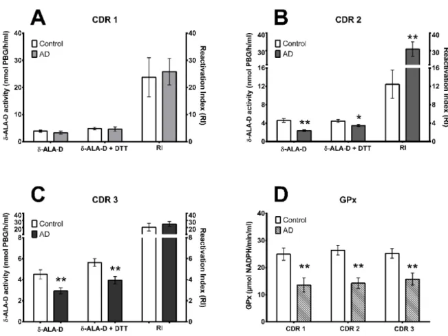
We assessed the enzymes δ-ALA-D activ- ity, which plays a role in the heme biosynthe- sis pathway and GPx which is involved in ox- idative damage repair (Figure 2). Raw data from subjects can be assessed in supplemen- tary material (Supplementary Table 1). We detected a decrease in δ-ALA-D activity in patients ranging from moderated (CDR2) to advanced (CDR3) AD stage (Figure 2A, B, and C, p< 0.05 or p< 0.001, unpaired two- tailed t-test). However, the reactivation index only was significantly increased in AD pa- tients in the CDR2 stage (Figure 2B, p<
0.001, unpaired two-tailed t-test). Addition- ally, GPx activity was decreased in patients from all AD-CDR stages when compared to the control group (Figure 2D, p< 0.001, un- paired two-tailed t-test).
Figure 2: Blood δ-Amino δ-ALA-D and GPx activity in different stages of Alzheimer Disease (AD). δ- ALA-D activity with or without DTT addition and its reactivation index (RI) in AD stages CDR1, CDR2, and CDR3 (A, B and C, respectively) and GPx activity in AD stages CDR1, CDR2, and CDR3 (D). *p<
0.05 or **p< 0.001 from control group
Correlation
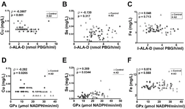
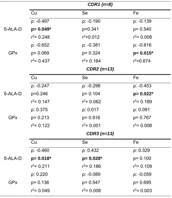
We performed a correlation test using the δ-ALA-D activities and GPx data from all tested subjects (control group and AD pa- tients) and blood measures of Fe, Cu and Se levels (previously published by Vaz et al., 2018) using the Pearson’s correlation analysis (Figure 3A-F). We detected a significant neg- ative (weak) correlation between δ-ALA-D activity and blood concentrations of Cu (Fig- ure 3A, p<0.001, ρ= -0.3907, r2=0.19, Pear- son correlation method). Furthermore, we found that GPx activity is negatively corre- lated with Cu levels (Figure 3D, p<0.05, ρ= - 0.282, r2=0.079) and positively correlated with Se levels in the blood (Figure 3E, p<0.05, ρ= -0.269, r2=0.072). We also ex- plored the correlation of both enzymes’ activ- ities with the metal levels in the blood in each AD-CDR stage separately (Table 1). Interest- ingly, it was detected a negative correlation between δ-ALA-D activity and Cu levels in patients on the CDR1 and CDR3 stages. A positive correlation was found between δ- ALA-D activity and Fe levels in AD-CDR2
patients and between δ-ALA-D activity and Se levels in AD-CDR3 patients. Moreover, we detected a positive correlation between GPx activity and Fe levels in patients in the AD-CDR1 stage.
DISCUSSION
In this study, we observed that AD pa- tients showed an alteration in the δ-ALA-D and GPx activities. In addition, we observed a correlation between the inhibition of these en- zymes and the blood levels of copper (Cu), iron (Fe), and selenium (Se).
The inhibition of the enzyme δ-ALA-D has been extensively studied because of its in- volvement in the oxidative stress biomarkers increase, being present in several pathological and physiological conditions, such as gesta- tional diabetes mellitus (Rodrigues et al., 2018), preeclampsia (de Lucca et al., 2016), lung cancer (Zanini et al., 2014), chronic renal failure (da Silva et al., 2007), and type 2 dia- betes (Bonfanti et al., 2011).
Figure 3: Correlations between the activity of δ-ALA-D (A, B and C) or GPx (D, E and F) with metals concentrations in the blood from control subjects and Alzheimer Disease (AD) patients according to Pearson correlation method. ρ: Pearson’s correlation coefficient
Recently, Baierle et al. (2010, 2014) sug- gested that the δ-ALA-D inhibition is an ad- ditional factor to the cognitive decline associ- ated with aging. To the best of our knowledge, we demonstrated for the first time that AD pa- tients showed significant δ-ALA-D inhibition when compared with age-matched control subjects. The inhibitory effects vary with dis- ease progression: δ-ALA-D activity is main- tained in AD-CDR1 patients while in AD-
CDR2 and AD-CDR 3 patients, the enzyme was inhibited.
However, the enhanced reactivation index in the second stage of AD, but not in the third stage, may indicate that the enzyme δ-ALA- D oxidation state deteriorated with the AD progression.
Table 1: Correlations between the activity of δ-ALA-D or GPx with Cu, Se and Fe concentrations in the blood from Alzheimer Disease (AD) patients according to Pearson correlation method. ρ: Pearson’s correlation coefficient; r2: coefficient of determination
CDR1 (n=8)
Cu Se Fe δ-ALA-D
ρ: -0.497 ρ: -0.190 ρ: -0.139
p= 0.049* p=0.341 p= 0.540
r2= 0.248 r2=0.012 r2= 0.008 GPx
ρ: -0.652 ρ: -0.381 ρ: -0.816
p= 0.069 p= 0.324 p= 0.015*
r2= 0.437 r2= 0.164 r2=0.674 CDR2 (n=13)
Cu Se Fe δ-ALA-D
ρ: -0.247 ρ: -0.298 ρ: -0.453
p=0.246 p= 0.104 p= 0.022*
r2= 0.147 r2= 0.082 r2= 0.189 GPx
ρ: 0.375 ρ: 0.017 ρ: 0.091
p= 0.213 p= 0.916 p= 0.767
r2= 0.122 r2= 0.001 r2= 0.008 CDR3 (n=13)
Cu Se Fe δ-ALA-D
ρ: -0.460 ρ: 0.432 ρ: 0.329
p= 0.018* p= 0.028* p= 0.100
r2= 0.211 r2= 0.186 r2= 0.109 GPx
ρ: 0.220 ρ: -0.089 ρ: -0.059
p= 0.136 p= 0.547 p= 0.695
r2= 0.049 r2= 0.008 r2= 0.003
Metals can inhibit the enzyme δ-ALA-D and this effect has been described both in vitro and in vivo studies (Baierle et al., 2010;
Klimaczewski et al., 2018; Mesquita et al., 2016; Oliveira et al., 2014; Pauza et al., 2005;
Peixoto et al., 2004; Rocha et al., 2004, 2012;
Vargas et al., 2013). In our study, we ob- served a negative correlation between the δ- ALA-D activity and the Cu levels in the blood. Interestingly, Baierle et al. (2010) ob- served the same pattern in elderly women and Klimaczewski et al. (2018), using in silico tools, observed that Cu(II) could enter in the δ-ALA-D active site, binding in the thiolate group of C135, the carboxyl moieties of D131 and E136, and the hydroxyl group of S179, interacting and oxidizing the thiol group. In this context, the reactivation of the enzyme by DTT, observed in our study, corroborates with these in silico findings.
We also analyzed the enzyme GPx status in AD patients. GPx is an antioxidant seleno- enzyme that catalyzes the reduction of hydro- gen peroxide (H2O2) and lipid peroxides by glutathione (GSH) (Michiels et al., 1994;
Papp et al., 2007). Alteration in the GPx ac- tivity has been reported in several human comorbidities, for instance, diabetes (Martín- Gallán et al., 2003), renal disorders (El-far et al., 2005), leukemia (Zuo et al., 2006) and amyotrophic lateral sclerosis (Przedborski et al., 1996). Regarding AD, there are incon- sistent results concerning GPx activity in AD patients. Some authors observed that the en- zyme is inhibited in the blood (serum and/or erythrocytes) of AD patients (Jeandel et al., 1989; Padurariu et al., 2010; Rinaldi et al., 2003; Vural et al., 2010) and other authors re- ported that GPx activity is maintained in AD patients (Bourdel-Marchasson et al., 2001;
Casado et al., 2007; Ceballos-Picot et al., 1996). In this study, we observed an inhibi- tion in the GPx activity of AD patients. The conflicting results found in the literature may be explained by the Se status of the patients, since the GPx is a Se-dependent enzyme (Rotruck et al., 1973). In fact, we observed a positive correlation between the enzyme ac- tivity and the Se levels. Taken together, ours
results point out that GPx status evaluation may be better interpreted when combined with Se levels data.
CONCLUSION
In summary, the decreased blood δ-ALA- D and GPx enzymes found in AD patients could be related to a deregulation in the blood homeostasis of Cu and Se, respectively. Fur- thermore, the pro-inflammatory and pro-oxi- dant scenario found in AD could be important factors contributing to deregulate δ-ALA-D and GPx activities as well. Our results indi- cate for the first time that blood δ-ALA-D ac- tivity is significantly inhibited in AD patients.
Therefore, δ-ALA-D may be applied as an earlier AD marker.
Declaration of interest
The authors declare that they have no competing interests.
Acknowledgments
The authors would like to thank the finan- cial support from Coordination for Improve- ment of Higher Education Personnel (CAPES/PROEX- Finance Code 001), the National Council for Scientific and Techno- logical Development (CNPq), the Rio Grande do Sul Foundation for Research Support (FAPERGS - Brazil), and the Instituto de Pesquisa Pelé Pequeno Príncipe.
REFERENCES
Angeli JPF, Conrad M. Lipoxygenases - killers against their will? ACS Cent Sci. 2018;43:312-4.
Baierle M, Valentini J, Paniz C, Moro A, Barbosa F Jr, Garcia SC. Possible effects of blood copper on hema- tological parameters in elderly. J Bras Patol Med Lab.
2010;46:463-70.
Baierle M, Charão MF, Göethel G, Barth A, Fracasso R, Bubols G, et al. Are delta-aminolevulinate dehydra- tase inhibition and metal concentrations additional fac- tors for the age-related cognitive decline? Int J Environ Res Public Health. 2014;11:10851-67.
Ballard C, Gauthier S, Corbett A, Brayne C, Aarsland D, Jones E. Alzheimer's disease. Lancet. 2011;377:
1019-31.
Bennett DA, Schneider JA, Arvanitakis Z, Kelly JF, Aggarwal NT, Shah RC, et al. Neuropathology of older persons without cognitive impairment from two com- munity-based studies. Neurology. 2006;66:1837-44.
Berlin A, Schaller KH. European standardized method for the determination of delta-aminolevulinic acid de- hydratase activity in blood. Z Klin Chem Klin Bio- chem. 1974;389-90.
Bonfanti G, Ceolin RB, Valcorte T, De Bona KS, de Lucca L, Gonçalves TL, et al. δ-Aminolevulinate de- hydratase activity in type 2 diabetic patients and its as- sociation with lipid profile and oxidative stress. Clin Biochem. 2011;44:1105-9.
Bourdel-Marchasson I, Delmas-Beauvieux MC, Peuchant E, Richard-Harston S, Decamps A, Reignier B, et al. Antioxidant defences and oxidative stress markers in erythrocytes and plasma from normally nourished elderly Alzheimer patients. Age Ageing.
2001;30:235-41.
Casado Á, López-Fernández ME, Casado MC, de La Torre R. Lipid peroxidation and antioxidant enzyme activities in vascular and Alzheimer dementias. Neuro- chem Res. 2007;33:450-8.
Castora FJ. Mitochondrial function and abnormalities implicated in the pathogenesis of ASD. Prog Neuro- psychopharmacol Biol Psychiatry. 2019;92:83-108.
Ceballos-Picot I, Merad-Boudia M, Nicole A, Thevenin M, Hellier G, Legrain S, et al. Peripheral an- tioxidant enzyme activities and selenium in elderly subjects and in dementia of Alzheimer’s type - Place of the extracellular glutathione peroxidase. Free Rad Biol Med. 1996;20:579-87.
Costa CA, Trivelato GC, Pinto AM, Bechara EJ. Cor- relation between plasma 5-aminolevulinic acid concen- trations and indicators of oxidative stress in lead-ex- posed workers. Clin Chem. 1997;43:1196-202.
da Silva AC, Rocha JBT, Morsch AL, Zanin RF, Kaizer R, Maldonado PA, et al. Oxidative stress and delta-ALA-D activity in chronic renal failure patients.
Biomed Pharmacother. 2007;61:180-5.
de Lucca L, Rodrigues F, Jantsch LB, Kober H, Neme WS, Gallarreta FMP, et al. Delta-aminolevulinate de- hydratase activity and oxidative stress markers in preeclampsia. Biomed Pharmacother. 2016;84:224-9.
Demasi M, Penatti CA, DeLucia R, Bechara EJ. The prooxidant effect of 5-aminolevulinic acid in the brain tissue of rats: implications in neuropsychiatric mani- festations in porphyrias. Free Radic Biol Med. 1996;
20:291-9.
Demasi M, Costa CA, Pascual C, Llesw S, Bechara EJH. Oxidative tissue response promoted by 5-amino- levulinic acid promptly induces the increase of plasma antioxidant capacity. Free Rad Res. 1997;26:235-43.
Di Domenico F, Tramutola A, Butterfield DA. Role of 4-hydroxy-2-nonenal (HNE) in the pathogenesis of alzheimer disease and other selected age-related neu- rodegenerative disorders. Free Radic Biol Med.
2017;111:253-61.
Dubois B, Feldman HH, Jacova C, Dekosky ST, Bar- berger-Gateau P, Cummings J, et al. Research criteria for the diagnosis of Alzheimer's disease: revising the NINCDS-ADRDA criteria. Lancet Neurol.
2007;6:734-46.
El-far MA, Bakr MA, Farahat SE, El-Fattah EAA. Glu- tathione peroxidase activity in patients with renal dis- orders. Clin Exp Nephrol. 2005;9:127-31.
Flohe L, Günzler WA, Schock HH. Glutathione perox- idase: A selenoenzyme. FEBS Lett. 1973;32: 132-4.
Halliwell B. Reactive oxygen species and the central nervous system. J Neurochem. 1992;59:1609-23.
Halliwell B. Antioxidants in human health and disease.
Annu Rev Nutr. 1996;16:33-50.
Halliwell B. Oxidative stress and neurodegeneration:
where are we now? J Neurochem. 2006;97:1634-58.
James BD, Bennett DA. Causes and patterns of demen- tia: An update in the era of redefining Alzheimer's dis- ease. Annu Rev Public Health. 2019; 40:65-84.
Jeandel C, Nicolas MB, Dubois F, Nabet-Belleville F, Penin F, Cuny G. Lipid peroxidation and free radical scavengers in Alzheimer’s disease. Gerontology. 1989;
35:275-82.
Klimaczewski CV, Nogara PA, Barbosa NV, Rocha JBT. Interaction of metals from group 10 (Ni, Pd, and Pt) and 11 (Cu, Ag, and Au) with human blood δ-ALA- D: in vitro and in silico studies. Environ Sci Pollut Res Int. 2018;25:30557-66.
Lin MT, Beal MF. Mitochondrial dysfunction and ox- idative stress in neurodegenerative diseases. Nature.
2006;443:787-95.
Malhotra PA. Impairments of attention in Alzheimer's disease. Curr Opin Psychol. 2018;8;29:41-8.
Markesbery WR, Carney JM. Oxidative alterations in Alzheimer's disease. Brain Pathol. 1999;9:133-46.
Martín-Gallán P, Carrascosa A, Gussinyé M, Domínguez C. Biomarkers of diabetes-associated oxi- dative stress and antioxidant status in young diabetic patients with or without subclinical complications.
Free Radic Biol Med. 2003;34:1563-74.
Mesquita M, Pedroso TF, Oliveira CS, Oliveira VA, Santos RF, Bizzi CA, et al. Effects of zinc against mer- cury toxicity in female rats 12 and 48 hours after HgCl2
exposure. EXCLI J. 2016;15:256-67.
Michiels C, Raes M, Toussaint O, Remacle J. Im- portance of Se-glutathione peroxidase, catalase, and Cu/Zn-SOD for cell survival against oxidative stress.
Free Radic Biol Med. 1994;17:235-48.
Oliveira VA, Oliveira CS, Ineu RP, Moraes-Silva L, de Siqueira LF, Pereira ME. Lactating and non-lactating rats differ in sensitivity to HgCl2: Protective effect of ZnCl2. J Trace Elem Med Biol. 2014;28:240-6.
Padurariu M, Ciobica A, Hritcu L, Stoica B, Bild W, Stefanescu C. Changes of some oxidative stress mark- ers in the serum of patients with mild cognitive impair- ment and Alzheimer’s disease. Neurosci Lett.
2010;469:6–10.
Paglia DE, Valentine WN. Studies on the quantitative and qualitative characterization of erythrocyte glutathi- one peroxidase. J Lab Clin Med. 1967;70:158-69.
Papp LV, Lu J, Holmgren A, Khanna KK. From sele- nium to selenoproteins: synthesis, identity, and their role in human health. Antioxid Redox Signal.
2007;9:775-806.
Pauza NL, Cotti MJ, Godar L, de Sancovich AMF, Sancovich HA. Disturbances on delta aminolevulinate dehydratase (ALA-D) enzyme activity by Pb2+, Cd2+, Cu2+, Mg2+, Zn2+, Na+, K+ and Li+: analysis based on coordination geometry and acid-base Lewis capacity. J Inorg Biochem. 2005;99:409-14.
Peixoto NC, Roza T, Pereira ME. Sensitivity of delta- ALA-D (E.C. 4.2.1.24) of rats to metals in vitro de- pends on the stage of postnatal growth and tissue. Tox- icol In Vitro. 2004;18:805-9.
Prohaska JR. Functions of trace elements in brain me- tabolism. Physiol Rev. 1987;67:858-901.
Przedborski S, Donaldson D, Jakowec M, Kish SJ, Guttman M, Rosoklija G, et al. Brain superoxide dis- mutase, catalase, and glutathione peroxidase activities in amyotrophic lateral sclerosis. Ann Neurol.
1996;39:158-65.
Rinaldi P, Polidori MC, Metastasio A, Mariani E, Mattioli P, Cherubini A, et al. Plasma antioxidants are similarly depleted in mild cognitive impairment and in Alzheimer’s disease. Neurobiol Aging. 2003;24:915-9.
Rocha JBT, Tuerlinckx SM, Schetinger MR, Folmer V.
Effect of Group 13 metals on porphobilinogen synthase in vitro. Toxicol Appl Pharmacol. 2004;200:169-76.
Rocha JBT, Saraiva RA, Garcia SC, Gravina FS, Nogueira CW. Aminolevulinate dehydratase (δ-ALA- D) as marker protein of intoxication with metals and other pro-oxidant situations. Toxicol Res. 2012;1:85- 102.
Rocha JBT, Piccoli BC, Oliveira CS. Biological and chemical interest in selenium: a brief historical ac- count. Arkivoc. 2017;2017:457-91.
Rodrigues F, de Lucca L, Neme WS, Gonçalves TL.
Influence of gestational diabetes on the activity of δ- aminolevulinate dehydratase and oxidative stress bi- omarkers. Redox Rep. 2018;23:63-7.
Rotruck JT, Pope AL, Ganther HE, Swanson AB, Hafeman DG, Hoekstra WG. Selenium: Biochemical role as a component of glutathione peroxidase. Sci- ence. 1973;179:588-90.
Scheiber IF, Mercer JF, Dringen R. Metabolism and functions of copper in brain. Prog Neurobiol. 2014;
116:33-57.
Schneider JA, Aggarwal NT, Barnes L, Boyle P, Ben- nett DA. The neuropathology of older persons with and without dementia from community versus clinic co- horts. J Alzheimers Dis. 2009;18:691-701.
Selkoe DJ. Alzheimer's disease: genes, proteins, and therapy. Physiol Rev. 2001;81:741-66.
Solovyev ND. Importance of selenium and selenopro- tein for brain function: From antioxidant protection to neuronal signalling. J Inorg Biochem. 2015;153:1-12.
Trinh N-H, Hoblyn J, Mohanty S, Yaffe K. Efficacy of cholinesterase inhibitors in the treatment of neuropsy- chiatric symptoms and functional impairment in Alz- heimer disease: A meta-analysis. JAMA.
2003;289:210-6.
Vargas LM, Soares MB, Izaguirry AP, Lüdtke DS, Braga HC, Savegnago L, et al. Cadmium inhibits the ovary δ-aminolevulinate dehydratase activity in vitro and ex vivo: protective role of seleno-furanoside. J Appl Toxicol. 2013;33:679-84.
Vaz FNC, Fermino BL, Haskel MVL, Wouk J, de Freitas GBL, Fabbri R, et al. The relationship between copper, iron, and selenium levels and Alzheimer dis- ease. Biol Trace Elem Res. 2018;181:185-91.
Vural H, Demirin H, Kara Y, Eren I, Delibas N. Alter- ations of plasma magnesium, copper, zinc, iron and se- lenium concentrations and some related erythrocyte antioxidant enzyme activities in patients with Alz- heimer’s disease. J Trace Elem Med Biol. 2010;24:
169-73.
Ward RJ, Zucca FA, Duyn JH, Crichton RR, Zecca L.
The role of iron in brain ageing and neurodegenerative disorders. Lancet Neurol. 2014;13:1045-60.
Younus H. Therapeutic potentials of superoxide dis- mutase. Int J Health Sci (Qassim). 2018;12(3):88-93.
Zanini D, Pelinson LP, Schmatz R, Pereira LB, Martins CC, Baldissareli J, et al. δ-Aminolevulinate dehydra- tase activity in lung cancer patients and its relationship with oxidative stress. Biomed Pharmacother. 2014;68:
603-9.
Zuo XL, Chen JM, Zhou X, Li XZ, Mei GY. Levels of selenium, zinc, copper, and antioxidant enzyme activ- ity in patients with leukemia. Biol Trace Elem Res.
2006;114:41-54.