ARTICLE
Mechanisms governing the pioneering and redistribution capabilities of the non-classical pioneer PU.1
Julia Minderjahn 1 , Andreas Schmidt 2 , Andreas Fuchs 3 , Rudolf Schill 4 , Johanna Raithel 1 , Magda Babina 5 , Christian Schmidl 6 , Claudia Gebhard 1,6 , Sandra Schmidhofer 1,7 , Karina Mendes 1 , Anna Ratermann 1,8 , Dagmar Glatz 1,9 , Margit Nützel 1 , Matthias Edinger 1,6 , Petra Hoffmann 1,6 , Rainer Spang 4 , Gernot Längst 3 , Axel Imhof 2 & Michael Rehli 1,6 *
Establishing gene regulatory networks during differentiation or reprogramming requires master or pioneer transcription factors (TFs) such as PU.1, a prototype master TF of hematopoietic lineage differentiation. To systematically determine molecular features that control its activity, here we analyze DNA-binding in vitro and genome-wide in vivo across different cell types with native or ectopic PU.1 expression. Although PU.1, in contrast to classical pioneer factors, is unable to access nucleosomal target sites in vitro, ectopic induction of PU.1 leads to the extensive remodeling of chromatin and redistribution of partner TFs. De novo chromatin access, stable binding, and redistribution of partner TFs both require PU.1 ’ s N-terminal acidic activation domain and its ability to recruit SWI/SNF remodeling complexes, suggesting that the latter may collect and distribute co-associated TFs in con- junction with the non-classical pioneer TF PU.1.
https://doi.org/10.1038/s41467-019-13960-2 OPEN
1 Department of Internal Medicine III, University Hospital Regensburg, 93053 Regensburg, Germany. 2 Biomedical Center, Protein Analysis Unit, Faculty of Medicine, Ludwig-Maximilians-Universität München, Großhaderner Strasse 9, 82152 Planegg-Martinsried, Germany. 3 Biochemistry Centre Regensburg (BCR), University of Regensburg, 93053 Regensburg, Germany. 4 Statistical Bioinformatics Department, Institute of Functional Genomics, University of Regensburg, 93053 Regensburg, Germany. 5 Department of Dermatology and Allergy, Charité Universitätsmedizin Berlin, Berlin, Germany. 6 Regensburg Center for Interventional Immunology (RCI), University Regensburg and University Medical Center Regensburg, 93053 Regensburg, Germany.
7Present address: AstraZeneca, Tinsdaler Weg 183, 22880 Wedel, Germany.
8Present address: Rentschler Biopharma SE, 88471 Laupheim, Germany.
9Present address: Chromatin Structure and Cellular Senescence Research Unit, Maisonneuve-Rosemont Hospital Research Centre, Montréal, QC, Canada H1T 2M4.
*email: michael.rehli@ukr.de
1234567890():,;
C ellular differentiation requires so-called master or pio- neering transcription factors (TFs) to establish access to regulatory elements embedded in chromatin 1 . Similar to the vast majority of TFs, they usually recognize specific DNA motifs ranging from 6 to 12 base pairs (bp) in length, implying the existence of roughly a million potential binding sites throughout the genome 2 . How the actually bound sites, which usually range in the thousands, are selected from the vast array of putative binding sites is largely unknown.
The ETS family TF PU.1 (encoded by SPI1) is a well-studied master regulator of the hematopoietic compartment and is required for the generation of common lymphoid and granulocyte-macrophage (MAC) progenitor cells, as well as later stages of monocyte (MO)/MAC and B-cell development 3 . It controls the expression of hundreds of genes that include growth factor receptors, adhesion molecules, TFs, and signaling compo- nents 4 , and is able to initiate myeloid gene expression programs in heterologous cell types including T cells and fibroblasts 5 . The ability of PU.1 to shape chromatin landscapes and re-program cells and its role in regulating cell type-specific gene expression make it a prototypic pioneer factor 6 . Yet, how this factor interacts with chromatin to access its binding sites de novo to date has not been elucidated.
The first genome-wide analyses of PU.1 occupancy observed cell type-specific binding-site selection in murine MACs and B cells 7,8 , as well as in human MOs and MO-derived MAC 9 . Cell type-specific binding sites depended on the co-occurrence of sequence motifs for other cell type-specific TFs, including members of the C/EBP and AP-1 family TFs in human MO or murine MAC, EGR2 in human MO-derived MAC, or E2A, EBF, and OCT2 in B cells 8,9 . The exact mechanisms behind this cooperativity between PU.1 and other TFs are not well under- stood and may include direct protein–protein interactions, interactions between TFs that are facilitated by DNA, DNA- mediated interactions in the absence of TF interactions, and indirect cooperativity involving competition between nucleo- somes and TFs 10 . The requirement for TF cooperativity is inversely correlated with motif affinity: high-affinity motifs are frequently bound by PU.1 alone, whereas low-affinity motifs are only bound when other factors co-bind nearby 11 . Known physical interaction partners of PU.1 include general TFs such TFIID and TBP (TATA-box binding protein), cell type-specific TFs such as interferon regulatory factor 4 and 8 (IRF4 and IRF8), the proto- oncogene c-Jun (JUN, a component of the AP-1 TF), and early hematopoietic TFs such as GATA-binding protein 2 (GATA2) and runt-related TF1 (RUNX1) 12–17 . In early T cells, PU.1–RUNX1 interactions lead to a redistribution of RUNX1 binding, highlighting the importance of TF interactions as well as TF protein levels in binding-site selection 18 . These interactions, however, do not explain how PU.1 exerts its presumed pioneering role or how it selects its binding sites in chromatin in the first place.
Here we systematically analyze the ability of PU.1 to access its binding sites in vitro and in vivo. By profiling PU.1 binding across a large atlas of hematopoietic cell types, we show that PU.1 only occupies a fraction of its potential binding sites, and that cell type-specific binding is not exclusively explained by TF co- association. In vitro studies further show that PU.1 binding to DNA is subject to both epigenetic and chromatin constraints. It is unable to bind CpG-methylated or nucleosome-bound DNA, suggesting that PU.1 may not act as a classical pioneer factor, which are defined by their ability to recognize their binding sites in nucleosomal DNA. Despite these constraints, introduction of PU.1 into heterologous model cell lines lacking endogenous PU.1 expression leads to extensive de novo remodeling of chromatin at PU.1-binding sites and rapid initiation of a myeloid gene
expression program. Functional analysis of several mutant PU.1 variants indicates that efficient binding of PU.1 to de novo- remodeled sites depends on the N-terminal acidic activation domain (AAD), suggesting that the latter is strictly required for accessing binding sites de novo. Further analyses including in vivo proximity-dependent biotinylation (BioID) and co- immunoprecipitation (CoIP) show that the N-terminal acidic domain mediates the interactions of PU.1 with the SWI/SNF family of chromatin remodeling complexes (BAF complex).
Hence, the ability of PU.1 to shape regulatory landscapes and to act as a non-classic pioneer factor requires the AAD and its interaction with SWI/SNF. The redistribution of partner TFs by PU.1 also requires the SWI/SNF-interacting acidic domain, sug- gesting that the remodeler complex may act as part of a hub to collect and distribute co-associated TFs in a PU.1-dependent manner.
Results
PU.1 binding across multiple cell types in vivo and in vitro. To better understand what distinguishes PU.1-bound sequences from unbound sequences, we first determined its DNA-binding profiles across a large array of different cell types. PU.1 DNA-binding maps have already been generated in a number of studies, but comparative analyses were generally restricted to few cell types.
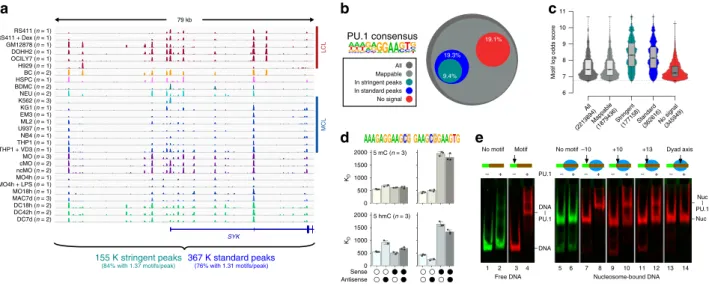
For a more comprehensive view of PU.1-binding patterns, we collected publicly available occupancy data and generated several additional PU.1 binding maps in various lymphoid and myeloid cell lines, primary human cells and several MO-derived cell types (Fig. 1a). Summaries of published and generated chromatin immunoprecipitation sequencing (ChIP-seq) data are provided in Supplementary Table 1 and Data File 1. With reference to a previously defined PU.1 consensus sequence 11 , PU.1 binding (defined by standard or stringent peak calling criteria) was only detected at a fraction (<20%) of possible binding sites (Fig. 1b).
Bound sites were generally characterized by higher motif scores (Fig. 1c). Cell type-restricted binding events (comprising 27% of stringent peaks) were enriched for co-associated sequence motifs, suggesting that combinatorial TF interactions support binding at low-affinity sites (Supplementary Fig. 1d). Notably, high-affinity sites (high motif log odds scores) are frequently bound across the large majority of cell types (Supplementary Fig. 1e) and are almost absent from the group of motifs showing no evidence of ChIP-seq signals (Fig. 1c). Nevertheless, the observed dependency on DNA sequence motifs (both for PU.1 and partner TFs) was not mandatory, suggesting that PU.1 binding is controlled on additional levels.
Obvious candidate mechanisms include the possible restriction of PU.1 binding to DNA via the covalent modification of DNA (e.g., DNA methylation) or via the competition with nucleo- somes. To test this in vitro, we performed binding assays with recombinant PU.1 on either (hydroxy-)methylated or nucleosome-bound double-stranded DNA (dsDNA). DNA (hydroxy-)methylation was found to interfere with PU.1 binding when present close to the GGAA-core sequence, but not further downstream, and only when occurring on the sense strand (Fig. 1d). Of all sequences covered by the PU.1 consensus, only a small fraction of about 20 K (10% of which are located in promoters) contain a proximal CpG that might affect binding.
Nucleosomes also presented a barrier to PU.1 binding. Although
sites located around to the nucleosome dyad axis were not bound
at all, sites proximal to the nucleosome entry site were at least
partially accessible to PU.1 (Fig. 1e). This is in line with published
data for the highly homologous ETS domain of SPIB, which
showed a clear binding preference for linker and nucleosome
entry sites in the NCAP-SELEX assay 19 . These in vitro binding
studies suggested that PU.1 alone is indeed restricted by both DNA methylation and chromatin. Given the well-documented pioneering role of this TF and its ability to access a large fraction of high-affinity sites in vivo, PU.1 must be able to overcome epigenetic and chromatin constraints at least to some extent.
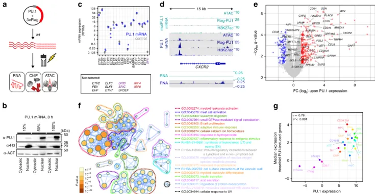
Immediate consequences of de novo PU.1 expression. PU.1 occupancy maps across multiple cell types were useful to explore cell type-specific binding preferences, but less informative regarding rules of de novo binding-site selection. To better understand how PU.1 selects binding sites in vivo and which auxiliary factors may help to overcome epigenetic and chromatin constraints, we established a system where PU.1 expression is rapidly induced in a PU.1-negative acute lymphoblastic leukemia cell line (CTV-1) using mRNA transfection (Fig. 2a, b). CTV-1 cells neither express endogenous PU.1 nor its related class III ETS family members SPIB and SPIC (Fig. 2c), suggesting that recog- nition sites of class III ETS factors may be “untouched” in these cells. IRF4 and IRF8, which may form heterodimers with PU.1, were also not detected in CTV-1 cells (Fig. 2c). At its peak, PU.1 protein expression levels in CTV-1 after mRNA transfection exceeded those of natively high expressing cell types (such as MO-derived dendritic cells (DCs) or THP-1 cell line; Supple- mentary Fig. 2a).
In this model system, we observed de novo chromatin remodeling and transcription (Fig. 2d, e) upon PU.1 expression.
Induced genes (measured 24 h after PU.1 mRNA transfection) were associated with Gene Ontology (GO) terms such as myeloid activation and differentiation (Fig. 2f), and expression levels of
these genes correlated with PU.1 expression across hematopoietic cells types (Fig. 2g).
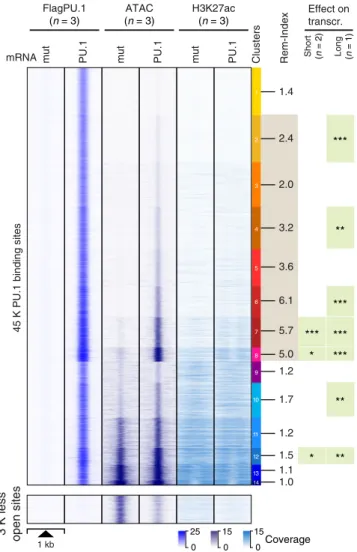
After establishing that transient PU.1 mRNA transfection in CTV-1 cells affects relevant target genes, we examined the effects of PU.1 expression on chromatin. Relative to control-transfected cells (PU.1mut), we detected 45 K PU.1-binding sites (using stringent peak calling criteria) by ChIP-seq. Of those, 80% or 94%
overlapped with stringent or standard peaks sets derived from all natively PU.1-expressing cell types (in Fig. 1a), respectively. The number of detected peaks was comparable to other highly PU.1- expressing cell types (e.g., 51 K stringent peaks in DC, 35 K stringent peaks in MO-derived MACs, or 63 K stringent peaks in phorbol 12-myristate 13-acetate (PMA) and Vitamin D3-treated THP-1 cells; peak counts of all samples are listed in Supplemen- tary Table 1 and Data File 1). As shown in Fig. 3, clustering of PU.1 peaks based on corresponding ATAC-seq data (a measure of chromatin accessibility) separated PU.1 peaks into groups of peaks with different degrees of accessibility before and after PU.1 induction and revealed extensive chromatin remodeling upon PU.1 expression at PU.1 peak clusters 1–8, comprising the large majority of PU.1-binding sites. PU.1 peak clusters with highly remodeled sites (e.g., PU.1 peak clusters 6–8) were significantly co-associated with the induced expression of neighboring genes, either after a single PU.1 induction (short), or seven cycles of PU.1 mRNA electroporation over 7 days (long).
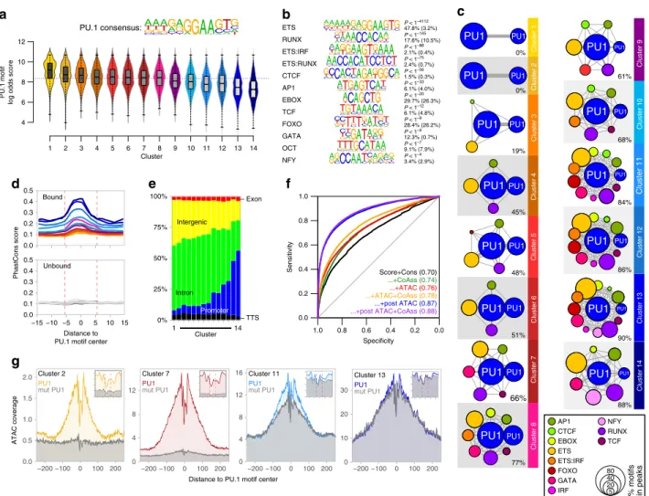
To further characterize the properties of PU.1 peaks in clusters defined by chromatin accessibility (Fig. 3), we first analyzed their motif composition. We found that de novo-remodeled sites generally associate with higher-affinity motifs compared with PU.1-binding sites that were located in accessible chromatin prior
– DNA – 79 kb
SYK
LCLMCL
155 K stringent peaks
(84% with 1.37 motifs/peak)
367 K standard peaks
(76% with 1.31 motifs/peak)
All Mappable In stringent peaks In standard peaks No signal
PU.1 consensus
a b c
9.4%
19.3%
19.1%
d e
KDKD
PU.1
– + – + – + + – +
1 2 3 4 5 6
– + – + –
7 8 9 10 11 12 13 14
–10 +10 +13 Dyad axis
Free DNA Nucleosome-bound DNA
– Nuc – No motif
No motif Motif
DNA
| PU.1
Nuc
| PU.1 RS411 (n = 1)
RS411 + Dex (n = 1) GM12878 (n = 1) DOHH2 (n = 1) OCILY7 (n = 1) H929 (n = 1) BC (n = 2) HSPC (n = 1) BDMC (n = 2) NEU (n = 2) K562 (n = 3) KG1 (n = 1) EM3 (n = 1) ML2 (n = 1) U937 (n = 1) NB4 (n = 1) THP1 (n = 1) THP1 + VD3 (n = 1) MO (n = 3) cMO (n = 2) ncMO (n = 2) MO4h (n = 1) MO4h + LPS (n = 1) MO18h (n = 1) MAC7d (n = 3) DC18h (n = 2) DC42h (n = 2) DC7d (n = 2)
All (2213894)
Mappable (1879436)
Stringent (177158)Standard
(362616)No signal (345949) 6
7 8 9 10 11
Motif log odds score
0 500 1000 1500 2000 0 500 1000 1500 2000
5 hmC (n = 3) 5 mC (n = 3)
Sense Antisense
Fig. 1 PU.1 occupancy in vivo and binding constraints in vitro. a Comparison of PU.1 ChIP-seq data across various human lymphoid and myeloid cell lines
(LCL and MCL, respectively) and primary cells (BC B cells, BDMC breast skin-derived mast cells, cMO classical MO, DC dendritic cells, HSPC
hematopoietic stem and progenitor cells, MAC, macrophages, MO monocytes, ncMO non-classical MO, NEU neutrophils) at an exemplary locus. For
replicated data sets (as indicated), averaged coverage tracks are shown. Total motif occurrences in standard and stringent peaks are summarized below
the tracks. b Fraction of PU.1 motifs residing in either standard or stringent PU.1 peaks (3.61 × 10 5 and 1.77 × 10 5 , respectively) compared with all motif
occurrences (2.21 × 10 6 ) across the genome, motifs fi ltered for mappability (1.88 × 10 6 ), as well as motifs that showed no evidence of binding (<3 per 10 7
reads within 200 bp motif-centered window) across all samples (no signal, 3.48 × 10 5 ). c Distribution of motif scores across total occurrences,
mappability- fi ltered motifs, motifs in peaks, and no-signal motifs. d Microscale thermophoresis-derived dissociation constants ( K
Dvalues, bars represent
the mean of n = 3 experiments ± SD, individual data points are shown as black dots) for the interaction between recombinant full-length PU.1 and the
indicated double-stranded oligonucleotides representing PU.1 motifs with a high motif log odds score (left panels, 9.4; right panel 8.8). Open circles
indicate unmethylated cytosines. Black circles represent methylated or hydroxymethylated cytosines (top panels, 5mC or bottom panels, 5hmC,
respectively). e Gelshift assays demonstrating position-dependent binding of PU.1 to nucleosome-associated DNA. The relative position of the PU.1-binding
site (arrow) to the nucleosome boundary is indicated. Nucleosomes are positioned by the 601 nucleosome-positioning sequence marked in red. The
positions of free DNA (DNA) and nucleosomes (nuc), as well as of DNA-PU.1 and nuc-PU.1 complexes are indicated. a – d Source data are provided as a
Source Data fi le.
to PU.1 induction (Fig. 4a and Supplementary Fig. 2b). The increasing chromatin accessibility through PU.1 peak clusters 1–14 was accompanied by a linear drop in average PU.1 motif log odds scores and the increasing presence of nearby motifs for co- associated TFs such as RUNX or GATA (Fig. 4b, c and Supplementary Fig. 2c), which likely support the binding of PU.1 at less-affine sites. In line with previous observations 11 , a high degree of accessibility also correlated with evolutionary conservation (Fig. 4d) and genomic annotation as promoters (Fig. 4e), whereas de novo-remodeled sites often showed little conservation across species and were mostly inter-/intragenic (Fig. 4d, e). Although the above suggested an important role of sequence-related features in the PU.1-binding-site selection, modeling revealed that motif scores, conservation, and nearby presence of co-associated motifs poorly discriminated PU.1- bound and -unbound sites (Fig. 4f). Models including chromatin accessibility in control cells showed a marginally improved predictive power (Fig. 4f), suggesting that additional features (such as chromatin structure or epigenetic modifications) must have a significant impact on PU.1-binding-site selection. The best predictor included chromatin accessibility data after PU.1 induction (Fig. 4f), which is in line with the ability of PU.1 to increase accessibility at a large majority of binding sites.
Further analysis of chromatin accessibility data at single- nucleotide resolution revealed common footprints across PU.1 motif-centered peaks in PU.1 peak clusters (examples are shown in Fig. 4g). Notably, in PU.1 peak clusters with pre-accessible chromatin (peak clusters 12–14), the footprints across PU.1 motifs pre-existed. The lack of change in accessibility, despite the
de novo binding of PU.1 suggests replacement or competition with other (likely ETS family) TFs at these elements (Fig. 4g, right histogram) rather than assisted loading as observed for GR 20 .
PU.1 induction also caused the rapid disappearance of ~3 K accessible sites (Fig. 3, bottom panel), which were highly enriched for consensus TF motifs (like RUNX, ETS, or GATA) that were also identified in PU.1-induced accessible sites (Fig. 5a). The loss of corresponding footprints at former and their gain at latter sites (Fig. 5b–d) suggests the PU.1-induced redistribution of these partner TFs, as recently observed for RUNX1 and SATB1 in early T cells 18 .
To further confirm the marked impact of PU.1 induction on chromatin landscapes, we analyzed two additional heterologous cell lines (T-cell acute lymphoblastic leukemia-1 (TALL1) and the hepatocellular carcinoma cell line HepG2). As observed in CTV-1 cells, PU.1 induction in either cell line caused the rapid reorganization of chromatin landscapes, which showed similar associations with either high-affinity motifs or the presence of cell type-specific co-associated motifs, such as RUNX, IRF, and E2A motifs in TALL1 cells, and FOXA1, HNF1, and HNF4 motifs in HepG2 cells (Supplementary Fig. 3a–e). The predictive power of individual features shown in Supplementary Fig. 3f was also similar to CTV-1 cells, indicating similar constraints for PU.1 binding. Using published whole genome DNA methylation data for HepG2 cells, we could additionally confirm the almost exclusive binding of PU.1 to unmethylated GGAA-core sequences (Supplementary Fig. 3g), as predicted from the in vitro binding studies (shown in Fig. 1d). Redistribution of cell type-specific TFs was also clearly evident in both cell types (Supplementary
PU.1 + 3×Flag
ivt
ATAC ChIP RNA
AAAAAAAAAAAAA
a e
FC (log2) upon PU.1 expression
−log10q−value
d
CXCR2 ATAC Flag-PU1 H3K27ac 15 kb
b
(kDa) 25 50 50 20 α-PU.1
α-H3 α-ACT
Cytosolic Nuclear Cytosolic Nuclear Cytosolic Nuclear
15% 50% 100%
PU.1 mRNA, 8 h
–10 –25
– 0.25 ––0.25 ––0.25 ATAC Flag-PU1 H3K27ac
RNA RNA PU.1 mRNAmutPU.1 mRNA
f
P-value
10–2 10–3 10–4 10–6 10–10
10–20 GO:0034644: cellular response to UV
R-HSA-2129379: molecules associated with elastic fibres GO:0090311: regulation of protein deacetylation GO:0046717: acid secretion
GO:0030073: insulin secretion GO:0002573: myeloid leukocyte differentiation R-HSA-202733: cell surface interactions at the vascular wall GO:0001975: response to amphetamine
GO:2000378: negative regulation of reactive oxygen species metabolic process R-HSA-198933: Immunoregulatory interactions between a Lymphoid and a non-Lymphoid cell R-HSA-2142691: synthesis of leukotrienes (LT) and eoxins (EX)
GO:0002437: inflammatory response to antigenic stimulus GO:0033194: response to hydroperoxide GO:0006874: cellular calcium ion homeostasis GO:0002250: adaptive immune response GO:0042100: B cell proliferation
GO:0007264: small GTPase mediated signal transduction GO:0050900: leukocyte migration
GO:0045576: mast cell activation GO:0002274: myeloid leukocyte activation
CD84 GSN PLAC8
BTK
CTSS CNR2 RASSF2
AMICA1 LRMP EVI2B AIF1
CD244 MANBA
GPR183 ARHGAP9
CXCR2 GLA
CD38
TRPM4
PDE1B GAB3
CD33 GAPT
FGL2 LST1 METTL7A LYL1 SYT1
HSH2D CEBPE SELL
CD300A CD1C
GPR97 IL18RAP CAMK4 BCL6
HHLA2 EMR2 S100A13
0 2 4 6
0 4 8
HPC CD34.BM
CMP GMP
Baso
Eo Mast
Neu ncMo cMo
iMo
Mac iDC pDC
mLC iLC CD19.B
nTconv
mTconv nTreg CD8.T
NK r = 0.78
P < 0.001
–2 0 2 4
–5 0 5 10
PU.1 expression Median expression threefold PU1-induced genes
g
Not detected:
mRNA expression (RPKM)
ETV2 FEV EHF
c
ELF3 ELF5 ETV7
SPIB SPIC SPDEF
IRF4 IRF8 0.125
0.25 0.5 1 2 4 8 16 32 64 128
ETS1 ETS2ETV1 ETV3ETV4 ETV5 ELK1 ELK3 ELK4 ERF ERGFLI1 GABPAELF1 ELF2 ELF4ETV6 SPI1
control PU.1 mRNA
–10 –10 –25 –10
–0.25
Fig. 2 Transient PU.1 expression induces a myeloid expression program. a Schematic of the experimental setup. b Immunoblotting con fi rming the
translation and nuclear location of full-length PU.1 in transfected cells using increasing amounts of IVT mRNA. c Relative mRNA expression levels of ETS
family members in PU.1-expressing or control cells measured by RNA-seq ( n = 2 each). d Example tracks for a PU.1-activated gene. Coverage represents
the mean of n = 3 or n = 2 experiments for ATAC/ChIP-seq or RNA-seq data, respectively. e Volcano plot of signi fi cantly PU.1 up- or downregulated genes
(red or blue, respectively). Genes with known function in myeloid or B-cell biology are labeled. f Metascape-derived network of Gene Ontology (GO) terms
associated with PU.1-induced genes in CTV-1 cells. The signi fi cance of enrichment of a particular term is depicted with the log10 of the p -value and
indicated by node coloring. g Comparison of median expression levels of PU.1-induced genes (threefold) with PU.1 expression levels across various human
blood cell types data based on CAGE (Cap Analysis of Gene Expression) expression data from the FANTOM Consortium 35 . Related cell types in the
hematopoietic tree share similar colors. The Pearson ’ s correlation as well as the corresponding p -value are indicated. b, c, e – g Source data are provided as a
Source Data fi le.
Fig. 3h–k), suggesting that PU.1 operates similarly in different cell types.
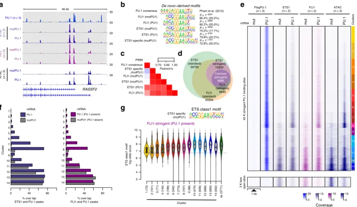
Interaction among ETS family factors. As part of the motif enrichment analysis (Fig. 4c), we noted that de novo-remodeled PU.1 peaks frequently contained two (or more) PU.1 motifs.
Across peaks, these homotypic motif pairs were significantly enriched (compared with non-bound motifs) in a range of 12–50 bp (Supplementary Fig. 4a), which resembles early findings of clustered PU.1-binding sites in many myeloid-specific pro- moters 21 . Notably, homotypic motif pairs were preferentially found in the de novo-remodeled fraction of peaks (PU.1 peak clusters 1–8). Their appearance correlated with the degree of remodeling across PU.1 peak clusters (Supplementary Fig. 4b–f), suggesting that PU.1 motif pairing could assist binding-site
selection. However, the motifs in pairs also showed significant overlaps with consensus sequences for other ETS class family members (Supplementary Fig. 4g), suggesting that de novo- remodeled “homotypic” motif pairs are either bound by PU.1 alone or in combination with another ETS family member expressed in CTV-1 cells.
As homotypic or heterotypic interactions of PU.1 may influence binding-site selection, we asked whether and how the distribution of other ETS factors would be affected by PU.1 expression and to which extent binding sites of PU.1 overlapped with other ETS factors. We generated occupancy maps for two prominent, representative ETS factors (FLI1, ETS1) in CTV-1 cells before and after PU.1 expression (an exemplary locus is shown in Fig. 6a). Both factors share an almost identical ETS class 1a motif, which was clearly different from the PU.1 consensus site (Fig. 6b, c). Correspondingly, the largest fraction of FLI1 and ETS1 target sites overlapped (Fig. 6d), suggesting that they compete for the majority of genomic binding sites. The small fraction of ETS1-specific peaks was primarily associated with two co-associated motifs (ZNF143 and a composite ETS:RUNX motif, see Supplementary Fig. 5a, b), indicating that corresponding TFs may favor ETS1 at these sites.
The induction of PU.1 had a major impact on the genomic distribution of FLI1 and ETS1 in CTV-1 cells (Fig. 6e, f and Supplementary Fig. 5c). As already indicated by the footprints across PU.1 motifs observed at pre-accessible PU.1-binding sites (as shown for PU.1 peak cluster 13 in Fig. 4g), PU.1 joined the competition of ETS factors at a large fraction of pre-existing ETS- binding sites (across clusters 9–14). Correspondingly, the ChIP- seq coverage of ETS1 and FLI1 at pre-accessible PU.1-binding sites was reduced after PU.1 induction (Supplementary Fig. 5d).
Likewise, both ETS factors joined PU.1 at a major subset of de novo-remodeled fraction of peaks (Fig. 6e, f and Supplementary Fig. 5c, f) across PU.1 peak clusters 1–8. Motif scores of ETS factors and PU.1 at their binding sites showed an inverse correlation across de novo-remodeled PU.1 peak clusters 1–8 (Fig. 4a and 6g, and Supplementary Fig. 5e). The predicted recognition motif resembled the ETS motif at PU.1-binding sites co-bound by ETS factors (both at single and paired motif sites), whereas sites without evidence of ETS binding resembled the PU.1 consensus motif (Supplementary Fig. 5g). This suggests that the ETS factor distribution is driven at least in part by motif affinities of individual factors. At sites bound by PU.1 alone, chromatin accessibility changes were limited, regardless of the presence of single or paired sites (Supplementary Fig. 5h), suggesting that binding at these motif pairs is likely restricted to a single position. At present, we cannot say whether the recruit- ment of ETS factors (or other partner factors) to de novo- remodeled sites actively contributes to the process of remodeling or whether it stabilizes the accessible space between two nucleosomes created in the course of PU.1 binding. Nevertheless, it is clear that at these sites, PU.1 is required to allow for ETS factor binding, which is not observed in the absence of PU.1.
In line with the redistribution of other partner TFs (as shown in Fig. 5a–d), the binding of ETS1 and FLI1 was also reduced at the disappearing ~3 K sites that were accessible prior to PU.1 induction (Fig. 6e, bottom panel), further corroborating the ability of PU.1 to redistribute other TFs.
PU.1 domains and interactors required for de novo binding.
Next, we sought to characterize the mechanism underlying PU.1’s ability to change chromatin accessibility and redistribute other TFs across the genome. Hypothesizing that specific interactions of PU.1 with other proteins are involved in these changes, we tested the effects of deleting different known protein–protein interaction
PU.1
1 kb
Clusters Rem-Index
1.4
2.4
2.0
3.2
3.6
6.1 5.7 5.0 1.2 1.7
1.2 1.5 1.1 1.0
45 K PU.1 binding sites mut PU.1 mut PU.1 mut
FlagPU.1 (n = 3)
ATAC (n = 3)
H3K27ac (n = 3)
mRNA
Short (n= 2) Long (n = 1)
Effect on transcr.
***
**
***
***
***
***
*
**
**
*
0 25
0 15
0 15
Coverage
13 11
12 10 9
14 8 7 6 5 4 3 2 1
3 K less open sites
Fig. 3 PU.1-induced changes in chromatin accessibility. The distribution of average PU.1 ChIP-seq, ATAC-seq, and H3K27ac ChIP-seq signals of PU.1 vs. PU.1mut mRNA-transfected cells 8 h after electroporation are plotted across 1 kb windows and 45 K PU.1-binding sites (top panel) or 3 K regions that lost accessibility after PU.1 induction (bottom panel) in CTV-1 cells.
PU.1 peaks are ordered according to K-means clustering of peak-centered ATAC-seq signals. PU.1 peak clusters are indicated by the color bar on the right, along with the average remodeling index (Rem-Index) of each cluster.
The asterisks on the right indicates the signi fi cant induction of mRNA
expression across genes associated with PU.1 peaks in the indicated peak
cluster (*** P < 0.001; ** P < 0.01; * P < 0.05; paired Wilcoxon ’ s test) in PU.1-
expressing CTV-1 cells. The coloring of PU.1 peak clusters is kept consistent
in all following analyses based on the clustering. Source data are provided
as a Source Data fi le.
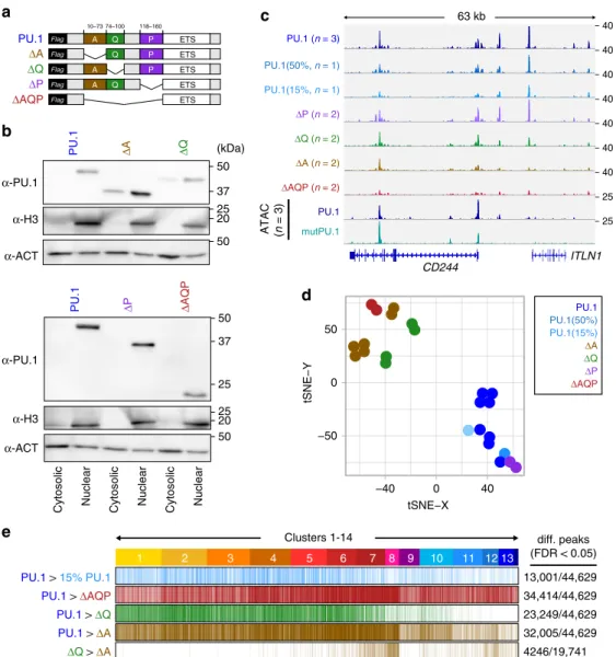
domains of PU.1 on its ability to access chromatin de novo. To this end, we generated PU.1 expression constructs devoid of each one or all of the acidic (A), glutamine-rich (Q), and PEST (P) domains, and tested them in our model compared with wild-type PU.1 (Fig. 7a). All mutant proteins were expressed and translo- cated to the nucleus as expected (Fig. 7b). Detected peak sets of PU.1 mutants generally represented subsets of the wild-type and in three of four cases, binding profiles differed significantly from wild-type PU.1 (Fig. 7c, d). Most pronounced changes in peak coverage were observed for the isolated ETS domain (ΔAQP, 77%
of wild-type PU.1 peaks showed significantly reduced signals) and the mutant lacking the acidic domain (ΔA, 72% of wild-type PU.1 peaks showed significantly reduced signals), followed by the less-
affected mutant lacking the glutamine-rich domain (ΔQ, 52% of wild-type PU.1 peaks showed significantly reduced signals), which partially resembled a lower dose of PU.1 (15% PU.1, 29%
of wild-type PU.1 peaks showed significantly reduced signals).
The ΔP mutant did not reveal significantly altered binding pat- terns. Interestingly, binding profiles of ΔA and ΔQ mutants were particularly different at highly remodeled clusters (Fig. 7e, clusters 7/8), suggesting that the ΔA mutant may specifically lack remo- deling capacity.
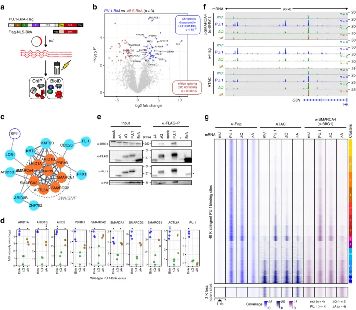
To identify interaction partners of PU.1 that may explain the observed effect of PU.1 mutant proteins, we adapted the BioID approach 22 to biotinylate and identify candidate interacting proteins in the proximity of PU.1 (Fig. 8a). We initially validated
PU1
PU1AP1 CTCF EBOX ETS ETS:IRF FOXO
PU1 PU1
PU1PU1
PU1PU1
PU1
PU1PU1
PU1PU1
PU1PU1
PU1PU1
PU1PU1
PU1PU1
PU1PU1
PU1PU1
PU1PU1
PU1Cluster 14Cluster 13Cluster 12Cluster 11Cluster 10Cluster 9
Cluster 8Cluster 7Cluster 6Cluster 5Cluster 4Cluster 3Cluster 2Cluster 1
80 40 20
5 % motifs in peaks
GATA IRF
NFY RUNX TCF 0%
0%
19%
45%
48%
51%
66%
77%
61%
68%
84%
86%
90%
88%
c
0 10 20 30
0 4 8 12
0.0 0.5 1.0 1.5 2.0
ATAC coverage 4
8 12 16
Distance to PU.1 motif center
g
Cluster 2 PU1 mut PU1
Cluster 7 PU1 mut PU1
Cluster 11 PU1 mut PU1
Cluster 13 PU1 mut PU1 4
6 8 10 12
PU.1 motif log odds score
Distance to PU.1 motif center
PhastCons score
0%
25%
50%
75%
100%
1 14
Cluster – Exon
Intergenic
Intron Promoter
– TTS
d a
PU.1 consensus:
1 2 3 4 5 6 7 8 9 10 11 12 13 14
Cluster
b
AP1 CTCF
EBOX ETS
ETS:IRF ETS:RUNX
FOXO GATA
NFY OCT RUNX
TCF
P < 1–4112 47.8% (3.2%) P < 1–145 17.6% (10.5%)
P < 1–56 1.5% (0.3%) P < 1–88 2.1% (0.4%)
P < 1–4 3.4% (2.9%) P < 1–33 6.1% (4.0%) P < 1–75 2.4% (0.7%)
P < 1–9 12.3% (0.7%) P < 1–9 28.4% (26.2%) P < 1–20 29.7% (26.3%) P < 1–12 6.1% (4.8%)
P < 1–7 9.1% (7.9%)
0.0 0.1 0.2 0.3 0.4
−15 −10 −5 0 5 10 15 0.5
0.0 0.1 0.2 0.3 0.4 0.5
Bound
Unbound
e f
Score+Cons (0.70) ...+CoAss (0.74) ...+ATAC (0.76) ...+ATAC+CoAss (0.78) ...+post ATAC (0.87) ...+post ATAC+CoAss (0.88)
Specificity
Sensitivity
0.0 0.2 0.4 0.6 0.8 1.0
0.0 0.2 0.4 0.6 0.8 1.0
−200 −100 0 100 200 0
−200 −100 0 100 200 −200 −100 0 100 200
−200 −100 0 100 200
Fig. 4 Genomic features of PU.1-induced accessible sites. a The PU.1 motif log odds score distribution of the consensus MAC PU.1 motif, generated
earlier, is shown for each cluster. The median of the speci fi c distribution across all clusters is depicted inside the bean with a conventional boxplot. b De
novo-derived co-associated motifs enriched across ATAC-seq-derived PU.1 peak clusters. The best motifs corresponding to known factor families derived
from individual clusters are shown. Signi fi cance of motif enrichment (hypergeometric test) and the fraction of motifs in peaks (background values are given
in parenthesis) correspond to their distribution across all PU.1 peak regions. c Motif co-enrichment networks for individual clusters are shown. The size of
each node represents the motif enrichment (fraction of peaks) and co-associated TF motifs (PU.1 masked) are indicated by coloring. The second PU.1 node
corresponds to the fraction of peaks containing at least two PU.1 motifs. Edge thickness indicates the frequency of motif co-association within the PU.1
peak. The fraction of PU.1 peaks overlapping with co-associated (PU.1 masked) TF motifs is given below each network. d Evolutionary conservation of the
PU.1 motif across the K-means clusters, color coded as in Fig. 3 as illustrated by the PhastCons score. Corresponding sequence-matched random control
sets of non-bound motifs are shown in the bottom histogram (in dark gray to light gray). e Genome ontology analysis across the K-means clusters is shown
in a stacked bar chart. The association with the individual regions is given as the fraction of peaks in each cluster. f Predictability of PU.1 binding from motif
score, conservation score, nearby presence of co-associated motifs, and chromatin accessibility before and after PU.1 induction. Shown are ROC curves of
logistic models with different sets of predictors (AUC in parentheses), trained and evaluated on separate subsets of the data. g Representative ATAC-seq
footprints across motif-centered, cluster-associated peaks (as indicated by coloring). Corresponding footprints of control cells (mutPU.1) are shown in
gray. Smaller histograms in the upper right corner zoom into the central part of the main graph. The position of the PU.1 motif is indicated by two vertical
dashed lines. a – g Source data are provided as a Source Data fi le.
that the BirA fusion constructs were expressed, functional, and recruited to the same locations compared with wild-type constructs (Supplementary Fig. 6a–c). BioID experiments in CTV-1 cells transfected with wild-type PU.1-BirA and BirA carrying a nuclear localization sequence (NLS) as control (NLS- BirA) followed by mass spectrometry of biotin-ligated proteins revealed significant enrichment of proteins associated with the SWI/SNF complex (Fig. 8b, c) in the neighborhood of PU.1. We obtained similar results with constitutively PU.1-expressing THP- 1 and K-562 cells, confirming that this was not confined to CTV- 1 cells (Supplementary Fig. 6d). Performing BioID analysis with the ΔA and ΔQ deletion mutants, we found that the ΔA mutant, but not the ΔQ mutant, specifically lost proximity to the SWI/
SNF remodeling complex relative to full-length PU.1 (Fig. 8d).
The specific interaction between PU.1 and SWI/SNF was also observed in CoIP experiments. Focusing on the central ATP- dependent chromatin remodeler SMARCA4 (BRG1), we observed its specific interaction with PU.1 independent of the presence or absence of DNA (Supplementary Fig. 6e), and both in FLAG-PU.1 and BRG1 CoIPs (Fig. 8e and Supplementary Fig. 6f).
We also detected two additional SWI/SNF components (ARID2 and SMARCE1) in CoIP westerns (Supplementary Fig. 6g, h).
However, we could not detect FLI1 or LDB1 in CoIPs, suggesting that, although being proximal to PU.1, they do not interact with PU.1 directly, or that the interaction is not stable during CoIP.
Although SWI/SNF components were reproducibly detected in CoIPs with wild-type PU.1, these interactions were slightly reduced with the ΔQ mutant and strongly reduced or absent with the ΔA mutant (Fig. 8e and Supplementary Fig. 6f-h).
We further confirmed that PU.1 interacts with SWI/SNF by blocking experiments and ChIP-seq of SMARCA4 (BRG1) in CTV-1 cells after induction of PU.1 or its ΔQ and ΔA mutants.
Inhibition of SMARCA4 (BRG1) led to a dose-dependent
reduction of PU.1 binding across the entire genome (Supple- mentary Fig. 6i, j), suggesting that its inactivation may have a general effect on TF binding. ChIP-seq experiments after induction of PU.1 clearly demonstrated the recruitment of SMARCA4 (BRG1) to PU.1-remodeled sites (Fig. 8f, g). As exemplified by the GSN locus (Fig. 8f) and across all PU.1- binding sites (Fig. 8g), the ΔQ mutant retained some of the remodeling capacity of wild-type PU.1. However, the ΔA mutant, although retaining some of its binding capacity, was neither able to alter chromatin accessibility nor did it recruit SMARCA4 (BRG1) to the otherwise de novo-remodeled sites.
In addition, as shown in the bottom panel of Fig. 8g, the redistribution of partner TFs was disabled in the ΔA mutant.
Notably, the PU.1-induced redistribution of partner factors also included SMARCA4 (BRG1), which was depleted upon PU.1 induction at the ~3 K sites that lose accessibility. Upon induction of the ΔA mutant, however, the remodeler remained at those sites.
Discussion
The present work provides a detailed analysis of mechanisms allowing the master regulator PU.1 to shape regulatory land- scapes. We show that its N-terminal AAD interacts with the SWI/
SNF remodeling complex and that this interface is required for PU.1 to access and remodel chromatin de novo. However, despite its potentially strong impact on chromatin landscapes, PU.1 is not a pioneer factor in its classical definition. In vivo PU.1-binding profiles as well as in vitro binding studies suggest that PU.1 binding is constrained by chromatin and epigenetic mechanisms, in particular by nucleosome positioning. Hence, it likely initiates remodeling primarily at those binding sites (and prepares them for other factors) that it can access. Given that even classical
0 4 8 12 16
0 3 6 9
0.0 2.5 5.0 7.5 10.0
0 4 8 12 16
0 5 10 15
−200 0 200
Distance to RUNX motif center
ATAC Coverage
0 3 6 9 12
−200 0 200
Accessibility ↓ Accessibility ↑
GATA P < 1
–12935.25% (16.3%) P < 1
–14643.1% (21.3%) P < 1
–18126.6% (8.1%)
RUNX P < 1
–45552.4% (15.0%)
P < 1
–11618.7% (6.0%) TCF
ETS
AP1
De novo–derived motifs in less accessible chromatin
a b
−200 0 200
Distance to TCF motif center
−200 0 200 −200 0 200
Distance to GATA motif center
−200 0 200
RUNX PU.1
Accessibility ↓ Accessibility ↑ TCF
PU.1
Accessibility ↓ Accessibility ↑ GATA
PU.1
PU.1 mutPU.1
PU.1 mutPU.1
PU.1 mutPU.1
ATAC coverage ATAC coverage
d c
Fig. 5 Motif signatures at sites loosing accessibility. a De novo-derived motifs enriched across regions that lost accessibility upon PU.1 induction in CTV-1
cells. The best motifs corresponding to known factor families derived from individual clusters are shown. The signi fi cance of motif enrichment
(hypergeometric test) and the fraction of motifs in peaks (background values are in parenthesis) are given for the top fi ve motifs corresponding to known
motif families. b – d Representative ATAC-seq footprints across enriched motif-centered peaks that either lost or gained accessibility upon PU.1 induction in
CTV-1 cells. Footprints of control cells (mutPU.1) are in grey, footprints of PU.1 mRNA-transfected cells are colored. Smaller histograms in the upper right
corner zoom into the central part of the main graph. The position of the PU.1 motif is indicated by two vertical dashed lines. a – d Source data are provided as
a Source Data fi le.
pioneer factors, which are able to bind nucleosomal target sites, depend on epigenetic and chromatin landscapes 23,24 , we propose a new class of non-classical pioneer factors, such as PU.1, which share pioneering functions with classical factors but lack the ability to access nucleosome-bound target sites.
A prerequisite for pioneering is the ability to recruit remo- deling complexes. Our data show that the N-terminal AAD of PU.1, which was originally defined as an activation domain using reporter assays 25 mediates the interaction with SWI/SNF. This is in concordance with earlier work demonstrating that AADs of other TFs in yeast, including VP16, Gcn4, Swi5, and Hap4, interacted directly with purified SWI/SNF complex and with the SWI/SNF complex in whole-cell extracts 26 . There is also abun- dant evidence for a crucial role of SWI/SNF remodeling com- plexes in the establishment and maintenance of lineage-specific enhancers 27–29 , and their recruitment to target loci is believed to require interaction with DNA-associated TFs. Our work clearly shows that PU.1 is one of those factors that recruits SWI/SNF to its binding sites. This will allow PU.1 to fulfil its role as a master regulator and to participate in the establishment of enhancers across many hematopoietic cell types.
In a given cell type, PU.1 usually occupies a small fraction (5–10%, depending on the statistical stringency of peak calling) of its potential binding sites. Previous work indicated that PU.1- binding-site selection in individual cell types depends on its expression level and the cell type-specific mix of partner TFs 7–9 . The current work suggests that these factors only partially explain
PU.1-binding-site selection in individual cell types. In line with chromatin and epigenetic constraints observed in vitro, PU.1 cannot access all high-affinity sites in all cell types, despite its ability to recruit the remodeling machinery. The current work suggests that its pioneering role depends on cell type-specific chromatin structures, and that the ability of PU.1 to establish novel regulatory elements is likely restricted to accessible binding sites. According to our in vitro experiments, the latter may lack DNA methylation and locate to nucleosomal linker regions or sequences proximal to nucleosome entry sites. Although detailed nucleosome (and corresponding DNA methylation) maps will be required to prove this model, a restricted pioneering role of PU.1 perfectly explains the diverse and manifold PU.1 binding patterns observed across cell types. The frequent observation of shallow ChIP-seq signals (below peak detection) could be owed to the fact that nucleosome positions are not fixed across the large part of the genome and rarely synchronized across populations of cell types, which may create ample opportunities for PU.1 to bind particular recognition sequences in a (variable) sub-fraction of cells, where these sites are accessible.
Interestingly, the expression of PU.1 not only affected reg- ulatory elements containing PU.1-binding sites. Hosokawa et al. 18 recently showed that PU.1 regulates gene expression in early T- cell development both by recruiting TFs RUNX1 and SATB1 to its own binding sites and by depleting them from the binding sites that they occupied in the absence of PU.1 18 . Our data extend their model to PU.1 partner proteins in general and implicate SWI/
PU.1
1 kb
Clusters
45 K stringent PU.1 binding sites mut PU.1mut PU.1mut
FlagPU.1 (n = 3)
ETS1 (n = 2)
FLI1 (n = 2)
mRNA
e
PU.1
mut
ATAC (n = 3)
14 13 12 11 10 9 8 7 6 5 4 3 2 1
PU.1 (PU.1 present) mutPU1 (PU.1 absent)
% over lap FLI1 and PU.1 peaks
0 40 80
14 13 12 11 10 9 8 7 6 5 4 3 2 1
PU.1 mutPU1
% over lap ETS1 and PU.1 peaks
Cluster
0 40 80
0 25
0 15
0 0
15 15
Coverage
g a
RASSF2
–35
–20
–20
–20
–20
–30
–30 46 kb
PU.1 (n = 3)
PU.1 mutPU.1 ATAC (n = 3)
PU.1 mutPU.1 FLI1 (n = 2)
PU.1 mutPU.1
ETS1 (n = 2) 3 K less open sites
13 12 11 12 10 9
14 8 7 6 5 4 3 2 1
0.70 1.00
Pearson‘s
FLI1 (stringent)
9949 ETS1 (stringent)
15830
FLI1 (standard)
43358 ETS1 (standard)
58793
b
d
Common (stringent) 8886
ETS1 (PU1) P < 1–3181
79.8% (22.2%) P < 1–3059 75.0% (17.7%) P < 1–2892 82.2% (20.0%)
FLI1 (mutPU1) P < 1–3291
85.4% (20.2%)
P < 1–1757 72.8% (20.0%) PU.1 consensus
ETS1 (mutPU1) FLI1 (PU1)
ETS1-specific (mutPU1)
Pham et al. (2012) De novo–derived motifs
ETS1 (PU1) FLI1 (mutPU1) PU.1 consensus
ETS1 (mutPU1)
FLI1 (PU1) ETS1 specific (mutPU1)
f
PWM
c
0.85
3 4 5 6 7 8 9 10
Cluster ETS class1 motif log odds score
ETS1 specific (mutPU1)
ETS class1 motif
1 (73) 2 (101) 3 (71) 4 (145) 5 (189) 6 (342) 7 (773) 8 (707) 9 (98) 10 (475) 11 (946) 12 (966) 13 (990) 14 (302) sp (3771) FLI1-stringent (PU.1 present)
mRNA mRNA
Fig. 6 Overlap between ETS1/FLI1 and PU.1-binding sites. a IGV genome browser tracks of the RASSF2 locus showing average PU.1 (blue), ETS1 (purple),
and FLI1 (pink) ChIP-seq coverage in control (mutPU.1) and PU.1-expressing cells. ATAC-seq coverage of PU.1-transfected and control cells are depicted in
blue below the ChIP-seq tracks. b De novo-derived motif enrichment across the indicated ChIP-seq peaks. c Correlation matrix heatmap for position weight
matrices (PWM) of the motifs shown in b. d Venn – Euler diagram showing the overlap of ETS1 and FLI1 ChIP-seq peaks (using stringent and standard peak
calling). e Distribution of PU.1, ETS1, and FLI1 ChIP-seq, as well as ATAC-seq signals before and after PU.1 expression plotted across the ATAC-seq-derived
PU.1 peak clusters (top panel), as well as regions that lost accessibility after PU.1 induction (bottom panel) in CTV-1 cells, as introduced in Fig. 3. f Bar plots
displaying the overlap of stringent ETS1 (left panel) and FLI1 (right panel) peaks in PU.1-transfected (blue/purple bars) and control CTV-1 cells not
expressing PU.1 (gray bars) with PU.1 peaks across the PU.1 peak clusters introduced in Fig. 3. g Motif log odds score distribution of the consensus ETS
class 1 motif is shown for FLI1-overlapping peaks across ATAC-seq-derived PU.1 peak clusters along with FLI1 speci fi c (sp) peaks. The median of each
distribution is depicted inside the bean with a conventional boxplot. b – g Source data are provided as a Source Data fi le.
SNF remodelers in the process of TF redistribution. Based on our footprinting and ChIP-seq analyses, we observe the same type of factor redistribution in all three cell systems (CTV-1, TALL1, and HepG2) studied here. The PU.1-induced redirection primarily affected TFs occupying lineage-specific cis-modules, such as RUNX1 and GATA-family factors in T-cell lines and HNF- and FOXA-family factors in the liver cell line. Interestingly, the redistribution of partner TFs required the acidic domain of PU.1, which is not required for direct protein–protein interactions with RUNX1, GATA-, AP1-, or C/EBP-family factors 14 – 17 . Hence, the direct binding to partner proteins may not be sufficient for PU.1 to sequester partner proteins. The fact that the N-terminal acidic domain of PU.1 interacts with SWI/SNF implicates remodeling complexes in the redirection of partner proteins. The decom- missioning of TF-bound cis-modules after PU.1 induction could be mediated through the reallocation of limiting SWI/SNF remodeling complexes by PU.1, which are generally required to
maintain the accessible state of regulatory elements such as lineage-specific enhancers 27–30 . As many of the identified PU.1 partner proteins have already previously been shown to interact with components of SWI/SNF remodeling complexes 31–34 , the latter may act as part of a hub increasing the probability of co- binding of PU.1 partner proteins at de novo-remodeled binding sites. In conclusion, our systematic analysis of de novo TF binding reveals important mechanistic details and provides more comprehensive understanding of a master regulator that shapes regulatory landscapes during hematopoiesis, has known repro- gramming capabilities, but is different from “classical” pioneer factors.
Methods
Cell culture. CTV-1 (DSMZ: #ACC 40), HepG2 (DSMZ: #ACC 180), K-562 (DSMZ:
#ACC 10), TALL1 (DSMZ: #ACC 521), and THP-1 cells (DSMZ: #ACC 16) were grown in RPMI 1640 (Gibco) routinely supplemented with 2 mM
L-glutamine PU.1
Flag A Q P ETSQ P ETS
Flag
A Q P ETS
Flag
A Q ETS
Flag
ETS Flag
ΔA ΔQ Δ P Δ AQP
PU.1(15%) Δ A Δ Q Δ P Δ AQP PU.1(50%) PU.1
–
40
–
40
–
40
–
40
–
40
–
40
–
40
–
25
–
25 PU.1(15%, n = 1)
Δ A (n = 2) Δ Q (n = 2) Δ P (n = 2)
Δ AQP (n = 2) PU.1(50%, n = 1)
CD244 PU.1 (n = 3)
PU.1 mutPU.1 ATAC ( n = 3)
63 kb
a c
e
d
Clusters 1-14
PU.1 > 15% PU.1 PU.1 > Δ AQP PU.1 > Δ Q PU.1 > Δ A Δ Q > Δ A
diff. peaks (FDR < 0.05) 13,001/44,629 34,414/44,629 23,249/44,629 32,005/44,629 4246/19,741 α -PU.1
α -H3
Cytosolic Nuclear Cytosolic Nuclear Cytosolic Nuclear
(kDa)
25 50 50
20
PU.1 Δ A Δ Q
Δ P Δ AQP
PU.1
37
25 50 37
25 50 20
b
10–73 74–100 118–160
tSNE−X
tSNE−Y
13 11 12 10 9 8 7 6 5 4 3 2 1 α-ACT
α-PU.1
α -H3 α -ACT
−50 0 50
−40 0 40
ITLN1
Fig. 7 Effect of PU.1 protein domains on chromatin access. a Design of PU.1-deletion mutants. The DNA-binding domain (ETS domain) was kept in all constructs and a 3 × -FLAG-tag was added N-terminal for detection. b Immunoblot con fi rming the nuclear expression of PU.1-deletion mutants compared with full-length PU.1 in transfected cells using IVT mRNA. c IGV genome browser tracks of the CD84 locus showing PU.1 ChIP-seq coverage of CTV-1 cells transfected with varying amounts of PU.1 IVT mRNA as well as PU.1 mutant mRNA. ATAC-seq coverage of CTV-1 cells transfected with PU.1 (blue) and mutPU.1 (turquoise) IVT mRNA is depicted in the two bottom rows. d Two-dimensional visualization of the distribution of the indicated samples across annotated PU.1 peaks using tSNE embedding. Replicates of the same mRNA transfections are indicated by coloring. e Differential ChIP-seq peaks of WT vs.
less PU.1 (15%, light blue), WT vs. Δ AQP (red), WT vs. Δ Q (green), and WT vs. Δ A (brown), as well as differential peaks between Δ Q vs. Δ A (brown) are
shown across ATAC-seq-derived PU.1 peak clusters (introduced in Fig. 3). b – e Source data are provided as a Source Data fi le.
(Biochrom), 1 mM sodium pyruvate (Sigma), 50 U ml
−1penicillin/streptomycin, 0.4×
vitamins (Sigma), 1× non-essential amino acids (Sigma), 50 μM b-mercaptoethanol (Gibco), and 10% or 15% (TALL1) heat-inactivated fetal calf serum (FCS) (Gibco) at 37 °C, 5% CO
2, 95% humidity. Cells were split every 2–3 days and resuspended in fresh medium. Adherent HepG2 cells were detached using 1× Trypsin-EDTA (Thermo Fisher Scientific). After 20 passages, cells were discarded and a new batch of cells was used. For differentiation of THP-1 cells into MAC-like cells PMA (10
−8M;
Calbiochem) and 1,25-dihydroxyvitamin D3 (VD3, 10
−7M; Sigma) were added, cells were incubated for 3 days at 37 °C, 5% CO
2, 95% humidity, and adherent cells were detached using Accutase solution (Sigma). Collection of blood cells from healthy donors was performed in compliance with the Helsinki Declaration. All donors signed an informed consent. The leukapheresis procedure and subsequent puri fi cation of hematopoietic cell types were approved by the local ethical committee (reference number 12-101-0260). Separation of peripheral blood cell types and in vitro
differentiation of MOs into MACs or DCs were performed as described
previously
9,35,36. B cells were purified as described
37. Neutrophils were purified from whole blood after lysis of erythrocytes with ACK (Ammonium-Chloride-Potassium) lysis buffer (5 ml per ml blood of 155 mM NH
4Cl, 100 mM KHCO
3, 0.1 mM EDTA) and puri fi ed by fl uorescence-activated cell sorting (FACS) based on size and granu- larity. Human mast cells were purified from the skin that was obtained from cosmetic breast-reduction surgeries
38with informed consent of the patients. Mast cell pre- parations were performed in compliance with the Helsinki Declaration and were approved by the ethics committee of the Charité Universitätsmedizin Berlin (reference number EA1/204/10).
Transfection of in vitro transcribed mRNA. Synthetic DNA templates (gBlocks) for wild-type PU.1, it’s mutated version, all PU.1-deletion mutants, and all BirA*
ivt
AAAAAAAAAAAAAAAAAAAAAA AAAAAAAAAAAAAAAAAAAAAA AAAAAAAAAAAAAAAAAAAAAA
ChIP
a
A Q P ETS 27aa BirA Flag
PU.1-BirA-Flag
BioID
log2 fold change
−log10P
b
SWI/SNF
PU.1-BirA vs. NLS-BirA (n = 3)
FlagNLS BirA
Flag-NLS-BirA
f
GSN PU.1
ATACα-Flagα-SMARCA4 (α-BRG1)
– 30
– 30 – 30
– 25
– 25 – 25 – 20
– 30
– 25 mut
ΔA PU.1 mut
ΔA PU.1 mut
ΔA
PU.1
1 kb
45 K stringent PU.1 binding sites mut PU.1 ΔA
mut
α-Flag mRNA
g
PU.1
mut
ATAC
0 25
0 25
0 Coverage 15
ΔQ
ΔA
α-SMARCA4 (α-BRG1)
3 K less open sites Clusters
13 11 12 10 9
14 8 7 6 5 4 3 2 1
d c
ΔA
ΔQ
ΔQ
ARID3B KMT2C
ZNF792 ARID5BSMARCA4
ARID1A
SMARCA2 LDB1
SPI1
ACTL6A SMARCD2 ARID2
SMARCE1 ARID1B
PBRM1 KMT2D
RFX1 CDC20
FLI1
ACTL6A ARID1A
ARID1B ARID2 ARID3B
FLI1 KMT2C KMT2D
LDB1 PBRM1 SMARCA2 PDHX SMARCA4 SMARCD2
SMARCE1
SPI1
ZBTB4
0 2 4
−5 0 5 10
96 kb
– 20 – 20 ΔQ – 20
ΔQ
ΔQ mRNA
mRNA splicing (GO:0000398) q < 0.00005 Chromatin disassembly (GO:0031498) q < 10–9
0 3 6 9
PU.1
1 2 3
SMARCE1
0 1 2 3
SMARCD2
0 1 2 3
PBRM1
0 1 2 3
ARID2
1 2 3 4
ARID1B
0 1 2 3 4
ARID1A
1 2 3 4
MS intensity ratio (log2)
ACTL6A
0 1 2 3
SMARCA4
0 1 2
SMARCA2
BirA ΔQ ΔA BirA ΔQ ΔA BirA ΔQ ΔA BirA ΔQ ΔA
BirA ΔQ ΔA
BirA ΔQ ΔA
BirA ΔQ ΔA
BirA ΔQ ΔA
BirA ΔQ ΔA
BirA ΔQ ΔA
Wild-type PU.1-BirA versus
** * **
* * * ** * * * * * * * *
mock ΔA ΔQ PU.1 BirA
Input
α-FLAG
α-H3 α-BRG1
α-PU.1
15 250
37 50
37 50
(kDa) ΔA ΔQ mock PU.1 – BirA α-FLAG-IP
e
*
n = 4 n = 4
n = 4 n = 2
n = 4 n = 4
n = 4 n = 2
n = 4 n = 4
n = 4 n = 2
PU.1 (n = 4) mut (n = 4) ΔQ (n = 2)
ΔA (n = 4)