J Clin Periodontol. 2020;47:863–874. wileyonlinelibrary.com/journal/jcpe | 863
1 | INTRODUCTION
Periodontal disease is among the most prevalent human diseases with a prevalence of about 796 million affected adults world- wide (James et al., 2018). Furthermore, periodontitis is regarded to be the most common cause for tooth loss (Tonetti, Jepsen,
Jin, & Otomo-Corgel, 2017) and a limitation of oral health-re- lated quality of life (Buset et al., 2016; Kinane, Stathopoulou, &
Papapanou, 2017).
While the first and foremost aim of any periodontal treat- ment is to resolve dysbiotic microbial communities in subgingival plaque and the concomitant periodontal inflammation (Bostanci Received: 19 November 2019 | Revised: 21 March 2020 | Accepted: 2 May 2020
DOI: 10.1111/jcpe.13302
C L I N I C A L P E R I O D O N T O L O G Y
Tooth survival and clinical outcomes up to 26 years after
guided tissue regeneration therapy in deep intra-bony defects:
Follow-up investigation of three randomized clinical trials
Fabian Cieplik1 | Insa Ihlenfeld1 | Karl-Anton Hiller1 | Andreas Pummer1 | Gottfried Schmalz1,2 | Wolfgang Buchalla1 | Michael Christgau1,3
This is an open access article under the terms of the Creative Commons Attribution-NonCommercial License, which permits use, distribution and reproduction in any medium, provided the original work is properly cited and is not used for commercial purposes.
© 2020 The Authors. Journal of Clinical Periodontology published by John Wiley & Sons Ltd Cieplik and Ihlenfeld contributed equally.
1Department of Conservative Dentistry and Periodontology, University Hospital Regensburg, Regensburg, Germany
2Department of Periodontology, School of Dental Medicine, University of Bern, Bern, Switzerland
3Private Practice, Düsseldorf, Germany
Correspondence
Fabian Cieplik, Department of Conservative Dentistry and Periodontology, University Hospital Regensburg, Franz-Josef-Strauß- Allee 11, 93053 Regensburg, Germany.
Email: fabian.cieplik@ukr.de Funding information
Fabian Cieplik received funding from the Medical Faculty of the University of Regensburg (ReForM B program).
Abstract
Aim: To investigate tooth survival and clinical long-term outcomes up to 26 years fol- lowing guided tissue regeneration (GTR) therapy in deep intra-bony defects.
Methods: Patients from three prospective clinical split-mouth studies, which inves- tigated the outcomes of GTR therapy, were re-evaluated 21–26 years after surgery independent of the membrane type used, and tooth survival was assessed according to several site-specific and patient-related factors.
Results: About 50 patients contributing 102 defects were available for this long-term follow-up. After up to 26 years (median 23.3 years), 52.9% of the teeth were still in situ. The median survival of the extracted teeth was 13.8 years. Patients with dia- betes mellitus and/or smoking history lost significantly more teeth in the long term.
Compared to the 1-year situation, there was no new median CAL loss after up to 26 years in the teeth which were still in situ.
Conclusions: Within the limitations of this study, our data show that more than 50%
of the initially seriously diseased teeth were still in situ up to 26 years following GTR therapy despite an overall limited adherence to SPT. In the majority of these teeth, the CAL gain 1 year after GTR could be maintained over this long period.
K E Y W O R D S
guided tissue regeneration, intra-bony defects, long-term results, periodontal surgery
& Belibasakis, 2017), regeneration of the lost periodontal tissues in terms of reconstitution of their original architecture and func- tion represents the ultimate challenge in clinical periodontology (Cortellini & Tonetti, 2015; Sallum, Ribeiro, Ruiz, & Sallum, 2019).
In this context, guided tissue regeneration (GTR) and the use of enamel matrix derivatives are scientifically accepted and clinically available treatment concepts for predictive regenerative periodon- tal therapy in deep intra-bony periodontal defects (Cortellini &
Tonetti, 2015; Kao, Nares, & Reynolds, 2015; Reynolds et al., 2015;
Sallum et al., 2019; Wu, Lin, Song, Su, & Tu, 2017). GTR therapy is based on the use of cell-occlusive barrier membranes that prevent down-growth of epithelial cells along the root surface and maintain a protected space within the periodontal defect that allows for pro- liferation of the slowly migrating periodontal ligament (PDL) cells (Karring, Nyman, Gottlow, & Laurell, 1993; Larsson et al., 2016;
Sallum et al., 2019). While conventional non-surgical or surgical periodontal treatment approaches result in the formation of a long-junctional epithelium as a reparative tissue, numerous histo- logical studies demonstrated regeneration of PDL, cementum and alveolar bone following GTR therapy (Karring et al., 1993; Larsson et al., 2016; Sallum et al., 2019).
Clinical outcomes after regenerative periodontal therapy still exhibit high variability due to several factors, which can either be related to the individual patient, the defect or the surgical tech- nique used (Cortellini & Tonetti, 2000, 2015). This may particularly hold true for GTR, which is known to be a technically sensitive and highly demanding technique (Bashutski, Oh, Chan, & Wang, 2011;
Cortellini & Tonetti, 2015; Sallum et al., 2019). As GTR therapy is a very complex and also rather cost-intensive procedure, clinical data on its long-term efficacy are desirable for sufficient information of patients and healthcare providers (Cortellini, Buti, Pini Prato, &
Tonetti, 2017; Elangovan, 2016).
Consequently, the aim of this study was to perform a follow-up examination of three randomized controlled clinical trials, which had been carried out between 1992 and 1996 and examined the clinical healing results following GTR in deep intra-bony periodontal defects with different types of barrier membranes (Christgau et al., 2002;
Christgau, Bader, Schmalz, Hiller, & Wenzel, 1998). In particular, the present study focused on investigating tooth survival and long-term treatment outcomes up to 26 years following GTR therapy in gen- eral, independent of the membrane type used.
2 | MATERIAL AND METHODS
2.1 | Study design
The present study is a follow-up investigation of three controlled randomized prospective clinical split-mouth studies (Christgau et al., 2002; Christgau et al., 1998) and investigates the long-term out- comes up to 26 years following GTR therapy. These original studies investigated the clinical performance of
• non-resorbable expanded polytetrafluoroethylene membranes (ePTFE; Gore-Tex Periodontal Material, W.L. Gore & Associates Inc., Flagstaff, AZ, USA) compared to resorbable Polyglactin-910 membranes (PG-910; Vicryl-Netz Parodontal-Zuschnitt, Ethicon, Norderstedt, Germany; study 1; Christgau, Schmalz, Reich, &
Wenzel, 1995),
• polylactic acid membranes (PLA; Guidor bioabsorbable matrix barrier, Guidor AB, Huddinge, Sweden) compared to PG-910 membranes (Vicryl-Netz Parodontal-Zuschnitt; study 2; Christgau et al., 1998) or
• experimental polydioxanone membranes (PDS; Mempol, Ethicon) compared to PLA membranes (Guidor bioabsorbable matrix bar- rier; study 3; Christgau et al., 2002).
Due to non-avoidable drop outs, the original split-mouth design and the comparison between different membrane types could not be followed in the present follow-up. Instead, the focus was set on tooth survival and long-term stability of the attachment gain fol- lowing GTR therapy in general, independent of the membrane type used. The present study design was approved by the ethics com- mittee of the University of Regensburg (reference: 18-897-101) in accordance with the 1964 Helsinki Declaration and its later amend- ments and comparable ethical standards. After detailed description of the proposed clinical examinations, written informed consent was obtained from each individual participant included in this follow-up.
2.2 | Patient population
The three studies included in this follow-up originally comprised 74 patients that had been recruited from the patient pool of the Department of Conservative Dentistry and Periodontology of the University Hospital Regensburg (Germany). For inclusion in the original studies (Christgau et al., 1995, 1998, 2002), all patients had
Clinical Relevance
Scientific rationale for the study: To investigate the long- term outcomes up to 26 years after GTR therapy in deep intra-bony defects.
Principal findings: In teeth, which survived, the 1-year CAL gain remained stable. However, almost 50% of the teeth were lost most probably due to an irregular and insufficient SPT. Diabetics and smokers showed significantly less tooth survival.
Practical implications: Our data indicate that regenerated tissue can be maintained over a very long period of time.
However, the implementation of an adequate SPT and con- trol of the known risk factors smoking and diabetes seem to be crucial in those patients.
to have a pair of contra-lateral deep interproximal intra-bony peri- odontal defects with a probing pocket depth (PPD) of at least 6 mm and radiographic evidence of angular bone loss of at least 4 mm at baseline. Full-mouth supra- and subgingival scaling and root plan- ing as well as splinting of highly mobile teeth had to be successfully completed at least 4–6 weeks before surgery. For inclusion in this follow-up, patients had to have received the intended treatment and successfully completed clinical examinations at baseline as well as after 1 year. Only defects without any furcation involvement at baseline were included in this follow-up.
2.3 | Clinical therapeutic procedures
All surgical interventions were performed by one experienced sur- geon (MC) according to the principles of GTR therapy between April 1992 and December 1996 and have been described in de- tail in the respective original publications (Christgau et al., 1995, 1998, 2002). Thus, only a brief summary is given here, as follows:
buccal and oral mucoperiosteal flaps were elevated after sulcular incisions. Vertical releasing incisions were avoided in order not to interfere with the flap vascularization. Subsequently, a thorough defect debridement was performed. The surgical sites in each pa- tient were randomized for treatment with one of the two different barrier membranes in the respective study. The barrier mem- branes were adjusted to the individual defect morphology, and the surgical sites were closed tension-free by coronally repositioned flaps to ensure primary membrane coverage. In the case of the non-resorbable ePTFE membranes, a second surgical procedure was performed after 4–6 weeks for membrane removal. For peri- operative infection prophylaxis, systemic antibiotic therapy was performed (studies 1 and 2 [Christgau et al., 1995, 1998]: doxy- cycline for 10 days; study 3 [Christgau et al., 2002]: amoxicillin or clindamycin for 2 days). During the first 6 post-operative weeks, all patients had to suspend mechanical oral hygiene procedures in the area of surgery, rinse with 0.2% chlorhexidine solution and undergo weekly inspections including tooth cleaning. All patients were scheduled in a strict supportive periodontal therapy (SPT) programme at the Department of Conservative Dentistry and Periodontology of the University Hospital Regensburg for the first post-operative year with visits every 2–3 months. After that pe- riod, patients were encouraged to further participate in this SPT programme but most of them returned to their referring dentists.
The remaining patients were scheduled for SPT twice a year in the undergraduate programme of the Department of Conservative Dentistry and Periodontology.
2.4 | Clinical examinations
The clinical examinations of this follow-up study were performed by two blinded examiners (FC; AP), who had previously been cali- brated to the principal investigator (MC). The examiners were not
involved in the treatments and not aware of the treatment mo- dality used in the individual defects. Oral hygiene was assessed by the approximal plaque index (API; Lange, Plagmann, Eenboom,
& Promesberger, 1977) and gingival inflammation by the papillary bleeding index (PBI; Saxer & Mühlemann, 1975). The following clinical parameters were recorded for the assessment of the peri- odontal situation using a PCP-UNC-15 periodontal probe (PCP- UNC-156; Hu-Friedy): gingival recession (REC) as the distance between the cemento-enamel junction (CEJ) or the cervical mar- gin of a restauration to the gingival margin, probing pocket depth (PPD) as the distance from the gingival margin to the fundus of a periodontal pocket, and clinical attachment level (CAL) as the dis- tance from the CEJ or the cervical margin of a restauration to the fundus of a periodontal pocket. Furthermore, bleeding on probing (BOP) was recorded. In addition, the vertical relative attachment gain (V-rAG) was calculated as the percentage of the CAL gain re- lated to the baseline depth of the osseous defect measured intra- operatively (Christgau et al., 1995, 1998, 2002). Furthermore, the total number of residual teeth at this follow-up was recorded for each patient.
Apart from the clinical parameters, a detailed anamnesis of the medical history of the individual patient was obtained, with particu- lar emphasis on the aspects of smoking habits and history of diabe- tes mellitus. Smoking habits were recorded as pack-years. History of diabetes comprised both type I and type II diabetes. However, there were no data available on the quality of metabolic control during this long period of time. The timepoint of a possible loss of a treated tooth was obtained either from the dental charts or by interview of the respective patient. Furthermore, the frequency of SPT participation was examined based on the dental charts (for individuals still being patients at the University Hospital) or the self-reported information provided by the patients. SPT participa- tion at least twice per year (irrespective of the SPT provider) was classified as “regular SPT.”
2.5 | Data analysis
The single defect was regarded as statistical unit in this study. As discussed previously (Christgau, Schmalz1, Wenzel, & Hiller, 1997), clinical measurements are reported as median values (with 1st and 3rd quartiles) and were statistically evaluated using chi-square tests and non-parametric Mann–Whitney tests on a significance level of α = 0.05. The Kaplan–Meier procedure was applied for survival analysis followed by log-rank tests for determining significant dif- ferences between survival curves (α = 0.05). All statistical analyses were performed using SPSS for Windows, version 25 (SPSS Inc.).
3 | RESULTS
For inclusion in this follow-up study, patients had to have re- ceived the intended treatment and successfully completed clinical
F I G U R E 1 Flowchart of the study outline
examinations at baseline as well as after 1 year. Therefore, 2 out of the initially recruited 74 patients had to be excluded due to missing baseline or 1-year data. Further 22 patients could not be re-exam- ined because the surgical site either did not correspond to the inclu- sion criteria of this study with regard to defect classification (i.e. no furcation involvement) or the patient was not available.
From the remaining 50 patients contributing 102 surgical sites, 34 (contributing 69 surgical sites) could be scheduled for clini- cal examination at the Department of Conservative Dentistry and Periodontology of the University Hospital Regensburg, while the other 16 patients (contributing 33 surgical sites) were only available for an oral interview by telephone. Consequently, tooth survival analyses could be performed in 102 surgical sites in 50 patients.
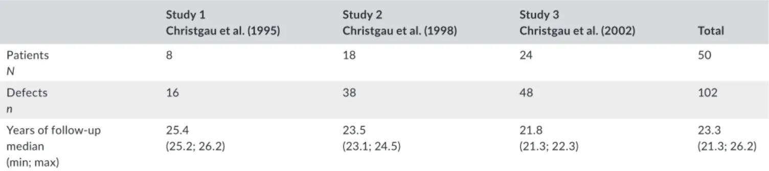
Figure 1 shows the flow of participants through the stages of this study. Table 1 gives an overview of the patients and defects that were available for this follow-up.
3.1 | Compliance with SPT
The calculation of the SPT frequencies was carried out based on the dental charts (for individuals still being patients at the University Hospital) and the information provided by the 50 patients who were available for tooth survival analysis. After the first post-operative year, 40.2% of all surgical sites received SPT only at the University Hospital, 13.7% only at their referring dentists and 46.1% alternating between the University Hospital and the referring dentists. 53.9% of the surgical sites received SPT at least twice a year (irrespective of the SPT provider), while 46.1% of the surgical sites received SPT less than twice a year.
3.2 | Tooth survival analysis
Tooth survival analysis was performed in 50 patients contributing 102 surgical sites. Up to 26 years after GTR therapy, 52.9% of the teeth were still in situ, while 47.1% had been extracted (Figure 2).
Table 2 shows a frequency distribution of all included teeth with re- gard to tooth type and CAL at baseline. 19 patients did not lose any of the treated teeth, whereas 16 patients lost at least one tooth, and 15 patients lost all treated teeth. The median (1st; 3rd quartile) duration of tooth survival was found to be 23.4 (21.9; 23.8) years for
teeth which were still in situ at this follow-up examination and 13.8 (6.2; 17.2) years for extracted teeth.
When classifying the defects according to tooth-related and site-specific factors, there was a tendency for more tooth loss in pre- molars (p = .132) and molars (p = .105) as compared to anterior teeth (Figure 3a) and in initially deeper defects with CAL ≥ 12 mm at baseline as compared to defects with CAL ≤ 8 mm (p = .08) or CAL 9–11 mm (p = .078; Figure 3b). Considering the 1-year data, there was significantly more tooth loss in surgical sites still exhibiting CAL ≥ 7 mm at 1 year as compared to surgical sites with CAL ≤ 4 mm (p = .045) and CAL 5–6 mm (p = .043; Figure 3c). Furthermore, there was a tendency for more tooth loss in surgical sites with residual PPD ≥ 6 mm at 1 year as compared to sites with 1-year PPD ≤ 3 mm and 1-year PPD 4–5 mm (Figure 3d).
Taking patient-related factors into account, that is age at baseline (Figure 4a), history of diabetes (Figure 4b), smoking habits (Figure 4c) and frequency of SPT participation between 1 and 26 years after GTR therapy (Figure 4d), there was significantly more tooth loss in diabetic patients as compared to non-diabetics and in patients with smoking history of at least 10 pack-years as compared to non-smokers and patients with smoking history of less than 10 pack-years. In diabetic patients, only 20% of the treated teeth were still in situ, whereas in non-diabetics 61% of the treated teeth were still in situ (p = .001). In non-smokers, 57.5% of the treated teeth could be preserved, while in smokers with history of at least 10 pack-years only 25% of the treated teeth were still in situ (p = .011). There was a slight tendency for more tooth loss in patients with an age of at least 51 years at baseline com- pared to the younger patient groups. On the contrary, no statistically significant association could be found between tooth loss and fre- quency of SPT participation between 1 and 26 years.
Additionally, the total number of residual teeth at the long-term fol- low-up was recorded in 44 patients contributing 89 surgical sites. GTR- treated teeth, which were still in situ, were accompanied by 25 (18.8;
27) residual teeth in median (1st; 3rd quartile), while sites, where the GTR-treated tooth was lost, were accompanied by only 12 (4; 21) re- sidual teeth. This difference was found statistically significant (p = .000).
3.3 | Clinical examinations
Clinical examinations could be performed in 34 patients, originally contributing 69 defects, which showed a median (1st; 3rd quartile)
TA B L E 1 Numbers of patients and defects available for this follow-up from each original study and the respective follow-up durations Study 1
Christgau et al. (1995)
Study 2
Christgau et al. (1998)
Study 3
Christgau et al. (2002) Total Patients
N
8 18 24 50
Defects n
16 38 48 102
Years of follow-up median
(min; max)
25.4
(25.2; 26.2) 23.5
(23.1; 24.5) 21.8
(21.3; 22.3) 23.3
(21.3; 26.2)
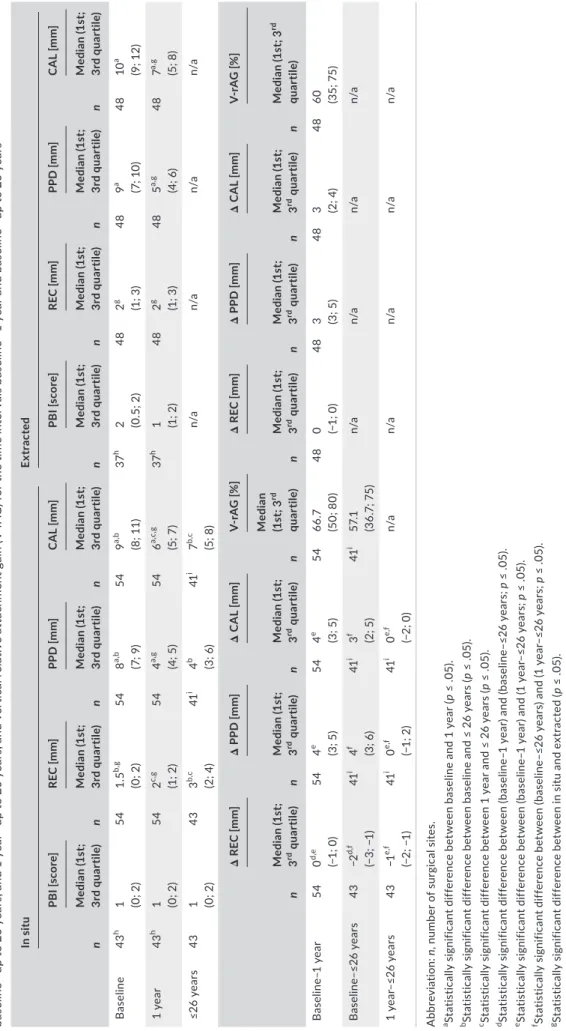
API of 33% (20%; 44%). Table 3 reports the clinical periodontal data for teeth still in situ as well as for the extracted teeth. Furthermore, the respective changes of periodontal parameters after 1 and up to 26 years are reported.
The median baseline PPD was 8.0 mm (in situ) or 9.0 mm (ex- tracted) and decreased to 4.0 mm or 5.0 mm, respectively, after 1 year. After up to 26 years, no increase in PPD was found for the teeth that were still in situ. Median baseline REC of teeth still in situ was 1.5 mm and increased to 2.0 mm after 1 year and 3.0 mm after up to 26 years. In contrast, median REC of extracted teeth was found constant at 2.0 mm at baseline and the 1-year follow-up. The me- dian baseline CAL was 9.0 mm (in situ) or 10.0 mm (extracted), both exhibiting a significant CAL gain of 3.0 mm resulting in 6.0 mm (in situ) or 7.0 mm (extracted) at the 1-year examination. For the teeth found still in situ, there was no median change in CAL between the 1-year examination and the follow-up after up to 26 years. The clin- ical 1-year data differed statistically significantly between the teeth
still in situ and those extracted over time in median PPD (p = .03), median REC (p = .008) and median CAL (p = .022). The median V-rAG was found 66.7% (in situ) and 60.0% (extracted) between baseline and 1 year, and 57.1% in the in situ-group between baseline and the follow-up after up to 26 years.
4 | DISCUSSION
There are only very few studies reporting on long-term outcomes after regenerative periodontal therapies, as it was recently con- cluded by Wu et al. (2017) in their systematic review. With regard to GTR (or combined GTR/graft therapy) in intra-bony defects, there are only five studies with follow-up periods of 10 years (Nickles, Ratka-Krüger, Neukranz, Raetzke, & Eickholz, 2009; Nygaard- Østby, Bakke, Nesdal, Susin, & Wikesjö, 2010; Pretzl, Kim, Holle,
& Eickholz, 2008; Pretzl et al., 2009; Sculean et al., 2008), one study with 13-year follow-up evaluating additional application of autogenous platelet concentrate (Cieplik et al., 2018) and two studies with follow-up periods of 20 years (Cortellini et al., 2017;
Petsos et al., 2019). These latter two studies are prospective clini- cal studies comparing GTR therapy and open flap debridement (OFD) but comprise only 41 defects (in 41 patients; parallel design;
Cortellini et al., 2017) or 38 defects (in 12 patients; split-mouth design; Petsos et al., 2019), respectively. In the present study, pa- tients from three prospective randomized clinical trials (Christgau et al., 1995, 1998, 2002), which each originally had been designed to compare clinical healing outcomes of two different types of barrier membranes in a split-mouth approach, have been pooled to conduct a long-term follow-up investigation of up to 26 years (median follow-up duration 23.3 years) after GTR therapy. A com- mon drawback of long-term studies with follow-up periods of 20 or even more years is that only parts of the initial patient popula- tions are still available due to non-avoidable drop outs (e.g., move and death). Therefore, in the present study the split-mouth de- sign of the original studies and the comparison between different F I G U R E 2 Results of tooth survival analysis: cumulative survival
(thick lines) including 95% confidence limits (hairlines) of all surgical sites included in this study, irrespective of all other parameters
Time under risk [years]
Total In situ Extracted
Tooth type Incisors, canines 21
20.6%a
15 71.4%b
6 28.6%b
Premolars 35
34.3%a 17
48.6%b 18
51.4%b
Molars 46
45.1%a
22 47.8%b
24 52.2%b
CAL at baseline ≤8 mm 27
26.5%a
16 59.3%b
11 40.7%b
9–11 mm 47
46.1%a
27 57.4%b
20 42.6%b
≥12 mm 28
27.5%a
11 39.3%b
17 60.7%b
aPercentages of the respective tooth types or CAL at baseline categories.
bPercentages of in situ or extracted teeth within the respective tooth type or CAL at baseline category.
TA B L E 2 Frequency distribution of 102 surgical sites in 50 patients according to tooth type and CAL at baseline with regard to whether teeth were still in situ after up to 26 years or had been extracted
membrane types were not followed anymore, particularly as none of the three original studies had found significant differences in clinical parameters between the respective membrane types in- vestigated (Christgau et al., 1995, 1998, 2002). Instead, the focus of the present study was set on tooth survival and clinical long- term stability, independent of the respective type of barrier mem- brane used. Therefore, not the individual patient but the single surgical site was chosen as statistical unit for this study. Since patients contributed different numbers of defects, dependencies among the surgical sites in one patient cannot be excluded. The authors are aware of this problem, but accepted it in order to not further reduce the numbers of surgical sites for this long-term follow-up.
Tooth survival could be assessed in 102 surgical sites (in 50 patients), and clinical examinations were possible in 69 surgical sites (in 34 patients). Although the present study lacks an OFD control group, it reports the longest follow-up after GTR therapy with the largest number of re-examined surgical sites in the liter- ature, so far.
4.1 | Compliance with SPT
Regular participation in a SPT programme is known to be cru- cial for long-term success of periodontal treatment (Axelsson &
Lindhe, 1978; Axelsson, Nyström, & Lindhe, 2004; Mombelli, 2019) and particularly of regenerative periodontal therapy (Cortellini, Prato, & Tonetti, 1996; Cortellini & Tonetti, 2004). All patients in- cluded in this follow-up had participated in a strict SPT programme at the University Hospital within the first post-operative year. Due to the wide catchment area of the University Hospital Regensburg and the geographical context of the Oberpfalz Region, many pa- tients subsequently preferred to attend their referring local dentists rather than to accept long distances and shoulder additional travel costs to reach Regensburg. Furthermore, the principal investigator, who had performed all surgical procedures (MC), left the University Hospital Regensburg in 2002, which may further have deteriorated patients’ adherence, whereas Cortellini et al. showed that a high degree of SPT adherence can be achieved, if patients are treated by the same dentist over a long period of time (Cortellini et al., 2017).
F I G U R E 3 Results of tooth survival analysis: cumulative survival (thick lines) including 95% confidence limits (hairlines) of all surgical sites included in this study grouped according to tooth-related factors: tooth type (a), CAL at baseline (b), CAL at 1-year (c) and PPD at 1-year (d), irrespective of all other parameters. Asterisks depict statistically significant differences (p < .05) between survival curves
(a) (b)
(c) (d)
8 mm 9-11 mm
12 mm Incisors, canines
Premolars Molars
Time under risk [years] Time under risk [years]
CAL at baseline
Time under risk [years] Time under risk [years]
4 mm 5-6 mm 7 mm CAL at 1-year
3 mm 4-5 mm 6 mm PPD at 1-year
*
All in all, only 53.9% of the surgical sites received at least two SPT appointments per year (including periodontal examinations and supra- and subgingival tooth cleaning) over the whole examination period of up to 26 years. However, these data are at least partly based on the information provided by the patients. Although big efforts were made for a detailed and thorough questioning of the patients, the self-reported data could not be controlled and thus must be interpreted with some caution. Petsos et al. also described that their patients were not permanently kept in regular SPT dur- ing their 20-year follow-up (Petsos et al., 2019). On the contrary, Cortellini et al. (2017) reported that their patients received SPT in a specialist practice with appointments every three months and that 41 out of 45 patients complied with this programme for more than 20 years. Such good compliance with SPT protocols is highly desirable after periodontal therapy, particularly following regen- erative periodontal procedures such as GTR, but may not be gener- ally realizable (Elangovan, 2016). Accordingly, two recent reviews pointed out that the adherence to SPT is highly variable with only
about 30% of the patients following the recommended SPT regi- mens in the long term (Amerio et al., 2020; Echeverría, Echeverría,
& Caffesse, 2019).
4.2 | Tooth survival analysis
As suggested by Hujoel et al., true end points like tooth loss should be used for evaluating periodontal treatment approaches (Hujoel, Armitage, & García, 2000). In this study, 52.9% of the initially heav- ily diseased teeth were still in situ at the follow-up examination up to 26 years after GTR therapy. This stands in marked contrast to Cortellini et al. who described that all teeth that had received GTR therapy were still in function after 20 years (Cortellini et al., 2017) and to Petsos et al. who reported a 20-year tooth survival of about 79%
in the sites treated with GTR (Petsos et al., 2019). This difference may be attributed to several factors: as outlined above, only about half of the defects included here received SPT with at least two regular F I G U R E 4 Results of tooth survival analysis: cumulative survival (thick lines) including 95% confidence limits (hairlines) of all surgical sites included in this study grouped according to patient-related factors: age at baseline (a), history of diabetes mellitus (b), smoking habits (c) and SPT frequency between 1 and up to 26 years (d), irrespective of all other parameters. Asterisks depict statistically significant differences (p < .05) between survival curves
Non-smokers
Smokers (< 10 pack years) Smokers ( 10 pack years)
40 years 41-50 years
51 years Age at baseline
Non-diabetics Diabetics
2 per year
< 2 per year SPT frequency
*
*
Time under risk [years]
Time under risk [years] Time under risk [years]
Time under risk [years]
(a) (b)
(c) (d)
TABLE 3 Papillary bleeding index (PBI), gingival recession (REC), probing pocket depth (PPD) and clinical attachment level (CAL) at all surgical sites at baseline, after 1 year and after up to 26 years as well as the respective changes in gingival recession (REC), probing pocket depth (PPD) and clinical attachment level (CAL) at the surgical sites for the time intervals baseline—1 year baseline—up to 26 years, and 1 year—up to 26 years, and vertical relative attachment gain (V-rAG) for the time intervals baseline—1 year and baseline—up to 26 years In situExtracted n
PBI [score] n
REC [mm] n
PPD [mm] n
CAL [mm] n
PBI [score] n
REC [mm] n
PPD [mm] n
CAL [mm] Median (1st; 3rd quartile)Median (1st; 3rd quartile)Median (1st; 3rd quartile)Median (1st; 3rd quartile)Median (1st; 3rd quartile)Median (1st; 3rd quartile)Median (1st; 3rd quartile)Median (1st; 3rd quartile) Baseline43h 1 (0; 2)541.5b,g (0; 2)548a,b (7; 9)549a,b (8; 11)37h 2 (0.5; 2)482g (1; 3)489a (7; 10)4810a (9; 12) 1 year43h 1 (0; 2)542c,g (1; 2)544a,g (4; 5)546a,c,g (5; 7)37h 1 (1; 2)482g (1; 3)485a,g (4; 6)487a,g (5; 8) ≤26 years431 (0; 2)433b,c (2; 4)41i 4b (3; 6)41i 7b,c (5; 8)n/an/an/an/a n
Δ REC [mm] n
Δ PPD [mm] n
Δ CAL [mm] n
V-rAG [%] n
Δ REC [mm] n
Δ PPD [mm] n
Δ CAL [mm] n
V-rAG [%] Median (1st; 3rd quartile)Median (1st; 3rd quartile)Median (1st; 3rd quartile)
Median (1st; 3rd quartile)Median (1st; 3rd quartile)Median (1st; 3rd quartile)Median (1st; 3rd quartile)Median (1st; 3rd quartile) Baseline–1 year540d,e (−1; 0)544e (3; 5)544e (3; 5)5466.7 (50; 80)480 (−1; 0)483 (3; 5)483 (2; 4)4860 (35; 75) Baseline–≤26 years43−2d,f (−3; −1)41i 4f (3; 6)41i 3f (2; 5)41i 57.1 (36.7; 75)n/an/an/an/a 1 year–≤26 years43−1e,f (−2; −1)41i 0e,f (−1; 2)41i 0e,f (−2; 0)n/an/an/an/an/a Abbreviation:n, number of surgical sites. aStatistically significant difference between baseline and 1 year (p ≤ .05). bStatistically significant difference between baseline and ≤ 26 years (p ≤ .05). cStatistically significant difference between 1 year and ≤ 26 years (p ≤ .05). dStatistically significant difference between (baseline–1 year) and (baseline–≤26 years; p ≤ .05). eStatistically significant difference between (baseline–1 year) and (1 year–≤26 years; p ≤ .05). fStatistically significant difference between (baseline–≤26 years) and (1 year–≤26 years; p ≤ .05). gStatistically significant difference between in situ and extracted (p ≤ .05). hIn 11 teeth from one study (Christgau et al., 1995), no PBI was recorded at baseline and after 1 year. iOne patient contributing two defects refused measurement of PPD, CAL and PPD.
recall appointments per year over the whole study period. The lack of a clear association between tooth loss and irregular SPT participation found here may be attributed to the at least partially self-reported data from the patients and a potentially insufficient quality of SPT provided by the referring dentists which both could not be controlled by the investigators. Accordingly, tooth survival was found slightly higher for patients who continuously received SPT at the University Hospital, which did not reach the level of statistical significance (data not shown). Therefore, besides the fact that teeth with very deep de- fects were treated, the consistent loss of teeth observed in this study may be at least partly attributed to the irregular and probably insuf- ficient SPT over this long study period.
Another aspect may be the higher proportion of molars (46.2%) in the present study as compared to both other studies (about 11%
or 32% of teeth treated with GTR for Cortellini et al. (2017) or Petsos et al. (2019), respectively). Accordingly, when evaluating tooth sur- vival with regard to the site-specific factor tooth type and CAL at baseline, there was a trend for higher tooth loss in premolars and molars and defects with higher CAL at baseline. Helal et al. also re- ported significantly higher risk of tooth loss for molars (odds ratio (OR): 4.22, 95% confidence intervals (CI): 2.12–8.39) and teeth with high PPD (OR: 3.19; 95% CI: 1.7–5.98) in their systematic review on predictors of tooth loss in periodontitis patients (Helal et al., 2019).
Multi-rooted teeth may be, in general, more prone to periodontal disease due to potential furcation involvement, but also may have higher risk for endodontic complications and may be more heav- ily restored as compared to single-rooted teeth (Helal et al., 2019).
When stratifying according to the 1-year data, we also found sig- nificantly less tooth survival in teeth still showing CAL ≥ 7 mm and a tendency for less tooth survival in teeth with a residual PPD ≥ 6 mm after 1 year. Thus, surgical sites which did not optimally respond to GTR therapy and still exhibited persistent pockets 1 year after sur- gery had a higher risk for tooth loss.
When evaluating tooth survival according to patient-related fac- tors, we found significantly more tooth loss in patients with diabetes and in smokers with a history of at least 10 pack-years. This also is consistent with Helal et al. who reported significantly higher risk of tooth loss for smokers (OR: 1.98; 95% CI: 1.58–2.48) and diabet- ics (OR: 1.8; 95% CI: 1.26–2.57). Smoking and diabetes are known as important risk factors for onset and progression of periodonti- tis (Knight, Liu, Seymour, Faggion, & Cullinan, 2016; Kocher, König, Borgnakke, Pink, & Meisel, 2018; Ryder, Couch, & Chaffee, 2018) and have also been included as grade modifiers in the new classifica- tion of periodontal diseases (Papapanou et al., 2018). There was also a slight trend for higher tooth loss in patients with an age of at least 51 years at baseline. Age has been shown to be a constant risk factor for tooth loss in general, particularly due to an impaired ability to manage oral hygiene properly with increasing age (Grönbeck Lindén, Hägglin, Gahnberg, & Andersson, 2017). With respect to the present study, this seems reasonable given the fact that patients aged 51 at baseline have now reached their mid-70s.
Interestingly, we found that, if a GTR-treated tooth was still in situ, it was accompanied by significantly more residual teeth in the
respective patient (median 25 teeth) as compared to sites, where the GTR-treated tooth was already lost (median 12 teeth). Since many patients returned to their referring dentists after the 1-year fol- low-up, the reasons for extractions are not transparent and may be very subjective according to the approach of the respective general practitioner.
4.3 | Clinical examinations
As discussed above, a strongly reduced statistical power due to non-avoidable drop outs up to 26 years after GTR therapy impeded sufficient comparisons between the distinct types of barrier mem- branes. Therefore, the original split-mouth design of the included studies was left for a joint analysis of all defects. The median CAL gain obtained 1 year after periodontal surgery measured 4.0 mm for defects found still in situ after up to 26 years and 3.0 mm for defects that had been extracted in the meanwhile. These results are in line with those from previous studies on GTR therapy in intra-bony defects (Murphy & Gunsolley, 2003). For teeth still in situ after up to 26 years, the median CAL gain was 3.0 mm as compared to baseline. Interestingly, there was no new median CAL loss between the 1-year examination and this follow-up. However, this finding must be interpreted with some caution because only a selection of “successful” teeth could be re-examined after up to 26 years. The large part of failing teeth (probably showing CAL loss) was already extracted before this follow-up examination.
Petsos et al. (2019) recently reported a mean CAL loss of 0.53 mm only between their 1-year and their 20-year examination. Likewise, Cortellini et al. found mean new CAL losses of 0.1 mm or 0.5 mm between 1 and 20 years, depending on the surgical technique used (modified papilla preservation technique or access flap, respectively).
5 | CONCLUSIONS
Up to 26 years following GTR therapy, 52.9% of the treated teeth were still found in situ. The loss of 47.1% of the treated teeth may be at least partially attributed to the irregular and probably insufficient SPT. Diabetic patients and individuals with smoking history of at least 10 pack-years showed statistically significant worse tooth survival, while there were also trends for worse tooth survival for premolars and molars as well as for defects with base- line CAL ≥ 12 mm. Teeth that were found still in situ after up to 26 years exhibited no new median CAL loss as compared to the 1-year follow-up.
CONFLIC T OF INTEREST
The authors declare that they have no conflicts of interest.
ORCID
Fabian Cieplik https://orcid.org/0000-0002-1750-7380