• Keine Ergebnisse gefunden

Visualizing the radical-pair mechanism of molecular magnetic field effects by magnetic resonance induced electrofluorescence to electrophosphorescence interconversion

N/A
N/A
Protected

Academic year: 2021

Aktie "Visualizing the radical-pair mechanism of molecular magnetic field effects by magnetic resonance induced electrofluorescence to electrophosphorescence interconversion"

Copied!
5
0
0

Wird geladen.... (Jetzt Volltext ansehen)

Volltext

(1)

Visualizing the radical-pair mechanism of molecular magnetic field effects by magnetic resonance induced electrofluorescence to electrophosphorescence interconversion

Hermann Kraus,

1

Sebastian Bange,

1

Felix Frunder,

1

Ullrich Scherf,

2

Christoph Boehme,

3

and John M. Lupton

1,*

1

Institut für Experimentelle und Angewandte Physik, Universität Regensburg, Universitätsstrasse 31, 93053 Regensburg, Germany

2

Macromolecular Chemistry Group, Chemistry Department and IfP, Bergische Universität Wuppertal, Gauss-Strasse 20, 42097 Wuppertal, Germany

3

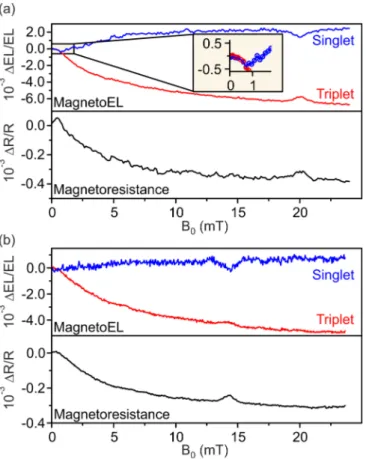
Department of Physics and Astronomy, University of Utah, Salt Lake City, Utah 84112, USA (Received 13 February 2017; revised manuscript received 25 April 2017; published 14 June 2017) We probe the interconversion of spin permutation symmetry of weakly bound electron-hole carrier pairs in an organic light-emitting diode by monitoring the changes in yield of recombinant species—singlet and triplet excitons—through fluorescence and phosphorescence, respectively. Spin mixing occurs by spin precession in local hyperfine fields and is suppressed by an external magnetic field, leading to an anticorrelation of fluorescence and phosphorescence yield, which follows the same functionality as magnetoresistance. A resonant radio-frequency field reverses this effect, enhancing spin mixing to raise the phosphorescence and lower the fluorescence. The experiment offers a direct simultaneous optical probe of the two interconverting spin states in the radical-pair mechanism, which features prominently in models of biological magnetoception.

DOI: 10.1103/PhysRevB.95.241201

Many molecular reactions exhibit strong magnetic-field effects at room temperature with regard to a specific reaction yield [1]. A physical picture put forward to explain some of the observations, the radical-pair mechanism [2], involves the modification by an external magnetic field of the precession of the spins of positive and negative charges in local hyperfine fields. Since such a radical—or electron-hole—pair is Coulom- bically bound, it can ultimately recombine, with the rate of recombination depending on the spin permutation symmetry of the pair. Many examples of the influence of magnetic fields on reaction yields based on such underlying pair processes have been reported, with measurable magnetic-field effects on scales as small as a few microtesla [3]. Such processes have also been invoked to explain the magnetoceptive abilities of some birds [3]. While the precise chemical and physiological origin of this ability remains subject to debate, the sensitivity of birds to magnetic fields was recently underlined by the demonstration of an influence of ambient electromagnetic noise on navigational ability [4]. The fact that extremely weak oscillatory fields of nanotesla amplitude [4] disrupt magnetoception suggests that spin-dependent reaction yields are influenced by very long spin-coherence times [5], possibly of 0.1 ms duration [6]. Although much of the research interest in spin-dependent radical-pair processes stems from such biological or model synthetic chemical phenomena, a crucial limitation of investigations is that it is only the reaction yield which is measured rather than the permutation symmetry of the spin pair [7–10]. Analogous spin-pair processes can also arise in the solid state, and have been invoked to explain magnetic resonance phenomena and magnetic-field effects in the conductivity of amorphous silicon [11] and molecular organic semiconductor films [12–18]. Such measurements are, in principle, sensitive down to the single-charge level. We recently reported a direct probe of electronic spin coherence in thin films of organic semiconductors incorporated in an organic

*

john.lupton@ur.de

light-emitting diode (OLED) configuration by measuring the magnetic resonance signature in device currents [19–21].

The appeal of OLEDs is that the entire operational principle is based on spin-dependent recombination of injected charges to yield light. Uncorrelated carrier pairs are generated in a spin- statistical ratio of 1:3 singlets to triplets. The two carrier-pair states have different recombination and dissociation rates, so that changes in spin statistics impact the number of free charges and hence conductivity. Time-resolved measurements under magnetic resonance conditions allow the direct quantification of spin-coherence times and reveal the dynamics of hyperfine interactions in the time domain [20]. Much of the evidence reported to date is consistent with magnetic-field effects and associated magnetic resonance phenomena arising from inter- mixing between singlet and triplet-pair states [22]. However, device current—like the reaction yield in spin chemistry—is ultimately a secondary integral quantity which reports on changes in spin-pair permutation symmetry without directly probing singlet or triplet pair population density. Magnetic resonance has, of course, been probed in organic molecules by monitoring the fluorescence of singlets [21–23] and the phosphorescence of triplet excited states [24–26]. However, there are no reports of simultaneous probing of the two radiative recombination channels which would be required to monitor interconversion between spin species. Here, we demonstrate how to directly probe the density of singlet and triplet pair products in an OLED by monitoring the radiative decay of the molecular excitons formed by pair recombination:

phosphorescence from the triplet and fluorescence from the singlet. Anticorrelated changes in the two luminescence chan- nels are observed under an external magnetic field and under magnetic resonance conditions, demonstrating that singlet pairs do indeed convert into triplets and vice versa.

Figure 1(a) illustrates the dominant electron-hole recombi-

nation channels in an OLED. Electrons and holes are injected

from opposite electrodes to form bound carrier pairs of singlet

or triplet spin configuration. Interconversion between the two

species can occur by driving one or both of the carrier spins

coherently under magnetic resonance conditions, or through

(2)

FIG. 1. Spectrally resolved optically detected magnetic reso- nance of a dual singlet-triplet emitting OLED. (a) Free injected charge carriers in the OLED attract Coulombically to form weakly coupled spin pairs. The overall spin permutation symmetry of the pair can oscillate between singlet and triplet configuration by either spin precession in the local hyperfine fields or by coherent driving in an external magnetically resonant radio-frequency driving field.

Ultimately, the carrier pairs either dissociate again or recombine to form tightly bound molecular singlet or triplet exciton species.

Singlets decay by fluorescence and triplets by phosphorescence.

(b) EL spectrum of an OLED made of PhLPPP, the structure of which is shown in (c), with R

1

: n-decyl, R

2

: n-hexyl. The EL is characterized by distinct fluorescence (singlet) and phosphorescence (triplet) features, which are separated spectrally by a combination of dichroic beam splitters and spectral filters. The spectral regions defined by the setup are indicated. (d) Illustration of the copper coplanar waveguide structure used for magnetic resonant excitation of the OLED charge-carrier states. The dimensions of the waveguide are h = 1.6 mm,t = 35 μm,s = 0.5 mm,w = 4 mm, with the FR-4 circuit-board substrate dielectric ε

s

= 4.5.

spin precession in the local hyperfine fields [27]. Typically, however, such precession is very slow, so that recombination mostly follows the spin-statistical limit [28]. The carrier pairs recombine into either singlet or triplet molecular excitons. In pure hydrocarbon compounds, luminescence is dominated by fluorescence from the singlet, with most triplet excitons under- going nonradiative decay [29]. In contrast, in organometallic complexes with strong singlet-triplet intersystem crossing in

the excited state, radiative recombination from the higher-lying singlet is suppressed so that phosphorescence dominates [30,31]. Few materials actually show dual singlet-triplet luminescence, and mostly this can only be identified under time-resolved detection at low temperatures [29–32]. Very few reports of distinguishable dual singlet-triplet luminescence exist for OLEDs [33–37]. Since we wish to probe small changes in singlet and triplet exciton yield due to either magnetic resonance or a static magnetic field, we require a material with strong dual electroluminescence (EL) in which singlet and triplet can be spectrally separated very clearly. A suitable such material is phenylene-substituted ladder-type poly(para-phenylene) [PhLPPP; Fig. 1(c)] which has a very small (<100 ppm) concentration of palladium atoms present in its hydrocarbon framework, left over as catalytic residue [28,38]. Both triplet and singlet excitons are highly mobile in this material [39], which has a high fluorescence quantum yield in the solid state. However, since triplet excitons are longer-lived than singlets, they more readily reach the palladium atoms where intersystem crossing occurs and radiative T

1

S

0

recombination becomes possible. In contrast, and unlike conventional organometallic compounds [31], since the concentration of palladium atoms is so low, excited-state intersystem crossing (S

1

T

1

) is barely modified by the heavy-element atom [40]. Figure 1(b) shows a typical EL spectrum of this material in an OLED of the struc- ture indium tin oxide/poly(styrene sulfonate)-doped poly(3,4- ethylenedioxythiophene) (PEDOT:PSS)/PhLPPP/Ba/Al. The device is encapsulated with epoxy and a glass cover slip.

Two distinct spectral features are seen: an emission peaking at 460 nm, followed by a pronounced vibronic progression, which arises from the singlet exciton transition; and a second feature at 590 nm of nearly identical shape from radiative decay of the triplet—phosphorescence. In pristine material, these two features are clearly separable, and the blue and red areas marked in the figure indicate the spectral regions defined by a dichroic beam splitter and color filters. Following oxidative stress of the material under prolonged device operation, however, a pronounced defect band appears between the fluorescence and phosphorescence at around 550 nm [38,40].

This defect, which arises from the formation of ketones on the ladder-type structure [41], can complicate spectral separation of singlet and triplet, as discussed below.

To provide a magnetic field, the OLED is mounted at the center of a Helmholtz coil and is driven by a Keithley 238 source-measure unit under constant current conditions.

EL is collected with an optical fiber in close proximity

to the pixel (1 × 3.2 mm

2

in size), passed through a beam

splitter, and recorded either on a charge-coupled device

spectrometer, or filtered spectrally for singlet and triplet, and

detected by two silicon photodiodes coupled to low-noise

amplifiers (Femto OE-200-SI). To drive magnetic resonance,

the OLED is mounted on the coplanar waveguide structure

[42] shown in panel (d) with the cathode side of the device

placed on the waveguide. Radio-frequency (rf) radiation is

generated by an HP 83732A high-frequency source and a

20 W Mini-Circuits ZHL-20W-13+ amplifier and fed into

the waveguide, which is terminated by a 50 load. From

numerical calculations, we estimate that an rf magnetic field

of approximately 100–180 μT amplitude (i.e. the B

1

field)

(3)

FIG. 2. Spectrally resolved steady-state magnetoelectrolumines- cence and magnetoresistance under magnetic resonant excitation of charge carrier spins. Data points show the average of up and down sweeps in a magnetic field. (a) The magnetoresistance correlates directly with an increase in fluorescence and a decrease in phosphorescence, which is reversed under magnetic resonance at 20 mT (560 MHz). The inset shows the region of the ultrasmall magnetic-field effect below 1 mT. In this region, singlet and triplet yield follow exactly the same functionality, with the increase in magnetoresistance correlating with a decrease in luminescence.

Individual measurement data points are shown enlarged for clarity.

(b) A second OLED device with the resonance feature at 14.3 mT (400 MHz). In this example a weak EL contribution from the keto defect limits the quantitative match between singlet enhancement and triplet quenching.

is incident on the OLED pixel. The measurement procedure allows for simultaneous recording of magnetoresistance and magnetoelectroluminescence, with and without an rf field applied. Since the overall sensitivity to changes in EL is of order 90 ppm, we can detect EL and resistance in direct current [43], without the need of modulation and lock-in procedures, providing truly steady-state measurements and adiabatic field-sweep conditions.

Figure 2 shows two examples of simultaneous magnetore- sistance and spectrally resolved magnetoelectroluminescence, with incident rf fields at frequencies of 560 MHz (a) and 400 MHz (b). The data result from a single up and down sweep of magnetic field which takes a total of 10 min. The magnetoresistance shows an initial increase with magnetic field below 1 mT, a phenomenon referred to as the “ultrasmall magnetic-field effect” [44], and subsequently decreases. A

popular model of the magnetoresistance invokes the precession of electron and hole spins in the local hyperfine fields [45,46], which leads to mixing between singlet and triplet multiplicity of the carrier pair [27]. An external magnetic field suppresses this mixing. Various forms of this process have been discussed in the literature with regard to OLEDs, and our data appear to follow the established functionalities [47]. A magnetic resonant driving field in turn counteracts this suppression effect by promoting spin mixing, leading to an overall increase of magnetoresistance on resonance, which is clearly seen in both plots. With our dual emitter, the effect of spin mixing in the electron-hole pair exciton precursor state can be visualized directly. An increase in fluorescence with rising magnetic field matches near quantitatively the suppression of phosphorescence. Under magnetic resonance, the effect is partially reversed. This reversal is limited because the resonance line is broader than the driving-field amplitude, which defines the spectral width of the resonant excitation.

Charge-carrier spin-resonance linewidths are governed by local hyperfine fields, which are determined by the abundant hydrogen. The limitation of the excitation power which can be applied will therefore ultimately limit the size of the subensemble of charge carriers which can be brought into magnetic resonance simultaneously [42]. Nevertheless, the data demonstrate directly that magnetoresistance, and the associated magnetoelectroluminescence, originates from interconversion between carrier-pair spin species and not, for example, from changes with magnetic field in carrier-pair recombination or dissociation rates [22]. This relation has previously only been considered for the singlet [44] or the triplet EL channel [48] individually, but not in both within one material at the same time. As we discuss below, the fact that in the example in panel (b) the relative static magnetic-field effect appears slightly weaker in the singlet than in the triplet channel, but the resonance feature is somewhat less pronounced in the triplet, can be attributed to the influence of the parasitic spectral defect band [38]. This emission is of singlet character but overlaps both singlet and triplet excitonic features and therefore introduces random shunt rates to the detected singlet and triplet transition rates. We stress that both singlet and triplet EL correspond to spin-1/2 species, i.e., the individual carrier spins [43]. This phosphorescence-detected magnetic resonance should not be confused with the spin-1 detection of the triplet exciton resonance, which has been reported previously in molecular emitters but probes spin conversion within the triplet manifold rather than singlet-triplet transitions [24,25]. We also note that in our early measurements of magnetoelectroluminescence on PhLPPP we were not able to resolve these very subtle spectral differences due to limitations in sensitivity [28].

The data in Fig. 2(a) reveal a further subtle feature in the

ultralow magnetic-field effect. The inset shows a close-up of

the magnetoelectroluminescence in this region. Singlet and

triplet exciton yields follow each other exactly for fields below

1 mT before diverging: the ultrasmall magnetic-field effect

[44,45] with its characteristic inversion of field dependency

only arises in the resistance and in the singlet channel, but

not in the triplet. A similar trend is apparent in panel (b),

although with reduced significance due to increased noise. This

observation implies that the ultrasmall magnetic-field effect

(4)

FIG. 3. Fluorescence and phosphorescence magnetic resonance features measured under lock-in detection to remove the steady-state magnetoelectroluminescence background. (a) The resonance curves are described by the same sum of two Gaussians with standard deviations of 0.38 and 0.72 mT, indicated by the dashed green lines.

The resonance amplitudes of singlet and triplet do not match perfectly because a third luminescence species, the broad keto-defect emission band [38], contributes to EL after prolonged device operation.

This defect band leads to a sign inversion of the phosphorescence resonance (red squares) with time, as plotted in (b).

differs fundamentally from the effect on larger magnetic-field scales. This effect has been tentatively attributed to the zero- field splitting of the carrier pair arising from either anisotropic hyperfine interactions [44] or dipolar spin-spin coupling [49].

The zero-field splitting lifts the degeneracy of the three triplet states and therefore changes mixing rates within the triplet manifold. We propose that the observed congruence of ultrasmall magnetic-field effect for singlet and triplet could arise from different transition rates of the three triplet states of the carrier pair, which define the triplet symmetry of the exciton—T

+

,T

or T

0

—to the singlet ground state S

0

. In phosphorescent emitters, the superposition triplet T

0

couples most effectively to the superposition state S

0

. The yield of this superposition triplet species therefore appears to not be affected by the field on very small magnetic-field scales of the order of the zero-field splitting of the carrier pairs.

Finally, we demonstrate the data quality achievable by em- ploying lock-in detection to study the resonance spectra more closely, demodulating against a square-wave full amplitude modulation of the rf source at 23 Hz. Figure 3(a) shows spectra measured at three different frequencies. All resonances are accurately described by the superposition of two Gaussians with standard deviations of 0.38 and 0.72 mT. These two Gaussians correspond to the hyperfine-field distributions expe- rienced by the electron and hole spins [21,43] and are shown in green in the rightmost singlet resonance curve for illustration

purposes. However, in a perfect pair process, the areas of both Gaussian peaks must be identical, since resonant excitation of electron and hole each map onto the same observable of differential current or EL [21]. This is not the case here, and evidently even the overall singlet and triplet resonance peaks have different areas. This discrepancy arises because of the fact that a spectrally broad defect band is always superimposed on the narrow singlet and triplet EL peaks and cannot be separated fully by spectral filtering [38]. The influence of this feature depends on material quality and device age. Figure 3(b) plots the change with time of the differential EL signal on resonance for singlet and triplet spectral features. Only at the outset of the measurement do singlet and triplet actually have the opposite resonance signs. After 2 h of continuous operation, the signal associated with the phosphorescence resonance vanishes entirely for a short time before both resonance features become negative. The amplitude of the resonance from the red part of the spectrum, the superposition of phosphorescence and keto defect, changes faster with time than the blue part of the spectrum, the superposition of singlet fluorescence and defect emission. The inversion of resonance sign with prolonged device operation time arises because the contribution of the broad keto-defect emission band to the EL spectrum increases with time and the defect itself acts as a charge-carrier trap [50] with a distinct magnetic-field sensitivity [51]. This observation of an inversion of resonance amplitude with time contains further information on material- specific recombination kinetics: it demonstrates that the broad keto-defect band has singlet character rather than mirroring the pure triplet species. This is an interesting conclusion since delayed luminescence experiments have demonstrated that triplet excitons can feed into the defect band, which is common to many polycyclic aromatic hydrocarbons, on time scales of seconds [32]. The keto defect can therefore harvest long-lived triplet excitons [32], but when carriers recombine directly on the defect it acts as a pure singlet fluorophore.

OLEDs offer highly sensitive and versatile conditions to probe spin-dependent recombination processes and their sen- sitive responses to magnetic fields. The experiments presented here were carried out under dc OLED operation conditions and time-averaged photodetection. With gated luminescence detection we expect to be able to separate fluorescence and phosphorescence signatures even more clearly. In combination with pulsed magnetic resonance excitation [20] it should be possible to directly probe, in the time domain, coherent singlet- triplet interconversion in the change in OLED luminescence color. Such experiments would constitute alternatives to conventional singlet-triplet qubit systems based on coupled quantum dots [52–54] which are considered as building blocks for quantum information processing architectures.

The authors are indebted to Dr. Kipp van Schooten and Dr. Hans Malissa for helpful discussions and to the DFG for funding through SFB 689. C.B. acknowledges support through the US Department of Energy, Office of Basic Energy Sciences, Division of Materials Sciences and Engineering under Award No. DESC0000909.

[1] U. E. Steiner and T. Ulrich, Chem. Rev. 89, 51 (1989).

[2] T. Ritz, S. Adem, and K. Schulten, Biophys. J. 78, 707

(2000).

(5)

[3] K. Maeda, K. B. Henbest, F. Cintolesi, I. Kuprov, C. T. Rodgers, P. A. Liddell, D. Gust, C. R. Timmel, and P. J. Hore, Nature (London) 453, 387 (2008).

[4] S. Engels, N. L. Schneider, N. Lefeldt, C. M. Hein, M. Zapka, A. Michalik, D. Elbers, A. Kittel, P. J. Hore, and H. Mouritsen, Nature 509, 353 (2014).

[5] J. M. Cai and M. B. Plenio, Phys. Rev. Lett. 111, 230503 (2013).

[6] E. M. Gauger, E. Rieper, J. J. L. Morton, S. C. Benjamin, and V. Vedral, Phys. Rev. Lett. 106, 040503 (2011).

[7] C. A. Dodson, C. J. Wedge, M. Murakami, K. Maeda, M. I.

Wallace, and P. J. Hore, Chem. Commun. 51, 8023 (2015).

[8] J. R. Woodward, C. R. Timmel, K. A. McLauchlan, and P. J.

Hore, Phys. Rev. Lett. 87, 077602 (2001).

[9] K. B. Henbest, P. Kukura, C. T. Rodgers, P. J. Hore, and C. R.

Timmel, J. Am. Chem. Soc. 126, 8102 (2004).

[10] T. Ritz, P. Thalau, J. B. Phillips, R. Wiltschko, and W. Wiltschko, Nature (London) 429, 177 (2004).

[11] D. Kaplan, I. Solomon, and N. F. Mott, J. Phys. Lett. 39, L51 (1978).

[12] E. L. Frankevich, I. A. Sokolik, D. I. Kadyrov, and V. M.

Kobryanskii, JETP Lett. 36, 486 (1982).

[13] V. Dyakonov, G. Rosler, M. Schwoerer, and E. L. Frankevich, Phys. Rev. B 56, 3852 (1997).

[14] K. Morgan and R. Pethig, Nature (London) 213, 900 (1967).

[15] P. Desai, P. Shakya, T. Kreouzis, W. P. Gillin, N. A. Morley, and M. R. J. Gibbs, Phys. Rev. B 75, 094423 (2007).

[16] T. L. Francis, O. Mermer, G. Veeraraghavan, and M.

Wohlgenannt, New J. Phys. 6, 185 (2004).

[17] J. Kalinowski, M. Cocchi, D. Virgili, P. Di Marco, and V. Fattori, Chem. Phys. Lett. 380, 710 (2003).

[18] J. M. Lupton and C. Boehme, Nat. Mater. 7, 598 (2008).

[19] W. J. Baker, T. L. Keevers, J. M. Lupton, D. R. McCamey, and C. Boehme, Phys. Rev. Lett. 108, 267601 (2012).

[20] H. Malissa, M. Kavand, D. P. Waters, K. J. van Schooten, P. L.

Burn, Z. V. Vardeny, B. Saam, J. M. Lupton, and C. Boehme, Science 345, 1487 (2014).

[21] M. Kavand, D. Baird, K. van Schooten, H. Malissa, J. M. Lupton, and C. Boehme, Phys. Rev. B 94, 075209 (2016).

[22] S. P. Kersten, A. J. Schellekens, B. Koopmans, and P. A. Bobbert, Phys. Rev. Lett. 106, 197402 (2011).

[23] L. S. Swanson, J. Shinar, A. R. Brown, D. D. C. Bradley, R. H.

Friend, P. L. Burn, A. Kraft, and A. B. Holmes, Phys. Rev. B 46, 15072 (1992).

[24] D. A. Antheunis, J. Schmidt, and J. H. van der Waals, Chem.

Phys. Lett. 6, 255 (1970).

[25] J. Schmidt, W. G. van Dorp, and J. H. van der Waals, Chem.

Phys. Lett. 8, 345 (1971).

[26] G. Li, J. Shinar, and G. E. Jabbour, Phys. Rev. B 71, 235211 (2005).

[27] R. C. Roundy and M. E. Raikh, Phys. Rev. B 88, 125206 (2013).

[28] M. Reufer, M. J. Walter, P. G. Lagoudakis, B. Hummel, J. S.

Kolb, H. G. Roskos, U. Scherf, and J. M. Lupton, Nat. Mater. 4, 340 (2005).

[29] A. Köhler, J. S. Wilson, and R. H. Friend, Adv. Eng. Mater. 4, 453 (2002).

[30] M. A. Baldo, D. F. O’Brien, Y. You, A. Shoustikov, S. Sibley, M. E. Thompson, and S. R. Forrest, Nature (London) 395, 151 (1998).

[31] S. Haneder, E. Da Como, J. Feldmann, J. M. Lupton, C.

Lennartz, P. Erk, E. Fuchs, O. Molt, I. Muenster, C. Schildknecht et al., Adv. Mater. 20, 3325 (2008).

[32] D. Chaudhuri, H. Wettach, K. J. van Schooten, S. Liu, E.

Sigmund, S. Höger, and J. M. Lupton, Angew. Chem., Int. Ed.

49, 7714 (2010).

[33] S. Hoshino and H. Suzuki, Appl. Phys. Lett. 69, 224 (1996).

[34] C. Garditz, A. G. Muckl, and M. Cölle, J. Appl. Phys. 98, 104507 (2005).

[35] D. Chaudhuri, E. Sigmund, A. Meyer, L. Roeck, P. Klemm, S.

Lautenschlager, A. Schmid, S. R. Yost, T. Van Voorhis, S. Bange et al., Angew. Chem. Int. Ed. 52, 13449 (2013).

[36] W. Ratzke, L. Schmitt, H. Matsuoka, C. Bannwarth, M. Retegan, S. Bange, P. Klemm, F. Neese, S. Grimme, O. Schiemann et al., J. Phys. Chem. Lett. 7, 4802 (2016).

[37] J. M. Lupton and J. Klein, Chem. Phys. Lett. 363, 204 (2002).

[38] J. M. Lupton, A. Pogantsch, T. Piok, E. J. W. List, S. Patil, and U. Scherf, Phys. Rev. Lett. 89, 167401 (2002).

[39] M. Reufer, P. G. Lagoudakis, M. J. Walter, J. M. Lupton, J. Feldmann, and U. Scherf, Phys. Rev. B 74, 241201 (2006).

[40] S. Bange, U. Scherf, and J. M. Lupton, J. Am. Chem. Soc. 134, 1946 (2012).

[41] L. Romaner, G. Heimel, H. Wiesenhofer, P. Scandiucci de Freitas, U. Scherf, J.-L. Brédas, E. Zojer, and E. J. W. List, Chem. Mater. 16, 4667 (2004).

[42] W. J. Baker, K. Ambal, D. P. Waters, R. Baarda, H. Morishita, K.

van Schooten, D. R. McCamey, J. M. Lupton, and C. Boehme, Nat. Commun. 3, 898 (2012).

[43] D. P. Waters, G. Joshi, M. Kavand, M. E. Limes, H. Malissa, P. L. Burn, J. M. Lupton, and C. Boehme, Nat. Phys. 11, 910 (2015).

[44] T. D. Nguyen, B. R. Gautam, E. Ehrenfreund, and Z. V. Vardeny, Phys. Rev. Lett. 105, 166804 (2010).

[45] T. D. Nguyen, G. Hukic-Markosian, F. J. Wang, L. Wojcik, X.

G. Li, E. Ehrenfreund, and Z. V. Vardeny, Nat. Mater. 9, 345 (2010).

[46] Y. Sheng, D. T. Nguyen, G. Veeraraghavan, O. Mermer, M.

Wohlgenannt, S. Qiu, and U. Scherf, Phys. Rev. B 74, 045213 (2006).

[47] A. J. Schellekens, W. Wagemans, S. P. Kersten, P. A. Bobbert, and B. Koopmans, Phys. Rev. B 84, 075204 (2011).

[48] K. Xu, D. Z. Yang, Y. B. Zhao, J. S. Chen, and D. G. Ma, Org. Electron. 15, 590 (2014).

[49] K. J. van Schooten, D. L. Baird, M. E. Limes, J. M. Lupton, and C. Boehme, Nat. Commun. 6, 6688 (2015).

[50] J. M. Lupton and J. Klein, Phys. Rev. B 65, 193202 (2002).

[51] M. Cox, M. H. A. Wijnen, G. A. H. Wetzelaer, M. Kemerink, P. W. M. Blom, and B. Koopmans, Phys. Rev. B 90, 155205 (2014).

[52] J. R. Petta, A. C. Johnson, J. M. Taylor, E. A. Laird, A. Yacoby, M. D. Lukin, C. M. Marcus, M. P. Hanson, and A. C. Gossard, Science 309, 2180 (2005).

[53] F. H. L. Koppens, J. A. Folk, J. M. Elzerman, R. Hanson, L. H.

W. van Beveren, I. T. Vink, H. P. Tranitz, W. Wegscheider, L. P.

Kouwenhoven, and L. M. K. Vandersypen, Science 309, 1346 (2005).

[54] B. M. Maune, M. G. Borselli, B. Huang, T. D. Ladd, P. W.

Deelman, K. S. Holabird, A. A. Kiselev, I. Alvarado-Rodriguez,

R. S. Ross, A. E. Schmitz et al., Nature 481, 344 (2012).

Abbildung

FIG. 1. Spectrally resolved optically detected magnetic reso- reso-nance of a dual singlet-triplet emitting OLED
FIG. 2. Spectrally resolved steady-state magnetoelectrolumines- magnetoelectrolumines-cence and magnetoresistance under magnetic resonant excitation of charge carrier spins
FIG. 3. Fluorescence and phosphorescence magnetic resonance features measured under lock-in detection to remove the steady-state magnetoelectroluminescence background

Referenzen

ÄHNLICHE DOKUMENTE

We perform numerical simulations of the stochastic Landau-Lifshitz-Gilbert equation of motion for an atomistic spin model and calculate the magnon accumulation in linear

In our case, the equivalence of the structure factors for orbit 1, k 1 , and orbit 2, k 2 , with the basis functions listed in Table III can be easily seen if we consider the

In: Progress in Nuclear Magnetic Resonance Spectroscopy 38.1 (2001), pp. Sidles et al. “Magnetic Resonance Force Microscopy”. Sidles and Daniel Rugar. “Signal-to-Noise Ratios

These mag- netic thin films with an easy axis of magnetisation perpendicular to the surface have attracted particular attention due to their potential advantage in high-

By using NV centers in diamond as a spin sensor, we perform the Qdyne measurement technique, which resembles the quadrature detec- tion done in NMR spectroscopy, to detect

a Present address: Radiological Clinic, University of T¨ubingen, Germany Reprint requests to Prof. All integrals can be computed analytically and are derived from one single

In field emission experiments with spin polarized electrons a magnetic field is superposed on the electric emission field to define the preferred spin direction.The m

For pulse durations much shorter than the timescale of the elec- tron spin evolution, the re-encounter probability can be described by a peaked distribution p re ðtÞ / dðt s re Þ,