β-trace protein (prostaglandin D synthase) - a stable and reliable protein in perilymph
β-trace Protein (Prostaglandin D Synthase) - ein stabiles und signifikantes Protein in Perilymphe
Abstract
Objective: Beta-trace protein (β-TP) has been analysed in human cerebrospinal fluid (CSF) and other body fluids. Beta-trace protein is a
Olaf Michel
1Stephan Bamborschke
2very sensitive and specific clinical marker and can confirm reliably the
presence of CSF in patients with a suspected CSF leakage.
Marko Nekic
2Gregor Bachmann
3Design:Perilymph specimens from the scala vestibuli (n=10) and from the lateral semicircular canal (n=4) were taken from patients undergoing stapedotomy or surgery for acoustic neuroma. During post-mortem ex-
1 University of Cologne, Department of aminations perilymph specimens from the scala vestibuli (n=70), the
scala tympani (n=11), endolymph specimens (n=21) and CSF specimens
Otorhinolaryngology, Head (n=17) were obtained. All specimens were analyzed by a one-dimension-
al immunoelectrophoresis using a polyclonal, monospecific antibody. and Neck Surgery, Cologne, Germany
Results:Specimens from live surgery showed a mean concentration of
51.5 ± 48.9 mg/l β-TP in scala vestibuli perilymph. Specimens from 2 University of Cologne, Department of Neurology, Cologne, Germany post-mortem examinations revealed a mean concentration of 49.1 ±
17.7 mg/l in CSF, 71.9 ± 29.3 mg/l in perilymph and 68.0 ± 21.7 mg/l
3 University of Tromsoe, Department of in endolymph. There was no evidence of a circadian alteration of β-TP
in CSF or inner ear fluids.
Otorhinolaryngology, Tromsoe, Norway Conclusions:Our results demonstrated clearly that β-TP is contained
in human perilymph and endolymph. This is the first published data that point out the aptitude of the β-TP-test in verifying traces of perilymph, a valuable diagnostic tool for the existence of perilymphatic leaks.
Keywords:beta-trace protein, perilymph, endolymph, cerebrospinal fluid, prostaglandin D synthase
Zusammenfassung
Ziel:Beta-trace Protein (β-TP) wurde in der menschlichen Hirnflüssigkeit (Liquor) und in anderen Körperflüssigkeiten nachgewiesen. Beta-trace Protein ist ein sehr sensitiver und spezifischer klinischer Marker und kann die Anwesenheit von Liquor cerebrospinalis in Patienten mit dem Verdacht auf ein Liquorleck sicher nachweisen.
Methodik:Perilymph-Probenfrom aus der Scala vestibuli (n=10) und vom lateralen Bogengang (n=4) wurden Patienten abgenommen, die stapedotomiert oder wegen eines Akustikusneurinoms labyrinthekto- miert wurden. Auch wurden post mortem humane Perilymph-Probenfrom aus der Scala vestibuli (n=70), der Scala tympani (n=11), Endolymphe (n=21) und Liquorproben (n=17) entnommen. Alle Proben wurden mit einer eindimensionalen Immunoelektrophorese und einem polyclonalen, monospezifischen Antikörper untersucht.
Ergebnisse:Die Proben, die während der chirurgischen Eingriffe gewon- nen wurden, wiesen eine mittlere Konzentration von 51,5 ± 48,9 mg/l β-TP in der Perilymphe der Scala vestibuli auf. Proben, die post mortem gewonnen wurden, wiesen eine mittlere Konzentration von 49,1 ± 17,7 mg/l im Liquor, 71,9 ± 29,3 mg/l in der Perilymphe und 68,0 ± 21,7
mg/l in der Endolymphe auf. Ein Hinweis auf einen zirkadianen Rhyth- mus wurde weder im Liquor noch in der Perilymphe gefunden.
Schlussfolgerung:. Unsere Ergebnisse weisen nach, dass β-TP ohne Ausnahme in der menschlichen Perilymphe und Endolymphe in hoher Konzentration vorkommt. Dies sind die ersten veröffentlichten Daten, die zeigen, dass der β-TP-Test geeignet ist, auch Spuren von Perilymphe nachzuweisen. Damit steht erstmals ein klinischer Test zum sicheren Nachweis von Perilymphfisteln zur Verfügung.
Schlüsselwörter:Beta-trace Protein, Nachweis in Perilymphe, Nachweis von Perilymphfisteln
Introduction
The incidence of perilymphatic fistula has been subject to controversial discussions since they were first de- scribed by Simmons [1]. The controversy arose due to difficulties in diagnosing a perilymph fistula at tympano- tomy. Unless there is profuse gushing of fluid, there is no reliable, objective, intraoperative indicator of a perilymphatic fistula [2], [3]. The search of specific protein markers [4], [5] or the intravenous application of fluores- cein [6] has so far not been successful. Also β2-transferrin was reported to be unique in human perilymph [7], but in a study of 22 patients more than half of them had known perilymph leaks β2-transferrin was falsely negative [8].
Since Felgenhauer [9] demonstrated beta-trace protein (β-TP) to be after albumin the second most abundant protein in human cerebrospinal fluid (CSF) it has become a reliable marker for CSF leaks.
β-TP is completely different to β2-transferrin. With a mo- lecular mass of 23.500 Dalton β-TP is identical to gluta- thion-independent prostaglandin D synthase [10], which seems to play an important role in regulating the sleep and waking state [11]. So far, the highest concentrations of β-TP were found in CSF and seminal fluid [12]. In ser- um, the level of β-TP is below the detection limit (< 5 mg/l) of rocket immunoelectrophoresis. Only participants with severe renal filtration barrier pathology would show a detectable concentration of β-TP.
With the more sensitive immunofluorometric assay, which is based on an enzymatic amplified time-resolved fluoro- metry, the mean levels of β-TP were 0.3 mg/L in human serum and 17 mg/L in human CSF and the detection limit was 5 mg/L [13]. β-TP has the highest CSF/serum ratio of all CSF specific proteins.
β-TP was found in aqueous humor, sperm and urine, too [14]. β-TP was detected in homogenate of the rat cochlea [15] and in the lateral wall of the cochlea of the Mongolian gerbil [16].
Based upon our experience with β-TP in the diagnosis in CSF-leaks, the purpose of this study was to investigate whether β-TP occurs in human inner ear fluids and if positive, in which concentrations compared to CSF.
Subjects and methods
To clarify the occurrence of β-TP we conducted analysis of perilymphatic fluid obtained from post-mortem exam- inations and from inner ear surgery. In post-mortem ex- aminations 81 perilymph, 21 endolymph specimens and 17 CSF specimens were collected.
14 perilymph samples were sampled from patients under- going stapedotomy or labyrinthectomy at the Department of Otorhinolaryngology, University of Cologne. The perilymph samples were taken from the lateral semicircu- lar canal (n=4) or from the oval window (n=10). Also pooled CSF taken by lumbar puncture from 125 patients with non-inflammatory diseases of the CNS was studied.
35 serum samples were collected from patients without renal disease. Subjects with a renal insufficiency or acute glomerulonephritis were not enrolled in this study.
Technique of sampling in post-mortem examination
After partial mastoidectomy, removal of the posterior ear canal and the tympanic membrane including malleus and incus, the stapes was exposed and the footplate carefully removed. Samples of perilymph were taken from the scala vestibuli with precision capillary bores (Wiretrol 1-5 μl, Drummond®, Broomall, USA - Pennsylvania 19008) under microscopic view (Figure 1). To prevent contamination with CSF, the samples were taken without active aspira- tion only by the capillary power. The volume was limited to a minimum of 4 μl and a maximum of 5 μl. The capillary bores were placed near the upper edge of the vestibular window, the membranous labyrinth was initially not touched. After the sampling of a second and third perilymph specimen of 5 μl, the utricle was punctured and endolymph was obtained. If feasible, a second en- dolymph specimen of 5 μl was taken. CSF specimens were obtained from the third ventricle. The period between death and autopsy as well as the cause of death were documented.
Figure 1: Precision capillary bore (1-5 µl), placed at the edge of the oval window under microscopic view (Magn.: 12 x), FN:
facial nerve, TN: tympanic nerve, OW: oval window
Technique of sampling at stapes surgery
14 perilymph samples were obtained from patients un- dergoing stapedotomy or labyrinthectomy. The perilymph samples were taken from the oval window or from the lateral semicircular canal using sterilized precision bore.
The volume of the fluid specimen was restricted to a maximum of 5 μl. Because of the high specificity of β-TP, small blood contamination or small dilution with local anesthetic could be ignored.
In addition to the post-mortem CSF samples, pooled CSF taken by lumbar puncture from 125 patients with non- inflammatory diseases of the CNS was studied. 35 serum samples were collected from patients without renal dis- ease.
Rocket immunoelectrophoresis
All specimens were investigated by a rocket immunoelec- trophoresis using a monospecific, polyclonal antibody, used in rabbits [9]. Investigation took place either imme- diately or after storage of maximum 7 days at - 40°C.
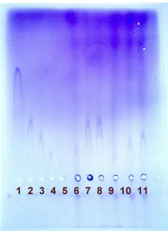
The standard electroimmunoassay as described by Laurrell [16] was performed using a gel composition of 1.5% Litex agarose (Litex, Denmark), 0.02 M barbital buffer pH 8.6, 1% antiserum, 1% polyethylenglycol 6000 (Merck, Darmstadt, Germany) and an electrophoresis buffer of 0.02 M barbital buffer, pH 8.6. Electrophoresis was performed during 3 hours at 18° C at a constant voltage of 250 V. Semiquantitative measurements were performed using a standard row with serial dilution of purified β-TP isolated from pooled human CSF (Figure 2) as described by Felgenhauer et al. [9].
Figure 2: Rocket electrophoresis with standard row on the left (1-5). Specimen from surgery (6 and 9-10): scala vestibuli perilymph. Specimen from post-mortem examination (7-8):
scala vestibuli perilymph. Specimen from post-mortem examination (11): endolymph.
Results
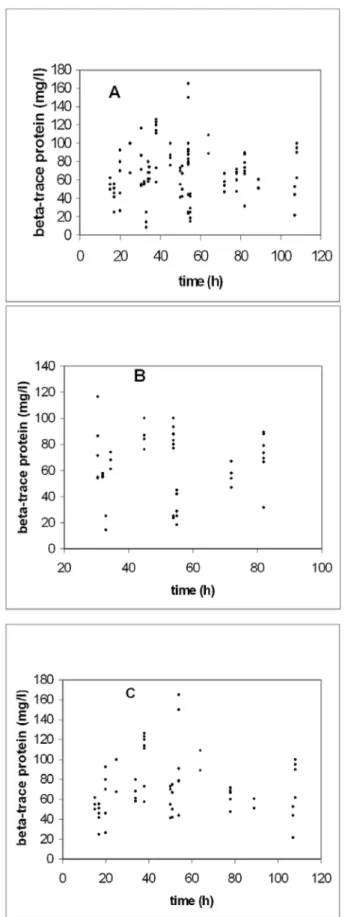
By immunoelectrophoresis the perilymph, endolymph and CSF specimens from post-mortem examination precipit- ated clearly (Figure 2). In correlation with the time of death, there was no clear evidence for a circadian altera- tion (Figure 3). Up to 117 hours post mortem a significant change of the β-TP concentration in CSF and inner ear fluids was not observed in males or females (Figure 4A, B, C ). The values ranged between 8.5 mg/l and 165 mg/l.
Figure 3: Correlation of β-TP concentration in human inner ear fluids and time of death. Scatterplot of β-TP concentration in
human perilymph and endolymphvs. time post-mortem.
Maximum between 10:00 and 12:00 p.m.
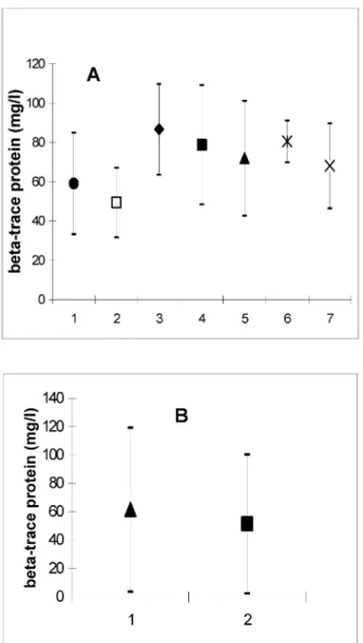
As depicted in Figure 5A , the mean concentration of β- TP was 49.1 ± 17.7 mg/l in CSF and 59.0 ± 25.8 mg/l in perilymph (scala tympani). In the first drawn specimen of scala vestibuli perilymph β-TP was found in a concen- tration of 71.9 ± 29.3 mg/l. In the second specimen from the scala vestibuli 78.7 ± 30.4 mg/l and in the third specimen 86.4 ± 23.1 mg/l were detected. In endolymph, samples the values were 68.0 ± 21.7 mg/l (first speci- men) and 80.5 ± 10.6 mg/l (second specimen). The first perilymph specimen from the labyrinthine vestibule showed a lower concentration compared to the second or third specimens, which originated from deeper parts of the scala vestibuli. The β-TP concentration in the second endolymph specimens was also higher in compar- ison with the values from the first specimens.
The samples obtained during inner ear surgery were all positive for β-TP. The specimens revealed a β-TP-concen- tration of 61.5 ± 57.8 mg/l in perilymph from the lateral semicircular canal and 51.4 ± 48.9 mg/l in perilymph from the scala vestibuli (Figure 5B ).
In pooled CSF taken by lumbar puncture from 125 pa- tients with non-inflammatory diseases, a β-TP-concentra- tion of 28.4 mg/l was determined. All serum samples were negative for β-TP.
Figure 4: A: Scatterplot of time after deathvs. β-TP concentrations (all specimens);
B: Scatterplot of time after deathvs. β-TP concentrations in males;
C: Scatterplot of time after deathvs. β-TP concentrations in females.
Figure 5: A: β-TP concentrations in CSF and different inner ear compartments from post-mortem examinations. 1 = scala tympani perilymph (59.0 ± 25.8; n=11); 2 = III. ventricle CSF
(49.1 ± 17.7; n=17); 3 = third scala vestibuli perilymph specimen (86.6 ± 23.1; n=7); 4 = second scala vestibuli perilymph specimen (78.7 ± 30.3; n=27); 5 = first scala vestibuli perilymph specimen (71.9 ± 29.2; n=36); 6 = second endolymph specimen (80.5 ± 10.6; n=2); 7 = first endolymph
specimen (68.0 ± 21.6; n=19);
B: β-TP concentrations in different inner ear compartments from surgical procedures. 1 = perilymph from the lateral semicircular canal (61.5 ± 57.7; n=4); 2 = scala vestibuli perilymph from the oval window (51.4 ± 48.9; n=10)
Discussion
The issue of whether perilymphatic fistula is the cause of hearing loss remains enigmatic unless a specific marker exists for reliable proof. With β-TP, such a specific marker could exist.
In human CSF, the occurrence and the concentration of β-TP have been investigated by several authors and were re-evaluated in this study (Table 1). β-TP has become a reliable indicator for CSF-leak with advantages over β2- transferrin. Although it is detectable in perilymph [17],
clinical marker for perilymph in the operative setting be- cause of its little sensitivity with 29% positive results [8].
This is partly due to the property of this protein as a brain- specific variant of transferrin that lacks neuraminic acid.
It can be distinguished from serum transferrin by electro- phoretic procedures only.
Our study now makes obvious that β-TP is detectable in inner ear fluids in even higher concentration compared to CSF. Nevertheless, our results are the first to quantify β-TP in inner ear fluids. Tachibana and coworkers [15]
described prostaglandin D synthase (PGD-S) activity in rat inner ear homogenate. With this method it is not possible to distinct where predominantly PGD-S could be found in the inner ear.
The analysis of our entire specimen revealed a high inter- individual variation between 8.5 and 165 mg/l, which cannot yet be explained due to the unknown physiological role of β-TP. With the upcoming new nephelometryic assay it will be more feasable in the future to analyse β-TP under various conditions. Nevertheless, β-TP proved to be a very stable protein. Until 117 hours after sampling, no signi- ficant variation in the β-TP concentration in the diverse samples was detected. The sensitivity was 100% in n=81 perilymph and n=21 endolymph samples. The concentra- tion of β-TP showed no significant divergence comparing both inner ear fluids.
We did not find clear evidence for a circadian alteration of the β-TP-level in inner ear fluids as it could be assumed from its role in the sleep/awake rhythm and as detected in CSF [18]. The highest concentrations were found when death had occurred around midnight. But a high interin- dividual variation and not excludable post-mortem changes hindered a statistical proof for a circadian rhythm in our series. Nevertheless, the high interindividual vari- ation as observed cannot be.
By this clear confirmation for the occurrence of β-TP in inner ear fluids and particularly in perilymph, assessment for clinical use is feasible without circumstantial and bi- asing handling, which has been described for the β2- transferrin test [19].
This is the first published data that point out the aptitude of the β-TP-test in verifying traces of perilymph in the middle ear, a valuable diagnostic tool for the existence of perilymphatic leaks as we have already shown in two cases of an vestibulocochlear disorder [20]. Nowadays, an immediate intraoperative check for β-TP is possible by laser-nephelometry, which takes only 20 minutes de- tection time [21].
Table 1: β-TP concentrations in human CSF
References
1. Simmons FB. Theory of membrane breaks in sudden hearing loss. Arch Otolaryngol. 1968;88(1):41-8.
2. Vartiainen E, Nuutinen J, Karjalainen S, Nykanen K. Perilymph fistula--a diagnostic dilemma. J Laryngol Otol. 1991;105(4):270- 3.
3. Shea JJ. The myth of spontaneous perilymph fistula [editorial]
[see comments]. Otolaryngol Head Neck Surg. 1992;107(5):613- 6.
4. Schweitzer VG, Woodson BT, Mawhinney TD, Rarey KE, Bauman MJ, Raymer SL, Peterson. Free amino acid analysis of guinea pig perilymph: a possible clinical assay for the PLF enigma?
Otolaryngol Head Neck Surg. 1990;103(6):981-5.
5. Thalmann I, Kohut RI, Ryu J, Comegys TH, Senarita M, Thalmann R. Protein profile of human perilymph: in search of markers for the diagnosis of perilymph fistula and other inner ear disease.
Otolaryngol Head Neck Surg. 1994;111(3 Pt 1):273-80.
6. Poe DS, Gadre AK, Rebeiz EE, Pankratov MM. Intravenous fluorescein for detection of perilymphatic fistulas. Am J Otol.
1993;14(1):51-5.
7. Weber PC, Kelly RH, Bluestone CD, Bassiouny M. Beta 2- transferrin confirms perilymphatic fistula in children. Otolaryngol Head Neck Surg. 1994;110(4):381-6.
8. Levenson MJ, Desloge RB, Parisier SC. Beta-2 transferrin:
limitations of use as a clinical marker for perilymph.
Laryngoscope. 1996;106(2 Pt 1):159-61.
9. Felgenhauer K, Schädlich HJ, Nekic M. Beta-trace protein as marker for cerebrospinal fluid fistula. Klin Wochenschr.
1987;65(16):764-8.
10. Hoffmann A, Conradt HS, Gross G, Nimtz M, Lottspeich F, Wurster U. Purification and chemical characterization of beta-trace protein from human cerebrospinal fluid: Its identification as prostaglandin D synthase. J Neurochem. 1993;61(2):451-6.
11. Hayaishi O, Matsumura H, Urade Y. Prostaglandin D synthase is the key enzyme in the promotion of physiological sleep. J Lipid Mediat. 1993;6(1-3):429-31.
12. Link H, Olsson JE. Beta-trace protein concentration in CSF in neurological disorders. Acta Neurol Scand. 1972;48(1):57-68.
13. Melegos DN, Diamandis EP, Oda H, Urade Y, Hayaishi O.
Immunofluorometric assay of prostaglandin D synthase in human tissue extracts and fluids. Clin Chem. 1996;42(12):1984-91.
14. Giacomelli S, Leone MG, Grima J, Silvestrini B, Cheng CY.
Astrocytes synthesize and secrete prostaglandin D synthetase in vitro. Biochim Biophys Acta. 1996;1310(3):269-76.
15. Tachibana M, Fex J, Urade Y, Hayaishi O. Brain-type prostaglandin D synthase occurs in the rat cochlea. Proc Natl Acad Sci U S A.
1987;84[21]: 7677-80.
16. Laurell CB. Electroimmunoassay. Scand J Clin Lab Invest.
1972;29(Suppl 124):21-3.
17. Skedros DG, Cass SP, Hirsch BE, Kelly RH. Beta-2 transferrin assay in clinical management of cerebral spinal fluid and perilymphatic fluid leaks. J Otolaryngol. 1993;22(5):341-4.
18. Pandey HP, Ram A, Matsumura H, Satoh S, Hayaishi O. Circadian variations of prostaglandins D2, E2, and F2 alpha in the cerebrospinal fluid of anesthetized rats. Biochem Biophys Res Commun. 1995;213(2):625-9.
19. Skedros DG, Cass SP, Hirsch BE, Kelly RH. Sources of error in use of beta-2 transferrin analysis for diagnosing perilymphatic and cerebral spinal fluid leaks. Otolaryngol Head Neck Surg.
1993;109(5):861-4.
20. Bachmann G, Petereit H, Djenabi U, Michel O. Messung von beta- trace-Protein zum Nachweis von Perilymphfisteln. HNO.
2002;50(2):129-33.
21. Bachmann G, Petereit H, Djenabi U, Michel O. Predictive values of beta-trace protein (prostaglandin D synthase) by use of laser- nephelometry assay for the identification of cerebrospinal fluid.
Neurosurgery. 2002;50(3):571-6.
22. Tumani H, Reiber H, Nau R, Prange HW, Kauffmann K, Mader M, Felgenhauer K. Beta-trace protein concentration in cerebrospinal fluid is decreased in patients with bacterial meningitis. Neurosci Lett. 1998;242(1):5-8.
Corresponding author:
Prof. Dr. med. Olaf Michel
Klinik und Poliklinik für Hals-Nasen-Ohren-Heilkunde, Universität zu Köln, 50924 Köln, Tel.: +49-221-478- 86635, Fax: +49-221-478-86636
michel@uni-koeln.de
Please cite as
Michel O, Bamborschke S, Nekic M, Bachmann G. β-trace protein (prostaglandin D synthase) - a stable and reliable protein in perilymph.GMS Ger Med Sci. 2005;3:Doc04.
This article is freely available from
http://www.egms.de/en/gms/2005-3/000022.shtml Published:2005-06-23
Copyright
©2005 Michel et al. This is an Open Access article distributed under the terms of the Creative Commons Attribution License
(http://creativecommons.org/licenses/by-nc-nd/3.0/deed.en). You are free: to Share — to copy, distribute and transmit the work, provided the original author and source are credited.