J.Perinat.Med.
7 (1979) 85
Behavioral states and state related heart rate and motor activity patterns in the newborn infant and the fetus antepartum — A comparative study.
L Technique, Illustration of recordings, and general results H. D. Junge
Department of Obstetrics and Gynecology, University of Würzburg, Germany
Numerous proposals for ante partum assessment of fetal wellbeing have been published in the past years and recently special attention has been directed towards refined methodsof FHRevaluation äs well äs methods of fetal movement count [8— 11, 13, 14, 17, 23,24,26-29,32,39,41-47,49,51, 54, 55, 59]. Unfortunately in these publications possible influences of spontaneous changes of central nervous coordination on FHR and fetal motor activity have not been taken into account.
The existence of spontaneous changes in CNS co-.
ordination, i.e. the existence of behavioral states or states of „sleep" and „wakefulness" in the fetus has been postulated for some time [14, 18, 60].
But there are some reasons why we do not have precise knowledge about the relation between behavioral states in the human fetus and state specific FHR and motor activity patterns. First of all fetal state behavior cannot be assessed by direct observation. Therefore indirect methods have to be applied in this research field. Drawbacks of animal experiments on this topic [3,4,21, 25,40]
are differences in species cerebral maturation and species specific heart rate pattern. Another indirect method would be to compare state specific heart rate and motor activity patterns of healthy human newborn infants with patterns seen in the unstres- sed fetus.
Neuropediatricianshave accumulated avast amount of knowledge about state behavior in the newborn
infant by combination of direct observation of the infant and polygraphic recording of a set of para- meters including EEG, BOG, EMG, respiration and heart rate [5, 6, 12, 19, 30, 31, 33-35, 37, 37a, 48, 50,52, 53,62].
According to PRECHTL [19,35] five states are defined:
State l: eyes closed, no movements under the lids, regulär respiration, no movements except sudden generalized startles
State 2: eyes closed, eye movements under the closed lids, irregulär respiration, small muscular twitches, no gross movements (State 2/4: same äs above but interspersed gross
movements)
State 3: eyes open, no gross movements
State 4: eyes open, movements of head, limbs and trunk
State 5:Crying
(State l grossly corresponds to NREM-sleep, quet sleep or regulär sleep, state 2 grossly corresponds to REM-sleep, active sleep or irregulär sleep [19].
In several publications state related changes of the newborn infant's heart rate pattern have been demonstrated, but neuropediatricians usually evaluate heart rate [36,37,37a, 57, 58] on a beat- to-beat-interval basis and statistic data presented are measures of location and dispersion of beat-to- beat-interval histograms or measures of time series 0300-5577/79/0007-0085502.00
© by Walter de Gruyter & Co.. Berlin . New York
analyses. In only one publication spectra for heart rate variability in cycles per min are demonstrated [16]. Contrary to that obstetricians evaluate FHR in respect to baseline level, amplitude and frequency of macrofluctuation and deviations from baseline, i.e. accelerations and decelerations. Therefore typical state related heart rate patterns in the new- born infant have to be reanalysed in respect to baseline level s well s amplitude and frequency of macrofluctuation and deviations. A comparative search for identical heart ratepatterns in the human fetus, if successful, would prove the influence of CNS function on FHR pattern and this would im- prove our knowledge for correct Interpretation of FHR patterns.
l Material and methods
18 patients were selected for this evaluation (see Tab. I). Six patients were hospitalized because of mild HEP-syndrome (in all of them at the time of FHR recording Symptoms were restituted to nor- mal after bed rest and in some of them after medication of diuretics for some days). Nine
Tab. I. Patient data
patients were postterm, being 41/4 to 42/5 weeks of gestation. One had history of caesarean section, in one there was suspicion of cervical incompetence. In two anencephaly had been diagnosed, and bothfetuses died duringparturition.
In one of our earlier studies on postmaturity we had seen, that statistical relevant effects of post- maturity (i.e. signs of fetal distress), if any, could only be seen in patients more than one week over- due. We again subdivided according to this border- line.
The 16 babies of our selected patients served s control group for neonatal heart rate recording.
As to these infants no special events have to be reported except that in 5 infants intermittent hyperbilirubinemia occurred. In 4 of these bili- rubin was back to normal at the time of recording, in one (008) it was still persistent and light therapy was applied.One baby ( l7)was slightly premature.
l.l Recording procedure
For the time of FHR recording patients were kept in bed. They were allowed to lay either side or, for
CASENR
001
002
003
004 005 006 007 ΟΟΘ 009 010 011 012 013 OH 01S 016
017
o:u
GROUP
B
A
A A A A A
A B B A B A C
c
B
A B
INI- TIAL
J.W.
K.M.
M.B.
R. V.
H.H.
S.B.
V.L.
G. K.
H.ST.
N. E.
R.W.
H.T.
H. . l. Ft.
L.G.
E.B.
R.O.
R.G AGZ
25
24
25
19 23 26 31
19 24 29 28 22 23 27 25 27
24 23
PARITY
I
II
II
I II I II
I I II II I I I II
I
I I
HISTOKY or
PRBGMANCY
κυ&τηκΜ SUSPICION
OT GEXVICAL INCOMPETENCI
fAESAREAH SBCTION IN HISTORY POSTTERM POSTTERM POSTTERM HE-SYNDROMEMILD
HE-SYNDRCMEMILD POSTTERM POSTTERM HE-SYNDROMEMILD
POSTTERN MILD HE-SYNDROME ANENCEPHALY ANENCEPHALY HE-SYNDROMEMILD POSTTERM HE-SYNDROMEMILD
POSTTERM
MEDICATXOM
./.
./.
./.
./.
./.
./.
DIURETICS DIURETICS ./.
./.
DIURETICS ./.
DIURETICS ./.
./.
./.
./.
./.
oesnuci.
MIN/MAX
8.7- 9.0
6.1-19.6
./.
-/.
28.3 ./- 5.2-18.7 10.3-14.1 13. 0-17. J 15.5 I I . 7-23. 8
PARTUS (HEEK) GESTATIONor
42/4
42/4
41/4
41/4 41/5 41/4 42/2 41/1 42/2 42/1 40/7 10.1-15.2 42/5 7.5-19.5 ' 40/5 ./. - 39/2 ./. 23/6
, ./.
11.2-21.8 8.5-22.5
42/4
38/5 42/4
MODUS
SP
SP
C.S.
SP SP SP SP
VE C.S.
SP SP VE SP SP SP
C.S.
SP VE
^ er er 9 99
σ*
9 9 9
? 9
σ·
9
σ*
? 9
WEICHT/
LENGTB
4000/54
3050/49
3830/52
3980/47 3970/51 3080/48 3450/48 3380/48 3400/49 3040/47 3950/52 3880/53 3900/53 2380/44 420/27
3810/51
2840/48 3730/52
APCAR
8/10/10 9/10/10
9/10/10 9/10/10 8/ 9/10 9/10/10 8/ 9/10 8/ 9/10 9/10/10 9/10/10 9/10/10 9/10/10 9/10/10
3/10/10
10/10/10 9/10/10
pH* BE*
./.
7.12/-20.7
7.23Λ 9.5
./.
7.34/- 9.1 7.2Θ/- 8.2 7.23/-10.4 7.26/-10.2 7.11/-14.4 7.26/-10.9 7.22/- 6.3 7.25/- 8.8 7.26/-10.7
./.
./.
7.08/-14.0
7.30/- 8.6 7.3S/- 5.2
MEIGTB PLACENTA
./.
510
./.
./.
755 450 580
-/.
600 540 ' ./.
550 650 400 ./.
490
500 570
TIME OF RECOBDXNG FETUS «BBC OF GESTATION
42/4
39/4*
41/3
41/3 41/4 41/2 41/4
39/4 41/7 41/7 38/2 42/3 39/3 36/2 23/1 42/1
37/4 42/4
DAYS
»irre
<1
2l
.
1 1 2
s
9
2 l 19
2 ' · 9 ./.
./.
3
• 8
.
TXMC or
MCOBDXMG IN DAYS
5
7
13
S 7 S 5
4 S S 6 5 5 ./.
./.
g
S 7
HYPER- BILIRUBIN-
AENIA DAY
2-4
3-4 LIGHT THERAf3-5
3
2-4
J.Perinat. Med. 7(1979)
shorter periods, on their back. Positions were noted on the strip chart.
Instantaneous fetal heart rate, derived from ab- dominal fetal ECG and the external tocogram were recorded on HEWLETT-PACKARD 8021 A cardio- tocograph strip chart with l cm/min time base.
Fetal motor activity was recorded by the patient marking every fetal movement she feit with a push button causing a l volt spike from a battery box.
FECG, a heart beat synchronous pulse, fetal heart rate, tocogram and fetal movements were recorded on analog tape (AMPEX PR 2200). Recording lasted 8 hours, beginning in the morning and ending in the afternoon.
At least 4 days after parturition, when adaptation to extrauterine life had led to central nervous and cardiovascular homoiostasis and when the influence of intrapartum medication was eliminated the same individuae, now newborn infants, were ob- served for assessment of newborn infant state behavior and state related heart rate and motor activity patterns. Electrodes were attached to the ehest for recording of instantaneous heart rate via ECG andrespirationvia impedance pneumography.
The babies were swaddled comfortiy and trans- fered into an incubator to assure constant environ- mental conditions with temperature at 30° to 32 °C and humidity at 55%. Again electrodes were connected to a HEWLETT-PACKARD 8021 A cardiotocograph for recording of heart rate in the same way äs in the fetus and they were connected to an impedance pneumograph (HELLIGE). ECG, a heart beat synchronous pulse, heart rate and respiration again were recorded on analog tape (AMPEX PR 2200) too. The recording procedure, was begun after feeding in the evening and ended in the early morning, again lasting 8 hours. In all newborn except 001 during the whole time close observation and protocol of the newborn's be- havioral states and all interesting events was per- formed along with the recording.
1.2 Analysis
After each recording procedure compressed strip chart records of heart rate, movement marks and tocogram (fetus) or heart rate and respiration (newborn) were gene- rated by play back from tape. Secondly all analog recordings of heart rate were digitized with 2 Hz sample
rate and data of sequential segments of l min (= 480 Segments per recording) were analysed in respect to base- line level äs well äs amplitude and frequency of macro- fluctuation. This was done according to quantification methods developed by the author [12]. The Computer program (hp BASIC) for automatic control of l min- segment-by-segment play back from tape via SYSTRON DONNER Model 8140 Tape Search Unit, digitizing (INTERTECHNIQUE Model Physiosope) and Computing of baseline level, amplitude and frequency of macro- cluctuation (HEWLETT-PACKARD Model 9830 A Mini- computer) had been written by the author. (Because of restriction in hard wäre power the author's program did not automaticaliy define and sort out deviations from baseiine level, i.e. acceierations and decelerations). Out- put of data was done in the form of tables (HEWLETT- PACKARD 9866 Printer) and sequential histogram plots (HEWLETT-PACKARD 9862 Plotter). Output data were stored on tape casette for further statistical analysis äs well. The latter was performed on the 9830 A Computer with HEWLETT-PACKARD Standard statistical programs, whose input-output files in some cases were modified to allow direct, i.e. faster interprogram I/O Operation.
Combined evaluation of original äs well äs compressed strip chart records of the newborn infants' heart rate and respiration in connection with the protocols of direct observation of state behavior and motor activity allowed Identification of NREM-sleep, REM-sleep and wakefulness and definition of state related patterns of heart rate. (The states of wakefulness were not evaluated in detail because discrimation between these states is dubious in the fetus for obvious reasons).
Statistical analysis of state related differences in baseline level äs well asamplitudeandfrequencyofmacrofluctuation supported our definitions. (Statistics were performed on the HEWLETT-PACKARD Model 9830 A Minicomputer with the One Sample Nonparametric Program of the HEWLETT-PACKARD Statistics Library).
After we had learned to identify states in our recordings of newborn infants by visual analysis and by statistical evaluation of heart rate, the same procedure was applied to FHR recordings and we were able to identify FHR segments, that were characteristic and compaiable to new- born heart rate in NREM-sleep, REM-sleep and wakeful- ness. Definition of states according to typical heart rate patterns in the fetus was supported by taking into account fetal movement marks too.
2 Results
2.1 Evaluation of the newborn infant's state behavior
2.1.1 The newborn infant's state behavior and
state related heart rate patteras illustrated
Polygraphie recordings of state related patterns of
physiological parameters äs heart rate, respiration
and others in the newborn have been demonstrated
elsewhere in detail [35, 53]. Still, there is reason to
STATE SCORING
( _ j Jl/TM^jy ii fF
l.L+inP-::::*::.:..« M·'!
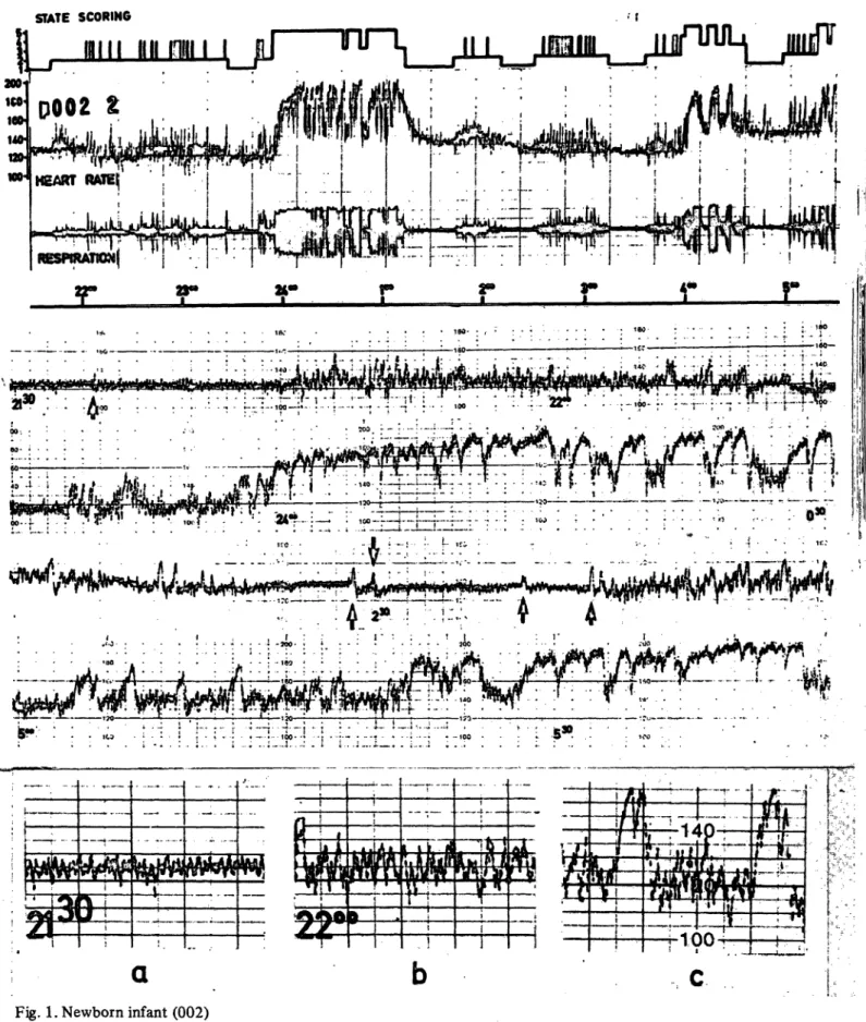
Fig. l. Newborn infant (002)
Top: Compressed writeout of 8 hour recording of heart rate and respiration together with state scoring from obser- vational notes.
In the compressed record well defined changes of heart rate and respiration coincide with changes in observational state scoring. Changes between NREM-sleep and REM-sleep occur in a fairly regulär sequence, although sometimes a transition from REM-sleep seems to be going on but is not followed by NREM-sleep at last (-22.25 h). Gross body movements J.Perinat. Med. 7(1979)
demonstrate some of our recordings here. The deceleration pattems are synchronous with startles, main aim of this work was to search for state a phenomenon that has been described by neuro- behavior and state related heart rate patterns in pediatricians in detaü [11, 35]. During REM-sleep the human fetus on the basis of comparison and it is more difficult to define baseline visually, analogy. This can be done more easily, when both because overall baseline is less stable. During state fetal and newborn heart rate tracings are recorded 4 and state 5 heart rate is grossly altered. During with the same recording machine, i.e. when tracings wakefulness with gross body movements acceler- are identical in respect to time base and amplific- ations of large amplitude, synchronous with motor ation. Moreover, because results will be most inter- activity, are repetitive and often merging so that esting and important for the obstetrician, äs they heart rate rises abruptly and the high level can will aid him in correct visual analysis and inter- often be taken äs a sequence of peaks of these pretation of FHR recording, both fetal and new- repetitive and merging accelerations with their born heart rate tracings were recorded with the interspersed downward and upward slopes. In the HEWLETT-PACKARD cardiotocograph, a machine vigorously crying infant a marked and more or less used in many obstetric departments. straightlined tachycardia is present. In our opinion For a detailed demonstration of all peculiarities of this elevated heart rate should not be taken äs some patterns, copies from original recordings may "baseline" in strict sense.
be less suitable. In that case the technique of play- Changes in the pattern of macrofluctuation related back from tape and writeout of compressed or to sleep state changes can be seen clearly. From extended records is of great advantage. When app- the original heart rate writeout at l cm/min shown lying this method the illustrating effect is enhanced below the compressed records it can be seen that by adding corresponding Segments of the original in general the macrofluctuation pattern in NREM- record for comparison. sleep is of lower amplitude (in beats per min) and From the compressed records in Fig. l a cyclic higher frequency (in cycles per min) compared to change of pattern in heart rate and respiration can the macrofluctuation pattern in REM-sleep. Besides be seen. As evaluated from the observational notes a higher amplitude and a slightly lower frequency these changes are synchronous with changes in the in REM-sleep there is more Variation in both and newborn infant's state. identifying small accelerations within this pattern First of all from the compressed records äs well äs is difficult unless an obvious state 2/4 occurs, i.e., from the original strip charts it can be seen that when interspersed gross movements of body and/
during NREM-sleep baseline in general is stable or or limbs give rise to accelerations with quite large slowlylowering. Small accelerations oracceleration- amplitude and duration. The heart rate pattern
during REM-sleep (state 2/4) are marked by upstrokes in the state scoring. Periods of wakefulness occur about midnight and in the early morning, when the baby is fed and swaddled.
Center: 4 Segments of original heart rate writeout (HEWLETT-PACKARD machine at l cm/min speed), each of 45 min duration.
In the first segment at the left a NREM-sleep heart rate pattern is seen. One startle, marked by an arrow occurs. At 21.45 h a change to pure REM-sleep pattern occurs, gross body movements and with that accelerations of larger amplitude are absent.
The second segment again Starts with a NREM-sleep pattern. Three minutes later REM-sleep begins. Body movements are noted and with them accelerations can be seen. 2 min before midnight the baby wakes up and it Starts crying almost immediately. Heart rate rises and the pattern can be taken äs a sequence of peaks of repetitive and merging accelerations with their interspersed downward and upward slopes. Sometimes between accelerations heart rate goes down to normal baseline level for short periods. The third segment Starts with a transition from REM- to NREM-sleep and during the following NREM-sleep pattern 4 small accelerations synchronous with startles are marked by arrows. Immediately after the last marked startle a change to REM-sleep pattern occurs. The first half of the last segment shows a REM-sleep pattern with interspersed accelerations synchronous with body movements (state 2/4). This is followed by wakefulness again.
Bottom:
a) Macrofluctuation pattern during NREM-sleep b) Macrofluctuation pattern during REM-sleep
c) Macrofluctuation pattern during REM-sleep with interspersed accelerations from gross body movements (state 2/4).
during state 4 and state 5, consisting of repetitive or merging accelerations to our opinion should not be taken äs macrofluctuation. Macrofluctuation during states 4 or 5 can only be identified, when between accelerations heart rate goes down to the normal baseline level for short periods.
Alterations of these typical macrofluctuation patterns in the newborn infant's heart rate may be caused by additional influences such äs periodic respiration or respiratory arrhythmia. (These alterations of macrofluctuation pattern together with identical macrofluctuation patterns seen in FHR recordings will be presented in a separate publication).
2.1.2 Overall distribution of state duration in
* the newborn infant in 8 hours recordings According to literature [19, 53] the newborn infant is asleep 17 to 20 hours a day, which is 70 to 83% of time, spending 75% of total sleeping time in REM-sleep.
In Tab. II total and relative times spent in NREM- sleep, REM-sleep and wakefulness are given for
all newborn infants. Because there was no statistical difference in data of groüp A and B, they were pooled. Our data are in complete agreement with literature data. In 76.9% of total recording time (14 X 8 hours) the babies were asleep and 26.6%
of total sleeping time was defined äs NREM-sleep.
2. l .3 Duration of sleep cycles and ratio of NREM- and REM-sleep within sleep cycles
According to literature [12,19,50, 52] in the new- born infant duration of a sleep cycle (a cycle of NREM- + REM-sleep) is 45 min to 2 hours. Periods of NREM-sleep last 10 to 20 min and periods of REM-sleep last 20 to 45 min.
In Tab. III mean values äs well äs minimal and maximal values for absolute duration of sleep cycles and for absolute durations and relative fractions of NREM- and REM-sleep periods are given. In our recordings mean duration of sleep cycles was 61.9 min, mean duration of NREM-periöds was 18.5 min (= 29.8% of total duration of cycle) and mean duration of REM-periods was 43.2 min. Again
Tab. II. Distribution of state duration in 8 hour recordings newborn infantsGASE NR
002 003 004 006 007 008 009 010 011 012 013 016 017 018 TOTAL
STATE 1
113 min (23.5 %) 69 min (14.3 %) 140 min (29. 1 %)
43 min ( 8.9 %) 113 min (23.5 %) 94 min (19.5 %) 124 min (25.8 %) 66 min (13.7 %) 73 min (15.2 %) 95 min (19.7 %) 117 min (24.3 %) 90 min (18.7 %) 135 min (28.1 %) 106 min (22.0 %) 1378 min (20.5 %)
STATE 2
231 min (48.1 %) 158 min (32.9 %) 210 min (43.7 %) 240 min (50.0 %) 292 min (60.8 %) 318 min (66.2 %) 274 min (57.0 %) 296 min (61.5 %) 363 min (75.6 %) 359 min (74.7 %) 293 min (61.0 %) 202 min (42.0 %) 315 min (65.6 %) 242 min (50.4 %) 3793 min (56.4 %)
STATE 3-5
136 min (28.3 %) 253 min (52.7 %) 130 min (43.7 %) 197 min (41.0 %) 75 min (15.6 %) 68 min (14.1 %) 82 min (17.0 %) 118 min (24.5 %) 44 min ( 9.1 %) 26 min ( 5.4 %) 70 min (14.5 %) 188 min (39.1 %) 30 min ( 6.2 %) 132 min (27.5 %) 1549 min (23.0 %)
TOTAL SLEEPING TIME
344 min (71.6 %) 227 min (47.2 %) 350 min (72.9 %) 283 min (58.9 %) 406 min (84.5 %) 412 min (85.8 %) 398 min (82.9 %) 362 min (75.4 %) 436 min (90.8 %) 454 min (94.5 %) 410 min (85.4 %) 292 min (60.8 %) 450 min (93.7 %) 348 min (72.5 %) 5172 min (76.9 %)
STATE 1 HITHIN TOTAL SLEEPING TIME
32.8 % 30.3 % 40.0 % 15.1 % 27.9 % 22 '.8 % 31.1 % 18.2 % 16.7 % . . 20.9 % ' '
28.5 % 30.8 % 30.0 % 30.4 % 26.6 %
J. Perinat. Med. 7 (1979)
Tab. III. Duration of sleep cycles, absolute and relative duration of state l and state 2 periods within sleep cycle Newborn infants
CASE NR
002 003 004 006 007 008 009 010 011 012 013 016 017 018
NUMBE* Of SLEEP CYCLES
BVALUATEO
5 3 5 4 4 5 6 5 4 3 6 2 5 7 TOTAL
64
MINIMUM OURATION SLEEP CYCLESor
36 Bin 49 ein 36 Bin 27 Bin 45 ain 46 ain 41 Bin 23 Bin 38 ain 32 ein 32 Bin 28 Bin 37 ain 16 Bin
MAXIMUM DURATION
or
SLEEP CYCLES
123 ain 65 ain 69 ain 78 ein 185 ain 104 Bin 86 ain 120 ein 138 ein 117 ain 78 ain 43 Bin 91 ain 94 ain
OURATION SLEEP CYCLESer
64.0 Bin 59.6 Bin 56.6 Bin 59.2 ain 86.5 Bin 66.6 Bin 59.5 ein 55.4 Bin 81.5 ein 87.6 ain 50.5 ain 35.5 Bin 67.4 ain 47.4 ain
WEICHTED MEAN 61.9 Bin
MINIMUM OURATION
or
STATE 1
17 Bin 10 Bin 10 Bin 9 Bin 17 ain 7 Bin 17 Bin 5 ain 6 ain 16 aln 9 min 5 ain 12 Bin 10 ain
MAXIMUM OURATION
or
STATE 1
24 Bin 19 Bin 44 Bin 15 Bin 34 Bin 25 Bin 24 ein 25 min 24 Bin 31 min 25 ein 26 Bin 26 Bin 24 Bin
MEAN OURATION STATE 1or (\ or CYCLE)
19.6 Bin (36.4 %) 15.3 Bin (26.9 %) 27.2 nin (45.8 %) 10.7 ein (21.4 %) 24.2 ain (37.3 %) 18.0 ain (29.3 *) 20.6 Bin (35.8 %) 13.2 min (31.3 %) 18.2 Bin (23.2 %) 22.6 Bin (31.7 «) 19.5 min (43.2 %) 15.5 ain (39.1 %) 19.2 Bin (24.0 %) 15.1 ein (37.5 %)
WEIGHTED MEAN 18.5 ein (29.8 %)
MINIMUM OURATION
or
STATE 2
21 Bin 30 ain 25 ein 17 ain 22 Bin 21 ain 24 ain 11 Bin 32 Bin 16 Bin 10 ain 17 Bin 16 ain 6 Bin
MAXIMUM OURATION
or
STATE 2
105 ain 55 ain 35 ain 69 Bin 162 Bin 80 Bin 62 ein 111 Bin 117 Bin 96 min 56 Bin 23 ain 75 aln 74 ein
MEAN OURATION
or
STATE 2 (% CYCLE)
44.4 ain (63.5 %) 44.0 Bin (73.0 %) 29.4 Bin (54.1 %) 48.5 Bin (78.5 %) 62.2 ain (62.7 %) 48.6 Bin (70.6 %) 38.8 Bin (64.1 %) 42.2 Bin (68.6 %) 63.2 Bin (76.7 %) 65.0 Bin (68.2 %) 31.0 ein (56.6 %) 20.0 Bin (60.8 %) 48.2 Bin (71.5 %) 32.2 Bin (62.3 %)
WEICHTED MEAN 43.2 Bin (69.9 %)
data are in good agreement with data cited from literature.
2.1.4 Evaluation of motor activity in the new- born infant in respect to sleep state
Close observation of the newborn infants during 8 hours recordings revealed a striking difference in motor activity during different states. During NREM-sleep, motor activity generally is absent except for the occurrence of startles, fully or abortive, single or sometimes repetitive. Gross movements in NREM-sleep with larger accelerations are very rare. During REM-sleep motor activity is enhanced. Small muscular twitches do not alter the typical heart rate pattern of a pure REM-sleep but besides these small twitches single or sequences of gross movements of limbs and even trunk do occur. This has led to the definition of state 2/4 in literature [19, 35], Synchronous to these movements accelerations of heart rate can be seen and, äs there is a wide ränge äs to the amount and pattern of movements there is a wide ränge of acceleration patterns in respect to amplitude, duration and frequency. The ratio of movement counts per duration of NREM- and REM-states
was .129 and .391 respectively. The difference is statisticaUy significant: motor activity is 3 X higher in REM-sleep compared to NREM-sleep.
3. Evaluation of fetal state behavior
As has been said in our introduction fetal state behavior cannot be assessed by direct observation.
Our approach to solve the problem must be in- direct and it is based on the assumption that comparable neonatal and fetal heart rate and motor activity patterns are related to comparable states in the newborn infant and the fetus. To prove that comparable heart rate patterns do exist in the neonate and in the fetus, first of all some examples of our FHR recordings will be presented here. Secondly general results of comparing neonatal heart rate and FHR patterns will be presented. A presentation of our Computer analysis in respect to heart rate patterns will be published elsewhere [22].
3.1 Fetal heart rate patterns and assumed related state behavior illustrated
Compressed 8 hour FHR records first of all show
known technical limitations of abdominal FECG-
STATE SCORING
^ ^?. ^-^.^..^, ..„.^..^ ,,,.„
' ä i v c
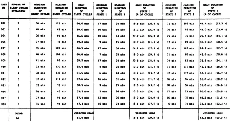
Fig. 2. Fetus (013)
Top: Compressed writeoutof 8 hours recording of FHR, movement marks and external tocogram together with assumed state scoring.
In the FHR recording a regulär change in macrofluctuation pattern can be seen clearly. The low amplitude macrofluc- tuation pattern corresponds to a heart rate pattern seen in the newborn infant during NREM-sleep and the pattern with higher amplitude and accelerations corresponds to a heart rate pattern seen in the newborn during REM-sleep (state 2/4). Thus identical or comparable states may be assumed in the fetus. Movement marks are state related: they are seen in REM-sleep and are absent (or rare) in NREM-sleep. A FHR pattern corresponding to a newborn heart rate pattern during wakefulness is not seen in this case.
J. Perinat. Med. 7(1979)
STATE SCORINO
P jnmrn na^ ninimqj mm j
EXTERNALJLOCOCÄAMt T T
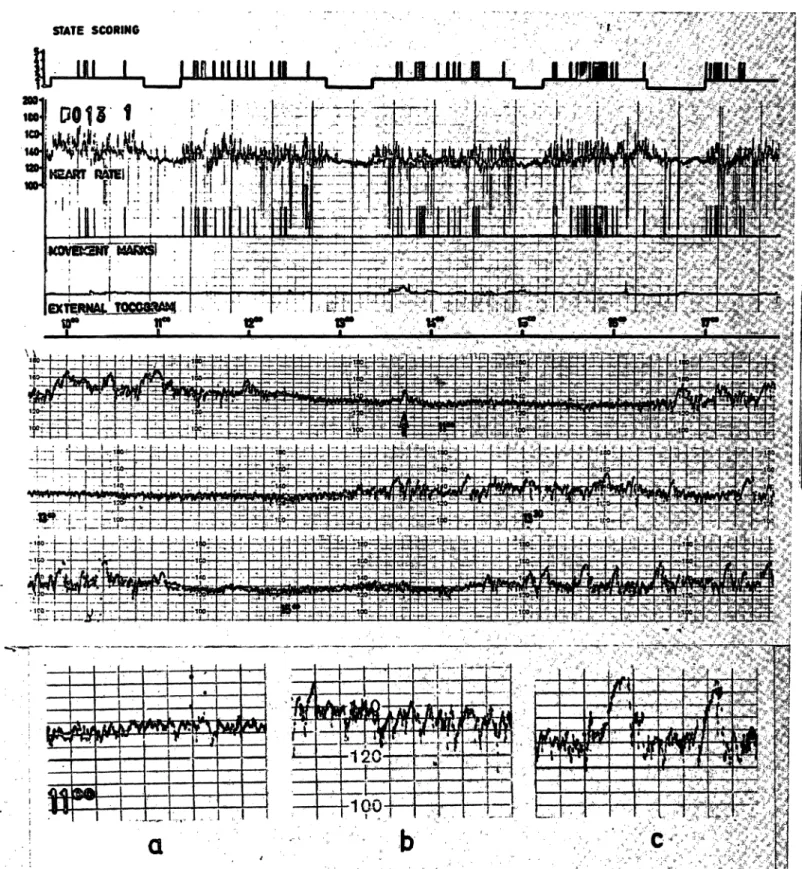
Fig. 3. Fetus (018)
Top: Compressed writeout of 8 hour recording of FHR, movement marks and external tocogram together with assumed state scoririg.
The fetus is postterm (group B). Besides a regulär change in FHR macrofluctuation äs in the recordings demonstrated before, a long lasting period of FHR alteration, identical to the newborn heart rate pattern during wakefulness is seen (marked by in the state scoring). Movement marks are much more frequent compared to tracings shown before.
Bottom:
3 45-min-segments of original FHR writeout (HEWLETT-PACKARD machine at l cm/min speed).
Upper tracing: a REM-sleep macrofluctuation pattern with interspersed accelerations (state 2/4) is followed by a NREM-sleep pattern and again by a REM-sleep pattern. Middle tracing and left side of lower tracing: FHR pattern of
"wakefulness".
Center: 3 Segments of 45 min duration of original FHR writeout (HEWLETT-PACKARD machine at l cm/min speed).
FHR macrofluctuation pattern during assumed NREM-sleep (middle part of upper segment, left side of middle segment, middle part of lower segment) and assumed REM-sleep is seen. One small acceieration in the upper segment during NREM-sleep (arrow) isassumed to be caused by a startle. Accelerations during REM-sleep (rhight side of lower segment) are synchronous with movement marks (state 2/4).
Bottom:
a) Macrofluctuation pattern during assumed NREM-sleep b) Macrofluctuation pattern during assumed REM-sleep
c) Macrofluctuation pattern dujing assumed REM-sleep with interspersed accelerations from body movements (state 2/4).
STATE SCORINO
HEIM III l III 0 II II
SXTERHAL TOCOCKAMj
r
t3*- i - M - ! - : f . -r frr"-·:—l—r-r—r*r«f
F
·'
|: !.:;r: r:! :r α:^
i-IK-t*ii.LJL'iiaBiiL»
' · ' U*' ' «Λ!:-j -:-U.t- '..-[. U ίΓΠ ΓΤ^.-
ί » " ! ' ' f ! ' i " »" ' " l l"" *" · * » "*
; . v . i . ; . i4o- » - 4->~ i- - -*- - -^—f- ' · * '
;j i_:: ι ίΑ; *i:_::-t:; i-;f-H-t-r
J. Perinat. Med. 7 (1979)
derived FHR recording. Interspersed trigger errors appear äs downstrokes. Clusters of trigger errors occurred with enhanced maternal (or fetal) motor activity.
In fetuses of group A FHR patterns comparable to neonatal NREM- and REM-patterns could be seen clearly (Fig. 2) but a FHR pattern comparable to neonatal heart rate during wakefulness was absolutely rare.
One fetal recording showed a macrofluctuation pattern identical to that seen in a neonate with periodic respiration.
In recordings of group B fetuses (Fig. 3) some remarkable alterations could be seen. Firstly and surprisingly heart rate patterns seen during neonatal wakefulness were seen in FHR recordings of this group in the same percentage äs in the newborns.
They were associated with markedly enhanced motor activity, äs can be seen from the density of movement marks. Secondly enhancedmotor activity in group B fetuses modified FHR patterns during assumed sleep äs well: A higher number of acceler- ations of largeramplitude and duration in connection with movements, i.e. a state 2/4 occurred in REM- sleep and similar accelerations even occurred in NREM-sleep. One could speak of a state l/4.There- fore differentiation of sleep states turned out to be difficult or even dubious in 2 cases (010,016).
Last not least in only one of our group B recordings (009) FHR alterations, that are known to be signs
of fetal distress could be seen. In assumed NREM- sleep macrofluctuation was minimal and these states were of slightly longer duration. In assumed REM-sleep macrofluctuation amplitude was dmini- ished too (Fig. 4).
3.2 Overall distribution of state duration in the fetus in 8 hour recordings
3.2.1 The fetus near term (group A)
In Tab. IV total and relative times spent in assumed NREM-sleep, REM-sleep and "wakefulness" are given for the fetuses of group A. It can be seen that in 97.6% of total recording time (10 X 8 hours minus 19 min) FHR showed a pattern iden- tical to the newborn NREM- or REM-sleep heart rate pattern. In 26.3% of total "sleeping time" a NREM-sleep heart rate pattern was found. A pattern corresponding to wakefulness in the neonate was seen in only 1.2% of total recording time. In l case a severe deceleration with conse- cutive reactive FHR alteration occurred when the patient sät up in bed to empty her bladder.
3.2.2 The postterm fetus (group B)
A striking difference in the overall distribution of states in 8 hour recordings was found comparing group A and B (see Tab, V). A heart rate pattern corresponding to neonatal wakefulness occurred
Fig. 4: Fetus (009)
Top: Compressed writeout of 8 hour recording of FHR, movement marks and external tocogram together with assumed state scoring.
The fetus is postteim (group B) and routine FHR monitoring prior to our recording gave rise to suspicion of beginning distress. A regulär change in FHR pattern and with that assumed state change is seen. Motor activity is raised in the first half. In the second haft it is less, but definitely present.
Center: 5 segments of original FHR writeout (HEWLETT-PACKARD machine at l cm/min speed), each of 45 min duration.
Segment 1: Starts with several minutes of a REM-sleep pattern, then a NREM-sleep pattern with 2 accelerations from movements and at last the beginning of a pattern of "wakefulness" is seen.
Segment 2: pattern of "wakefulness" throughout.
Segment 3: at the beginning and at the a REM-sleep pattern. Inbetween a NREM-sleep pattern of 33 min duration with minimal macrofluctuation amplitude. No accelerations.
Segment 4: REM-sleep pattern with decreased macrofluctuation amplitude, but accelerations from movements are present.
Segment 5: NREM-sleep pattern of 39 min duration with minimal macrofluctuation amplitude. One small acceleration from a single movement (startle) is marked by an arrow.
Bottom:
a) Macrofluctuation pattern during assumed NREM-sleep
b) Macrofluctuation pattern during assumed REM-sleep with interspersed accelerations from body movements (state 2/4).
Tab. IV. Distribution of state duration in 8 hour recordings fetuses group A
GASE NR
002 003 004 005 006 007 008 011 013 017
\ TOTAL
STATE 1
117 min (24.3 %) 150 min (31.2 %) 103 min (21.4 %) 167 min (34.7 %) 175 min (36.4 %) 109 min (22.7 %) 129 min (26.8 %) 98 min (20.4 %) 112 min (23.3 %) 76 min (15.8 %)
1236 min (25.7 %)
STATE 2
335 min (69.7 %) 330 min (68.7 %) 366 min (76.2 %) 313 min (65.2 %) 302 min (62.9 %) 366 min (76.2 %) 351 min (73.1 %) 325 min (67.7 %) 368 min (76.6 %) 396 min (82.5 %)
3452 min (71.9 %)
STATE 3-5
28 min (5.8 %)
25 min (5.2 %)
8 min (1.6 %)
61 min (1.2 %)
DBCELERATZON + REACTIVE FHR ALTBRATION
32 min (6.6 %)
32 min (0.6 %) -·.·.>
REOORDZNG DISTURBANGE
NO STATE SCORING POSSIBLE
11 min
3 min 5 min
19 min
TOTAL SLEEPING TIME
452 min ( 94.1 %) 480 min (100.0 %) 469 min ( 97.7 %) 480 min (100.0 %) 477 min ( 99.3 %) 475 min ( 98.9 %) 480 min (100.0 %) 423 min ( 88.1 %) 480 min (100.0 %) 472 min ( 98.3 %)
4688 min ( 97.6 %)
STATE 1 HITHIN TOTAL SLEEPING TIME
25.8 % 31.2 % 21.9 % 34.7 % 36.6 % 22.9 % 26.8 % 23.1 % 23.3 % 16.1 %
26.3 %
Tab. V. Distribution of state duration in 8 hour recordings fetuses group B
CASE NR
001 009 010 012 016 018
TOTAL
STATE 1
92 min (19.1 %) 116 min (24.1 %) 109 min (22.7 %) 150 min (31.2 %) 104 min (21.6 %) 82 min (17.0 %)
653 min (22.6 %)
STATE 2
234 min (48.7 %) 261 min (54.3 %) 218 min (45.4 %) 294 min (61.2 %) 347 min (72.2 %) 186 min (38.7 %)
1540 min (53.4 %)
STATE 3-5
154 min (32.0 %) 103 min (21.4 %) 153 min (31.8 %)
212 min (44.1 %)
622 min (21.5 %)
DECELERATION + REACTIVE FHR ALTERATION
36 min (7.5 %) 29 min (6.0 %)
65 min (2.2 %)
TOTAL SLEEPING TIME
326 min (67.9 %) 377 min (78.5 %) 327 min (68.1 %) 444 min (95.5 %) 451 min (93.9 %) 268 min (55.8 %)
2193 min (76.1 %)
STATE 1 WITHIN TOTAL SLEEPING TIME
28.2 % 30.7 % 33.3 % 33.7 % 23.0 % 30.5 %
29.7 %
for longer periods, relative duration of this pattern was 21.5%. With that relative duration of assumed REM-sleep FHR pattern diminished to 53.4%.
The relative duration of assumed NREM-sleep FHR pattern remained nearly constant: 22.6%.
Thus in l'6.1% of total recording time a "sleeping"
FHR pattern was seen and in 29.7% of "sleeping time" a NREM-sleep FHR pattern was identified.
Thus state distribution in group B was found to be
nearly identical to state distribution in the neonate.In 2 cases severe decelerations with consecutive FHR alterations occurred.
3.3 Duration of sleep cycles and ratio of NREM- and REM-periods within sleep cycles
Despite the striking difference between group A- and group B-fetuses in respect to the ratio Qf
"sleep and wakefulness" no significant difference was found for both groups in respect to dunr&on of sleep cycles and relative fractions of assumed NREM-sleep and REM-sleep within sleep cycles.
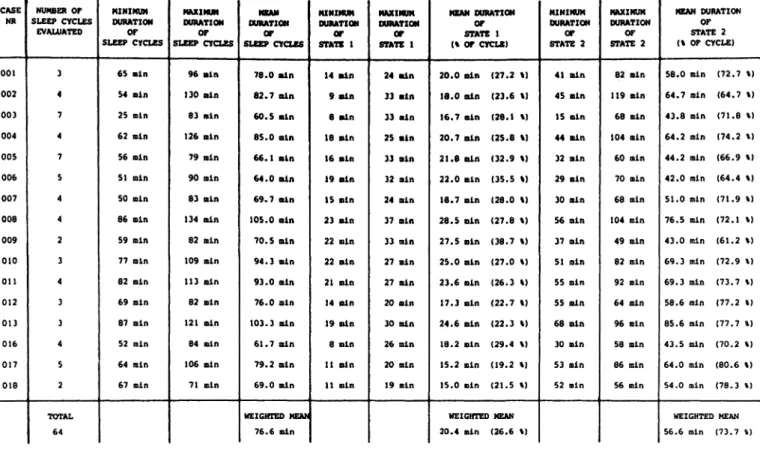
Because of this fact data were pooled. According to Tab. VI mean duration of sleep cycles was 76.6 min, mean duration of NREM-periods within
J. Perinat. Med. 7(1979)
Tab. VI. Duration of sleep cycles, absolute and relative duration of state l and state 2 periods within sleep cycle fetuses
CASE NR
001 002 003 004 OOS 006 007 008 009 010 011 012 013 016 017 018
NUMBER OF SLEEP CYCLES
EVALUATEO
3 4 7 4 7 S 4 4 2 3 4 3 3 4 5 2
TOTAL 64
MINIMUM DURATION SLEEP CYCLES
65 min 54 min 25 min 62 Bin 56 ein 51 Bin SO Bin 86 Bin 59 Bin 77 ein 82 Bin 69 aln 87 Bin 52 min 64 Bin 67 Bin
MAXIMUM DURATION
OF SLEEP CYCLES
96 ain 130 ein 83 ein 126 Bin 79 Bin 90 Bin 83 Bin 134 Bin 82 Bin 109 ain 113 Bin 82 ain 121 ain 84 Bin 106 Bin 71 Bin
MEAN DURATION
OF SLEEP CYCLES
78.0 Bin 82.7 Bin 60.5 ain 85.0 Bin 66.1 ain 64.0 Bin 69.7 ain 105.0 ain 70.5 Bin 94.3 ein 93.0 aln 76.0 ein 103.3 Bin 61.7 Bin 79.2 Bin 69.0 Bin
WEZGHTED MEAN 76.6 Bin
MINIMUM OURATIOM
OF STATE 1
14 ain 9 ein 8 ain 18 Bin 16 ain 19 aln 15 aln 23 aln 22 Bin 22 aln 21 ein 14 Bin 19 Bin 8 Bin 11 aln U Bin
MAXIMUM DURATIOM
OF STATE 1
24 Bin 33 ain 33 aln 25 aln 33 aln 32 aln 24 aln 37 Bin 33 Bin 27 ein 27 Bin 20 ein 30 Bin 26 Bin 20 Bin 19 min
MEAN DURATION OF STATE 1 (% OF OfCLE)
20.0 aln (27.2 %) 18.0 aln (23.6 %) 16.7 ain (28.1 %) 20.7 aln (25.8 %) 21.8 aln (32.9 %) 22.0 Bin (35.5 %) 18.7 aln (28.0 %) 28.5 Bin (27.8 %) 27.5 Bin (38.7 %) 25.0 ain (27.0 %) 23.6 Bin (26.3 %) 17.3 ein (22.7 %) 24.6 Bin (22.3 %) 18.2 Bin (29.4 %) 15.2 ein (19.2 %) 15.0 min (21.5 %)
WEIGHTED MEAN 20.4 min (26.6 %)
MINIMUM DURATION
OF STATE 2
41 Bin 45 Bin 15 ain 44 Bin 32 aln 29 ein 30 aln 56 Bin 37 Bin 51 ain 55 min 55 ain 68 aln 30 Bin 53 min 52 min
MAXIMUM DURATION
OF STATE 2
82 Bin 119 ein 68 Bin 104 Bin 60 min 70 min 68 min 104 min 49 min 82 min 92 min 64 ein 96 min 58 min 86 min 56 min
MEAN DURATION OF STATE 2 (% OF CYCLE)
58.0 Bin (72.7 \) 64.7 min (64.7 %) 43.8 aln (71.8 %) 64.2 min (74.2 %) 44.2 min (66.9 %) 42.0 min (64.4 %) 51.0 min (71.9 %) 76.5 Bin (72.1 %) 43.0 min (61.2 %) 69.3 min (72.9 %) 69.3 min (73.7 %) 58.6 min (77.2 %) 85.6 min (77.7 %) 43.5 min (70.2 %) 64.0 min (80.6 %) 54.0 min (78.3 %)
WEIGHTED MEAN 56.6 min (73.7 %)
cycles was 20.4 min (26.6% of total duration of cycle) and mean duration of REM-periods within cycles was 56.6 min. Compared to values given above for the newborn infants, mean duration of sleep cycles and mean duration of NREM- and REM-sleep were slightly longer but values did not differ statistically.
3.4 Evaluation of fetal motor activity
Visual evaluation of our recording gave rise to the impression that in the group of postterm fetuses definitely more movement marks occurred comp- ared to the fetuses near term. Our impression was, that the higher percentage of "wakefulness"
within total recording time, which in some cases was accompanied by an abundant motor activity, was not the only reason and that motor activity was raised during "sleep" of postterm fetuses too.
Therefore analysis was döne in both respects.
Because of the unequal distributions of times spend in all states the ratio of movement counts per duration of states again is used for comparison.
3.4.1 Comparison of motor activity in 8 hour recordings in fetuses of group A and group B
The ratio of total movement counts per total recording time gives a relative measure of general motor activity for intergroup comparison. For group A a ratio of .230 and for group B a ratio of .448 was calculated. This means that there was about 2 X more general motor activity in the post- term fetuses.
3.4.2 Comparison of sleep state related motor activity in fetuses of group A and group B
In 1.4 it was demonstrated that motor activity of
the newborn infant is not evenly distributed in
respect to NREM- and REM-sleep. This holds true
for motor activity in the fetus äs well, but whereas
in the fetus near term (group A) ratios differ by a
factor of 10 (0.29 and .297), in the postterm fetus
the difference is less (.124 and .395) and values
are somewhat similar to those in the newborn in-
fants. Infact, motor activity is enhanced about red. The pattern seen thrp
fughout recordings was threefold in NREM-sleep of postterm fetuses similar to a REM-sleep or a transitional pattern, compared to fetuses near term. Heart rate control on the whole seemed to be less
stable.
3.4.3 Anencephaly
During the course of our investigations 2 cases of 4 Discussion
anencephaly, 23 and 36 weeks of gestation, were ^
General considerationsadmitted to the ward and 8 hour recordings were
done in both prior to termination of pregnancy. FHR analysis is a main diagnostic tool for the From Fig. 5 it can be seen that no distinct periodic perinatologist. In our opinion research in the field changes in FHR pattern and motor activity occur- of FHR analysis in the past years has been directed
-j._j.,_*—^.^.,^..^—.. ..4...^..,, -"h '.pj^-j^fef |·'·"·!· "· .f'.H'v l"··*1 Η''Π'i'"'"i^ ;*>-*?«rr-ita<- 4~—=4~r7ir ·γ-··'*;:'·*·- Γ—* · ···«··· ·—~~ ·« ·- * - - ·;·**- fc^^ '·
_νι<; · :.,, ,,Γ — ^:^^ . "··.._
φϊ£^^
?ί^^^
#> ':, ; .η, Λ;: ;.:±;^:j-^^ «t^t^
1\ 4- i ·· r *· ΙίΠ-. Pfc
Fig. 5. Fetus (014) v
Top: Compressed writeout of 8 hour rec^jrding of FHR and movement marks. Anencephalus of 36 weeks gestation.
No clear-cut changes of FHR pattern can be detected.
Bottom: 3 segments of original FHR writeout (HEWLETT- PACKARD machine at l cm/min speed).
FHR is similar to a REM-sleep pattern, but less stable.
Sometimes FHR looks like a transition from REM-sleep to NREM-sleep pattern, but no NREM-sleep follows.
J. Perinat. Med. 7 (1979)
mainly towards the relation between FHR alter- ations and fetal distress, whereas investigation of the "normal" variations of FHR patterns in the unstressed fetus and their regulating mechanisms hasbeenneglected.
The existence of fetal "sleep ' and "wakefulness"
(or rest and activity) with concomittant changes of FHR macrofluctuation pattern has been postulated for some time. But distribution of these states along time, i.e. their sequential properties were not known in detail. Moreover, descriptions of FHR pattern changes attributed to these state changes were rather unprecise. Last not least there are no precise ideas whether and how other factors influence FHR pattern in the normal undisturbed fetus, except the fact that baseline decreases with advancing gestation, i.e. with cerebral maturation [l, 20, 60, 61] and the fact that variability in- creases [60,61].
Only the knowledge and clear definition of the Variation of physiologic FHR patterns can lead to a clearcut definition of pathologic patterns. There- fore new approaches to investigate normal FHR patterns, their variations and their underlying regulatory mechanisms have to be looked for. In doing so one should Start to verify and investigate precisely factors, which according to our present knowledge seem to be of major importance and one of them is, to our mind, central nervous func- tion. The fact that according to literature state related changes of newborn heart rate pattern do occur and the fact that fundamental differences in CNS function in the mature fetus near term and in the newborn infant may not be assumed were the basis for our comparative investigation of fetal and neonatal heart rate patterns and their concomitant states of CNS function.
4.2 Comments on results
Presuming that identical heart rate patterns in the newborn infant and in the fetus derive.from identi- cal or at least comparable states of central nervous function the following may be stated.
In the mature and undisturbed fetus sleep cycles of about l hour duration occur sequentially and periods of NREM-sleep and REM-sleep occur alternating within this cycle, their duration being
roughly in a 1:2 to 1:3 ratio. Motor activity is unevenly distributed during a sleep cycle, fetal movements generally are associated with REM- sleep (= state 2/4) and they are rare (startles) in NREM-sleep. Central nervous arousal comparable to wakefulness in the newborn seems to be a rare phenomenon in the undisturbed fetus, at least when the mother is quietly lying in her bed. Synchron- ously with these sleep state changes Variation of FHR pattern occurs and according to our analysis the most distinct Variation is that of macrofluctu- ation amplitude whereas macrofluctuation fre- quency is less and baseline level inconsistently modulated. Because fetal movements are unevenly distributed in respect to state on one hand and on the other hand cause accelerations, Variation of FHR pattern by state changes is enhanced. There- fore, on the basis of the above given assumption it can be said that spontaneously changing central nervous coordination or arousal is the predominating factor governing FHR pattern and its Variation in fetal wellbeing.
Besides that there is suspicion that other factors, such äs respiratory activity may modify FHR pattern additionally and distinctly. Recent public- ations about antepartum FHR recording support thisidea[56,61].
The most striking Undings in the p os t term fetuses of group B were first of all the fact that the ratio of sleep and "wakefulness" changedconsiderably:
a state 3-5 like FHR pattern together with con- siderable motor activity was seen in about 20%
of total recording time. Secondly motor activity was enhanced even during sleep states: the ratio of movement counts per state duration was signif- icantly higher in REM-sleep (state 2/4) and in NREM-sleep compared to fetuses of group A.
Because fetal movements generally were asso- ciated with accelerations FHR pattern during sleep states was modified äs well. Contrary to that duration of sleep cycles and ratio of NREM- and REM-sleep were not altered significantly. At present we do not have a precise explanation for these findings of enhanced motor activity in post- maturity and it is in contrast to results of others [7,46].
Whereas in one of the two cases with suspicion of
beginning fetal distress (016) no alterations from
the FHR pattern described above could be seen during the 8 hour recordings (except a deceleration when the patient sät up to empty her bladder, which was seen in case 11 of group A too), in the other case (009) during sleep states macrofluctu- ation amplitude was minimal. Still, even in this fetus a state of "wakefulness" of nearly 2 hours duration occurred and Overall motor activity was raised.
The 2 cases of anencephaly were included in our
• investigation, because we excepted further insight in the relation of CNS function and FHR pattern.
Contrary to an earlier publication [15] in which complete loss of macrofluctuation was found in anencephaly, 8 hour recordings in these 2 anence- phalic fetuses showed macrofluctuation throughout
• Mhe recordings but no distinct periodic changes of FHR and motor activity. The macrofluctuation pattern seen was similar to a REM pattern. It has been stated that NREM-sleep is the state with the strongest homoiostatic control and that infants with CNS lesions are frequently not able to achieve this stable state or to continue it over more than a few minutes [48]. This Statement would be suppor- ted by our finding. Still one has to have in mind, that these 2 fetuses were 23 and 36 weeks of gestation and that absence of NREM-sleep may be
an effect of cerebral immaturity too.
4.3 Consequences for clinical routine FHR monitoring
Present management of clinical routine antepartum FHR monitoring aims at the early detection of FHR alterations caused by subacute or chronic hypoxia and is based on the fact, that hypoxia leads to diminution, at last to loss of macro- fluctuation amplitude and, if contractions are present to late deceleration.
For routine antepartum FHR recording generally a duration of 30 min is recommended, every two days to three times per day.
Our investigations revealed that diminution of macrofluctuation amplitude occurs cyclically in NREM-sleep for periods of up to 30 min and more, independent of hypoxia (see case 013). Therefore
a false suspicion of fetal distress may result from a 30 min routine FHR recording, when it is started just at the beginning of a normal NREM-sleep
period.
On the other hand at least in the beginning of fetal distress seemingly normal FHR patterns with en- hanced motor activity occur prior and after periods of defmitely suspect macrofluctuation (see case 009) and a false diagnosis of fetal wellbeing may result from a 30 min routine FHR recording, when it does not include the period of suspect macro- fluctuation pattern.
Therefore the presently recommended 30 min duration for ante partum FHR recording may be too short for safe discrimination of a normal NREM-sleep FHR recording and a FHR recording indicating hypoxia. General results of antepartum detection of hypoxia presumably will be better if at least one complete sleep cycle of NREM-sleep and REM-sleep is recörded in a Session of at least
l hour duration.
The same holds true for fetal movement counting:
because movements are rare to absentduringNREM- sleep in a healthy fetus fetal movement counting should always be done simultaneously with FHR recording, and fetal state behavior, according to macrofluctuation pattern, should be taken into
account.
At present we do not know why fetal motor activity was found to be enhanced in our group of postmature fetuses and if diminution would have occurred later with hypoxia. This at least could not be noticed in the 2 fetuses (009, 016) suspect of being slightly hypoxic at the time of recording.
In this respect our results are not in agreement with publications reporting diminution and/or cessation of fetal movements in the fetus in danger and further investigations are necessary.
In our opinion simultaneous recording of FHR, fetal respiration and fetal movements, similar to polygraphic recording in the newborn may result in a much better insight into fetal central nerv'öus control and its impairment by hypoxia.
We therefore have to develop methods of fetal polygraphy, practicable for clinical routine of fetal intensive care.
J. Perinat. Med. 7(1979)
Summary
In order to investigate the influence of spontaneous changes of CNS activity on ante partum FHR and fetal motor activity state specific heart rate and motor activity patterns of newborn in fan t s were compared with patterns seen in the fetus. Instantaneous FHR derived from ab- dominal FECG, the external tocogram and fetal move- ments äs indicated by the mother via push button were recorded continuously for 8 hours on strip chait and analog tape. At least 4 days after parturition the same individuae, now newborn (except 2 anencephaiy, who died during parturition) were observed for assessment of newborn infant state behavior. Heart rate and respiration again were recorded for 8 hours continuously and during the whole time close observation and protocoi of the newborns states and all interesting events was performed.
Visual evaluation and Computer analysis of recordings revealed that in the newborn infant state related heart rate pattern changes do occur and that identical heart rate patterns and their changes can be seen in the fetus too. Definition of states according to typical and com- parable heart rate patterns in the newborn and in the fetus was supported by taking into account newborn and fetal motor activity patterns.
Results
l. l Newborn infant:
Our results with respect to the distribution of sleep and wakefulness äs well äs with respect to duration of sleep cycles and the distribution of NREM-sleep and REM- sleep within sleep cycles äs defined from observational notes and recording of heart rate and respiration are in good agreement with literature data: In 76.9% of total recording time the babies were asleep and 26.6% of total sleeping time was defined äs NREM-sleep. From our recordings a mean duration for a sleep cycle of 61.9 min, a mean duration for a REM-period of 43.2 min was calculated.
Motor activity was rare in NREM-sleep (startles), but more frequent in REM-sleep (state 2/4). Mean ratios of movement counts per state duration were .129 and .391 respectively.
1.2 Fetus
Ante partum recordings were divided in 2 groups. Fetuses less than l week postterm were affiliated to group A.
Fetuses more than l week postterm were affiliated to group B.
1.3 Overall distribution of state duration in 8 hour recordings.
1.3.1 Group A
In 97.6% of total recording time FHR showed a pattern identical to the newborn NREM-sleep or REM-sleep heart rate patterns. In 26.3% of total "sleeping time" (which is total time of NREM- and REM-pattern) a NREM-sleep FHR pattern was found. A pattern corresponding to wakefulness in the neonate was seen in only 1.2% of total recording time.
1.3.2 Group B
A striking difference in the overall distribution of states was found in group B. Relative duration of FHR pattern comparable to the newborn heart rate pattern of wake- fulness increased to 21.5% and relative duration of REM- sleep pattern decreased to 53.4% The realtive duration of NREM-sleep pattern remained constant.
2 Duration of sleep cycles and ratio of NREM- and REM-periods within sleep cycles
Despite the above mentioned differences in group A and group B fetuses the duration of sleep cycles and the ratio of NREM- and REM-periods in both groups did not differ significantly. Because of this fact data were pooled and mean duration of sleep cycles was 76.6 min, mean duration of NREM-periods within sleep cycle was 20.4 min (= 26.6%
of total duration of sleep cycles) and mean duration of REM-periods was 56.6 min. Compared to values given above for the newborn infants, mean duration of sleep cycles and periods of NREM- and REM-sleep were slightly longer but values did not differ statistically.
3 Evaluation of fetal motor activity
Total movement counts per total recording time were .230 and .448 respectively for group A and group B fetuses. This means that there is more general motor activity in the postterm fetuses.
Calculation of NREM-sleep related motor activity in fetuses of group A and group B again demonstrates that there is less motor activity during NREM-sleep com- pared to REM-sleep. But whereas in group A the ratios of movement counts per state duration differ by a factor of 10 (.029 and .297 respectively), in the post- term fetus the difference is less (.124 and .359 respect- ively) and values are somewhat similar to those of the newborn infants.
4.1 Fetal distress
In two fetuses of group B suspicion of beginning fetal distress arose from routine clinical ante partum monitoring prior to our recording. In one of them no alterations from the FHR pattern described above could be seen, but in the other fetus during sleep states macrofluctuation amplitude was minimal. Still, even in this fetus a state of
"wakefulness" of nearly 2 hours duration occurred and overall motor activity was raised.
4.2 Anencephaiy
In two cases of anencephaiy no distinct periodic changes in FHR pattern and motor activity occurred. The pattern seen throughout the recording was similar to a REM-sleep or transitional pattern. Heart rate control on the whole seemed to be less stable.
5 Clinical consequences
Presuming that identical heart rate patterns in the new- born infant and the fetus derive from identical or at least comparable states of CNS activity it may be concluded