J. Perinat. Med.
10 (1982) 85
A contribution to the monitoring of fetal arrhythmias
R. Boos, L. Auer, H. Rüttgers, F. Kubli
Department of Gynecology and Obstetrics, University of Heidelberg
Fetal heart rate monitoring combined with fetal ECG can supply valuable Information concerning fetal Status, especially in cases of fetal arrhythmias.
The monitoring of fetal arrhythmias during the prenatal and intrapartum periods will be discussed with reference to the following case history.
l Case history
The patient is a 31 years old gravida V, para III, with a history of incompetant cervix, diagnosed in the 16th week of pregnancy and treated by cer- clage. An uncomplicated pregnancy ensued until a fetal arrhythmia resulted in the referral to oür unit during the 36th week of pregnancy.
On admission, a satisfactory fetal heart rate tracing was not obtainable with the three non-invasive methods attempted. (ÜOPPLER ultrasound, phono- cardiography, abdominal ECG) (Fig. 1).
Auscultation revealed a fetal rhythm of alternating bigeminy and trigeminy.
l .1 Antepartum monitoring
To obtain a tracing of the fetal arrhythmia, an electronical modification in the program of the HEWLETT PACKARD 8020 A cardiotocograph was necessitated. This modification was carried out äs follows: the HP 8020 A has certain electronical components which monitor the input values and then eliminate values which fall outside of the pre- programmed parameters (description to follow).
Interruption of these components caused elimina- tion of these parameters, which then resulted in the acceptance of all incoming values for fetal heart rate.
Curriculum vitae
Dr. med. RAINER BOOS, born in 1947. Studied in Heidelberg and was grad- uated from the University of Heidelberg in 1974.
1975 Internship. Since 1976 he is working äs resi- dent in the department of Obstetrics and Gynecology ofthe University of Heidel- berg. Field of interest:
Fetal monitoring.
Tliis modification in essence "turned off" the internal logic criterias. Continuous recording of the fetal heart rate, vaiying between 60 and 120 bpm, was then made possible (Fig. 2).
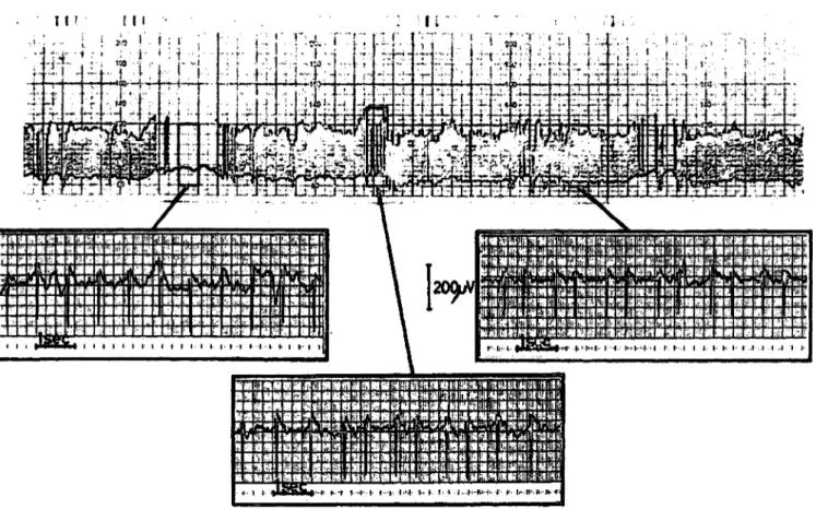
The exclusion of artefacts (e.g. alternating registra- tion of maternal and fetal heart rate) was elicited by simultaneous fetal abdominal ECG monitoring (Fig. 3). This confirmed the bigeminal or tri- geminal rhythms diagnosed by auscultation.
The abdominal ECG did not allow for an exact diagnosis äs to arrhythmia type because the P-wave was hidden in electrical interference. The QRS interval was prolonged at 0.085 seconds (normal ränge: 0.04 to 0.06 sec.), which was interpreted äs a sign of interventricular conduction delay.
A CTG was done at 2-3 days intervals, where the fetal heart rate was monitored with the internal logic of the HP 8020 A "turned off. The fetal arrhythmia persisted into the 38th week of preg- nancy.
0300-5577/82/0010-0085$02.00
ι ; , ' r : r o p r ι r f Γ: r r r ι 1 ϋ ··"' ο ο ο ο π ο s t a j 3 π.:ΐ :· π;~: η : ο.) J τ Vn ϋ$
.._
"· — ίη
.
Ρ — ,
*κ "Β"
—
TBj -1 -1 1
π
-1 Ηα
3fr
»- j J ΚΤ- 0- )0- 0-
ΟΗ
| Ν
—f
—
|
ι ι Τι.·*
J
Ι . ». C ι
π
Ier-11
•Η
Ρ
-10u
0- D- Kh 0"
ΙΟ- 0-
SP
W
u.
W
u u
4l
u V K
. -1
,*
u
R
H^i v„
| Λ
-a
Ι -1
-iJ
U
N
μ.(
Γ Q·
Γ
Os
10·
r
F Γ
v*
L
suc --'4
^\****
HX
ία**
t
·„
K
*
•M M) β
,,
)4
.yu
noT nnGO 1G
1r
1„T» i«•V..
1
C7T 1
fACrj«k>«CDC/i-PfiO d
• 1
1»
f t
Cf*OAOmp9370-oea
ECQ
eoo
*K v
TOO
K
9
«r.
•t*
yo
&
9·«
)5
-iAr
^
l* [
|
-Α-
Ι
ea*.foa^A m iau&
TUXtart
106
•wII
•
^
s
-K
-
ΊAh
n k
^
Fig. 1. Antepartal fetal heart rate curve in arrhythmia recorded on a conventional cardiotocograph. The same picture resulted in all conventional methods (ultrasound, phonographic, abdominal ECG). An Interpretation of the curve is not possible.
t » 11 l ·: l' f l 'i ;i *
i
•tea140
T T,**"·» £*«·-« ^«»t«
r^?iiM«««««j:^wt«iiKttStt«iiaiMi^iiat«»»^
^^if^llT?:^IIIBllP9HBI^{ll'|M«riM^'JR~·.**ΜΑ.··η*Ή7ϊ**£*ι£™?~*ι^ϊ*^^:^^
-*i**:>?«3;^^*i^*cr:^^^^
^<^^R1^^i'£'1!^i^
^»»»!^^^^
^«ai^
^«^K«^^" ' ~ "" " " ^ ' ^ ^ ' " ' - ' ^ ? r ^ ^ ! i i ^ ^f^;i5i?o^<s-r ^ ^ '
- T r **.· -
k^^
Fig. 2. Antepartal fetal heart rate recorded with ultrasound with Instrument logic switched off. The anhythmia is clearly to be seen in the middle part of the figure. The left and right part of the figure is disturbed by movement arte- facts.
FETUS
MOTHER FETUS
MOTHER
ΙΙΐΙΐΙΊΙΙΙ l
lllllllllllllllBIIHIIHJIIIIIIIlllfll ΙΙΙΜΙΙΰΠΊΐαίΙΠΙΙΙΙΙΙβΙΜ
ιιιιιιΐύΐΜι()ϊ!!!;ιιιΐ'!Μ^^
HllilllUlffllim^^
llIitfiiiUU«l(flllllljn l«iillN««lfflH
J. J.
1SEC
lliliiilllilHIIIPIIilH HilffllllliilHfllll!^
MlIlWilPftllftimilM fl!I ft!f(l«ra
• ΒΗΙΗΜΙΙΒ«^
flIHiiitlllliilJlllil·
•HB«iilflillliaiil«IW
•(aiiHiJiiflHiaiiiffln^
J
Fig. 3 The antepartal abdominal ECG clearly shows the bigeminal rhythms which can be heard by auscultation. Oniy the QRS complex is to be seen; other parameters such s the P or T wave cannot be recognized because of the super- imposed disturbances.
1.2 Intrapartum monitoring
Labor was induced in the 38th week of pregnancy after lung maturity had been verified with an L/S ratio of 3.5 and the cervix was found favorable for induction. Fig. 4 shows the intrapartum CTG tracings registered directly via scalp electrodes with logic on (A) and logic off (B). (C) reveals the simultaneous recordings of the maternal cardiac rate.
Thus s shown, the mean fetal heart rate was 70 bpm and the bradyarrhythmia became apparent only after the internal logic of the HP 8030 A had been turned off (Fig. 4b). Heart rate curves plotted via the fetal scalp ECG registered very few artefacts.
For this reason, some manufacturers have dis- pensed entirely with the internal logic in the direct cardiotocographic method (i.e. SIEMENS PARTE- CUST, COROMETRICS FM 112). In the Instrument 8030 A from HEWLETT PACKARD, tfie logic can be turned off via a switch for use in direct scalp CTG monitoring.
The fetal scalp ECG in our case history revealed an AV block with variable conduction (Fig. 5). Be- cause of the atypical CTG pattern, several fetal
scalp blood samples were obtained to assist in evaluation of fetal condition.
The intermittant MBA-pH measurements were supplemented by continuous subcutaneous tissue pH (tpH) measurements on the fetal scalp (R CHE 540 CTG/pH monitor) (Fig. 6).
A fetal hypoxia/asphyxia secondary to the fetal bradyarrhythmia could be excluded on the basis of the MBA pH or tpH values. A tissue pH of 7.24 corresponds to the initial value of the microsample pH of 7.29. The MBA pH did not change over a four hour period of labor (pH = 7.30), whereas the tissue pH feil a short time before delivery from the initial value to a tpH of 7.18, a drop of 0.6 units.
The tissue pH is closely correlated with the um- bilical arterial pH (UApH = 7.20, tpH = 7.18), and the capillary MBA pH is closely correlated with the umbilical vein pH (UVpH = 7.29, MBUpH = 7.30).
The fetal arrhythmia with varying frequencies is not detected by the CTG pH monitor. An extreme fetal bradycardia is falsely registered at normal tissue pH values (Fig. 6).
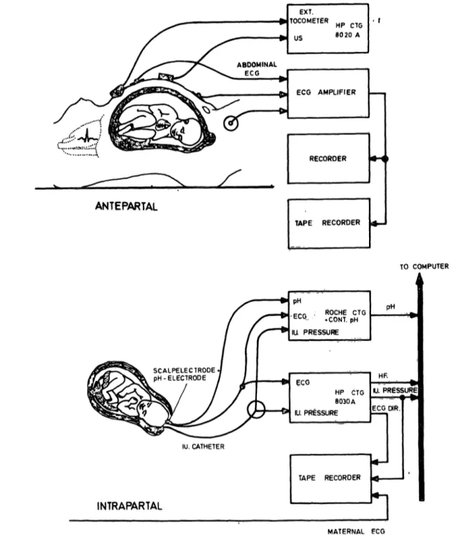
Fig. 7 gives an overall view of the antepartum and
.intrapartum devices.
233ΞΗΞΗΞΕΣΪ2ΞΗ33Ξ
• *f*VU!flr V»f.»Mne>
Fig. 4. A shows the heart rate curve registered during labor with Instrument logic switched on.
B shows the same curve with Instrument logic switched off. Here the arrhythmia present is clearly detected.
C simultaneously with fetal heart rate, the maternal heart rate was also registered. In this way, the possibility can be excluded that the maternal heart rate was registered in the bradycardiac phases of the arrhythmia.
1.3 Neonate
The 2850 g male newborn (arterial pH = 7.20, venous pH =7.29, APGAR 9/9/10) was transferred to the Dept. of Pediatrics because of a persistent bradycardia. Here the newborn showed AV-blocks varying from 1°—3° with the development of cardiac insufficiency. At first, suspicion of a ven- tricular septal defect was expressed, but cardiac catheterization did not reveal an organic heart defect. The child was kept for further observation in the pediatric cardiac division.
2 Discussion
In most cases of fetal arrhythmias, a technically satisfactory and interpretable continuous registra- tion of heart rate is hardly possible. The diagnosis is made by auscultation. According t KOM - ROMY [3], the most frequent fetal arrhythmias are extrasystoles and atrioventricular conduction disorders.
The problems of monitoring fetal arrhythmias are
discussed by way of the previous case history of a
fetal AV block.
u .-'l-i :·
mmmmmmmmmmmmmmmmmmmmmmmmmmmmmm
Ι&ίΊ&ΜάΙ&ίΚ^wa; π*ιι. TO (rannen taug*! m·· mm* im m« n» tiiti1$« m· min M ΜΜίικιΐίIHMm mn»m m
mmmmmmmmmmmmmmmmmmmmmmmmmmmmmmmm, , ^^^^
mmmi *mw®mmm\ s
Fig. 5. The assignment of the intrapartally recorded heart rate patterns to the corresponding ECG complexes is shown.
An AV block I-III could be diagnosed on the basis of the ECG complexes.
^ΗΓ«ΗΗΗ*η*ΗΗΚΗΗΗΗ^ τ·:-.:—.--κ—r H Fig. 6. In the lower part of the figure, the labor curve together with the subcutaneous tissue pH values measured on the scalp is to be seen. An application of the pH electrode is always worthwhile when an Interpretation of the heart rate curve in the conventional sense is not possible.
The heart rate curve recorded by the R che tocograph in the upper part of the figure falsely registers a pronounced bradycardia, cf. also Fig. 4b.
ANTEPARTAL
TAPE RECORDER
TO COMPUTER
SCALPELECTRODE pH-ELECTRODE
pH
•ECG IUL PRESSURE
R CHE CTG
*CONt. pH
pH
ECG
HP CTG 8 030 A IU. PRESSURE
HF.
IU. PRESSURE ECG CHR.
IU. CATHETER
INTRAPARTAL
TAPE RECORDER
MATERNAL FCG
Fig. 7. Schematic representation of the antepartal and intrapartal monitoring of the airhythmia described. Ante- partum peri d. Schematic representation of antepartum monitoring apparatus. HP 8020 A receives input from US (ultrasound heart rate monitor) and ext. tocometer. Simultaneous fetal abdominal ECG monitoring is amplified and recorded. Above data were stored on tape for documentation purposes. Intrapartum peri d. Schematic representa- tion of intrapartum (direct) monitoring apparatus. R che monitor receives input from continuous tpH measurement, ECG monitor and intrauterine pressure. HP CTG 8030 A receives ECG and intr uterine pressure input. Simultaneous maternal heart rate is monitored. Above data were also stored on tape and analysed by Computer.
-
Λ„ .
χ,
A,
r, i i GOCHBERG [21, this condition has an incidence of 2.1 Congenital AVblock
Λ. ~~
ΛΛΛί. , .
Λ Α 1.
ΛΓ,
Γ Α^
Γ6
l in 22000 live births. About 40 cases of AV con-
During pregnancy and labor, persistent fetal brady- duction block, diagnosed antepartum, are reported
cardia without signs of hypoxiaindicates the possi- in the literature [8]. Congenital heart malforma-
bility of a congenital AV-block. According to tions were found in about 40-50% of the cases. If
good.
Apart from frequent prenatal visits, answering parental questions about fetal cardiac defects and informing the pediatrician about the predicted date of delivery, further clinical consequences are not expected.
Congenital conduction disorders without cardiac malformations usually regress within days or weeks with normalization of heart rate.
2.2 Monitoring problems 2.2. l Antepartum period
Fetal arrhythmias are discovered mainly by auscul- tation or via the "audio" Output of the cardiotoco- graph. An interpretable heart rate curve can often not be obtained. In terms of differential diagnosis, the possibility of false registrations due to trigger uncertainties or maternal Signal artefacts must be considered [5].
With the Instrument logic criterias turned off and true beat-to-beat registration, the arrhythmia can be demonstrated by way of the heart rate curve.
In contrast to the direct (intrapartum) monitonng, where many commercially available cardiotoco- graphs posses the capability to turn off the inter- nal logic, only one monitor with the same capa- bility exists for external (antepartum) use, äs far äs we know (COROMETRICS FM 112). All others require electronic manipulation to "turn off" the internal logic criteria.
Another noninvasive technique (computation of systolic time intervals of the fetal cardiac cycle) to evaluate the cardiovascular performance of ar- rhythmic fetuses antepartum is described by SCHLOTTER [6].
2.2.2 Intrapartum period
Cardiotocographs with the capability of having the internal logic criterias turned off should be used for continuous monitoring of fetal bradyarrhyth- mias during delivery.
Since the exact detection of fetal heart rate is im- portant in fetal arrhythmia, some explanation äs to the way in which the cardiotocograph works is required:
change of the incoming heart rate. If this change falls outside of predetermined criteria, usually
± 14 bpm or ± 28 bpm, this value is rated äs an artefact, disregarded, and the previous value used (halt phase). Thus, an arrhythmia with the logic criteria on is not detected. Instead, a continuous heart rate curve with occasional halt phases is recorded. It is also possible to record a non- existent bradycardia.
If (äs in the present case) an arrhythmia with periodic beat-to-beat alterations is involved, then the two heart rate values have the same priority;*
the Instrument logic thus cannot make any distinc- tion for one of the frequency values. As a result, no registration takes place in such cases. In the cardiotocograph of the pH monitor (Fig. 6), in this case it is always the lower heart rate value which is detected äs correct. The frequency jump which follows it (ca. 40—60 bpm) is regarded äs an artefact; the Instrument then goes into a halt phase. The following value is then a jump to the lower heart rate again, which is regarded äs correct by the cardiotocograph. A constant alternation between registration and halt phase thus takes place here. In consequence, a bradycardia is falsely recorded because this Instrument logic always decides only for the lower heart rate value.
During labor a persistent bradycardia must arouse suspicion that the maternal heart rate is being picked up through the fetal scalp electrode. Simul- taneous maternal heart rate monitoring should be used to exclude this possibility.
The nature of the fetal arrhythmia can be recog- nized via the directiy registered fetal ECG (Fig. 5), since all ECG complexes are detected. This can be of importance for postpartum neonatal manage- ment.
Since the CTG cannot be interpreted äs usual during fetal arrhythmia, frequent scalp pH meas- urements must be performed to detect hypoxia äs a possible cause or consequence of the brady- arrhythmia. Vaginal delivery is possible with normal pH-values.
Additional Information äs to the fetal condition
can be obtained by use of continuous subcutane-
ous tissue pH measurements (Fig. 6). However, äs
a method to detect compromised fetuses, this can-
not yet be routinely used [1,4].
In our case, since the tissue pH values changed before birth, the tissue pH corresponded better to only slightly during birth, we dispensed with more the umbilical artery pH tüan to the scalp capillary frequent scalp pH samples (once an hour). Just pH.
Summary
.Antepartum and intrapartum monitoring pioblems are discussed with reference to a case history of a fetal AV- block detected during the 36th week of pregnancy. CTG Instruments where the internal logic criteria can be turned off are suitable for monitoring arrhythmias. During labor the direct fetal ECG permits exact diagnosis äs to the type
of arrhythmia. The necessity of antepartum diagnosis in regard to postpartum pediatric management and hypoxic states during labor justify the large expenditure on moni- toring apparatus. During labor continuous tissue pH meas- urement via the fetal scalp give ädded Information äs to fetal condition.
Keywords: Antepartum and intrapartum monitoring, fetal arrhythmia, AV-block.
Zusammenfassung
Ein Beitrag zur Überwachung fetaler Arrhythmien An Hand eines in der 36. SSW festgestellten fetalen A-V- Blockes wird die Problematik der ante- und intrapartalen apparativen Überwachung erörtert. Zur Arrhythmie- Überwachung sind CTG-Geräte geeignet, bei denen die logische Schaltung abgestellt werden kann. Die konti- nuierliche Gewebe-pH-Messung am fetalen Skalp unter
der Geburt gibt zusätzliche Informationen über den fe- talen Zustand. Das direkte fetale EKG unter der Geburt erlaubt eine exakte Diagnose des Arrhythmietyps, Die Notwendigkeit der Diagnosestellung für die postpartalen pädiatrischen Maßnahmen sowie die Erkennung asphyxie- bedingter fetaler Gefahrenzustände rechtfertigen den ho- hen apparativen Aufwand.
Schlüsselwörter: Apparative Überwachung antepartum und intrapartum, AV-Block, fetale Arrhythmie.
Resume
Une contribution au monitoring des aiythmies fetales Les problemes de monitoring antepaitales et intrapartales sont discutes en se referant a un cas d'un bloc A-V fetal trouve pendant la 36 semaine de grossesse.
Les appareils CTG presentant la possibilite de couper les contacts logiques internes sont appropries pour la surveill- ance des arythmies.
Pendant le travail, l'ECG fetal dkect permet une diagnose exacte du type d'arythmie. La necessite de la diagnose antepartale en regard du traitement pediatrique et k reconnaissance des etats hypoxiques durant le travail, justifient les larges depenses d'appaieils de monitoring.
Durant le travail k mesure continue du pH-tissulaire via le cräne fetal donne une inforrnation supplementaire sur k condition fötale.
Mots-cles: Arythmie fetale, monitoring fetal antepartale, monitoring fetal intrapartale.
Bibliography
[1] BOOS, R., H. RÜTTGERS, D. MULIAWAN, D.
HEINRICH, F. KUBLI: Continuous measurement of tissue pH in the human fetus. Arch. GynecoL 226
(1978)183
[2] GOCHBERG, S. H.: Congenital heart block. Amer. J.
Obstet. Gynec. 88 (1964) 238
[3] KOMÄROMY, B., J. GAAL, L. LAMPE: Foetal arrhythmia during pregnancy and labour. Brit. J.
Obstet. Gynaec. 84 (1977) 492
[4] KUBLI, F., H. RÜTTGERS, K. WERNICKE (eds.):
Proceedings of the fkst international Workshop on tissue pH measurements in obstetrics. Arch. Gynecol.
226 (1978) No. 1-2
[5] RÜTTGERS, H.: Technik, Registrierungsprinzipien und Registrierfehler von Kardiotokographen. In:
FISCHER, W. M. (ed.): Kardiotokographie, Lehr- buch und Atlas. Thieme, Stuttgart 1976
[6] SCHLOTTER, C. M.: Antepartale nichtinvasive Ab- klärung peristierender Rhythmusstörungen des feta- len Herzens Geburtsh. u. Frauenheilk. 41 (1981) 32 [7] SHENKER, L.: Fetal electrocardiography. Obstet.
Gynecol. Surv. 21 (1966) 367
[8] SHENKER, L.: Fetal cardiac arrhythmks. Obstet.
Gynecol. Surv. 34 (1979) 561
[9] STAMM, O. .U. LATSCHA, P. JANECEK, A. CAM- PANA: Kontinuierliche pH-Messung am kindlichen Kopf post partum and sub partu. Z. Geburtsh. Pe- rinat. 178 (1974) 368
Received May 12, 1981. Accepted January 27,1982.
Dr. med. Rainer Boos Universitäts-Frauenklinik Voßstraße 9
D-6900 Heidelberg FRG