Kolkenbrock et al.: Stromelysin from rheumatoid synovial fluid 625 Eur. J. Clin. Chem. Clin. Biochem.
Vol. 31, 1993, pp. 625-631
© 1993 Walter de Gruyter & Co.
Berlin · New York
A Trypsin Sensitive Stromelysin Isolated from Rheumatoid Synovial Fluid is an Activator for Matrix Metalloproteinases
By H. Kolkenbrock*, Adelheid Hecker-Kia
1, Dagmar Orgel
1, G. Buchlow
1, H. S rensen
2, W. Hauer
2and N. Ulbrich
11
Deutsches Rheuma-Forschungszentrum Berlin, AG Biochemie, Berlin, Germany
2
Immanuel-Krankenhaus GmbH, Rheumaklinik, Berlin, Germany
(Received April 16/June 21, 1993)
Summary: The processing of synovial fluids of patients suffering from rheumatoid arthritis led to the characterization of a neutral metalloproteinase with polymorphonuclear leukocyte progelatinase and poly- morphonuclear leukocyte procollagenase activating properties. The activator exhibits a relative molecular mass of M
t27000 and is an active form of Stromelysin. Thus, it reacts specifically with antibodies raised against human Stromelysin, splits polymorphonuclear leukocyte progelatinase in a manner characteristic of Stromelysin, and is inhibited by EDTA as well as by a tissue inhibitor of metalloproteinases (TIMP-2).
The activator shows a high specificity for the matrix metalloproteinases, polymorphonuclear leukocyte progelatinase and polymorphonuclear leukocyte procollagenase. It shows only weak hydrolysis of casein and gelatin, and it does not activate fibroblast Μ
τ72 000 progelatinase. Brief treatment with trypsin does not lead to a significant change in the activator's relative molecular mass, but induces a rapid loss of its activating activity for polymorphonuclear leukocyte progelatinase, while its proteolytic activity against the synthetic substrate, N-(2,4)-dinitrophenyl-Pro-Gln-Gly-Ile-Ala-Gly-Gln-Z)-Arg, is increased about 3-fold. The same tryptic treatment does not affect the activator's proteolytic activity towards casein and gelatin.
Introduction ., . „ ^ , .
Matrix metalloproteinases are secreted as latent pro- Matrix metalloproteinases play an important role in enzymes by different cell types, such as fibroblasts, remodelling of the extracellular matrix (1, 2). Since macrophages and polymorphonuclear leukocytes, in the structural elements of the extracellular matrix are response to certain stimuli. An important regulatory all substrates, the activity of these enzymes must be mechanism involves activation of the latent enzymes strictly regulated. In certain diseases, e. g. rheumatoid in the extracellular space. In vitro metalloproteinases arthritis, these control mechanisms do not function can be activated by organomercury compounds. Most properly, leading to destruction of the extracellular matrix metalloproteinases are also activated by tryp- matrix. In the special case of rheumatoid arthritis, sin.
destruction of the articular cartilage finally leads to j
tj
sassumed that the physiological activator for the the destruction of the whole joint. matrix metalloproteinases, collagenase
1) and Stromelysin
1), might be plasmin
1). Activated strome- ') Stromelysm EC 3.4.24.17 tysin (formerly called proteoglycanase) has a rather
Polymorphonuclear leukocyte gelatinase EC 3.4.24.35 broad spectrum of activities; it hydrolyses not only
Fibroblast collagenase EC 3.4.24 7 ^ ,
A^
A^ proteoglycan, but also other components of the ex-
Polymorphonuclear leukocyte collagenase EC 3.4.24.34 * „ , ' . , , . . „,
Trypsin EC 3.4.21.4 tracellular matrix, such as lamimn, fibronectm, gela-
Plasmin EC 3.4.21.7 tins and collagen types III, IV, V and IX, and it is Eur. J. Clin. Chem. Clin. Biochem. / Vol. 31,1993 / No. 10
626 Kolkenbrook et al.: Stromelysin from rheumatoid synovial fluid
also reported to be a "superactivator" for plasmin- activated collagenase, enhancing the collagenase ac- tivity about 10-fold (3-5). Recently, it was reported that stromelysin is an activator for polymorphonu- clear leukocyte progelatinase (6). Thus, stromelysin is not only an important proteinase with regard to its activity towards components of the extracellular ma- trix, but seems to play a key role in regulation of the activities of other matrix metalloproteinases.
Here we report some newly discovered properties of stromelysin (isolated from rheumatoid synovial fluid) that appear to extend the regulatory involvement of this enzyme.
Materials and Methods Materials
Rheumatoid synovial fluid originated from patients of the Rheuma-Klinik,' Immanuel-Krankenhaus, Berlin. Buffy coat was kindly supplied by Deutsches Rotes Kreuz, Berlin. Ultrogel AcA 44, blue-Trisacryl, heparin-Ultrogel and phorbol myristate acetate were purchased from Serva, Heidelberg. Gelatin-Se- pharose was prepared in our laboratory. Sepharose 4-B was obtained from Pharmacia, Freiburg. 4-Aminophenylmercury acetate, plasmin (3 U/mg) and molecular mass markers for gel electrophoresis were obtained from Sigma. Protein concentra- tions were determined with the bicinchoninic acid reagent (Pierce, USA) with bovine albumin as standard.
Enzyme assays
Matrix metalloproteinase activities were assayed with the syn- thetic substrate N-(2,4)-dinitrophenyl-Pro-Gln-Gly-Ile-Ala- Gly-Gln-Z)-Arg (dinitrophenyl-labelled peptide) (7). Activity against protein substrates, such as gelatin and casein was as- sayed by the fluorescamine method (8). Activator activity was analysed by incubating the activator with polymorphonuclear leukocyte progelatinase for 30 min at 37 °C in the presence of the dinitrophenyl-labelled peptide. The amount of substrate hydrolysed by the activated polymorphonuclear leukocyte ge- latinase was taken as a measure of activator concentration.
Isolation of matrix metalloproteinases
Polymorphonuclear leukocyte progelatinase and polymorpho- nuclear leukocyte procoilagenase were purified from buffy coat after stimulation with phorbol-myristate acetate. The secreted metalloproteinases were purified essentially as described (9,10).
The MT 72000 progelatinase and the MT 72000 progelatinase- TIMP-2 complex were isolated according to 1. c. (11).
Activator purification
To 100 ml of rheumatoid synovial fluid, saturated ammonium sulphate solution was added to 10% saturation. After 60 min in the cold the precipitate was removed by centrifugation at 40000g for 30 min, and the clear supernatant was adjusted to 50% ammonium sulphate saturation. After 24 h at 4 °C the precipitate was collected by centrifugation at 150000g for 20 min, washed twice with 60ml ammonium sulphate solution (313 g/1) and centrifuged again. The sediment was dissolved in 20 mmol/1 imidazple-HCl, pH 6.1, 0.5 g/1 Brij 35, 0.5 g/1 NaN3
and dialysed against 20 mmol/1 imidazole-HCl, pH 6.1, 5 mmol/1 CaCl2, 0.5 g/1 Brij 35,0.5 g/1 NaN3 (buffer A) followed
by dialysis against buffer A. The sample was chromatographed on carboxymethyl-trisacryl (5.5 cm χ 11 cm) equilibrated in buffer A. After washing with buffer A, bound protein was eluted with buffer A containing 0.5 mol/1 NaCl and stored at
—20 °C. After 300 ml of synovial fluid had been processed the carboxymethyl-Trisacryl eluates were combined, dialysed against 50 mmol/1 Tris-HCl, pH 7.0,200 mmol/1 NaCl, 5 mmol/1 CaCl2, 1 μηιόΐ/ΐ ZnCl2, 0.5 g/1 NaN3 (buffer B) and chromato- graphed on gelatin-Sepharose (4 cm χ 3 cm) equilibrated in buffer B. Unbound protein was concentrated by ultrafiltration (Amicon, YM-10 membrane) and applied to Ultrogel AcA 44 (3 cm χ 100 cm) equilibrated in buffer B. The activator-con- taining fractions were concentrated by ultrafiltration, dialysed against 20 mmol/1 Tris-HCl, pH 8.0; 5 mmol/1 CaCl2, 1 μιηοΐ/l ZnCl2, 0.5 g/1 Brij 35, 0.5 g/1 NaN3 and applied to blue- Sepharose (2.5 cm χ 7 cm) equilibrated in the same buffer. The activator was slightly retarded on blue-Sepharose, and activa- tor-containing fractions were concentrated by ultrafiltration, dialysed against buffer A and applied to heparin-Ultrogel (1.5 cm χ 8 cm) equilibrated with buffer A. The activator did not bind to heparin-Ultrogel and was further purified by gel filtra*
tion on a calibrated AcA 44 (2crn χ 100 cm) equilibrated in buffer B.
All following investigations of the matrix metalloproteinase activating properties of the activator were performed with poly- morphonuclear leukocyte progelatinase.
Demonstration of activator activity in SDS-PAGE The procedures employed are essentially those of Hibbs et al.
(9). To determine the polymorphonuclear leukocyte progelati- nase activating activity of the protein seen in polyacrylarnide gel electrophpresis in the presence of sodium dodecyl sulphate (SDS-PAGE), 1 μg activator was electrophoresed on a 12.5%
gel. After electrophoresis, the gel was washed twice for 15 min in buffer B, pH 7.6, containing 25 g/1 Triton X-100. After rinsing the gels briefly (5 min) in the above buffer without Triton X-100, the gel was sliced into 1 mm sections, and each slice was extracted with 200 ul buffer B for 24 h at 4 °C. 90 μΐ of each extract were mixed with 10 μΐ (2 μg) polymorphonuclear leukocyte progelatinase and 100 μί dinitrophenyl-labelled pep- tide and incubated at 37 °C. After 24 h the amount of hydro- lysed substrate was determined.
To examine the gelatinase activity of the protein seen on SDS- PAGE, 60 ng activator were electrophoresed on a 10% SDS- PAGE, containing 2 g/1 gelatin at 4 °C. The gel was washed as described above and the zymogram was developed in 50 mmol/1 Tris-HCl, 5 mmol/1 CaCl2, 1 μηιοΐ/ΐ ZnCl2, 10 g/l Triton X-100, 0.2 g/l NaN3, pH 7.6 for 24 h at 37 °C. The gels were stained with Coomassie brilliant blue.
pH-Optimum
Five μΐ (1 μg) polymorphonuclear leukocyte progelatinase were incubated with 5 μΐ (15 ng) activator and 40 μΐ buffer B adjusted to pH 6.0, 6.5, 7.0, 7.5, 8.0, 8.5 and 9.0. After l h at 37 °C, 50 μΐ of a solution containing 200 mmol/1 Tris-HCl, pH 7.0;
200 mmol/1 NaCl, 5 mmol/1 CaCl?, 0.5 g/1 NaN3 and 100 μΐ dinitrophenyHabelled peptide were added. After an incubation of 30 min at 37 °C the reaction was terminated and the amount of hydrolysed dinitrophenyl-labelled peptide was determined.
Sensitivity to trypsin
3 Activator (6 μg) in 198 μΐ buffer B was incubated with 22 μΐ bovine trypsin (1 g/1) at 37 °C. Periodically 3.3 μΐ were taken and added to 5 μΐ aprotinin (1 g/1) arid 81.7 μΐ buffer B to assay the activity against polymorphonuclear leukocyte progelatinase.
To measure the activity against the dinitrophenyl-labelled pep-
Kolkenbrock et al: Stromelysin from rheumatoid synovial fluid 627 tide, 25 μΐ of the mixture were added to 5 μΐ aprotinin and 70 μΐ
buffer B. The reaction was started with either 100 μΐ dinitro- phenyl-labelled peptide alone for 18 h (activity against dinitro- phenyl-labelled peptide) or with the same substrate and addi- tionally ΙΟμΙ (2μ§) polymorphonuclear leukocyte progelati- nase for 30 min (activator-activity).
Sensitivity to plasmin
The same procedure as described for the trypsin1) sensitivity was applied to the investigation on the activator sensitivity towards plasmin (l g/l).
Results
Activator purification
The amount of activator that can be isolated from synovial rheumatoid fluid depends largely on the kind of treatment the patients received. Treatment with glucosteroids leads to a very low content or even a lack of metalloproteinases in the synovial fluid. Using only synovial fluid of patients without glucosteroid treatment we isolated usually 30—100 μg activator from 100 ml synovia.
Employing the method described, an activator for polymorphonuclear leukocyte progelatinase and poly- morphonuclear leukocyte procollagenase with a mo-
Mr ^
66000 H*
45000 ^ 36000 «*·
29000 24000
B M
20100
Fig. 1.
14200
Polyacrylamide gel electrophoresis in the presence of sodium dodecyl sulphate and zymogram of purified activator.
Lane A: 1 μg activator on 12.5% polyacrylamide gel;
electrophoresis in the presence of sodium dodecyl sul- phate. Lane B: 60 ng activator on 10% polyacrylamide;
gel electrophoresis in the presence of sodium dodecyl sulphate containing 2g/l gelatin. Lane M: Molecular mass markers (MT): bovine albumin (66 000), ovalbumin (45 000), glyceraldehyde-S^phosphate dehydrogenase (36000), carbonic anhydrase (29000), trypsinogen (24000), trypsin inhibitor (20100) and a-lactalbumin (14200).
lecular mass of M
r27 000 was purified from human rheumatoid synovia. No activator could be detected in synovial fluid itself. The activator is detectable usually after Ultrogel AcA 44 chromatography, some- times earlier, after gelatin-Sepharose chromatogra- phy. Up to this purification step, identification of the activator is based on its ability to hydrolyse the di- nitrophenyl-labelled peptide.
The relative molecular mass of the activator was de- termined in different ways: on a calibrated Ultrogel AcA 44 column the activator was eluted in the range M
r26000-28000, in 12.5% polyacrylamide gel elec- trophoresis in the presence of sodium dodecyl sul- phate a main band at M
T27000 with one or two minor bands with lower relative molecular masses could be visualized and a gelatin-zymogram showed proteolytic activity at the same position (fig. 1). When larger quantities of activator were applied, an addi- tional band at M
T25 000 was observed on the zymo- gram. Assay of activator in eluates of sliced 12.5%
SDS-polyacrylamide gels showed activator activity associated with the region corresponding to M
r26000-28000.
Activity towards polymorphonuclear leuko- cyte procollagenase and fibroblast M
T72 000 progelatinase
The activator quickly activated polymorphonuclear leukocyte procollagenase, as already described (IS- IS). Fibroblast M
T72000 progelatinase was not acti- vated.
pH-Optimum
The activator shows a pH-optimum between pH 7.0 and 8.0 (data not shown).
Inhibition of the activator
The activator was inhibited by EDTA and the activity could not be restored by dialysing against buffer B.
It was also inhibited by a slight excess of the M\
72000 progelatinase-TIMP-2 complex (fig. 2), which acts in the same manner as TIMP-2 (11).
Trypsin-sensitivity
The activator displayed a pronounced sensitivity to trypsin; after 30 min incubation at 37 °C more than 80% of the activator-activity was lost. In contrast, under identical conditions, a 30 min incubation re- sulted in a threefold enhancement of the hydrolysis of the dinitrophenyHabelled peptide (fig. 3). Treat-
Eur. J. Clin. Chem. Clin. Biochem. / Vol. 31,1993 / No. 10
628 Kolkenbrock et al.: Stromelysin from rheumatoid synovial fluid A B C M
205000
116000
9740
°
{'j 66000
45000
Fig. 2. 8% Polyacrylamide gel electrophoresis in the presence of sodium dodecyl sulphate. Inhibition of activator by EDTA and by fibroblast Mr 72 000 progelatinase-TIMP- 2-complex.
2μ§ polymorphonuclear leukocyte progelatinase in 10 μΐ buffer Β were incubated for 45 min at 37 °C with 150 ng activator in 5 μΐ buffer B preincubated for 5 min at 37 °C with (Lane A) 800 ng MT 72000 progelatinase- TIMP-2 complex in 20 μΐ buffer B, (Lane B) 20 μΐ buffer B, and (Lane C) 20 μΐ of 100 mmol/1 Tris/HCl, 50 mmol/1 EDTA, 200 mmol/1 NaCl 0.5 g/l NaN3, pH 7.0.
(Lane M). Molecular mass markers (MT): myosin (205000), -galactosidase (116000), phosphorylase b (97400), bovine albumin (66000) and ovalbumin (45000).
300-
^
.-g* 200- T5COI
7= 10040 50 60
t [min]
Fig. 3. Trypsin-sensitivity of the activator activity.
6 μg activator in 200 μΐ buffer B were incubated with 22 |ig trypsin in 22 μΐ 1 mmol/1 HC1 at 37 °C. At the indicated times aliquots were withdrawn and activator activity towards polymorphonuclear leukocyte proge- latinase (open circles) and dinitrophenyl-labelled pep- tide (solid circles) was determined.
merit with trypsin for 25 min at 37 °C did not alter the proteolytic activity of the activator against casein and gelatin (fig. 4). The action of trypsin reduced the molecular mass of the activator by M
r1000-2000, as shown by Western-blot analysis (fig. 5). Prolonged incubation with trypsin led to the destruction of the activator.
150
t [min]
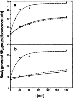
Fig. 4. Hydrolysis of casein (a) and gelatin (b) by the activator, trypsin-treated activator and by trypsin.
100 μg gelatin or casein, respectively, were incubated in a total volume of 200 μΐ buffer B with 1.2 μg activator (open circles), 1.2 μg activator preincubated with trypsin at 37 °C (solid triangles) or with 1.3 μg trypshi (solid circles). At the indicated times newly generated amino groups were determined with the fluorescamine method.
The tryptic treatment of the activator was stopped with 6 μg aprotinin after 25 min.
Plasmin-sensitivity
Under the conditions employed plasmin had no effect on the activator (data not shown). Even an incubation of 15 h at 37 °C did not change the activator's activity towards polymorphonuclear leukocyte gelatinase or the dinitrophenyl-labelled peptide.
Activation of polymorphonuclear leukocyte gelatinase
The treatment of polymorphonuclear leukocyte pro- gelatinase with the activator led via an M
r86000 intermediate to an active gelatinase with a molecular mass of M
r82000, (fig. 6) exactly as described for the action of Stromelysin on polymorphonuclear leu- kocyte progelatinase (6).
Substrate specificity
The specific activity of the activator against the di- nitrophenyl-labelled peptide was 40 mU/mg, which is about 4% of polymorphonuclear leukocyte gelatinase activity against that substrate (11). It can be deduced from figure 4 that the activator's activity against casein is about 3 times higher than against gelatin.
Compared with trypsin, however, the activator's acti-
vity towards these substrates is rather low, i.e. less
than 10% of the trypsin activity towards gelatin and
Kolkenbrock et al.: Stromelysin from rheumatoid synovial fluid 629
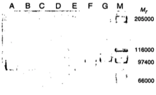
A B A J3_C D E F G M M
" ; f "J 205000
116000 96400 80400 55700 43700 38800
116000
• i · ; * ^ t / J 97400
ί ' 66000
Fig. 5. Immunoblotting of activator and of trypsin treated ac- tivator.
600 ng activator in 20 μΐ buffer Β (Lane A) and 600 ng activator in 20 μΐ buffer Β incubated with 2 μg trypsin in 2 μΐ 1 mmol/1 HC1 at 37 °C for 30 min (Lane B) were run on a 12.5% polyacrylamide gel in the presence of sodium dodecyl sulphate, blotted on a Millipore Im- mobilon membrane and blocked overnight with bovine serum albumin. The blots were incubated with an an- tiserum raised in sheep against human Stromelysin, fol- lowed by peroxidase-labelled anti-sheep-IgG. The re- active bands were visualized with 4-chloro-l-naphthol.
The tryptic treatment was stopped with 2 μg aprotinin in 2 μΐ distilled water. The position of the apparent molecular mass for prestained protein is indicated: β- galactosidase (116 000), fructose-6-phosphate kinase (96400), pyruvate kinase (80400), ovalbumin (55700), lactic dehydrogenase (43 700) and triosephosphate iso- merase (38 800).
about 20% of that towards casein. Trypsin and acti- vator are compared with respect to their polymor- phonuclear leukocyte progelatinase activating pro- perties in table 1, where it can be seen that the rela- tionship is reversed, i.e. the activator is nearly 30 times more efficient than trypsin in the activation of polymorphonuclear leukocyte progelatinase.
Tab. 1. Activation of polymorphonuclear leukocyte progelati- nase by activator and trypsin
2 μg polymorphonuclear leukocyte progelatinase in 100 μΐ buffer B and varying amounts of activator displaying a molecular mass of Mr 27 000 or bovine trypsin dis- playing molecular mass of Mr 24 000, respectively, were mixed with 50 μg dinitropheiayl-labeiled peptide in 100 μΐ buffer B and incubated for 30 min at 37 °C.
Activator (ng/200 μΐ) 300
4560 7590
Substrate hydrolysed (nmpl)
03.8 7.85.5 129.9
Trypsin (ng/200 μΐ)
6000 12001800 2400
Substrate hydrolysed (nmol)
02.5 7.95.2 11.2
45000
Fig. 6. Activation of polymorphonuclear leukocyte progelati- nase with activator.
l μg polymorphonuclear leukocyte progelatinase in 5 μΐ buffer B was incubated with 30 ng activator in 1 μΐ buffer B for 60 (Lane A), 45 (Lane B), 30 (Lane C), 20 (Lane D), 10 (Lane E), 5 (Lane F) and 0 (Lane G) min, respectively, and analysed on 8% polyacrylamide gel electrophoresis in the presence of sodium dodecyl sul- phate. Lane M: molecular mass markers (Mr): myosin (205 000), -galactosidase (116 000), phosphorylase 97400), bovine albumin (66000) and ovalbumin (45000).
Discussion
From the synovial fluid of patients suffering from rheumatoid arthritis, we isolated an activator for polymorphonuclear leukocyte procollagenase and polymorphonuclear leukocyte progelatinase, with a relative molecular mass of M
T27000. Furthermore, we demonstrated that the activator is an active form of the matrix metall proteinase Stromelysin; the ac- tivator is inhibited by EDTA and the M
T72000 ge- latinase-TIMP-2 complex, which acts as an inhibitor for matrix metalloproteinases the same way as TIMP (11), thereby establishing that the activator is a mem- ber of the family of matrix metalloproteinases. The activator reacts with antibodies raised against human Stromelysin, its pH-optimum lies in the neutral range, and it splits the polymorphonuclear leukocyte gela- tinase in a manner characteristic of Stromelysin.
The activator was not only found in synovia of pa- tients suffering from rheumatoid arthritis but also in the synovia of patients with osteoarthritis or systemic lupus erythematosus during an inflammatory attack.
The activator could not be detected in synovia from non-inflammatory joints, e. g. distorsion of the knee.
Synovia subsequently found to contain a high con- centration of activator also displayed a general en- hancement of matrix metall proteinase activity. Ac- tivator activity was not detectable in synovial fluids but became apparent after the Chromatographie step
Eur. J. Clin. Chem. Clin. Biochem. / Vol. 31,1993 / No. 10
630 Kolkenbrock et al.: Stromelysin from rheumatoid synovial fluid
on Ultrogel AcA 44, sometimes earlier, after gelatin- Sepharose. Latent prostromelyin is therefore activated during the purification procedure. Whether this is due to the action of other proteinases or to an autocata- lytic process is still unclear.
We believe that an activator for polymorphonuclear leukocyte collagenase and polymorphonuclear leu- kocyte gelatinase that was isolated from synovial fluid (12-14) and from the culture media of synoviocytes (15) many years ago is also stromelysin. However, this activator had a reported relative molecular mass of M
T35 000 and it lacked proteolytic activity against casein and gelatin. We could not detect activator activity in the relative molecular mass range of M
r35 000, either on a calibrated Ultrogel AcA 44, or by eluting protein from sliced SDS-polyacrylamide elec- trophoresis gels.
Polymorphonuclear leukocyte progelatinase, poly- morphonuclear leukocyte procollagenase, fibroblast M
r72000 progelatinase, casein, gelatin and dinitro- phenyl-labelled peptide were examined as substrates for the proteolytic activity of the activator. With the exception of the fibroblast M
r72000 progelatinase, the activator hydrolysed all these substrates, but showed a rather high degree of specificity for poly- morphonuclear leukocyte progelatinase and polymor- phonuclear leukocyte procollagenase. The activator was much less active than trypsin in the proteolysis of casein and gelatin. However, although trypsin is a potent activator for polymorphonuclear leukocyte progelatinase (16), the stromelysin activator is about 30 times more efficient. The activator displays a simi- lar efficiency in the activation of polymorphonuclear leukocyte collagenase, while the fibroblast M
T72 000 progelatinase is not a substrate for the activator. The high specificity for the matrix metalloproteinases dis- played by neutrophilic granulocytes may indicate a physiological role of this activator in the activation of these proteinases. This specificity may also explain why the M
T35 000 activator described earlier was not found to hydrolyse gelatin and casein; activator con- centrations too low to significantly hydrolyse gelatin and casein are still sufficient to rapidly activate the polymorphonuclear leukocyte progelatinase.
Hydrolysis of the dinitrophenyl-labelled peptide by the activator was slow compared with the rate of hydrolysis by polymorphonuclear leukocyte gelati- nase. However, a short tryptic treatment, that did not lead to a significant change in the molecular mass of the activator (fig. 5), enhanced the activator's activity against the dinitrophenyl-labelled peptide about threefold, destroyed its ability to activate polymor- phonuclear leukocyte progelatinase and did not affect
its activity against casein and gelatin. It might there- fore be concluded that a similar physiological proteo- lytic event may significantly increase the activator's activity against some components of the extracellular matrix. Plasmin, a proteinase with proteolytic proper- ties similar to trypsin, plays an important role under physiological conditions in the activation of procol- lagenase (MMP-1) and prostromelysin, but it had no effect on the activator.
It could be argued that the three different trypsin sensitivities of the activator might be due to the pres- ence of two different proteinases that react with tryp- sin in different ways:
1. the activator (active stromelysin) which hydrolyses the dinitrophenyl labelled peptide, casein/gelatin and the polymorphonuclear leukocyte progelati- nase, and which is inactivated by trypsin;
2. a latent metalloproteinase which, after activation by trypsin, could be responsible for the enhanced turnover of the dinitrophenyl labelled peptide, and which at the same time compensates the loss of the caseinolytic/gelatinolytic activity of the activator.
According to the relative molecular mass this me- talloproteinase could only be matrilysin (MMP-7), but so far this enzyme has only been found in the uterus (17) and in developing macrophages (18).
In addition, in synovial fluid, no such small latent metalloproteinase could be detected after chro- matography on Ultrogel AcA 44 (not shown).
The distinct sensitivity to trypsin of the activator's proteolytic activity towards different substrates sug- gests that during the transformation from prostro- melysin to active stromelysin at least one switch po- sition is passed. At this switch position a putative regulatory mechanism may have the option of quickly deciding whether stromelysin should function as an activator for matrix metalloproteinases, or conco- mitantly, with enhanced activity, as a proteinase of other substrates.
Recently, it was reported that treatment of prostro- melysin withp-aminophenylmercury acetate led to the formation of an activator for polymorphonuclear leu- kocyte progelatinase with a relative molecular mass of Μ
τ47000 (6). This activator was not investigated with regard to its sensitivity to trypsin and it would be interesting to know if this M
r47000 activator behaves in the same way as the Μ
τ27 000 activator . described in the present report.
The activity of matrix metalloproteinases in the ex-
tracellular space is subject to regulation on at least
three different levels — expression, activation, and
interaction with specific inhibitors. The present data
Kolkenbrock et al.: Stromelysin from rheumatoid synovial fluid 631
extend this regulatory repertoire by the observation that stromelysin may be further processed to active species displaying distinct and separate functions as activators or hydrolytic enzymes of macromolecules of the extracellular matrix.
We are now investigating whether a mechanism is present in the synovial fluid of patients afflicted by
rheumatoid arthritis, which under physiological con- ditions may initiate a cascade of events, producing and processing stromelysin as described here in vitro.
Acknowledgement
This work was supported by Hoechst AG, Frankfurt am Main, Germany.
References
1. Matrisian, L. M. (1992) The matrix-degrading metallopro- teinases. BioEssays 14, 455—463.
2. Birkedal-Hansen, H., Werb, Z., Welgus, H. & Van Wart, H. (eds.) (1992) Matrix Metalloproteinases and Inhibitors Gustav Fischer, Stuttgart, Jena, New York.
3. Murphy, G., Cockett, M. I., Stephens, P. E., Smith, B. J.
& Docherty, A. J. P. (1987) Stromelysin is an activator of procollagenase. A study with natural and recombinant en- zymes. Biochem. J. 248, 265-268.
4. Ito, A. & Nagase, H. (1988) Evidence that human rheu- matoid synovial matrix metalloproteinase 3 is an endoge- nous activator of procoUagenase. Arch. Biochem. Biophys.
2(57,211-216.
5. He, Gh., Wilhelm, S. M., Pentland, A. P., Manner, B. L., Grant, G. A., Eisen, A. Z. & Goldberg, G. J. (1989) Tissue cooperation in a proteoolytic cascade activating human interstitial collagenase. Proc. Natl. Acad. Sei. USA 86, 2632-2636.
6. Ogata, Y., Enghild, J. J. & Nagase, H. (1992) Matrix metalloproteinase 3 (stromelysin) activates the precursor of the human matrix metalloproteinase 9. J. Biol. Chem. 257, 3581-3584.
7. Masui, Y., Takemoto, T, Sakakibara, S. & Hori, H. (1977) Synthetic substrates for vertebrate coUagenase. Biochem.
Med. 17, 215-221.
8. Macartney, H. W. & Tschesche, H. (1980) Latent collage- nase from human polymorphonuclear leukocytes and ac- tivation to collagenase by removal of an inhibitor. FEBS Lett. 119, 327-332.
9. Hibbs, M. S., Hasty, Κ. Α., Sever, J. M., Kang, A. H. &
Mainardi, C. L. (1985) Biochemical and immunological characterization of the secreted forms of human neutrophil gelatinase. J. Biol. Chem. 250, 2493-2500.
10. Hasty, Κ. Α., Hibbs, M. S., Kang, A. H. & Mainardi, C.
L. (1986) Secreted forms of human neutrophil collagenase.
J. Biol. Chem. 257, 5645-5650.
11. Kolkenbrock, H., Orgel, D., Hecker-Kia, Α., Noack, W. &
Ulbrich, N. (1991) The complex between a tissue inhibitor of metalloproteinases (TIMP-2) and 72-kDa progelatinase is a metalloproteinase inhibitor. Eur. J. Biochem. 198,775 — 12. Kruze, D. & Wojtecka, E. (1972) Activation of leucocyte781.
collagenase proenzyme by rheumatoid synovial fluid.
Biochim. Biophys. Acta 285, 436-446.
13. Wize, J., Sopata, I., Wojtecka-Lukasik, E., Ksiezny, S. &
Dancewicz, A. M. (1975) Isolation, purification and prop- erties of a factor from rheumatoid synovial fluid activating the latent forms of collagenolytic enzymes. Acta Biochimica Polonica 22, 239-250.
14. Dancewicz, A. M., Wize, J., Sopata, I., Wojtecka-Lukasik, E. & Ksiezny, S. (1978) Specific and nonspecific activation of latent collagenolytic proteases of human polymorpho- nuclear leukocytes. In: Neutral Proteases of Human Poly- morphonuclear Leukocytes (Havemann, K. & Janoff, A., eds.) pp. 373 — 383, Urban & Schwarzenberg, Inc. Balti- more—Munich.
15. Wize, J., Abgarowicz, T, Wojtecka-Lukasik, E., Ksiezny, S. & Dancewicz, A. M. (1975) Activation of human leu- cocyte procollagenase by rheumatoid synovial tissue culture medium. Ann. Rheum. Dis. 34, 520-523.
16. Kolkenbrock, H., M.-Ali, H., Hecker-Kia, A., Buchlow, G., S rensen, H., Hauer, R. W. & Ulbrich, N. (1991) Characterization of a Gelatinase from Human Rheumatoid Synovial Fluid Cells. Eur. J. Clin. Chem. Clin. Biochem.
29,499-505.
17. Woessner, J. F. jr. & Taplin, C. J. (1988) Purification and properties of a small latent matrix metalloproteinase of the rat uterus. J. Biol. Chem. 253, 16918-16925.
18. Busiek, D. F., Ross, F. P., McDonnell, S., Murphy, G., Matrisian, L. M. & Welgus, H. G. (1992) The matrix metalloproteinase matrilysin (PUMP) is expressed in de- veloping human mononuclear phagocytes. J. Biol. Chem.
257, 9087-9092.
Dr. Hansj rg Kolkenbrock
Deutsches Rheumaforschungszentrum Berlin Ostpreu endamm 111
D-12207 Berlin Germany
Eur. J. Clin. Chem. Clin. Biochem, / Vol. 31,1993 / No. 10