Peripartum Hypothalmus-Pituitary-Adrenal axis plasticity and its prevention by high-fat diet intake:
Focus on the adrenal gland
DISSERTATION ZUR ERLANGUNG DES DOKTORGRADES DER
NATURWISSENSCHAFTEN (DR. RER. NAT.) DER FAKULTÄT FÜR BIOLOGIE UND VORKLINISCHE MEDIZIN DER UNIVERSITÄT REGENSBURG
vorgelegt von Clara Valentina Perani
aus Bergamo, Italien im Jahr 2014
Das Promotionsgesuch wurde eingereicht am: 18.02.2014
Die Arbeit wurde angeleitet von: Prof. Dr. rer. nat. Inga D. Neumann Unterschrift:
Dissertation
Durchgeführt am Institut für Zoologie der Universität Regensburg unter Anleitung von
Prof. Dr. rer. nat. Inga D. Neumann
I
Table of Contents
Prologue ... 1
Chapter 1: General Introduction ... 3
1.1. The stress response ... 3
1.2. The adrenal glands ... 7
1.2.1. The adrenal cortex ... 8
1.2.1.1. CORT synthesis ... 9
1.2.2. Adrenal cholesterol utilisation ... 11
1.2.2.1. Receptor mediated cholesterol endocytosis ... 13
1.2.2.2. Cholesterol selective uptake ... 14
1.2.2.3. Adrenal lipid droplets ... 15
1.2.2.4. Adrenal cholesterol synthesis ... 17
1.2.3. The adrenal medulla ... 18
1.3. HPA axis and hormonal changes across the estrous cycle ... 22
1.3.1. The rat estrous cycle ... 22
1.3.2. The human menstural cycle ... 25
1.3.3. Plasma ACTH and CORT levels across the rat estrous cycle ... 25
1.4. Hormonal and behavioural changes across the peripartum period ... 28
1.4.1. Pregnancy and lactation ... 28
1.4.1.1. Prolactin and Oxytocin ... 30
1.4.2. Changes in HPA axis function across the peripartum period ... 31
1.4.3. Adrenal plasticity might sustain the dissociation of plasma ACTH and CORT peripartum ………..35
1.4.4. Consequences of peripartum-associated changes on behaviour ... 36
1.5. Peripartum-associated mood and anxiety disorders and maternal obesity ... 38
1.5.1. Postpartum mood disorders ... 38
1.5.2. Risk factors for the development of postpartum mood and anxiety disorders ... 40
1.5.3. Obesity and HPA axis function ... 41
1.5.4. Maternal obesity ... 43
1.5.5. Animal models to study postpartum mood and anxiety disorders ... 43
1.5.5.1. Repeated stress based approaches... 43
1.5.5.2. High-fat diet based models ... 47
1.6. Aims of the thesis ... 49
Chapter 2: Adrenal gland plasticity across the estrous cycle and the peripartum period ... 50
II
2.2.1. Animals ... 54
2.2.2. Experimental design ... 54
2.2.3. Mating and verification of pregnancy ... 57
2.2.4. Killing - Blood and organ preparation ... 58
2.2.5. Oil red lipid staining ... 58
2.2.6. ACTH and OXT receptor autoradiography (RAR) ... 59
2.2.7. ACTH-R and OXT in situ hybridization ... 59
2.2.8. Western blot analysis ... 61
2.2.9. Establishment of an adrenal cell in vitro incubation protocol ... 61
2.2.10. Adrenal cell incubation across the peripartum period ... 62
2.2.11. Statistics ... 62
2.3. Results ... 63
2.3.1. ACTH and CORT levels across the estrous cycle and adrenal plasticity ... 63
2.3.1.1. Plasma measures ... 63
2.3.1.2. Adrenal weight, protein expression and cortical lipid droplets ... 64
2.3.2. Peripartum-associated changes in plasma ACTH and CORT and in vitro CORT production from isolated adrenal cells ... 66
2.3.2.1. Plasma CORT and ACTH ... 66
2.3.2.2. CORT release from isolated adrenal cells from V rats in vitro ... 67
2.3.2.3. CORT release from isolated adrenal cells in vitro from V, PD 21 and LD 8 animals ... 68
2.3.3. Peripartum-associated adrenal plasticity ... 68
2.3.3.1. Adrenal weight ... 68
2.3.3.2. ACTH-R mRNA and autoradiography across pregnancy and lactation ... 69
2.3.3.3. OXT mRNA and RAR ... 70
2.3.3.4. Adrenal lipid droplets ... 71
2.3.3.5. Adrenal protein expression ... 73
2.4. Discussion ... 75
Chapter 3: High-fat diet feeding and pregnancy chronic stress effects on maternal HPA axis- associated changes……….. ... 84
3.1. Introduction ... 84
3.2. Materials and Methods ... 87
3.2.1. Animals ... 87
3.2.2. Experimental design ... 87
3.2.2.1. High-fat diet study ... 87
III
3.2.2.2. Pregnancy-stress study ... 89
3.2.3. Diet ... 91
3.2.4. Maternal care... 91
3.2.5. Light-dark box (LDB) test ... 92
3.2.6. Jugular catheter implantation ... 93
3.2.7. Pregnancy chronic psychosocial stress procedure ... 95
3.2.8. Blood and organ preparation ... 96
3.2.9. Oil red lipid staining ... 96
3.2.10. Western blot analysis ... 96
3.2.11. ACTH-RAR ... 97
3.2.12. Adrenal glands cells isolation and incubation in vitro ... 97
3.2.13. Statistics ... 98
3.3. Results ... 99
3.3.1. HF diet effect on body and adrenal weight ... 99
3.3.1.1. Body weight ... 99
3.3.1.2. Absolute adrenal gland weight ... 100
3.3.2. Effects of HF diet feeding on maternal, retrieval and anxiety-related behaviour ... 100
3.3.2.1. Maternal care... 100
3.3.2.2. Anxiety-related behaviour ... 101
3.3.3. Effects of HF diet feeding on adrenal-related parameters (Cohort 1) ... 103
3.3.3.1. Adrenal ACTH-RAR and lipid droplets ... 103
3.3.3.2. Adrenal protein expression ... 104
3.3.3.3. Adrenal cells incubation in vitro ... 106
3.3.4. Effects of HF diet feeding on HPA axis (re)activity ... 107
3.3.4.1. Plasma ACTH and CORT before and after acute stress exposure (Cohort 2) ... 107
3.3.4.2. Plasma CORT before and after ACTH intravenous injection (Cohort 3)... 110
3.3.5. Chronic stress effects on body and adrenal weight ... 112
3.3.5.1. Body weight ... 112
3.3.5.2. Absolute adrenal glands weight ... 113
3.3.6. Chronic stress effects on adrenal parameters ... 114
3.3.6.1. Adrenal lipid droplets ... 114
3.3.6.2. Adrenal protein expression ... 114
3.4. Discussion ... 116
Chapter 4: Lactation-associated changes in adrenal cholesterol delivery in the mouse ... 126
4.1. Introduction ... 126
4.2. Materials and methods ... 129
4.2.1. Animals ... 129
IV
4.2.4. Blood and organ preparation ... 131
4.2.5. Oil red lipid staining ... 131
4.2.6. Western blot analysis ... 131
4.2.7. Statistics ... 132
4.3. Results ... 133
4.3.1. Basal ACTH and CORT levels in virgin and lactating mice ... 133
4.3.2. Adrenal weight and lipid droplets ... 133
4.3.3. Adrenal protein expression ... 134
4.4. Discussion ... 136
Chapter 5: General Discussion ... 140
5.1. Outlooks... 147
5.2. Summary - concluding remarks ... 150
References ... 151
Abbreviations ... 171
Acknowledgments ... 174
Curriculum Vitae ... 175
Publications ... 176
Author’s declaration - Eidesstattliche Erklärung ... 177
1
Prologue
The peripartum period represents a time of substantial plasticity in a wide-variety of systems of the maternal organisms in order to ensure the well-being of both the mother and the offspring. For its primary role in the maintenance of body homeostasis, the maternal Hypothalamus-Pituitary-Adrenal (HPA) axis profoundly adapts and animal models have been extensively used to study such plasticity given that particularly the findings from rodent studies were demonstrated to be highly translational to humans.
Through basic and human research, several peripartum changes in HPA axis–associated hormonal levels as well as alterations in brain and pituitary morphology and physiology have been already identified. However, to my knowledge, despite the importance of the adrenal glands for the overall HPA axis function as the site of glucocorticoids synthesis and secretion, glucocorticoids that are the main HPA axis peripheral effectors, direct evidence of peripartum adrenal plasticity is missing.
Emerging evidence indicates that interference with peripartum HPA axis plasticity participates in the aetiology of mood and anxiety disorders; among them postpartum depression (PPD) and anxiety. Indeed, while the peripartum period is generally associated with increased calmness and decreased stress responses, for a substantial subset of mothers this period represents a time of particular risk for the onset of psychiatric disorders.
Intriguingly, human and rodent studies demonstrated that dysregulation of the basal- and stress-induced neuroendocrine HPA axis peripartum (re)activity feature in multiple pathologies, among them PPD, anxiety and obesity. Specifically, the previously mentioned illnesses are often characterized by unbalanced CORT and adrenocorticotropic hormone (ACTH) basal and stress-induced levels, ACTH which is the main CORT
2
peripartum period, and if so, whether disruption of such changes may contribute to the aetiology of peripartum-associated pathologies.
In the following Chapter 1, I will first provide an overview of the stress response physiology with a detailed description of adrenal gland’s morphology, steroidogenic pathways and cholesterol delivery, as the main part of my thesis will focus on these aspects. Furthermore, I will describe the hormonal adaptations occurring across the estrous cycle and the peripartum period particularly regarding ACTH and CORT levels.
The last part of the general introduction chapter provides a description of the animal models that have been shown to induce dysregulation of the maternal HPA axis that are, therefore, commonly used to study potential mechanisms underlying PPD, anxiety and the mechanisms of comorbidity between obesity and these disorders.
The following three experimental chapters describe and discuss in detail the experiments I performed whereas in Chapter 5 a general discussion of the present thesis findings is provided, together with future perspectives and conclusions.
3
Chapter 1: General Introduction
1.1. The stress response
There are two major components that participate to mount an appropriate physiological response upon stress exposure, the HPA axis and the sympathetic nervous system. The former is involved in the regulation of several physiological functions, e.g. energy metabolism and immune function (Dallman et al., 1995, Straub et al., 2001, Ulrich-Lai and Herman, 2009) and its major role is to maintain body homeostasis and thus, concert the physiological response to external or internal changes or to threat exposure.
After exposure to a stressor, sensory information is driven to the brain to recruit the specific effectors to mount an appropriate response. Specifically, HPA axis activity is driven by corticotrophin-releasing factor (CRF) whereas its co-secretagogue vasopressin (AVP) is believed to amplify the stress response (Gillies et al., 1982); both CRF and AVP are released from the parvocellular neurons of the paraventricular nucleus (PVN) of the hypothalamus at the level of the median eminence. They reach anterior pituitary corticotropic cells through the hypophyseal portal system where they stimulate ACTH release and production mainly inducing calcium influx. ACTH is the main stimulator of adrenal steroidogenesis and reaches the adrenal glands via the circulatory system (Young and Akil, 1985, Familari et al., 1989, Aguilera and Rabadan-Diehl, 2000). The CRF- containing neuronal population of the PVN receives a great variety of projections, among them ascending brainstem fibers that promote HPA axis activation, angiotensinergic projections from the circumventricular organs that activate the HPA axis in case of osmotic stress and from several basal forebrain and hypothalamic pathways (Gillies et al., 1982). The main HPA axis peripheral effectors are glucocorticoids (cortisol in humans
4
and corticosterone in rodents; hereafter CORT), steroid hormones produced and secreted from the adrenal gland cortex in an intermittent manner (pulsatile and circadian) and acutely after stress exposure (Gan and Quinton, 2010) (details about adrenal morphology and steroidogenic pathways that are relevant for my thesis are provided in the introduction Section 1.2).
CORT binds two receptor subtypes that are widely distributed throughout the body, the mineralocorticoid receptor (MR) and the glucocorticoid receptor (GR). The former regulates several functions such as body mineral balance, while the latter participates to the regulation of a multitude of physiological processes, among them immune and cardiovascular function, reproduction and cognition and energy metabolism, particularly regarding glucose (Oakley and Cidlowski, 2013). MR and GR belong to the nuclear hormone receptors superfamily and are located in the cytosol when inactive although plasma membrane located receptors have also been identified; the membrane-located receptors are believed to mediate glucocorticoids effects that do not rely on genomic mechanisms (Zhang et al., 2012). As glucocorticoids are lypophilic they can freely cross the membrane barrier to bind to the receptor (MR or GR), which becomes activated and translocates into the nucleus where it modulates the activity of target promoter genes (Bamberger et al., 1996). Besides their peripheral effects, glucocorticoids exert several function at the brain and pituitary levels, effects that directly regulate HPA axis function.
Indeed, their action is of primary importance in the maintenance of basal HPA activity and its stress-induced activation and for the termination of the stress response via feedback mechanisms (De Kloet et al., 1998) (Figure (Fig.) 1).
In contrast to the HPA axis-related stress response activation, which takes several minutes, the sympathetic nervous system responds within seconds to challenges with its activity coordinated by the locus coeruleus and its effector being adrenaline released from the adrenal medulla (Chrousos and Gold, 1998) (Fig. 1). The locus coeruleus is the main
5 source of noradrenaline in the brain; it receives CRF projections from the central nucleus of the amygdala, the bed nucleus of the stria terminalis and from the PVN that directly modulate its activity and elicit its stress-induced activation, projections that terminate in the peri-coerulear region where locus coeruleus dendrites extend (Valentino and Van Bockstaele, 2008). The locus coeruleus projects to preganglionic sympathetic neurons, which are predominantly cholinergic, in the intermediolateral cell column of the thoracolumbar spinal cord. These preganglionic neurons project directly to the adrenal chromaffin cells or to prevertebral and paravertebral ganglia where they synapse with noradrenergic postganglionic neurons that, in turn, project to the target organs. Activation of the sympathetic nervous system is necessary to cope with stressful situations or threats and results in an increase of the circulating levels of adrenaline, mainly from adrenal chromaffin cells production, and noradrenaline, from sympathetic neuron’s terminal.
Through interactions with the two main adrenoreceptor classes, namely alpha- and beta- adrenoreceptors, adrenaline and noradrenaline coordinate the cardiovascular, respiratory, gastrointestinal, renal and endocrine systems function and increase energy mobilization (Molinoff, 1984).
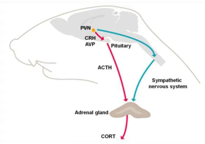
6
Figure 1. Schematic representation of the Hypothalamus-Pituitary-Adrenal (HPA) axis and sympathetic nervous system physiological response to stress
The sympathetic nervous system and the HPA axis orchestrate the stress response. The sympathetic pregnanglionic neurons from the toracolumbar spinal cord are activated upon stress exposure. These neurons innervate directly the adrenal medulla. HPA axis activation upon stress exposure results in corticotrophin releasing factor (CRF) and vasopressin (AVP) release from the paraventricular nucleus of the hypothalamus (PVN). CRH and AVP stimulate the release of adrenocorticotropic hormone (ACTH) from the anterior pituitary, ACTH that induces glucocorticoids (CORT) synthesis and release form the adrenal cortex. Adapted from www.kalsbeekgroup.nl/.
The experimental sections of the present thesis focus on studies that have been performed in rats and mice particularly at the adrenal gland level. Therefore, in the following introduction chapters, I will specifically describe some aspects of rodent physiology with a special focus on the adrenal glands and HPA axis-related hormones.
However, for completeness, cross references to the human physiology, particularly when
7 markedly divergent to the rodent condition are reported; although less details are available for humans.
1.2. The adrenal glands
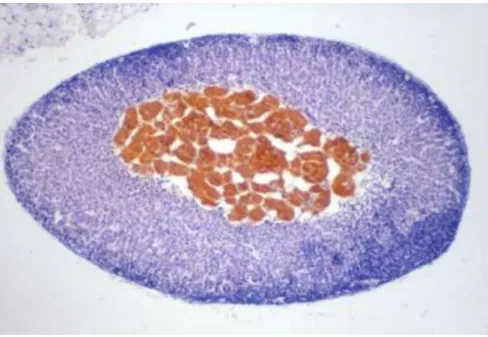
The adrenal glands are paired endocrine glands located in the retroperitoneal space close to the kidneys. The glands are macroscopically divided into two main regions, which are structurally and functionally distinct, the inner medulla and the outer cortex, and are surrounded by a connective tissue envelop termed the capsule (Fig. 2). Since the rat adrenal glands comprise the main body of my thesis, below I will describe the physiology, structure and function of these glands in detail.
Figure 2. Macroscopic structure of the rodent adrenal glands: the adrenal medulla and cortex
The chromaffin medullary cells constitute the adrenal glands core; in this photograph from a rat adrenal, the medulla is yellow coloured due to chromium staining. The area surrounding the medulla is the adrenal cortex. From www.pathbase.net
8
1.2.1. The adrenal cortex
As previously mentioned the cortex represents the adrenal structure located directly below the capsule and is dedicated to steroid hormone synthesis. Adrenal steroids are divided in two classes: glucocorticoids, with carbohydrate metabolism-regulating action and mineralocorticoids, with electrolyte balance-regulating functions. The adrenal cortex can be divided in three main regions depending on the hormonal production: the outer glomerulosa, the intermediate located fasciculata and the inner reticularis zone (Fig. 3).
The main product of the glomerulosa zone is aldosterone, a mineralocorticoid hormone that enhances renal sodium and water retention whereas the fasciculata and reticularis areas are responsible for CORT production (Rosol et al., 2001). In humans, the reticularis produces also androgens, specifically dihydroepiandosterone and androstenedione, hormones that in rodents are produced mainly by the gonads (Rodgers, 1990, Sharpe, 1990).
9 Figure 3. Photomicrographs of hematoxylin and eosin stained sections from the rat adrenal The adrenal medulla is surrounded by the innermost adrenal cortex area, the reticularis which is followed by the intermediate adrenal cortex area, the fasciculata (A). The outermost zone of the adrenal cortex is the glomerulosa (A) that is surrounded by the capsule (B). Adapted from Junqueira and Carneiro, Histologie, Springer, 2005.
1.2.1.1. CORT synthesis
As mentioned above, CORT is produced in the adrenal fasciculata and reticularis zones through a multiple-step reaction that is mainly controlled by ACTH. Optimal steroidogenesis is achieved upon ACTH binding to the adrenal ACTH receptor (ACTH-R), a seven transmembrane protein G coupled receptor whose activation increases cyclic adenosine monophosphate (cAMP) intracellular levels via adenylate cyclise activation that, in turn, activates protein kinase (PK) A (Gallo-Payet and Payet, 2003). Activation of the cAMP-PKA pathway is involved in both, the acute and chronic regulation of steroidogenesis and cholesterol is the precursor of all steroid hormones. The acute response occurs within minutes upon stimulation and induces cholesterol mobilization to the inner mitochondria membrane where the enzyme, cytochrome P450 (CYP) side-chain cleavage enzyme (CYP11A1 or P450scc), catalyzing the first steroidogenic reaction is located (see below). The chronic response to cAMP-PKA activation involves transcription of genes encoding for components of the steroidogenic machinery (Hu et al., 2010). The first step of adrenal steroidogenesis is, therefore, the transport of unesterified cholesterol to the inner mitochondrial membrane, transport that involves the steroidogenic acute regulatory (StAR) protein (Stocco, 2000). The two major classes of enzymes responsible for CORT biosynthesis are the cytochrome P450 (CYP) and the hydroxysteroid dehydrogenase (HSD) enzymes. Specifically, the P450 enzymes are membrane bound proteins associated with the mitochondrial membrane or the endoplasmic reticulum and
10
catalyze the hydroxylation and cleavage of the steroid substrates (Payne and Hales, 2004). On the other hand, HSD enzymes are involved in the reduction and oxidation of steroids and are membrane-bound enzymes located in the endoplasmic reticulum and in the mitochondria (Payne and Hales, 2004).
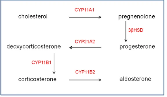
The first step in steroid hormone synthesis is catalyzed by the enzyme CYP11A1 that cleaves the carbon side-chain of the cholesterol molecule to produce pregnelonone (Fan and Papadopoulos, 2013) (Fig. 4). Pregnenolone is converted by the enzyme 3β-HSD into progesterone that, in turn, is converted in 11-deoxycorticosterone through the action of CYP21A2, enzyme located in all three adrenal cortex regions. The final step in CORT biosynthesis is mediated by the enzyme CYP11B1, which converts deoxycorticosterone in CORT (Mukai et al., 1993). In the glomerulosa zone, CORT is converted in aldosterone through the action of the enzyme aldosterone synthase (CYP11B2).
Figure 4. Schematic representation of the steroidogenic process in the rodent adrenal The rate-limiting step in CORT biosynthesis is the conversion of free cholesterol into pregnenolone through the action of the cytocrome P 11A1 (CYP11A1; P450scc) enzyme. 3β hydroxysteroid dehydrogenase (3βHSD) converts pregnenolone in progesterone that is, in turn, converted in deoxycorticosterone by CYP21A2. The final step in CORT production is catalyzed by the enzyme
11 CYP11B1 whereas, in the adrenal glomerulosa, CORT is converted to aldosterone by the enzyme CYP11B2.
The differential distribution of the steroidogenic enzymes confers the specificity of the hormonal production to each adrenal cortex area. Indeed, the enzyme CYP11B2 is exclusively located in the adrenal glomerulosa (Vinson, 2003). On the other hand, the steroidogenic enzyme expression pattern is species specific; this variability underlies the diversity in adrenal hormonal production across species. Thus, in rodents, the adrenal fasciculata and reticularis do not produce cortisol and androgens, which are the main hormonal products of the human adrenal gland. Indeed, mice and rats adrenals do not express the enzyme CYP17, which is involved in the conversion of pregnenolone and progesterone into cortisol and dehydroepiandrostenedione (Perkins and Payne, 1988, Rainey et al., 2002).
1.2.2. Adrenal cholesterol utilisation
As stated above, cholesterol is the substrate for steroid hormone synthesis and its availability for conversion into pregnenolone by the enzyme CYP11A1 represents the rate limiting step in steroidogenesis. Therefore, besides the importance of ACTH to stimulate CORT synthesis, cholesterol supply is also critical for optimal hormonal production (Hu et al., 2010).
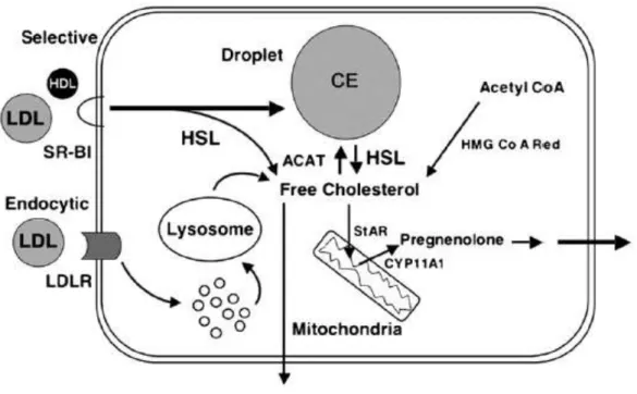
The adrenal gland’s cholesterol is supplied from several sources (Fig. 5): it can be uptaken through receptor-mediated endocytosis via low density lipoprotein receptor (LDLR) or selectively uptaken from LDL and high density lipoproteins (HDL); a process
12
mediated by the scavenger receptor class B type 1 (SRB1). Cholesterol can be de novo synthesised within the gland from acetyl CoA with being the rate limiting enzyme and, finally, adrenal cholesterol esters stores can be recruited (Kraemer, 2007, Hu et al., 2010).
Figure 5. Adrenal cholesterol delivery and storage dynamic
Cholesterol can be derived from three different sources. It can be selectively uptaken from plasma high density lipoproteins (HDL) and low density lipoproteins (LDL) through the action of scavenger receptor B1 (SRB1) whereas LDL cholesterol can also be recruited via LDLR internalization mediated by the LDL receptor (R). Cholesterol esters (CE), stored in adrenal lipid droplets and synthesised from free cholesterol by the enzyme acyl Coa cholesterol acyltransferase (ACAT), can be hydrolyzed by the hormone sensitive lipase (HSL) enzyme. Cholesterol can be de novo synthesised from Acetyl Co through a multiple-step reaction involving hydroxymethylglutaryl CoA reductase (HMG Co A Red) activity. Free cholesterol is transported to the inner mitochondrial membrane by steroidogenic acute regulatory (StAR) protein and the first step in steroidogenesis is catalyzed by the enzyme cytocrome P(CYP) 45011A1 that converts cholesterol into pregnenolone (Kraemer, 2007).
13 Cholesterol is transported through the aqueous circulatory system packed in plasma lipoproteins, which are complex aggregates of lipids and proteins. Four lipoprotein classes can be identified based on the density of the lipoproteins aggregates that form upon ultracentrifugation, chilomicrons, very low density lipoproteins (VLDL), low density lipoproteins (LDL) and high density lipoproteins (HDL), which differ in both composition and physical properties. The high steroidogenic activity of the adrenal cortex results in pronounced cholesterol metabolism and lipid exchange with the circulation. The adrenals express two lipoprotein receptors that uptake cholesterol via two distinct mechanisms, the LDLR and the SRB1, which are discussed in detail in the two next sections.
1.2.2.1. Receptor mediated cholesterol endocytosis
The LDLR, localized at the plasma membrane, interacts exclusively with LDL - and cholesterol uptake through this pathway is named receptor-mediated uptake. LDL directly interacts with the LDLR, and this complex migrates to coated pits, which are plasma membrane regions specialized in internalization of substances from the circulation. The coated pits first invaginate, and then come away from the plasma membrane as vesicles that fuse with early endosomes in the cytoplasm where the complex LDL-LDLR gets degraded because of the endosomal pH fall (Fielding and Fielding, 1996, Hu et al., 2010).
Here, the LDLR must undergo enough conformational change to release the ligand without getting denatured in the acidic endosomal environment. After dissociation, the LDLR leaves the endosome, apparently via incorporation into a membrane of a vesicle that buds from the endosome surface. These receptor-mediated endocytotic mechanisms allow reuse of the receptor every 10-20 minutes and an LDLR can make up to 150 trips through the endosomes without losing its function (Goldstein and Brown, 1985, Brown and Goldstein, 1986). The released (or internalised) LDL cholesterol esters are hydrolized in
14
the early endosomes and free cholesterol accumulates in the late endosomes/lysosomes.
Free cholesterol can then be esterified through the action of the enzyme acyl CoA:
cholesterol acyltransferase (ACAT) for storing in cytoplasmic lipid droplets or used immediately. There have been two ACAT subtypes identified to date, ACAT1 and ACAT2, but in adrenal cells ACAT1 is the major isoenzyme and contributes to more than 90% of the total ACAT activity (Chang et al., 2006). In addition to its storage into lipid droplets, cholesterol delivered via this LDLR-mediated pathway may be directed to the plasma membrane or to the mitochondria for steroid hormones synthesis (Sugii et al., 2003, Miller, 2007).
1.2.2.2. Cholesterol selective uptake
The SRB1 is responsible for “selective cellular cholesterol uptake”. This receptor is a ~ 82 kilodalton cell surface glycoprotein, which is highly expressed in tissues that are dependent on cholesterol from plasma lipoproteins for hormone production like the adrenal glands. Selective uptake via the SRB1 differs from the previously described receptor-mediated endocytosis as it occurs on the cell surface with cholesterol esters being internalized without lipoprotein endocytosis. Moreover, whilst the LDLR interacts exclusively with LDL, SRB1 binds a broader variety of ligands, among them HDL and native-, oxidized- and acetylated-LDL. SRB1 cholesterol ester uptake occurs in two steps:
binding of the lipoprotein to the receptor and transfer of the lipids from the lipoprotein to the plasma membrane. SRB1 was suggested to bind plasma lipoproteins via mechanisms involving direct protein-to-protein contact. Specifically, an amphiphathic α- elices motif present on different lipoproteins is hypothesised to be involved (Williams et al., 2000). The mechanisms involved in cholesterol ester transfer to the plasma membrane are poorly understood to date. One hypothesis is that the SRB1 forms a sort
15 of “channel” through which the cholesterol esters penetrate the cells driven by a concentration gradient (Vinals et al., 2003). Cholesterol esters, after being internalized in the adrenal cell, are hydrolyzed by a neutral cholesterol ester hydrolase (Sparrow and Pittman, 1990), which is believed to be hormone sensitive lipase (HSL) in adrenal cells (Kraemer and Shen, 2002). Existing literature reports that in murine adrenals and ovaries the fatty acids component of the cellular cholesterol esters differs from the circulating one;
strengthening the hypothesis that after uptake from plasma lipoproteins, cholesterol esters are hydrolyzed and then re-esterified before storage (Egwim and Kummerow, 1972). Free cholesterol from this selective uptake pathway may be driven to the mitochondria for steroidogenesis and in an unstimulated cell it would probably be stored in lipid droplets (details about adrenal lipid droplets dynamic are reported in the following Section) (Connelly and Williams, 2003).
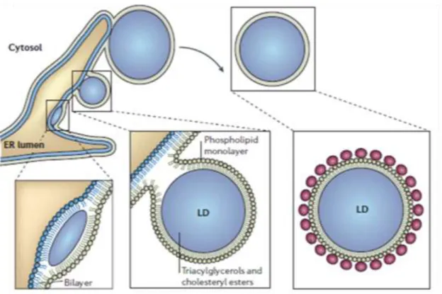
1.2.2.3. Adrenal lipid droplets
Cellular lipid droplets are considered to be a mobile, dynamic and complex organelle with a neutral lipid core surrounded by a monolayer of phospholipids and proteins (Martin and Parton, 2005). The primary function of lipid droplets is the storage and supply of cholesterol and fatty acids. In adrenal cells and other steroidogenic cells, the droplets are particularly enriched in cholesterol esters to provide the precursor for steroid hormone production. Lipid droplets form in the endoplasmic reticulum via mechanisms, which have been just partially addressed (Fig 6). It is for example still obscure whether they remain in physical contact with the endoplasmic reticulum after their formation or whether become totally independent organelles (Martin and Parton, 2006).
16
Figure 6. Model for cellular lipid droplet formation
Mature lipid droplets (LD) are hypothesised to bud from the endoplasmic reticulum (ER) to form mature organelle with a triacylglycerols and cholesterol esters core surrounded by a phospholipid monolayer. Adapted from (Martin and Parton, 2006).
Lipid droplets are rapidly formed and catabolized depending on the cell requirements for cholesterol and lipids. It is, therefore, intuitive how ACTH, the main adrenal tropic hormone, strongly affects adrenal lipid droplets physiology. For example, long term ACTH administration was shown to induce the depletion of glomerusa zone droplets, with parallel increase in plasma aldosterone (Mazzocchi et al., 1986). Interestingly, several acute and chronic stress paradigms were demonstrated to impact adrenal lipid droplets in animal models. Specifically, 60 minutes of high temperature exposure was reported to markedly increase plasma ACTH and CORT, together with depletion of adrenal fasciculata zone droplets in rats (Koko et al., 2004). The effect of chronic stress on the
17 pool of cholesterol esters within the adrenal appears to depend on the stress paradigm and species employed. In a study performed in rats subjected to chronic noise exposure, stress reduced adrenal fasciculata-associated lipid droplets (Oliveira et al., 2009), while chronic psychosocial stress in mice does not affect this parameter (Fuchsl et al., 2013).
Animal models of chronic stress are important to study the pathogenesis of diseases linked to HPA axis dysregulation like depression or anxiety (Slattery et al., 2012).
Therefore, understanding of possible adrenal participation in the pathology of these diseases is particularly relevant.
1.2.2.4. Adrenal cholesterol synthesis
The adrenal glands can also synthesise cholesterol from acetyl Co A via a multistep reaction. The enzyme catalyzing the rate-limiting step of the cholesterol production is HMGCR, a reductase that converts hydroxymethylglutaryl-Co A into mevalonate.
HMGCR is an important regulatory loci of cholesterol synthesis and both, sterol and non- sterol agents, were shown to impact on HMGCR function and expression. Cultured cells contain a maximal amount of HMGCR when incubated in the absence of serum. Addition of LDL to the culture suppresses HMGCR expression by 90% (Goldstein and Brown, 1977, Faust et al., 1982) suggesting that cholesterol uptake inhibits intracellular cholesterol de novo synthesis. Mevalonate is not exclusively a precursor for cholesterol synthesis. Several other non-sterol substances synthesised via mechanisms involving HMGCR may therefore, regulate HMGCR together with lipoproteins derived cholesterol (Brown and Goldstein, 1980).
Adrenal synthesis may supply adequate cholesterol for steroidogenesis. However, it is likely that plasma lipoproteins are recruited when steroidogenesis is sustained like during
18
the peripartum period, especially when considering that the maternal organism shows hyperlipidemia with plasma lipoprotein cholesterol increase that parallels the rise in CORT (Desoye et al., 1987).
1.2.3. The adrenal medulla
The adrenal medulla is located in the organ’s core and represents about 10% of the total gland size. This tissue is considered as a paraganglia because sympathetic pre- ganglionic neurons of the splanchnic nerve innervate the main medulla cell type, namely the chromaffin cells, via acetylcholine release. This neurotransmitter is synthesised through a multi-step reaction with the enzyme tyrosine hydroxylase (TH) catalysing the rate limiting step in catecholamines synthesis (Fernstrom and Fernstrom, 2007). TH is regulated acutely by phosphorylation at different sites whose phpsphorylation increase TH activity (Haycock and Wakade, 1992) chronically by protein synthesis. Adrenal TH mRNA, protein levels and phosphorylation are partially regulated by neurotransmitters released by the splanchnic nerve and, therefore, variation of the expression level are believed to mirror changes in the sympathetic firing to the adrenals (Brooks et al., 1997, Ong et al., 2011). Acetylcholine is hydrolyzed with consequent firing interruption through the action of the acetylcholinesterase enzyme (Fig. 7). Specifically, sympathetic pre- ganglionic neurons originating from the intermediolateralcell column of the spinal cord, enter the adrenal capsule and, after branching and travelling throughout the cortex forming an extensive neuronal network, innervate the adrenal medulla (Strack et al., 1989, Parker et al., 1993, Mravec, 2005). Normally, sympathertic pre-ganglionic neurons synapse in the ganglia with acetylcholine being the neurotransmitter involved, whereas the originating postganglionic neurons reach and innervate target organs via noradrenaline release. Chromaffin cells, which release adrenaline or noradrenaline upon
19 stimulation, are therefore, considered as modified post-ganglionic sympathetic neurons.
Postganglionic sympathetic fibers, mainly noradrenergic, have also been identified in the adrenal medulla and cortex, processes that have been found to be generally anatomically associated with blood vessels (Toth et al., 1997, Delarue et al., 2001).
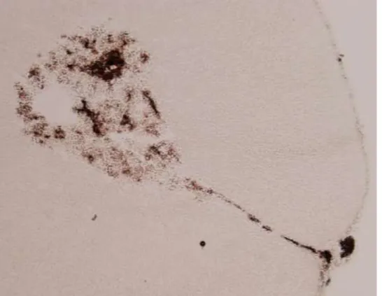
Figure 7. Rat adrenal glands acetylcholinesterase enzyme localization
The enzyme acethylcholinesterase localization can be visualized via the histochemical thiocholine method (Schober et al., 1997). The brown-coloured material results from acetylcholinesterase activity after adrenal slice exposure to the acetylcholinesterase substrate acetylcholine iodide.
Two sub-types of chromaffin cells can be distinguished depending on their secretory adrenergic or noradrenergic activity. In the adrenal medulla of the adult rat, 80-85% of the cells are adrenergic and primarily located adjacent to cortical cells and the remainder are therefore noradrenergic and mainly located in the centre of the medulla (Coupland, 1965).
20
Adrenaline and noradrenaline are stored in cytoplasmic secretory granules and released in response to a physiological signal by exocytotic mechanisms (Diaz-Flores et al., 2008).
Adrenergic and noradrenergic chromaffin cells are innervated by different sympathetic neurons that are functionally divided. This differential innervation of the two adrenal chromaffin cell populations is believed to underlie the selective release of adrenaline or noradrenaline in response to different insults. Thus, for example, insulin-induced hypoglicemia has been shown to activate adrenaline-secreting cells, whereas acute cold exposure has been reported to induce mainly noradrenaline release in the rat (Vollmer et al., 1992, Morrison and Cao, 2000). Brain areas that receive somatic and visceral information from the periphery including caudal raphe nuclei, ventromedial medulla, rostral ventrolateral medulla, A5 cell group, and PVN regulate the activity of the sympathetic neurons innervating the adrenal medulla (Mravec, 2005). Neurons whose cell body is located primarily in the adrenal medulla that innervate the adrenal cortex have also been identified. Specifically, vasoactive intestinal peptide-, substance P- and neuropeptide Y- containing ganglion cells have been recognised and they constitute the intrinsic adrenal innervation network (Engeland, 1998, Mravec, 2005). The rodent adrenal content of the above mentioned neuropeptide has been shown to vary depending, for example, on the age of the animal or after food deprivation (Chua et al., 1991). However, the physiological meaning and the mechanisms underlying these changes are still unclear (Bornstein and Ehrhart-Bornstein, 2000).
Steroidogenic and chromaffin cells have been to shown to be in physical contact (Fig. 8) and evidence of a mutual influence on the secretory activity of these different cells types has been reported by both in vitro and in vivo studies. For example, bovine adrenocortical cells basal CORT release in vitro was reported to be 10-fold higher in the presence of chromaffin cells; a coculture that also resulted in an increased expression of mRNA for several steroidogenic enzymes (Haidan et al., 1998). Adrenaline-mediated mechanisms
21 are hypothesised to be involved since stimulation of bovine cortical cells with adrenaline was reported to influence CORT secretion and accumulation of the mRNA encoding for the enzyme cholesterol side-chain cleavage cytochrome P450 (Ehrhart-Bornstein et al., 1991), the enzyme that catalyzes the first step of steroid hormones production (see Section 1.2.1.1). Moreover, ablation of the gene encoding for TH, the key enzyme involved in the synthesis of catecholamine, is associated with reduced basal CORT levels in vivo; despite no changes in plasma ACTH (Bornstein et al., 2000). On the other hand, adrenal steroids have also been demonstrated to influence the expression of enzymes involved in catecholamine synthesis (Haase et al., 2011). However, the exact mechanisms underlying the adrenal cortex and medulla cross-talk are poorly understood (Schinner and Bornstein, 2005).
Figure 8. Adrenal cortical-chromaffin physical contact
Light micrograph of the rat adrenal gland stained with hematoxylin-eosin; C cortex, M medulla, * medullary ray in the cortex (Pignatelli et al., 1998).
22
1.3. HPA axis and hormonal changes across the estrous cycle
1.3.1. The rat estrous cycle
In rodents, the estrous cycle, the ovarian cycle occurring in non-pregnant adult females, takes between three to five days resulting in multiple ovulations, while in women it takes between 25 and 30 days (called menstrual cycle) and only one oocyte is ovulated (Silberstein and Merriam, 2000, Westwood, 2008). The main hormones that are involved in the regulation of the estrous cycle-associated events are hypothalamic gonadotropin- releasing hormone (GnRH), pituitary luteinizing- and follicle-stimulating hormones (LH and FSH, respectively), progesterone and estrogens.
In the rat, female sexual maturity is reached immediately after the vaginal orifice opens around postnatal day 30, which is also when hormonal cycling begins. Hypothalamic GnRH neurons, mainly localised in the mediobasal hypothalamus, release GnRH in a pulsatile manner to the medial eminence, from where GnRH reaches the pituitary via the portal blood system, where it stimulates the synthesis and secretion of LH and FSH (Charlton, 2008). The estrous cycle is characterized by four alternating phases named diestrous, proestrous, estrous and metestrous (Westwood, 2008). During diestrous, the longest estrous cycle phase (50 to 55 hours), the reproductive tract prepares for the potential oocyte implantation. Usually, in the afternoon of the following 12 to 14 hours of the proestrous phase, LH and FSH levels that are otherwise low across the other estrous cycle phases, increase (Marcondes et al., 2001). These proestrous-associated hormonal changes are necessary to trigger ovulation that occurs during the 25 to 27 hours estrous phase. Specifically, ovulation takes place when prolonged and transitory exposure of the pituitary gland to high estrogens levels triggers the LH peak that, in turn, stimulates follicle rupture (Perez and Apfelbaum, 1992). During estrous, the female is receptive to the male
23 and, therefore, copulation may occur. After follicle rupture, the follicle differentiates into corpus luteum through a process named luteinisation. The corpus luteum is defined as a transient endocrine gland important for progesterone production (Stocco et al., 2007).
Progesterone, whose production is sustained by LH, is necessary for the uterus changes to allow the potential embryonic implantation. When conception does not take place, the corpus luteum loses its function and is converted into the corpus albicans; with the subsequent progesterone loss allowing a new cycle to begin (Stocco et al., 2007) (Fig. 9).
The estrous phase is followed by metestrous (6 to 8 hours), when estrous-associated changes in the reproductive tract subside in the absence of conception. In the morning of metesterous, a rise in estrogens synthesis from follicle production induces a decrease in FSH levels that results in follicle selection (Chaffin and Vandevoort, 2013). Progesterone levels increase during metesterous and diestrous and decline afterwards; progesterone levels peak a second time again at the end of the proestrous phase (Marcondes et al., 2001).
Figure 9. LH, FSH, progesterone and estrogens fluctuation across the estrous cycle
The estrous cycle is characterized by four phases: metestrous (M), diestrous (D), proestrous (P) and estrous (E). At proestrous, a peak in estrogens, LH and FSH is observed. Specifically, LH surge triggers ovulation and sustains progesterone proestrous-associated peak. After ovulation, the hormones return to baseline. Another progesterone rise is observed at diestrous whereas an estrogens peak is necessary to decrease FSH levels, events that result in follicle selection.
Adapted from (Emanuele et al., 2002)
24
Together with progesterone, the hormone prolactin (PRL) participates to induce corpus luteum involution. PRL is a polypeptide hormone secreted from various organs and tissues including the anterior pituitary, the brain and the mammary gland. Its release, particularly from the adenohypophyisis, follows an estrous cycle-dependent pattern characterized by a surge at proestrous that is necessary for luteolysis. PRL exerts a variety of important functions that allow reproduction and body homeostasis maintenance by participating to the immune response, osmoregulation and angiogenesis (Freeman et al., 2000). PRL exerts its effects through binding to numerous isoforms of the PRL receptor (PRL-R), a single membrane-bound protein (Freeman et al., 2000). These isoforms differ in the intracellular domain length and sequence whereas the extracellular structure is identical. A short, intermediate and long receptor form have been described in the rat and there is evidence that suggests that the different isoforms have independent biological activity (Binart et al., 2010). PRL-Rs are localized in several brain regions and peripheral tissues, among them the pituitary and adrenal and mammary glands, heart, uterus and ovary (Nagano and Kelly, 1994). Sex hormones, as well as oxytocin (OXT) and dopamine, are known to influence the P-associated PRL surge (Freeman et al., 2000). For example, administration of an antiserum to estradiol in the morning of diestrous has been shown to block the PRL surge (Neill, 1972), while a single estradiol injection was shown to increase plasma PRL following a time frame similar to the proestrous-associated PRL peak (Neill et al., 1971). Administration of a specific antiserum for OXT prior to the PRL surge was reported to reduce the systemic elevation in PRL concentrations in the afternoon of proestrous (Sarkar, 1988).
25
1.3.2. The human menstural cycle
In women, the menstrual cycle is divided in three phases: the proliferative (follicular) phase, ovulation (second phase) and the secretory (luteal) phase. The events that precede ovulation and the activities that result in follicle rupture are similar to the above described estrous cycle-associated events with the important exception that rodents are capable of multiple ovulations whereas women ovulate exclusively one oocyte per cycle.
On the other hand, the events associated with the maintenance of an active corpus luteum drastically diverge (Chaffin and Vandevoort, 2013). Specifically, in women, in the absence of conception a long functional luteal phase (from seven to twelve days) develops during which the corpus luteum actively engages in steroidogenic function and expresses inhibin A whose main function is to suppress FSH secretion preventing follicle growth. In rodents, a proper secretory phase is missing and, therefore, new follicle maturation takes place less than 24 hours after the previous ovulation (Chaffin and Vandevoort, 2013).
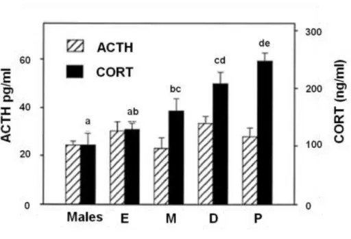
1.3.3. Plasma ACTH and CORT levels across the rat estrous cycle
Basal and stress-induced HPA axis (re)activity in terms of plasma ACTH and CORT levels have been shown to vary across the estrous cycle (Viau and Meaney, 1991, Atkinson and Waddell, 1997, Figueiredo et al., 2002). Sex hormone fluctuations were hypothesised to play a role taken that sex steroids have been demonstrated to influence adrenal steroidogenesis (Viau and Meaney, 1991, Nowak et al., 1995, Figueiredo et al., 2007). Interestingly, plasma ACTH and CORT have been shown to disconnect across the estrous cycle under basal conditions (Atkinson and Waddell, 1997) and also after acute stress exposure (Viau and Meaney, 1991) suggesting that adrenal- mediated mechanisms might participate to the estrous cycle associated changes in HPA axis (re)activity. Specifically, in an elegant and detailed study from 1997, Atkinson and colleagues
26
monitored the circadian release of ACTH and CORT across the estrous cycle in Wistar rats. An extra group of males was added to include sex comparison in the study. Circadian dependent rhythmicity in plasma CORT was observed in both males and females independently from the estrous cycle phase; the degree of the rhythm was also unaffected by sex and the estrous cycle.
On the other hand, the absolute CORT plasma levels markedly differed across the groups (Fig.
10). Specifically, average daily plasma CORT concentrations were higher at proestrous compared to all the other groups; the lowest concentrations were detected at estrous and in the following metestrous and diestrous phases the levels rose but remained significantly lower than the proestrous-associated average daily plasma CORT concentrations. Males CORT levels did not differ from the concentrations observed in females at estrous but were lower compared to those detected at metestrous, diestrous and proestrous; findings that have been recapitulated also by another study performed in Sprague Dawley rats (Figueiredo et al., 2002). The differences in the daily CORT averages observed were attributed to differences in the peak values since CORT trough-associated levels did not differ across the groups. Interestingly, whilst ACTH levels were reported to display a circadian rhythm with highest values detected before lights off, neither the sex nor the estrous cycle affected ACTH levels (Fig. 10).
Figure 10. ACTH and CORT daily average levels in male rats and across the estrous cycle
27 Average ACTH (striped columns) and CORT (full columns) daily levels are represented (n=five/nine per group). Hormonal levels measured in males and females at estrous (E), metestrous (M), diestrous (D) and proestrous (P) are represented. Groups without common notations differ significantly. Adapted from (Atkinson and Waddell, 1997).
Stress-induced ACTH and CORT levels have also been shown to vary across the estrous cycle (Viau and Meaney, 1991). Specifically, although 20 minutes restraint stress has been reported to increase ACTH and CORT levels in Long Evans rats at diestrous, proestrous and estrous, ACTH levels in the proestrous group were doubled compared to both diestrous and estrous at the end of the stress procedure. Moreover, plasma CORT was increased after stress termination and higher at proestrous compared to diestrous and estrous females. In contrast, the CORT differences observed across the groups were less pronounced compared to the marked diversity observed in the ACTH stress-induced levels. Estrous cycle associated changes in adrenal gland sensitivity to ACTH and sex hormones fluctuation have long been hypothesised to account for the previously mentioned disconnection between plasma ACTH and CORT. In line with this, estrogens through binding to the estrogens R α and β (Green et al., 1986, Nilsson and Gustafsson, 2011), have been shown to impact on adrenal steroidogenesis. Specifically, estradiol was reported to enhance basal, but not ACTH-induced, CORT secretion from isolated adrenocortical cells in vitro (Nowak et al., 1995). In contrast, an in vivo study from Figueiredo and colleagues suggested that estrogens potentiated adrenocortical response to stress in female rats with a mechanism hypothesised to involve the adrenal glands. In more detail, estradiol injection in ovariectomized females increased CORT secretion;
despite reduced PVN activation measured via c-fos mRNA activation and plasma ACTH levels after acute restraint stress exposure (Figueiredo et al., 2007). This discrepancy between in vitro and in vivo findings may be related to that fact that estrogens may affect also the synthesis of steroids hormones precursor, cholesterol. Specifically, the promoter
28
of the rat HMGCR, key enzyme for cholesterol synthesis, contains a tissue specific estrogen-responsive region (Di Croce et al., 1999).
LH has also been shown to influence adrenal steroidogenesis with LH overexpressing female mice displaying an 80% increase in adrenal size and 14-fold increase in serum CORT levels (Kero et al., 2000). Moreover, in vitro incubation of adrenal cells resulted in higher CORT production from LH overexpressing cells than controls, suggesting that adrenal mechanism may adapt to sustain the previously mentioned elevated CORT production in vivo (Kero et al., 2000). LH estrous cycle-associated fluctuations might, therefore, participate to the ACTH and CORT dissociation observed in females. Brain- mediated mechanisms may play a role as well in this imbalance. Choi and colleagues showed that lesion of the anteroventral region of the bed nucleus of the stria terminalis in male rats prevented the expected plasma ACTH peak after stress exposure without affecting CORT increase. However, it remains to be determined whether a similar response would be observed in female rats. Taken together, these results suggest that brain and adrenal mediated changes may bypass pituitary ACTH to stimulate CORT production (Choi et al., 2007).
1.4. Hormonal and behavioural changes across the peripartum period
1.4.1. Pregnancy and lactation
In all mammalian species, the peripartum period is associated with profound physiological, emotional and behavioural changes that act in concert to ensure the health and well-being of both, the mother and offspring. These changes include substantial alterations in the HPA- and hypothalamus-pituitary-ovary axes. In most mammalian species pregnancy-
29 associated estradiol and progesterone release follow the same pattern as in rodents (Rosenblatt et al., 1988). Specifically, estrogen and progesterone progressively increase during pregnancy to levels 50- and 10-fold higher than observed in virgins, respectively, and rapidly reverse to their pre-pregnancy levels after parturition. These levels drop within a few days of parturition, remain low during the first half of lactation (approximately 10 days in rodents and until a monthly cycle resumes in women - which can take up to 180 days) and begin to increase when follicular maturation starts again (Bridges, 1984, Brunton and Russell, 2010). In contrast, the progesterone profile differs between women and rodents, as it remains low throughout lactation in women but increases to levels observed in late pregnancy by the third day of lactation in rats (Grota and Eik-Nes, 1967, Taya and Greenwald, 1982, Rosenblatt et al., 1988, McNeilly, 2001). In the rat, the ovary produces hormones through pregnancy while in other species, including humans, fetoplacental production is more important (Taya and Greenwald, 1982). Specifically, the corpus luteum becomes in the rat a substantial site for progesterone production. This is a specific feature of the corpus luteum because the follicle indeed primarily produces estrogens. A constant cholesterol supply is needed to sustain progesterone synthesis throughout pregnancy with plasma lipoproteins being the primary source (see Section 1.2.2 for more details on cholesterol delivery for steroidogenesis). As previously mentioned, progesterone levels increase stating at pregnancy day (PD) 5 and they gradually rise to peak around PD 15-16 before rapidly decreasing around PD 19 (Pepe and Rothchild, 1973). Progesterone stimulates its own secretion and it protects corpus luteum cells from death (Telleria and Deis, 1994). Locally-produced estradiol stimulates progesterone production and promotes, particularly at mid-pregnancy, the vascularisation of the corpus luteum (Tamura and Greenwald, 1987).
30
1.4.1.1. Prolactin and Oxytocin
PRL exerts several important tasks during pregnancy and some of them at the corpus luteum level where PRL-R are expressed. Specifically, the longer PRL-R isoform is thought to mediate the effect of PRL on the luteal cells whereas the short isoform stimulates angiogenesis in the endothelial cells (Freeman et al., 2000). Importantly, LH induces an up-regulation of PRL-R during luteinisation and PRL-R-knockout (KO) mice display lack of implantation and consequent sterility (Grosdemouge et al., 2003). Pituitary lactotrophs, which synthesise PRL, represent the 20-50% of the pituitary cells depending on the sex and on the reproductive status of the animal. In the pituitary of neonatal rats another cell population called mammosomatotrophs secrets PRL. These cells differentiate into lactotrophs in the presence of estrogens and of a maternal signal that reaches the pups at early lactation throughthe milk (Porter et al., 1991, Freeman et al., 2000). Lactotrophs store a large amount of PRL that can be released via a calcium- dependent exocytotic mechanism. At early pregnancy, PRL is secreted following a circadian pattern characterized by two daily surges that cease after mid-pregnancy when pituitary PRL production stops and the decidua layer of the uterine lining takes over this task until the trophoblast begin the production of PRL-like hormones (Grattan and Averill, 1990, Prigent-Tessier et al., 1999). PRL levels remain low until they increase at late pregnancy before labour (Grattan and Averill, 1990). During lactation, the suckling physical stimulus induces PRL release and this release pattern is superimposed to the circadian rhythm. However, suckling-induced plasma PRL levels are increase when nursing occur at the time of the circadian peak (Freeman et al., 2000).
Together with the PRL system, OXT circuitry is highly activated in the peripartum period because of its involvement in the regulation of several pregnancy- and lactation- associated physiological processes. Blockade of these two systems was shown to
31 negatively interfere with phenomena like labour and lactation. Thus, blockade of OXT central signalling via central infusion of OXT selective antagonist at late gestation was reported to delay the suckling-induced systemic OXT release and to affect pups and dams weight (Lipschitz et al., 2003) while PRL and PRL-R KO mice failed to produce milk (Horseman et al., 1997, Ormandy et al., 1997). Involvement of brain OXT and PRL systems for the display of maternal behaviour is also known. Specifically, PRL-R KO mice (Lucas et al., 1998) and rats with reduced brain PRL-R expression induced by antisense infusion (Torner et al., 2002) show impaired maternal behaviour, while intracerbroventricular infusion of OXT antagonist reduces the display of arched back nursing between lactation day 2 and 5 (Bosch and Neumann, 2008) (see Section 1.4.4 for PRL and OXT involvement for the maternal stress hyporesponsiveness).
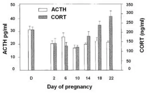
1.4.2. Changes in HPA axis function across the peripartum period
Maternal HPA axis function shows profound plasticity across the peripartum period with both basal and stress-induced ACTH and CORT plasma levels being profoundly affected.
Thus, basal CORT levels have been shown to be increased from mid-pregnancy despite minor changes in plasma ACTH whereas a marked reduction in the hormonal response elicited by acute stress exposure has been documented in several species including humans (Carr et al., 1981, Allolio et al., 1990, Douglas et al., 1998, Shanks et al., 1999, Brunton and Russell, 2003, Douglas et al., 2003, Windle et al., 2013).
Pregnant and lactating women have been shown to be less stress reactive compare to non-pregnant subjects with breastfeeding women appearing to be even less stress reacting when compared to non breastfeeding mothers (Rosenblatt et al., 1994, Heinrichs et al., 2001, Glynn et al., 2004, Heinrichs and Koob, 2004). Here, I go into detail of just
32
one example in which saliva CORT secretion was measured before and after 60 seconds (s) immersion of the dominant hand in ice cold water in women towards the end of pregnancy and eight weeks post-partum. Whilst non-pregnant women from the control group react to the stress with a significant increase in salivary CORT, neither pregnant nor lactating women showed differences in CORT release (Kammerer et al., 2002). Rodent studies reported that CORT secretion after exposure to 20 or 30 minutes restraint stress (da Costa et al., 1996, Johnstone et al., 2000), five minutes elevated plus maze exposure and 60 or 90 s forced swim (Neumann et al., 1998) was reduced in pregnant rats compared to virgins. Data collected from mice studies follow the same trend. Douglas and colleagues showed that pregnant mice were less responsive to novel environment exposure or forced swim in term of CORT secretion compared to virgins (Douglas et al., 2003). Evidence of reduced HPA axis responsiveness during lactation has also been reported. Specifically, lactating animals on LD 3 and 4 and between LD 10 and 14 displayed lower CORT levels compared to virgin animals when killed after 30 minutes restraint stress (da Costa et al., 1996). Lactating Sprague-Dawley rats did not show any change in ACTH and CORT plasma levels after noise stress exposure, whereas this stress procedure elicited an increase in both hormones in virgins (Windle et al., 1997).
Hillerer and colleagues observed reduced CORT response in dams on LD 8 compared to virgin rats after 60 s forced swim exposure. Interestingly, ACTH secretion did not differ among the groups pointing out that other mechanisms beyond changes in ACTH release might mediate the hypo-response in terms of CORT secretion, mechanisms that might involve adrenal glands plasticity (Hillerer et al., 2011). Peripartum changes in HPA axis stress-induced reactivity have been hypothesised to be mediated, at least in part, via alterations in the CRF system. Decreased CRF mRNA within the PVN in late pregnancy (Johnstone et al., 2000) and lactation (Fischer et al., 1995), decreased median eminence CRF content in late pregnancy (Ma et al., 2005), together with reduced pituitary CRF receptor binding from mid-pregnancy (Neumann et al., 1998), reflect changes that have