Biophysical properties of AMPA receptor complexes
D i s s e r t a t i o n
zur Erlangung des akademischen Grades Doctor of Philosophy (Ph.D.)
eingereicht an der
Lebenswissenschaftlichen Fakultät der Humboldt-Universität zu Berlin
von Irene Riva
Präsidentin der Humboldt-Universität zu Berlin:
Prof. Dr.-Ing. Dr. Sabine Kunst
Dekan der Lebenswissenschaftlichen Fakultät der Humboldt-Universität zu Berlin:
Prof. Dr. Bernhard Grimm
Gutachter/innen:
1. Prof. Andrew Plested 2. Prof. Peter Hegemann
3. Dr. Tanja Maritzen
Tag der mündlichen Prüfung: 24/02/2020
Hilfen und Hilfsmittel angefertigt zu haben. Ich habe mich anderwärts nicht um einen Doktorgrad beworben und besitze keinen entsprechenden Doktorgrad. Ich erkläre, dass ich die Dissertation oder Teile davon nicht bereits bei einer anderen wissenschaftlichen Einrichtung eingereicht habe und dass sie dort weder angenommen noch abgelehnt wurde. Ich erkläre die Kenntnisnahme der dem Verfahren zugrunde liegenden Promotionsordnung der Lebenswissenschaftlichen Fakultät der Humboldt-Universität zu Berlin vom 5. März 2015.
We i t e r h i n e r k l ä r e i c h , d a s s k e i n e Z u s a m m e n a r b e i t m i t g e w e r b l i c h e n Promotionsbearbeiterinnen/Promotionsberatern stattgefunden hat und dass die Grundsätze der Humboldt-Universität zu Berlin zur Sicherung guter wissenschaftlicher Praxis eingehalten wurden.
DECLARATION
I hereby declare that I completed the doctoral thesis independently based on the stated resources and aids. I have not applied for a doctoral degree elsewhere and do not have a corresponding doctoral degree. I have not submitted the doctoral thesis, or parts of it, to another academic institution and the thesis has not been accepted or rejected. I declare that I have acknowledged the Doctoral Degree Regulations which underlie the procedure of the Faculty of Life Sciences of Humboldt-Universität zu Berlin, as amended on 5th March 2015.
Furthermore, I declare that no collaboration with commercial doctoral degree supervisors took place, and that the principles of Humboldt-Universität zu Berlin for ensuring good academic practice were abided by.
Excitatory neurotransmission throughout the vertebrate central nervous system (CNS) is largely mediated by the α-amino-3-hydroxy-5-methyl-4-isoxazolepropionic acid receptors (AMPARs). AMPA receptors are glutamate-gated ion channels located at the postsynaptic membrane, where they compose the hub of macromolecular complexes with a number of auxiliary proteins that concertedly regulate the receptor function. Stargazin (or γ2) is the prototypical AMPAR auxiliary subunit, belonging, together with the related γ3, γ4, γ5, γ7 and γ8 isoforms, to the family of the transmembrane AMPA receptor regulatory proteins (TARPs).
TARPs show a bewildering array of effects on the trafficking, synaptic anchoring, gating kinetics and pharmacology of AMPARs. Despite the growing knowledge gathered about the structural features of the AMPAR-TARP complex, the molecular mechanisms underlying TARP modulation of AMPA receptors have not been fully revealed. Higher brain functions rely upon AMPAR activity and dysregulation of AMPA receptors has been associated to life- threatening CNS disorders. These observations provide motivation to unravel the molecular machinery behind AMPAR regulation and to identify AMPAR auxiliary proteins as potential pharmacological targets.
In the present study, AMPAR-TARP interactions were investigated using electrophysiology in human embryonic kidney (HEK) 293 cells. The role of TARP extracellular loops, Loop1 (L1) and Loop2 (L2), in the modulation of AMPAR gating was analysed. By modifying γ2 and γ8 L1 and L2, mutant TARPs that both lacked modulatory properties (“null” TARPs) and also those that had greatly enhanced modulation (“super” TARP) were obtained, without affecting formation of AMPAR-TARP complexes. A model for TARP modulation has been proposed, based on predicted state-dependent interactions of TARP L1 and L2 with the AMPAR ligand binding domain (LBD) and linkers between the LBD and the transmembrane domain (TMD).
L1 of γ8 emerged as a very powerful positive modulator of AMPAR gating, most likely by virtue of its length that enables extensive interactions with multiple sites of the receptor LBD.
Moreover, considering that native AMPARs in the brain mainly consist of heterotetrameric assemblies of four distinct subunits (GluA1-4), null and super TARP mutants were also tested on different AMPAR subunit compositions. Common as well as subunit-dependent mechanisms of AMPAR modulation by TARPs have been observed. In particular, in
In summary, these experiments provided evidence that TARP L1 and L2 are not involved in association of AMPAR-TARP complexes and can entirely account for the modulation of AMPAR gating by TARPs. Ultimately, by overexpression in organotypic brain slices, null and super TARP mutants may serve as useful tools to further examine, for the first time, the effects of TARP modulation on the function of synaptic AMPA receptors.
Im zentralen Nervensystem (ZNS) werden exzitatorische Signale zum großen Teil durch α- Amino-3-hydroxy-5-methyl-4-isoxazol-Propionsäure-Rezeptoren (AMPARs) vermittelt.
AMPARs sind Glutamat-gesteuerte Kationenkanäle und befinden sich in der postsynaptischen Membran. Dort bilden sie Komplexe mit „Hilfsproteinen“ (engl. Auxiliary Proteins), um die Rezeptorfunktion zu steuern. Der typische Vertreter, Stargazin (oder γ2), gehört zusammen mit seinen Isoformen γ3, γ4, γ5, γ7 und γ8 zur Familie der transmembranen AMPAR- regulierenden Proteine (TARPs). TARPs regulieren den Transport, die postsynaptische Insertion, die Steuerung (engl. Gating), sowie die Pharmakologie von AMPARs. Trotz der stetigen Aufklärung struktureller Merkmale von AMPAR-TARP-Komplexen, sind die molekularen Mechanismen, welche der TARP-Modulierung von AMPARs unterliegen, noch weitgehend unerforscht. Die normale Funktion von AMPARs ist Voraussetzung für die uneingeschränkte Hirntätigkeit, wohingegen Fehlfunktionen mit lebensbedrohlichen Störungen des ZNS in Verbindung gebracht werden konnten. Diese Beobachtungen bieten Grundlage und Motivation für die Aufklärung der molekularen Regulierung von AMPARs und der Identifikation von TARPs als potentielle pharmakologische Targets.
In dieser Arbeit wurden Interaktionen von AMPAR-TARP-Komplexen mithilfe elektrophysiologischer Methoden an HEK293-Zellen (engl. human embryonic kidney) untersucht. Insbesondere wurden die extrazellulären TARP-Schleifen (engl. Loops), Loop1 (L1) und Loop2 (L2) und deren Auswirkungen auf AMPAR-Gating studiert. Durch Mutationen von γ2- und γ8- L1 und L2 konnten TARPs generiert werden, die entweder keine modulierenden Eigenschaften an AMPARs aufwiesen („null“-TARP) oder eine verstärkte Modulierung zeigten („super“-TARP), ohne dabei die AMPAR-TARP-Komplexbildung zu beeinträchtigen. Zur Beschreibung der zustandsabhängigen Interaktionen von TARP- L1 und L2 mit der AMPAR-Ligandenbindedomäne (LBD), sowie den Linkern zwischen LBD und Transmembrandomäne (TMD), wurde ein Modell erstellt. Bezüglich AMPAR-Gating erwies sich γ8-L1 als verstärkender Modulator, der vermutlich aufgrund seiner Länge zahlreiche Wechselwirkungen mit der LBD eingehen kann. Da native AMPARs im Hirn vorwiegend als Heterotetramere mit unterschiedlichen Kombinationen der Untereinheiten (GluA1-A4) vorkommen, wurden die null- und die super-TARP-Mutanten an AMPARs mit verschiedenen
von AMPARs entstehen. Besonders in Komplexen von heteromerem GluA1 und mRNA- editiertem GluA2 AMPARs mit TARPs konnte ein dominantes Gating der GluA2- Untereinheit beobachtet werden.
Zusammenfassend konnte durch die Experimente gezeigt werden, dass L1 und L2 nicht bei der Bildung von AMPAR-TARP-Komplexen involviert sind, sondern vielmehr der Modulierung von AMPAR-Gating durch TARPs dienen. Durch Überexpression der null- und super-TARP-Mutanten in organotypischen Gehirnschnitten könnten diese zukünftig als nützliches Werkzeug dienen, um die Effekte von TARP-Modulationen erstmalig an synaptischen AMPARs zu studieren.
SUMMARY ...IV ZUSAMMENFASSUNG ...VI LIST OF ABBREVIATIONS ...XII
1. INTRODUCTION ...1
1.1 AMPA receptors ...1
1.1.1 Overview of AMPA receptor physiology ...1
1.1.2 AMPA receptor structure and role of the distinct domains ...3
1.1.3 Mechanisms of AMPA receptor gating ...7
1.2 AMPA receptor complex ...10
1.2.1 AMPA receptor auxiliary proteins ...10
1.2.2 Transmembrane AMPA receptor regulatory proteins (TARPs) ...11
1.3 TARPs regulation of the AMPA receptor ...13
1.3.1 TARPs mediate AMPA receptor trafficking and anchoring at the synaptic membrane ...13
1.3.2 TARP modulation of the AMPA receptor gating ...15
1.3.3 TARPs regulate AMPA receptor pharmacology ...18
1.4 AMPA receptor-TARP interaction ...22
1.4.1 Structures of the AMPA receptor-TARP complex ...22
Resting state ...22
Active and desensitised state ...23
Heteromeric and native complexes ...25
1.4.2 Molecular basis for TARP modulation of AMPA receptor gating ...26
1.5 TARPs in mutant mice and in human central nervous system disorders ...29
1.5.1 TARPs in mutant mice ...29
1.5.2 TARPs in human central nervous system disorders ...31
1.6 Transmembrane AMPA receptor auxiliary subunits beyond TARPs ...33
1.6.1 Cornichon homologs-2 and -3 (CNIH-2 and -3) ...33
1.7 Aim of the project ...35
2. MATERIALS AND METHODS ...37
2.1 HEK 293 cell culture ...37
2.1.1 HEK 293 cell solutions ...37
2.1.2 Cell cultivation ...37
2.1.3 Coverslips preparation ...38
2.1.4 DNA transfection ...38
2.2 Molecular biology ...39
2.2.1 Expression vectors ...39
2.2.2 Cloning of TARP mutant constructs ...40
2.2.3 Cloning of AMPA receptor mutant constructs ...41
2.3 HEK 293 cell electrophysiology ...42
2.3.1 Experimental setup ...42
2.3.2 Solutions for electrophysiological recordings ...43
2.3.3 Preparation of perfusion tools ...44
2.3.4 Preparation of patch pipettes ...45
2.3.5 Fast-perfusion outside-out patch-clamp recordings ...45
2.4 Data analysis ...47
2.4.1 Analysis of HEK 293 cell electrophysiology ...47
2.4.2 Calculation of abundance of tripartite AMPA receptor-TARP complexes ...49
2.5 Organotypic mouse brain cultures ...50
2.5.1 Organotypic slice solutions ...50
2.5.2 Preparation for mouse brain slice dissection ...52
2.5.3 Mouse brain slice dissection ...52
2.5.4 Organotypic slice cultivation ...53
2.6 Single-Cell Electroporation (SCE) in organotypic slices ...54
2.6.1 Solutions for SCE ...54
2.6.4 SCE procedure ...55
3. RESULTS ...58
3.1 Model of the AMPA receptor complex with TARPs ...58
3.1.1 Comparison of a GluA2-γ8 model with the GluA1/A2_γ8 complex structure ...58
3.2 TARP γ2 and γ8 extracellular loop mutants ...59
3.2.1 Membrane expression of TARP mutants ...59
3.2.2 Association with GluA2(Q) AMPA receptors ...59
3.3 Measuring modulation of GluA2(Q) gating by γ2 and γ8 extracellular loops ...61
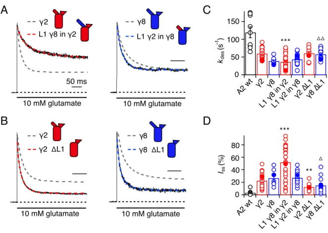
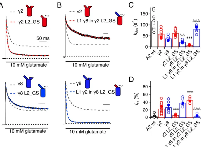
3.3.1 Desensitisation properties of TARP Loop1 mutants ...61
3.3.2 Desensitisation properties of TARP Loop2 mutants ...64
3.3.3 Role of Loop1 in AMPA receptor-TARP complex superactivation ...64
3.3.4 Role of Loop2 in AMPA receptor-TARP complex superactivation ...67
3.3.5 Effect of combined removal of Loop1 and Loop2 ...68
3.3.6 Modulation by the β4-TM2 acidic loop ...68
3.4 Testing site-specific AMPA receptor-TARP interactions ...71
3.4.1 GluA2(Q) mutants in linkers in close proximity to TARP Loop2 ...71
3.4.2 Functionality of GluA2(Q) linker mutants and assembly with TARPs ...72
3.4.3 Modulation of GluA2(Q) linker mutants by TARPs ...73
3.5 Subunit-dependent modulation of AMPA receptor gating by TARPs ...77
3.5.1 Association of TARP modulatory mutants with GluA1 and GluA2(R) homomeric receptors ...77
3.5.2 TARP mutants modulation of GluA1 and GluA2(R) homomers desensitisation 79 ... 3.5.3 TARP mutants modulation of GluA1 and GluA2(R) homomers superactivation 81 .. 3.5.4 Association of TARP modulatory mutants with GluA1/A2(R) heteromeric receptors ...84
3.5.5 TARP mutants modulation of GluA1/A2(R) heteromer desensitisation ...84
3.5.6 TARP mutants modulation of GluA1/A2(R) heteromer deactivation ...86
3.5.7 TARP mutants modulation of GluA1/A2(R) heteromer superactivation ...88
3.6 Single-Cell Electroporation of TARP mutants in organotypic hippocampal slices 95
3.6.1 Overexpression of wild-type and mutant TARPs in CA1 pyramidal neurons ...95
4. DISCUSSION ...100
4.1 Mutations of TARP γ2 and γ8 extracellular loops ...100
4.1.1 TARP extracellular loops are not involved in association with AMPA receptors 100 4.1.2 Characterisation of the role of TARP Loop1 and Loop2 in GluA2(Q) gating ...101
4.1.3 Inhibitory role of γ2 β4-TM2 loop in GluA2(Q) gating ...102
4.1.4 LBD-TMD linker region is critical for GluA2(Q) modulation by TARPs ...103
4.1.5 Proposed mechanism of AMPA receptor modulation by TARPs ...105
4.2 Subtype-dependency of TARP modulation ...107
4.2.1 TARP association with GluA2(R)-lacking and GluA2(R)-containing AMPARs 107 . 4.2.2 Loss of modulation of homomeric and heteromeric GluA1 and GluA2(R) receptors by null TARP mutants ...109
4.2.3 GluA1 resistance to potentiation by the super TARP and GluA2(R) dominance in heteromeric TARP complexes ...110
4.2.4 Super TARP facilitation of GluA1/A2(R) responses during train stimulation ...111
4.2.5 Predicting abundance of tripartite GluA1/A2(R)/TARP complexes ...113
5. FUTURE PERSPECTIVES: investigating the effects of TARP modulation on AMPA receptor-mediated synaptic transmission ...115
APPENDIX ...117
BIBLIOGRAPHY ...121
ACKNOWLEDGEMENTS ...142
3D three-dimensional
Å angstrom
ACSF artificial cerebrospinal fluid
AMPAR α-amino-3-hydroxy-5-methyl-4-isoxazolepropionic acid receptor
ATD amino-terminal domain
C- carboxyl-
C-tail cytoplasmic tail
Ca2+ calcium
CaMKII calcium/calmodulin-dependent protein kinase II CGN cerebellar granule neuron
CKAMP44 cystine-knot AMPAR modulating protein
CNIH-2 cornichon homologs-2
CNIH-3 cornichon homologs-3
CNQX 6-cyano-7-nitroquinoxaline-2,3-dione CNS central nervous system
Cryo-EM cryogenic electron microscopy CTD carboxyl-terminal domain
CTZ cyclothiazide
Cα alpha carbon
D1 lobe 1
D2 lobe 2
DDM n-Dodecyl-β-D-Maltopyranoside
DG dentate gyrus
DIC differential interference contrast
DIV days in vitro
DNQX 6,7-dinitroquinoxaline-2,3-dione
DsRed discosoma red
EBSS Earle’s balanced salt solution ECD extracellular segment
EPSC excitatory postsynaptic current
ER endoplasmic reticulum
Erev reversal potential
FBS fetal bovine serum
G conductance
G-V conductance-voltage
Glu glutamate
GS glycine-serine
GSG1L germ cell-specific gene 1-like protein
GΩ gigaohm
HEK human embryonic kidney
Hz Herz
I current
I-V current-voltage
iGluR inotropic glutamate receptor IRES internal ribosome entry site
Iss steady-state current
K+ potassium
KA kainate
kdeact deactivation rate
kdes desensitisation rate
L1 Loop1
L2 Loop2
LBD ligand binding domain
LTD long-term depression
LTP long-term potentiation
M1 membrane helix 1
M2 membrane helix 2
M3 membrane helix 3
MD molecular dynamics
MEM minimum essential medium
mEPSC miniature excitatory postsynaptic current mGluR metabotropic glutamate receptor
MW molecular weight
MΩ megaohm
N- amino-
NBQX 2,3-dioxo-6-nitro-1,2,3,4-tetrahydrobenzo[f]quinoxaline-7-sulfonamide
NMDA N-methyl-D-aspartate
NMDAR NMDA receptor
NP negative patch
P postnatal day
pA picoampere
PA polyamine
pC picocoulomb
PEG polyethylene glycol
PEI polyethylenimine
PEPA 4-[2-(phenylsulfonylamino)ethylthio]-2,6-difluorophenoxyacetamide
PKA protein kinase A
PKC protein kinase C
Popen open channel probability PP1 protein phosphatase 1 PP2B protein phosphatase 2B PSD postsynaptic density
PSD-95 PSD protein-95
PTFE polytetrafluoroethylene PVDF polyvinylidene fluoride
R resistance
RI rectification index
S1 amino acid segment 1
S2 amino acid segment 2
SCC Schaffer collateral/commissural SCE single-cell electroporation SEM standard error of the mean SNP single nucleotide polymorphism
TARP transmembrane AMPA receptor regulatory protein
TCM trichloromethiazide
tdeact deactivation time
tdes desensitisation time
TE tris-EDTA
TM1 transmembrane helix 1
TM2 transmembrane helix 2
TM3 transmembrane helix 3
TM4 transmembrane helix 4
TMD transmembrane domain
UPR unfolded protein response
V voltage
WT wild-type
1. INTRODUCTION 1.1 AMPA receptors
1.1.1 Overview of AMPA receptor physiology
Within the mammalian central nervous system (CNS), the vast majority of fast excitatory synaptic transmission is mediated by the α-amino-3-hydroxy-5-methyl-4-isoxazolepropionic acid receptors (AMPARs). Together with the NMDA (N-methyl-D-aspartate)- and kainate- type receptors, AMPA-type receptors belong to the family of the ionotropic glutamate receptors (iGluRs), ligand-gated ion channels located at excitatory synapses where they are activated by the neurotransmitter glutamate. Following binding of presynaptically released glutamate these three classes of iGluRs are characterised by different gating kinetics. The AMPAR produces a rapidly rising conductance that also rapidly decays (1-2 ms), whereas the NMDA and kainate receptors show much slower gating kinetics. In particular, the NMDA receptor (NMDAR) activates in milliseconds and deactivates between tens and thousands of milliseconds (Traynelis et al., 2010). Moreover, AMPA and kainate receptors undergo a rapid and pronounced desensitisation that occurs within milliseconds after activation; NMDARs instead are subject to weak or no desensitisation, whose rate and degree depend on the NMDAR subtype (Traynelis et al., 2010). The diversity in the gating kinetics of the iGluRs precisely defines the time course of synaptic currents and their role in synaptic physiology.
AMPARs sit at the postsynaptic density (PSD) of glutamatergic synapses, where binding of glutamate to their extracellular ligand binding domain (LBD) allows the influx of cations such as Na+ and Ca2+ through the channel pore, thus depolarising the postsynaptic membrane.
AMPAR signaling is typically mediated by tetrameric AMPARs (Rosenmund et al., 1998) assembled from differing combinations of the four subunits GluA1 to GluA4. AMPAR subunit composition is developmentally regulated, region and cell type-specific and activity- dependent (Henley & Wilkinson, 2016). In the adult brain diheteromeric GluA1-GluA2 and GluA2-GluA3 receptors are the most common assemblies (Wenthold et al., 1996; Lu et al., 2009; Zhao et al., Science 2019), whereas GluA4 is highly expressed in the early postnatal period and then down-regulated. GluA4 remains enriched in the adult in certain cerebellar cell types (Petralia & Wenthold, 1992; Zhu et al., 2000). A critical determinant of the AMPAR
function is dictated by the presence or absence of the GluA2 subunit. The vast majority of GluA2 RNA is post-transcriptionally modified by editing at a codon residing in the pore channel region that is converted from glutamine to arginine (Q/R site). By virtue of Q/R site editing, the permeation properties of GluA2-containing AMPARs are altered: they have low calcium (Ca2+) permeability, are insensitive to the voltage-dependent block by endogenous polyamines and have a low single-channel conductance (Sommer et al., 1991; Jonas &
Burnashev, 1995; Swanson et al., 1997). On the contrary, GluA2-lacking AMPARs, or AMPARs containing the unedited GluA2(Q) form, have high permeability to Ca2+, are blocked by endogenous polyamines and have higher single-channel conductance. During early postnatal development, expression of GluA2 is low compared to that of the other GluA subunits, but it increases rapidly from the first postnatal week (Pickard et al., 2000). This, coupled with the above-mentioned transient high expression of GluA4, renders many neonatal AMPARs Ca2+-permeable, suggesting that such receptors may play a role in neonatal synaptic functions (Isaac et al., 2007).
Thus, the kinetics and amplitude of the excitatory synaptic responses are crucially determined by the biophysical properties of the receptor subunit composition as well as by the density of the receptor expression (Traynelis et al., 2010). Activity-dependent changes in the composition and density of synaptic AMPARs are tightly regulated by dynamic receptor trafficking (i.e. lateral diffusion and vesicular trafficking) and intrinsically underlie some mechanisms of synaptic plasticity (Malinow & Malenka, 2002; Choquet & Triller, 2003;
Newpher & Ehlers, 2008). In fact, short periods of specific patterns of activity can trigger long-lasting changes in the function of excitatory synapses by modifying their efficacy to store information, either through a strengthening (long-term potentiation, LTP) or weakening (long-term depression, LTD) of the synaptic responses (Bliss & Lomo, 1973; Lynch et al., 1977). These mechanisms are widely believed to constitute the molecular basis of higher cognitive functions such as hippocampal-dependent memory formation and learning (Martin et al., 2000; Kessels & Malinow, 2009). Both LTP and LTD are thought to be expressed through the insertion or removal, respectively, of synaptic AMPARs (Malenka & Bear, 2004).
Especially, certain stimuli can induce the initiation of the LTP and LTD by activation of postsynaptic NMDARs (NMDAR-dependent LTP/LTD) and specific intracellular signaling cascades involving either protein kinases or phosphatases (Malenka, 1994) which determine,
respectively, an increase or a decrease in the number of synaptic AMPARs (Lüscher &
Malenka, 2012; Choquet, 2018). It was then proposed that expression of the LTP and LTD might be associated with bidirectional changes in phosphorylation of the C-terminus of the AMPAR subunits GluA1 and GluA2 (Lee et al., 2000; Chung at al., 2000). More recently this notion has been the subject of strong debate (Buonarati et al., 2019; see following section).
Centrally, AMPAR function at synapses is influenced by a number of auxiliary proteins that physically associate in a complex with the receptor and control the receptor activity at many different levels, considerably adding to the variation in AMPAR properties and hence to the diversity of information processing within the brain (Jackson & Nicoll, 2011).
1.1.2 AMPA receptor structure and role of the distinct domains
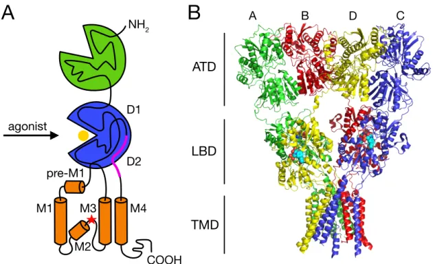
AMPA receptors are integral membrane proteins composed of the four GluA1-4 subunits that assemble to form a central ion channel pore. Each AMPAR subunit has a modular structure that includes four discrete domains (Fig. 1 A): the extracellular amino-terminal domain (ATD) that is involved in receptor subtype-specific assembly and synaptic anchoring (Leuschner &
Hoch, 1999; Ayalon et al., 2005; Watson et al., 2017; Díaz-Alonso et al., 2017), the extracellular ligand-binding domain (LBD) that binds the agonist and drives the activation gating (Baranovic et al., 2016), the transmembrane domain (TMD) that forms the membrane- spanning ion channel (Wollmuth & Sobolevsky, 2004) and an intracellular carboxyl-terminal domain (CTD) that takes part in synaptic localisation, trafficking and receptor regulation (Song & Huganir, 2002).
The first detailed crystallographic structure of a full-length GluA2 receptor in a closed antagonist-bound state showed that the receptor harbours an overall axis of two-fold symmetry, with the extracellular domains organised as pairs of local dimers and the ion channel domain exhibiting a four-fold symmetry (Sobolevsky et al., 2009; Fig. 1 B). The symmetry mismatch between the extracellular (ATD and LBD) and ion channel (TMD) domains as well as a subunit crossover (or domain swapping) from the LBD to the ATD give rise in the homotetrameric GluA2 receptor to two conformationally distinct types of subunits:
A/C and B/D. As a consequence of the swapping of domains, local ATD and LBD dimers are not formed by the same pairs of subunits, with the ATD dimers being formed by the A/B and C/D subunits and the LBD dimers by the A/D and B/C subunits. The ATD and LBD generate
two types of subunit-subunit contacts: within each dimer (A-B or C-D for the ATD and A-D and B-C for the LBD) and between dimers (mediated by residues at the B-D dimer-dimer interface for the ATD and at the A-C dimer-dimer interface for the LBD). The subunit crossover between the ATD and LBD layers is primarily mediated by the ATD-S1 amino acid linkers that connect the ATD with the first of the two amino acid segments (S1 and S2) that define the LBD. Similarly, the symmetry mismatch between the extracellular and ion channel
NH2
D1
M1 M3 M4
M2 pre-M1 agonist
D2
A
ATD
LBD
TMD
B D C
COOH
A B
Figure 1. Modular and crystal structure of an AMPAR. (A) Schematic representation of the modular structure of a single AMPAR subunit comprising the extracellular ATD (green) and LBD (blue), the pore-forming TMD (orange) and the intracellular CTD. The polypeptide chain is traced through the distinct domains. Two amino acid segments (S1 and S2) compose the upper (D1) and lower (D2) lobes of the LBD. Glutamate (yellow sphere) binds between the two LBD lobes. The TMD contains three membrane-spanning helices (M1, M3 and M4), preceded by a short helix oriented nearly parallel to the membrane (pre- M1), and a central pore-like helix (M2). The Q/R editing site (red star) is positioned in the membrane re-entrant loop of the M2. The location of the flip/flop cassette is indicated by a magenta line. (B) Crystal structure of the full-length homotetrameric rat GluA2 receptor bound to the competitive antagonist ZK200775 (PDB code: 3KG2, 3.6 Å resolution;
Sobolevsky et al., 2009). Each subunit is assigned a different colour. The extracellular ATD and LBD, organised as pairs of local dimers (A/B and C/D for the ATD, A/D and B/C for the LBD), show a two-fold symmetrical arrangement, while the TMD is characterised by a four-fold symmetry.
domains is mediated by the linkers connecting the LBD and TMD layers (S1-M1, M3-S2 and S2-M4 linkers). In both cases, these linkers adopt two different conformations corresponding to the A/C and the B/D subunits. It was demonstrated that mutations of all three LBD-TMD linkers and their swap between different iGluR classes produce drastic effects on receptor gating, thus indicating that their structure as well as their length are important for proper coupling of neurotransmitter signal and channel opening (Schmid et al., 2007; Talukder et al., 2010).
Around the 4-fold rotational symmetry axis of the TMD, each subunit is composed by three transmembrane helices (M1, M3 and M4), a central pore-like helix (M2) and a polypeptide pore-lining loop. At the apex of the M1, a short helix (pre-M1) is oriented nearly parallel to the membrane, residing at the top of the ion channel domain and acting like a cuff around it.
The M2 helix lies within the pore and at the apex of its reentrant loop it holds the Q/R editing site (residue 586). The M3 helices line the inside of the ion channel domain and in the antagonist-bound state they are crossing each other unambiguously closing the ion channel.
The M4 helices instead are located on the exterior, mediating extensive subunit-subunit interactions with the TMD of an adjacent subunit. It has been shown that AMPAR subunits lacking the M4 segment do not express at the plasma membrane because the interaction of a specific face of the M4 with the pore domain of a neighbouring subunit is necessary for AMPAR tetramerisation (Salussolia et al., 2011; Salussolia et al., 2013). Unlike AMPARs, the M4 segment in NMDAR subunits does not substantially contribute to their biogenesis (Amin et al., 2017). However, mutations of the M4 dramatically affect NMDAR gating and different missense mutations have been associated with neurological disorders (Amin et al., 2018).
The LBD is highly conserved within the distinct glutamate receptor classes, conversely the CTD is the most variable domain both in the amino acid sequence and in length (Traynelis et al., 2010). It encodes short docking motifs for intracellular PDZ domain-containing proteins, such as the glutamate receptor-interacting proteins GRIP1 and GRIP2 and the protein interacting with C kinase or PICK1 (Dong et al., 1997; Dong et al., 1999; Xia et al., 1999).
Furthermore, the CTD is thought to be important in the targeting and anchoring of AMPARs to specific synapses and therefore to be involved in mechanisms underlying synaptic plasticity (Buonarati et al., 2019). Nevertheless, since it has been excised in crystallographic structures to date, no structural details of this part of the receptor are available yet. Phosphorylation of
two sites, Ser831 and Ser845, in the CTD of the AMPAR subunit GluA1 by, respectively, the calcium/calmodulin-dependent protein kinase II (CaMKII) and the protein kinase A (PKA) has been shown to potentiate the receptor function (Roche et al., 1996; Barria et al., 1997;
Derkach et al.,1999) and to be reversibly modified during induction of LTP and LTD in the CA1 region of the hippocampus (Lee et al., 2000; Man et al., 2007). In particular, it was proposed that phosphorylation of these sites mediates an accumulation of GluA1-containing AMPARs into the perisynaptic space, which is thought to be located somewhere on the dendritic spines between the postsynaptic sites and the dendritic shaft, making them readily available to move to the PSD and contribute to LTP expression (Esteban et al., 2003; Lee et al., 2003; Lu et al., 2003; Oh et al., 2006; Yang et al., 2008; Gao et al., 2006; He et al., 2009).
Despite this long acknowledged role of GluA1 in synaptic plasticity, the level of phosphorylated GluA1 at Ser831 and Ser845 in the adult hippocampus was recently found to be almost negligible both upon basal transmission or LTP induction (Hosokawa et al., 2015).
Moreover, the requirement not only of GluA1 CTD for LTP (Granger et al., 2013) but also of AMPARs in general for LTD has been ruled out (Granger & Nicoll, 2014). Likewise, the phosphorylation of one site in the CTD of the AMPAR subunit GluA2 (Ser880) by protein kinase C (PKC) has been correlated to hippocampal LTD expression (Chung et al., 2000; Kim et al., 2001), nonetheless LTD was demonstrated to be intact in GluA2 knockout mice (Meng et al., 2003). Questioning again the role of GluA1 and GluA2 in synaptic plasticity, a study from last year made use of CTD replacement mutant mice to reveal that the CTDs of GluA1 and GluA2 are effectively necessary and sufficient for NMDAR-dependent LTP and LTD, respectively (Zhou et al., 2018). Zhou et al. conveyed that a major reason for the controversy of the previous studies might be the investigation of nonphysiological conditions (for example, using global or conditional AMPAR subunit knockout mice) that could perturb baseline AMPAR properties or synaptic function, making the interpretation of the data inconclusive.
In addition to the extensively studied role of the AMPAR CTD in synaptic trafficking, recent reports provided evidence that the ATD is also a central player in this process (Watson et al., 2017; Díaz-Alonso et al., 2017). The extracellular AMPAR ATD, accounting for nearly half of the receptor polypeptide, projects midway into the synaptic cleft. Similarly to the CTD, its sequence is highly diverse between the four AMPAR subunits, hence offering great ability for
subunit-specific control. Although it has been proposed to be involved in the subunit assembly of receptors into functional tetramers (Leuschner & Hoch, 1999; Ayalon et al., 2005) and modulate receptor gating (Möykkynen et al., 2014; Cais et al., 2014), ATD truncated subunits can form robust glutamate-activated channels (Tomita et al., 2007). The work from Watson (2017) and Díaz-Alonso (2017) and respective coauthors showed that AMPAR anchoring during LTP requires the ATD of GluA1, probably through the interplay with proteins of the synaptic cleft such as N-cadherin (Saglietti et al., 2007), neuronal pentraxins (Sia et al., 2007; Pelkey et al., 2015) and neuropilin-2 (Wang et al., 2017). How exactly these interactions might take place though remains to be elucidated.
1.1.3 Mechanisms of AMPA receptor gating
Crystallographic and cryo-electron microscopic (cryo-EM) structures of isolated domains or of the full-length AMPAR, alone or in complex with auxiliary proteins, in the presence of different kinds of agonists, antagonists and allosteric modulators have facilitated the understanding of the mechanisms underlying receptor gating. As already mentioned, an AMPAR hallmark is the ultrafast (sub-millisecond) gating kinetics, which shapes the fast component of excitatory neuronal signaling. The gating cycle develops as follows. From a resting state during which the receptor ion channel is closed, agonist binding rapidly activates the receptor into a conductive state. Subsequently, if the neurotransmitter release is short enough (within 1 ms) and the neurotransmitter is immediately cleared from the synaptic cleft, the receptor promptly deactivates moving back to the resting state. Otherwise, in the continuous presence of the neurotransmitter it profoundly desensitises into a non-conductive agonist-bound state. From desensitisation to return back to the closed resting state, the receptor must undergo conformational rearrangements in a process termed ‘recovery from desensitisation’.
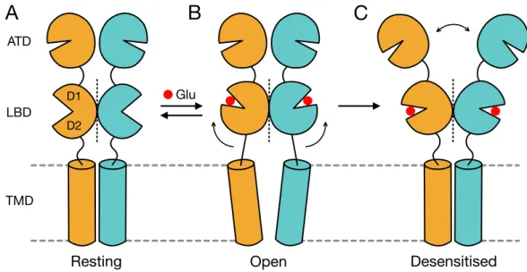
The AMPA receptor LBD shows a bilobed clamshell-like arrangement, in which the polypeptide segment S1 constitutes the upper lobe (D1) and the segment S2 the lower one (D2). In the LBD layer the dimers, arranged in a back to back manner, are mainly stabilised by the D1-D1 interface (Armstrong et al., 1998; Sun et al., 2002). Binding of full agonists, such as glutamate, quisqualate or AMPA, in a pocket located deep within the cleft between the two lobes D1 and D2 induces the closure of the clamshell and a movement of the D2 lobes
that leads to separation of the linkers connecting the LBD with the TMD (Fig. 2). The degree of the cleft closure is dependent on the size of the ligand (Armstrong & Gouaux, 2000). This transition results in a reorientation of the transmembrane helices and opening of the ion channel. When successive LBDs are closed, the pore generates subconductance levels of increasing amplitude, up to a main conductance when the receptor is fully ligated (Rosenmund et al., 1998; Smith & Howe, 2000). Within the conformational changes triggering AMPAR activation a critical role is played by the M3 helix and the M3-S2 linker.
In fact, upon agonist binding the movement of the M3-S2 linkers pulls apart the M3 helices at the bundle crossing fundamentally providing the energy required to open the ion channel (Sobolevsky et al., 2009). Given the 2-fold symmetry relating one LBD dimer to the other, the movements of the M3-S2 linkers are 2-fold symmetric, implying large conformational changes within dimers and smaller changes between dimers.
After activation, in the continuous presence of the agonist AMPARs rapidly undergo a profound desensitisation. Desensitisation can be triggered by just a single LBD closure and at similar rates independently of the number of receptor subunits bound to glutamate (Robert &
Howe, 2003). Recently though, a very surprising finding reported that homomeric Q/R edited
LBD
Glu ATD
TMD
Resting Open Desensitised
D1 D2
A B C
Figure 2. Structural transitions during AMPAR gating. Cartoon representing the movements that the distinct domains of the AMPAR (depicted here as a dimer) undergo to sustain the gating transitions from the (A) closed resting to the (B) open active state, and back, and from the active to the (C) agonist-bound desensitised state. Glutamate is shown as a red sphere.
GluA2 receptors still allow ions to flow when the receptors are desensitised, originating
‘conducting desensitised’ receptors that would account for the large steady-state current generated by GluA2(R) receptors both alone or in the presence of auxiliary proteins. This unusual phenomenon is attributable to the arginines at the Q/R site of the pore loop that would prevent desensitisation-mediated closure of GluA2(R) channels (Coombs et al., 2019). The conformational change onto which desensitisation is usually based is represented by the rupture of the LBD D1-D1 interface, followed by a rotation of the entire LBD to allow for the D2 domains and the LBD-TMD linkers to adopt a closed-state-like conformation (Sun et al., 2002). These rearrangements involve movements of the ATDs and ATD-LBD linkers. Under conditions that favour the stabilisation of a desensitised receptor conformation, structural studies of the homomeric GluA2 receptor have described that during desensitisation the LBD dimers accommodate strong rearrangements, losing their local 2-fold rotational symmetry towards a more 4-fold symmetrical architecture (Twomey et al., 2017). This was in agreement with previous findings about the kainate receptor subtype GluK2 that showed a pseudo-4-fold symmetric arrangement of the LBD upon desensitisation and suggested that a disruption of this ‘desensitisation ring’ might be the key mechanism for the restoration of the receptor resting state (Meyerson et al., 2016). Concerning the movements of the ATD layer sustaining the large LBD rearrangements during desensitisation, some three-dimensional (3D) classifications of GluA2 cryo-EM particles identified a separation, with a variable degree of displacement, of the ATD dimers, that was not observed for either the closed or active states nor for GluK2 receptors (Meyerson et al., 2014). Such separation was explained as the result of strain onto the ATD layer deriving from the symmetry switch of the LBD layer. However, this was not noticed according to other 3D classifications (Twomey et al., 2017) and there are still some discrepancies regarding the movements of the ATDs during AMPAR gating.
Another interesting recent study demonstrated that alternative splicing of the LBD flip/flop cassette, a region of 38 amino acids located at the C-terminal end of the LBD and known to influence desensitisation (Sommer et al., 1990; Mosbacher et al., 1994; Fig. 1 A), controls distant motions of the ATD in the resting state (Dawe et al., 2019). Specifically, it was shown that the flop variant of GluA2 (GluA2flop) imparts greater mobility of the ATD at rest compared to the flip variant (GluA2flip). Moreover, contrary to GluA2flip, GluA2flop receptors were found to be insensitive to regulation by anions, binding to a newly identified
pocket near the base of the LBD D1-D1 dimer interface, and also by auxiliary proteins.
Consistent with this result, alternative flip/flop splicing had previously been associated with differential sensitivity to allosteric modulators such as cyclothiazide (Partin et al., 1994;
Partin et al., 1995). The authors conclude that this intrinsic diversity in the nanoscale mobility of the resting state would favour/disfavour interactions that stabilise/destabilise the receptor open state and, in other words, predetermine the receptor gating behaviour in the presence of neurotransmitters, allosteric modulators and auxiliary proteins.
It is relevant to mention here that auxiliary proteins of the AMPAR can deeply affect the receptor gating, both positively and negatively, but this will be extensively addressed in the following paragraphs.
1.2 AMPA receptor complex
1.2.1 AMPA receptor auxiliary proteins
In the postsynaptic membrane AMPARs are not alone, but rather the hub of dynamic supramolecular complexes with other intracellular and transmembrane proteins that concertedly regulate the receptor function. In order to unravel the molecular machinery behind this regulation, over the past 30 years tremendous progress has been made towards the identification of AMPAR interacting proteins. As aforementioned, different cytosolic proteins (such as GRIP1, GRIP2 and PICK1) have been discovered to interact with the cytoplasmic tail (C-tail) of the AMPAR and to play a role in the receptor membrane trafficking and synaptic anchoring and in intracellular signaling cascades (Jackson & Nicoll, 2011). The first transmembrane protein described to regulate the AMPAR was actually discovered through a seemingly unrelated investigation. In fact, in a mouse inbred line a spontaneous mutation was found to be associated with a neurological phenotype reminiscent of human absence epilepsy.
This phenotype was particularly distinguished by a characteristic head-tossing behaviour, after which the mouse mutant was named stargazer (Noebels et al., 1990). The analysis of the stargazer mutation lead to the identification of a novel, brain-specific, tetraspanin membrane protein with structural homology to the gamma 1 (γ1) subunit of the skeletal muscle voltage- gated Ca2+ channel and therefore it was termed γ2, or stargazin (Letts et al., 1998). Stargazin though was displayed to only subtly modulate the functional properties of voltage-gated Ca2+
channel in heterologous systems (Letts et al., 1998; Klugbauer et al., 2000; Kang et al., 2001;
Rousset et al., 2001), but to be essential for the regulation of AMPARs in the cerebellum (Hashimoto et al., 1999; Chen et al., 2000). In cerebellar granule cells of the stargazer mouse, AMPAR-mediated synaptic as well as extrasynaptic currents were observed to be largely absent and to be both rescued by transfection with full-length recombinant stargazin. Hence, stargazin was proven to be required for surface expression and synaptic targeting of AMPARs in cerebellar granule cells.
Following the discovery of stargazin as the prototypical AMPAR auxiliary subunit, database mining revealed the existence of closely related γ subunits with widespread expression within the CNS (γ3, γ4, γ5, γ7, γ8) (Burgess et al., 1999; Klugbauer et al., 2000; Burgess et al., 2001) and together with stargazin they were collectively classified as “transmembrane AMPA receptor regulatory proteins” (TARPs). TARPs are present in the majority of AMPAR complexes in the brain (Fukata et al., 2005). However, in addition to TARPs, unrelated transmembrane proteins (such as SOL-1, CNIH-2 and 3, SynDIG1; CKAMP44, GSG1L) have been recently identified through genomic and proteomic analyses as other candidate AMPAR auxiliary subunits (Zheng et al., 2004; Schwenk et al., 2009; Kalashnikova et al., 2010; von Engelhardt et al., 2010; Shanks et al., 2012). These findings provided evidence for bewildering combinatorial possibilities of interaction of these auxiliary proteins with the AMPAR and between each other, moving the focus from the function of the receptor by itself to the joint action of a dynamic network of interacting proteins.
1.2.2 Transmembrane AMPA receptor regulatory proteins (TARPs)
TARPs are a family of four-pass transmembrane proteins composed of the γ subunits γ2 (or stargazin), γ3, γ4, γ5, γ7 and γ8. They are integral components of native AMPAR complexes (Fig. 3), with discrete patterns of expression throughout the brain, both in neurons and glial cells, depending on the neuronal cell-type and the course of development (Tomita et al., 2003). Stargazin shows highest levels of expression in the cerebellum, γ3 in the cerebral cortex, γ4 in the olfactory bulb and γ8 in the hippocampus. The highest levels of γ7 expression instead occur in cerebellar Purkinje cells (Kato et al., 2007), while γ5 is particularly enriched in hippocampal CA2 neurons and cerebrellar Bergmann glia (Kato et al., 2008). Moreover, it was shown that in the cerebral cortex of rat pups γ4 expression peaks at
postnatal day 6 (P6) and decreases through later development. In contrast, expression of stargazin, γ3 and γ8 appears later and progressively increases during animal maturation (Tomita et al., 2003).
Based on phylogenetic analyses of the primary sequence, the extended family of the γ subunits, comprising also the skeletal muscle γ1 and γ6 subunits (Burgess et al., 2001), can be divided into subgroups with higher homology: with stargazin, γ3, γ4 and γ8 forming one group, γ5 and γ7 forming another, and γ1 and γ6 being yet another. A second type of
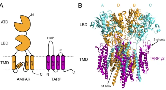
A D B C
TARP γ2 LBD
LBD ATD
AMPAR TARP
TMD M TMD
N
N
1 M
3 M M2 4
M1 ECD1
L2
β-sheets
α1 helix M2 M
3 M 4
C C
A B
Figure 3. Complex of the AMPAR with TARPs. (A) Cartoon depicting the structure of a monomeric AMPAR (orange) and next to it that of a TARP auxiliary subunit (purple). The TARP structure is formed by four transmembrane domains, intracellular amino (N)- and carboxyl (C)-terminus and two extracellular segments, of which the first one (ECD1) is the larger and includes Loop1 (L1) and the β2-TM4 loop (not indicated) and the second one is composed by the short Loop2 (L2). (B) Cryo-EM structure of the homomeric rat GluA2 receptor saturated with TARP γ2 subunits (PDB code: 5KK2, 7.3 Å resolution; Zhao et al., 2016). Extensive contacts take place between the transmembrane domains of the receptor (represented in cyan for subunit pair A/C and in orange for subunit pair B/D) and of γ2 (purple). γ2 extracellular loops (L1, β2-TM4 and L2) are not resolved, but density emanating from the extracellular β-sheets flanking the loops has been modelled, as well as from the pre-TM2 α1 helix following the β2-TM4 loop. β-sheets and α1 helix appear to be positioned in proximity to the receptor LBD and LBD-TMD linkers, allowing the extracellular loops to regulate these gating critical regions.
classification divides the TARP family into two categories fundamentally differing in the functional properties exerted on the AMPAR: type I (or canonical) TARPs including stargazin, γ3, γ4 and γ8 and type II TARPs comprising γ5 and γ7. Type I TARPs were the first ones to be studied and, even if with some differences between distinct subunits, they show to qualitatively similarly regulate the AMPAR by enhancing its function (Cho et al., 2007;
Milstein et al., 2007). γ5 and γ7 instead were initially thought to be unable to modulate the AMPAR function (Tomita et al., 2003; Tomita et al., 2004) and only later they were established as a separate class of TARPs with definite physiological features on specific AMPAR subtypes (Kato et al., 2007; Kato et al., 2008). The various modulatory effects of the different TARP subunits on the AMPAR will be thoroughly examined in the next section.
1.3 TARPs regulation of the AMPA receptor
1.3.1 TARPs mediate AMPA receptor trafficking and anchoring at the synaptic membrane
In both heterologous systems and neurons TARPs dramatically and selectively promote surface expression of mature AMPARs. It was revealed that in cerebellar granule cells of stargazer there was an approximate 75% decrease in the surface expression of the AMPAR subunit GluA2, whereas expression of the NMDAR subunit NR2A was not affected (Tomita et al., 2003). Remarkably, transfection of full-length stargazin as well as of the other type I TARPs γ3, γ4 and γ8 (but not of type II TARP γ5) in stargazer granule neurons could restore AMPAR surface expression and glutamate-evoked responses. This effect of TARPs on trafficking is specific to AMPARs and does not apply to structurally related receptors such as kainate-type channels (Chen et al., 2003). It was later proved that, similarly to type I TARPs, γ7 can promote AMPAR trafficking to the membrane surface and modestly enhance glutamate-evoked currents in transfected stargazer granule cells (Kato et al., 2007). Its effect though seems to be limited to channels containing the GluA1 or GluA2 subunits (Kato et al., 2008). Even more restrictively, γ5 was eventually discovered to be capable of modulating specific GluA2-containing AMPARs, with properties entirely dissimilar from those of canonical TARPs and without promoting receptor trafficking (Kato et al., 2008).
Concerning the molecular mechanisms underlying the deficit of AMPAR expression in stargazer cerebellar granule cells, it was found that a large proportion of GluA2 receptors did not receive mature endoplasmic reticulum (ER)-type glycosylation, a post-translational modification that allows exit of the receptors from the ER (Mah et al., 2005; Traynelis et al., 2010). This result suggested that stargazin plays a role in the early stages of the AMPAR biogenesis (Tomita et al., 2003). In support of this, it was also described that in TARP- deficient cells, such as heterologous cells or stargazer granule neurons, AMPAR expression induces the unfolded protein response (UPR), a homeostatic pathway activated by the accumulation of unfolded or misassembled proteins in the ER (Vandenberghe et al., 2005). In parallel, stargazin-mediated enhancement of AMPAR surface expression was verified not to be the result of the inhibition of constitutive receptor endocytosis (Vandenberghe et al., 2005).
Experiments using γ2-γ5 chimeras and γ2 deletion mutants displayed that the C-tail of γ2, as opposed to γ5, is necessary for the surface expression and the potentiation of glutamate- evoked responses of GluA1 receptors in heterologous systems (Tomita et al., 2004; Tomita et al., 2005; Turetsky et al., 2005). It was subsequently demonstrated that the C-terminus of stargazin encodes a membrane sorting signal that mediates AMPAR trafficking from the ER to specific membrane compartments (Bedoukian et al., 2008). Moreover, TARPs seem to associate exclusively with tetrameric AMPARs, thus being incorporated in nascent AMPAR complexes at some point between tetramerisation and ER export (Vandenberghe et al., 2005;
Shanks et al., 2010).
In neurons, not only are TARPs essential for the surface expression of AMPARs, but they also play a central role in anchoring and stabilising the receptors at synapses. In fact, TARPs regulate the lateral diffusion of AMPARs between extrasynaptic and synaptic sites, thank to the interaction of a PDZ binding motif on the TARP C-tail with PDZ domain-containing scaffolding proteins that are intrinsic components of the PSD (Chen et al., 2000; Bats et al., 2007; Sainlos et al., 2011). Such proteins include the PSD protein-95 (PSD-95) and the related members of the membrane-associated guanylate kinase (MAGUK) family (Schnell et al., 2002; Dakoji et al., 2003). The ability of TARPs to bind to PSD-95 is actually itself modulated by post-translational modifications of the TARP C-terminus. For instance, phosphorylation of a threonine (Thr321) located within stargazin PDZ binding motif was shown to be associated with opposing effects on AMPAR clustering during synaptic plasticity
depending on the kinase (PKA or the mitogen-activated protein kinase, MAPK) that phosphorylates it (Stein & Chetkovich, 2010). Furthermore, in its C-tail stargazin contains a series of nine serines, surrounded by a highly basic region, closely conserved in all four type I TARPs (Burgess et al., 2001; Klugbauer et al., 2000; Rousset et al., 2001) and constituting the phosphorylation substrate for CaMKII and PKC (Tomita et al., 2005; Tsui & Malenka, 2006).
Interestingly, under basal conditions in cultured cortical neurons these serines are phosphorylated. Dephosphorylation of the C-terminal poly-serine region by the protein phosphatase 1 (PP1) and 2B (PP2B) seems to be involved in NMDAR-dependent LTD in the hippocampal CA1 region. On the other hand, expression of a phospho-null stargazin construct prevents LTP induction. With these findings, Tomita et al. (2005) identified a key role for TARPs phosphorylation in the bidirectional regulation of synaptic plasticity. To explain how TARP C-tail controls AMPAR synaptic delivery in a phosphorylation-dependent manner, it was proposed that phosphorylation of the nine serines might inhibit the interaction of the surrounding basic residues with the negatively charged membrane lipid bilayer. This dissociation, that is likely to be graded by the multiplicity of phosphorylation sites, would enhance graded binding of the TARP C-tail to PSD-95 and thus regulate targeting of AMPAR/
TARP complexes at synapses (Sumioka et al., 2010). Therefore, synaptic anchoring of AMPARs relies both upon the interaction of TARPs PDZ-binding motif with PSD-95 and also of the AMPAR CTD with intracellular PDZ domain-containing proteins, although the true influence of this second kind of interaction is less clear (Buonarati et al., 2019).
1.3.2 TARP modulation of the AMPA receptor gating
In addition to their role in trafficking, both type I and II TARPs modulate AMPAR gating and pharmacology to varying degrees in a subtype-specific manner (Jackson & Nicoll, 2011).
Type I TARPs in general augment the functional properties of AMPARs. It is indeed well known from studies in heterologous expression systems that stargazin slows down the entry into desensitisation, enhances the steady-state current remaining upon desensitisation, slows down the rate of deactivation and accelerates the recovery from desensitisation of glutamate- elicited AMPAR currents (Yamazaki et al., 2004; Priel et al., 2005; Tomita et al., 2005;
Turetsky et al., 2005). Coexpression of distinct type I TARPs results in differential effects on the gating kinetics of AMPARs and for example γ4 and γ8 have been shown to slow
deactivation to a greater extent than stargazin and γ3 (Kott et al., 2007; Körber et al., 2007;
Milstein et al., 2007; Cho et al., 2007; Suzuki et al., 2008; Pierce & Niu, 2019). TARP subtype-dependent effects observed in heterologous systems were found to be largely mirrored in stargazer cerebellar granule neurons. Here, overexpression of specific TARP isoforms differentially modulates the amplitude, rise-time and decay of the AMPAR-mediated miniature excitatory postsynaptic currents (mEPSCs), with γ4 having the strongest effect in the slowing down of the mEPSCs decay (Milstein et al., 2007; Cho et al., 2007).
The effects of type II TARPs on AMPAR gating are complex and contradictory. Experiments in human embryonic kidney (HEK) 293 cells assessed that γ7 can enhance glutamate-evoked steady-state currents and modestly slow down the deactivation and desensitisation kinetics of GluA1 receptors. γ5 instead, originally showed unable to rescue AMPAR responses in stargazer granule cells (Tomita et al., 2003), displayed no significant effect (Kato et al., 2007). Other experiments in heterologous systems and neuron cultures indicated that, in contrast to the lack of effects on GluA1, γ5 induces an augment of the peak current of specific GluA2-containing AMPARs, without promoting receptor trafficking, coupled with a marked suppression of the steady-state current. γ7 instead would only enhance currents from channels containing the GluA1 or the GluA2 subunit (Kato et al., 2008). It was then found that γ5 increases single-channel conductance, comparably to the other TARP family members, but decreases the peak open probability (i.e. the probability that agonist-bound channels will open) of GluA1 and GluA4 receptors in tsA201 cells. Remarkably, the properties of recombinant AMPARs coexpressed with γ5 matched well those measured from outside-out patches of cerebellar Bergmann glial cells (Soto et al., 2009), neuronal cell type intensely expressing γ5 (Fukaya et al., 2005) and Ca2+-permeable AMPARs formed of GluA1 and GluA4 subunits (Burnashev et al., 1992; Wisden & Seeburg, 1993; Iino et al., 2001).
Additionally, γ5 was determined to preferentially modulate assemblies containing AMPARs with the longest C-tail (that are GluA1 and GluA4). Thus, γ5 was reestablished as an active member of the TARP family with unique properties in the selective regulation of long-form Ca2+-permeable AMPARs in the Bergmann glia.
Another kinetic signature imparted to AMPAR gating by specific TARP isoforms is superactivation. In the continuous presence of glutamate (several seconds), desensitisation is followed by a slow increase in the steady-state current. γ4, γ7 and γ8 can boost
superactivation of GluA1 receptors, while stargazin was shown to be ineffective on GluA1 (Kato et al., 2010). Like γ8 though, γ2 is able to superactivate unedited GluA2(Q) receptors (Carbone & Plested, 2016). This phenomenon was initially interpreted as a reversal of desensitisation and hence termed ‘resensitisation’ (Kato et al., 2010). It was later observed that removal of desensitisation actually produces a more profound TARP-induced increase in AMPAR steady-state current, indicating that this effect is independent of desensitisation (Carbone & Plested, 2016). It was also proven to be independent of the peak current amplitude. On the other hand, the renamed ‘superactivation’ was found to be strongly correlated with the rate of recovery from the desensitised state, with stargazin massively increasing the steady-state current of mutant receptors with fast recovery and thus more available open state. This correlation lead to reinterpret superactivation as a positive feedback mechanism dependent on availability of receptors in the open state. The physiological significance of superactivation still has to be assessed, but it might be hypothesised as a postsynaptic process for facilitation of excitatory neurotransmission. Interestingly, AMPAR- mediated responses in hippocampal pyramidal neurons do not exhibit superactivation.
However, γ8 overexpression in stargazer cerebellar granule neurons elicits superactivation and this is prevented by the cotransfection of the AMPAR auxiliary protein cornichon homolog-2 or CNIH-2 (Kato et al., 2010). The superactivating behaviour of native AMPARs is therefore likely to be governed by the synergetic effect of a number of interacting proteins that intercross to regulate the receptor.
At the level of unitary currents of single channels, AMPARs are known to visit multiple discrete conductance states (Rosenmund et al., 1998). It was demonstrated that stargazin increases the open channel probability (Popen) of GluA4 receptors by pushing the channel openings to large conductance states and that it prolongs the duration of bursts of channel openings (Tomita et al., 2005). Nonetheless, in line with previous findings (Soto et al., 2007), a recent report, in which GluA4 Popen was estimated based on channel-opening and -closing rates from whole-cell currents, showed that Popen is unchanged when stargazin, or γ4, is present (Pierce & Niu, 2019). Other single-channel data with the GluA1 subunit instead suggested that TARP γ4 produces a fundamental change in the receptor behaviour, by enhancing the amplitude of all conductance states including the largest one (Shelley et al., 2012). Nonstationary fluctuation analysis of currents evoked by ultrafast glutamate
application, a powerful method for estimating single-channel properties underlying macroscopic currents, revealed that type I TARPs differentially increase the mean channel conductance of recombinant GluA2-containing receptors, with γ8 enhancing it to the greatest extent (Jackson et al., 2011). In addition, it was proposed that TARP association generates an heterogenous population of receptors whose gating behaviour switches between a low-Popen
and a high-Popen mode (modal gating), resulting in ensemble currents with variable decay kinetics that may contribute to shape the multi-component decay of AMPAR synaptic currents (Zhang et al., 2014).
1.3.3 TARPs regulate AMPA receptor pharmacology
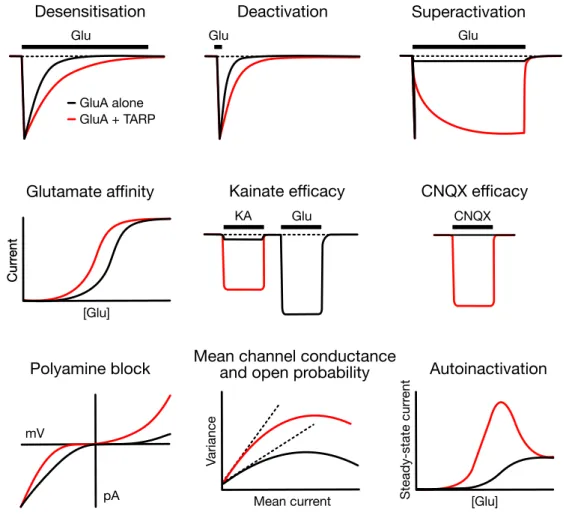
TARPs also modify the pharmacological properties of AMPARs. First of all, stargazin and the other type I TARPs potentiate AMPAR affinity to glutamate (Yamazaki et al., 2004; Priel et al., 2005; Tomita et al., 2005), by a degree that is dependent on the specific TARP subtype (Kott et al., 2007; Tomita et al., 2007; Kott et al., 2009). Recombinant AMPARs show a typical sigmoid glutamate dose-response curve and it is the same case when measuring the steady-state instead of the peak current (Robert & Howe, 2003). Conversely, native and TARPed recombinant AMPARs exhibit a bell-shaped steady-state dose-response curve that indicates a decline of the current at high concentrations of glutamate (above 10-100 µM), a phenomenon referred to as ‘autoinactivation’ (Raman & Trussell, 1992; Morimoto-Tomita et al., 2009; Semenov et al., 2012). Autoinactivation has first been ascribed to a desensitisation- induced complete dissociation of TARPs from AMPARs (Morimoto-Tomita et al., 2009), then rather to subunit-dependent structural rearrangements leading to the functional uncoupling of the AMPAR-TARP complex (Semenov et al., 2012). This concentration-dependent TARP action has been suggested to serve to modulate synaptic short-term plasticity upon neuronal overactivation or elevated levels of glutamate (Morimoto-Tomita et al., 2009). Subsequently, an alternative mechanism has been proposed to account for autoinactivation without implying physical dissociation of the AMPAR-TARP complex (Coombs et al., 2017). In fact, it has been observed that at low glutamate concentrations stargazin exerts a dual effect on AMPAR gating, overall shifting the balance from desensitisation to activation. On one hand, it reduces the sensitivity to desensitisation, by markedly slowing the desensitisation rate (at concentrations below 100 µM). On the other hand, it enhances gating of partially occupied
AMPARs, by increasing glutamate efficacy at low occupancy (i.e. fewer agonist events are necessary to gate the receptors). This behaviour has been related to a possible ability of TARPs of amplifying the synaptic signal transduction mediated by glutamate spillover, a phenomenon described by the diffusion of glutamate from the site of release to neighbouring synapses (Diamond, 2002).
Glu
GluA alone GluA + TARP
Glu Glu
[Glu]
CurrentCurrent
Mean current [Glu]
Variance Steady-statecurrent
Glu
KA CNQX
pA mV
Desensitisation
Glutamate affinity
Polyamine block Mean channel conductance and open probability
Deactivation
Kainate efficacy CNQX efficacy Superactivation
Autoinactivation
Figure 4. TARP modulation of AMPAR gating and pharmacology. Schematic summary of some of the various effects that TARP association (red) determines on the gating and pharmacology of AMPARs (alone, black). The following TARP-induced changes in AMPAR properties are illustrated from left to right, top to bottom: slowing in the desensitisation and deactivation kinetics, slow augment of the steady-state current upon long agonist exposure (superactivation), increase in affinity to the full agonist glutamate and in efficacy of the partial agonist kainate and the competitive antagonist CNQX, relief of the voltage-dependent block by intracellular polyamines, increase in the mean channel conductance and open probability and decline of the current at high glutamate concentrations (autoinactivation). Adapted from Jackson & Nicoll, 2011.
Furthermore, TARPs robustly enhance AMPAR kainate efficacy. Kainate is a partial agonist of AMPARs, meaning that even at saturating concentrations it only induces submaximal channel activation in the form of small, non-desensitising currents (Patneau & Mayer, 1991).
Structurally, partial agonism is due to an incomplete cleft closure of the LBD (Armstrong &
Gouaux, 2000; Jin et al., 2003). The presence of TARPs turns kainate into an almost full agonist in both heterologous cells and neurons (Tomita et al., 2005; Turetsky et al., 2005) and the ratio of kainate-evoked to glutamate-evoked currents (IKA/IGlu) has been a long-established indicator of TARP association in AMPAR measurements (Shi et al., 2009). Type II TARP γ7 slightly increases AMPAR kainate efficacy, while γ5 is ineffective (Tomita et al., 2005; Kato et al., 2007).
The quinoxalinediones are a family of compounds –such as 6-cyano-7-nitroquinoxaline-2,3- dione (CNQX), 6,7-dinitroquinoxaline-2,3-dione (DNQX), 2,3-dioxo-6-nitro-1,2,3,4- tetrahydrobenzo[f]quinoxaline-7-sulfonamide (NBQX)– widely used as competitive AMPAR antagonists (Honoré et al., 1988; Sheardown et al., 1990). By definition, competitive antagonists bind to the AMPAR LBD in the same pocket as agonists, thereby occluding agonist binding, but without leading to opening of the ion channel. It was then observed that CNQX application in neurons induces depolarising currents (McBain et al., 1992; Maccaferri
& Dingledine, 2002) that are blocked by the non-competitive AMPAR antagonist GYKI 53655 and potentiated by the allosteric AMPAR modulator trichloromethiazide (TCM), disclosing a new role for CNQX as an AMPAR agonist rather than antagonist (Menuz et al., 2007). DNQX was shown to behave similarly to CNQX, whereas NBQX purely acts as a competitive receptor antagonist. It was demonstrated that TARPs are responsible for converting CNQX competitive antagonism into a partial agonist activity, possibly by strengthening the coupling between the partial LBD closure induced by CNQX binding and channel opening (Menuz et al., 2007; Cokić et al., 2008; Kott et al., 2009). Subsequent work revealed instead that the degree of CNQX block is time-dependent and under non-equilibrium conditions (shortly after receptor activation, before CNQX and glutamate reach steady-state occupancy), which prevail at glutamatergic synapses, CNQX operates as a non-competitive antagonist and blocks AMPAR-TARP complexes with high affinity (MacLean & Bowie, 2011). Additionally, these authors suggested that the reduction of CNQX block by TARPs