Research Collection
Doctoral Thesis
CD4+ T cell-mediated immune control of cytomegalovirus infection in murine salivary glands
Author(s):
Oderbolz, Josua Publication Date:
2020
Permanent Link:
https://doi.org/10.3929/ethz-b-000465023
Rights / License:
In Copyright - Non-Commercial Use Permitted
This page was generated automatically upon download from the ETH Zurich Research Collection. For more information please consult the Terms of use.
ETH Library
DISS. ETH NO. 27235
CD4 + T cell-mediated immune control of
cytomegalovirus infection in murine salivary glands
A thesis submitted to attain the degree of DOCTOR OF SCIENCES of ETH Zurich
(Dr. sc. ETH Zurich)
presented by JOSUA ODERBOLZ
MSc Health Sciences and Technology, ETH Zurich
born on 16.09.1991
citizen of Lucerne (LU) and Wagenhausen (TG)
accepted on the recommendation of Prof. Dr. Annette Oxenius (examiner) Prof. Dr. Christian Münz (co-examiner)
Prof. Dr. Jens Stein (co-examiner) Prof. Dr. Federica Sallusto (co-examiner)
2020
i
Acknowledgments
Nine years ago, I started my journey at ETH Zurich. At that time, I did not think of completing my studies with a doctoral degree in natural sciences. Although these times were shaped by many unforgettable and valuable moments, I also faced intense and long-lasting study times, which were intellectually and mentally challenging. Nevertheless, I am grateful for many things that ETH Zurich taught me. I made many new (scientifically enthusiastic) friends, met my present wife, and developed personally further. Thus, I would like to dedicate my gratitude to all those who accompanied me the last decade and even longer.
First and foremost, I would like to thank Annette for giving me the opportunity to perform my doctorate in her laboratory, and for her generous and supportive guidance. I appreciated a lot being part of a dynamic team with many talented scientist and interesting personalities. I always felt very comfortable, enjoyed a lot of freedom and trust, and I was able to freely design my experimental work. Annette was not only always open for new ideas and suggestions, and provided important inputs, but also critically questioned my ideas, which helped me enormously improving the work. Her mindset, combined with her enthusiasm and "fire" for science, had major influence on my daily work, and strengthened my interest in immunology.
Next, I am very thankful for the well-appreciated scientific advice, the positive criticism, and the supportive feedback of my committee members, namely Annette Oxenius, Christian Münz, Jens Stein and Federica Sallusto, and for their participation in the yearly-hold committee meetings.
Furthermore, I would like to thank all lab members of the Oxenius lab for helpful discussions and enabling a casual and motivating atmosphere in the lab. Moreover, I am grateful for the scientific inputs from the Joller and Sallusto lab and for the valuable discussions during my lab meetings.
Special thanks goes to the lab technicians Nathalie Oetiker and Franziska Wagen who are an essential and central part of the lab by assisting in genotypings, performing multiple lab duties, and organizing social activities and the structure of the lab. I appreciated the nonscientific exchange during the lunches, the funny conversations, and their overall openness.
Next, I would like to thank Alessandro Pedrioli (Ale) and Ilaria Spadafora, my colleagues sharing the office G409 with me, for many funny moments, their scientific help, and the welcome
ii
atmosphere. I especially would like to thank Ale for his preference to communicate in GIFs, the relaxed attitude, and for becoming a good and valued colleague of me.
Furthermore, I am grateful to have met Niculò Barandun as he became a good friend of me during my last 1 ½ years in the lab. I will miss our intense rallies on the centre court, the mutual jokes and pranks, and our shared passion for sport, the gym, and the FC Bayern Munich.
I also would like to thank my former Master student and now friend Nathan Zangger who shared with me the innovative mindset and the passion for microscopy. I very much appreciated our daily discussions and your contribution to the project, and I like the way you think about science. I was very happy to supervise such an interested, motivated and talented student like you.
I also would like to thank all members of the staff of the Institute of Microbiology, in particular Markus Schlumberger for the management, Ilka Riedel and Susanne Soo Jin Surber for administrative and organizational concerns, Patrice Sutter (former member), Arian Zuta (former member), Aurel Schwitter, Joel Grand and Urs Blumentritt for their IT support, and Palmira Da Silva Duarte and Daniel Zogg for the infrastructural operations. Furthermore, big thanks goes to Judith Zingg for the coordination and successful execution of the MIM program, and for taking care of doctoral candidate's requests. I am furthermore thankful for the crucial daily work of our animal caretakers in the RCHCI and EPIC facilities, notably Manuela Graf, Corina Fusaro-Graf, Marion Hermerschmidt, Dennis Mollenhauer, Dominik Bacovcin, Martina Müller and Kerstin Sacher.
I would like to thank all of you my friends that I met since I have moved to Zurich. You significantly contributed to my path of life, encouraged me in difficult times, and spent with me many wonderful moments. You make me feel arrived in Zurich.
Finally yet importantly, I would like to thank my family for their endless help, patience, belief, incentive and love. I am grateful to have such amiable and wonderful parents from whom I learned the most important lessons of life. Growing up with three siblings: Benedikt, Tobias and Laura, I was blessed to experience a beautiful childhood. Mum & dad, I am exuberantly happy to have you in my life, and I will never forget what you have done for each of us children. Benedikt, Tobias and Laura, thank you for all the unbelievable lovely times we have spent so far together, and for your interest and participance in my life. Dankä villmol üch allne!!!
I dedicate the last words to my wife Linda who is since nearly seven years my companion, my missus, and my soulmate. I endlessly love you and I am lucky to have you in my life.
iii
List of abbreviations
(T)-CyCIF (Tissue)-cyclic immunofluorescence
2D Two-dimensional
3D Three-dimensional
AGECs Acinar glandular epithelial cells
AIDS Acquired Immune Deficiency Syndrome
APC Antigen presenting cell
AT Adoptive Transfer
B6 C57BL/6
BM Bone marrow
cCMV Congenital cytomegalovirus
CD4 Cluster of differentiation 4
CD8 Cluster of differentiation 8
CMV Cytomegalovirus
CODEX Co-detection by indexing
CTL Cytotoxic T lymphocyte
CTL Cytotoxic T lymphocyte
CXCR3 Chemokine receptor CXC motif 3
DC Dendritic cell
DNA Deoxyribonucleic acid
Dpi Days post infection
E Early
e.g. exempli gratia (for example)
EBV Epstein-Barr virus
ECS Entire cross section
f.p. Footpad
FOV Field of view
HCMV Human cytomegalovirus
HIV Human immunodeficiency virus
Hpi Hours post infection
HSV-1/2 Herpes simplex virus 1/2
i.e. id est (that is)
i.p. Intraperitoneal
iv
i.v. Intravenous
ICOS Inducible T cell costimulator
IE Immediate early
IFN Interferon gamma
IFNR Interferon gamma receptor
IL Interleukin
IMC Imaging mass cytometry
IS Immunological synapse
JAK Janus kinase
Kbp Kilo base pairs
kDa kilodalton
KSHV Kaposi’s sarcoma-associated herpesvirus
L Late
LCMV Lymphocytic choriomeningitis virus
LN Lymph node
MCK-2 Murine cytomegalovirus-encoded chemokine 2
MCMV Murine cytomegalovirus
MHC I/II Major histocompatibility complex class I/II
MS Multiple sclerosis
NIF Nodular inflammatory foci
NK Natural killer
NOS2 Nitric oxide synthase 2
ORF Open reading frames
PBMCs Peripheral blood mononuclear cells
PFA Paraformaldeyhde
PI Post infection
PM Patrolling monocytes
RIMS Refractive index matching solution
RNA Ribonucleic acid
SARS-CoV-2 severe acute respiratory syndrome coronavirus 2
SGs Salivary glands
STAT Signal transducer and activator of transcription
T-bet T-box expressed in T cells
TCR T cell receptor
v
Th1 Type-1 helper
TME Tumor microenvironment
TNF Tumor necrosis factor alpha
Treg Regulatory T helper cell
TRM Tissue-resident memory T cell
VIPRs Viral genes that interfere with antigen presentation
VZV Varicella-zoster virus
WSI Whole slide imaging
WT Wild type
Alpha
Beta
Gamma
Table of Contents
Acknowledgments... i
List of abbreviations ... iii
1. Summary ... 1
1.1 English summary ... 1
1.2 Deutsche Zusammenfassung ... 3
2. Introduction ... 6
2.1 Viruses ... 6
2.2 Herpesviruses ... 7
2.3 Epidemiological relevance of CMV infection ... 8
2.4 MCMV infection and in vivo Pathogenesis ... 9
2.4.1 MCMV as mouse model for CMV infection ... 10
2.4.2 Cell tropism & viral dissemination ... 10
2.4.3 NK cell response in BALB/c vs. C57BL/6 mice ... 12
2.5 Viral pathogenicity & lifelong survival strategies ... 13
2.5.1 CC chemokine homolog MCK-2 ... 14
2.5.2 MHC class I immune evasion ... 15
2.6 Adaptive immunity against CMV ... 16
2.6.1 CD8+ T cell response to CMV ... 16
2.6.2 CD4+ T cell response to CMV ... 17
3. Aims ... 20
4. Results ... 22
4.1 Abstract ... 24
4.2 Introduction ... 25
4.3 Results ... 26
4.4 Discussion ... 47
4.5 References ... 51
4.6 Materials & Methods ... 55
4.7 Supplementary Figures ... 66
5. Discussion ... 78
5.1 Versatile facets of CMV ... 78
5.2 Requirement of IFNsecreting CD4+ T cells during acute MCMV infection in the SGs ... 79
5.3 IL-10 provokes organ-wide virus persistence ... 83
1
5.4 CXCR3 - CXCL9/CXCL10 axis ... 85
5.5 IFN gradient-mediated protection ... 88
6. Unmask the unseen ... 91
6.1 Evolution of fluorescence microscopy ... 91
6.2 Towards high-dimensional imaging ... 92
6.3 MCMV triggered immune atlas of the SGs ... 93
7. References ... 95
8. Curriculum Vitae ... 109
1
1. Summary
1.1 English summary
Herpesviruses are ubiquitous and opportunistic pathogens that have successfully co-evolved with their respective hosts over millennia by acquiring species-specific survival strategies. They are responsible for a wide range of clinical pictures, such as fever blisters caused by herpes simplex virus 1, or Epstein-Barr virus provoked mononucleosis. Although the majority of herpesvirus infections proceed mainly asymptomatically in immunocompetent and healthy hosts, thus being of subclinical nature, immunocompromised individuals (e.g HIV patients or newborns) are at high risk of developing severe clinical manifestations or future abnormalities. Hence, herpesvirus infections represent a serious public health problem.
A hallmark of all herpesviridae family members is their ability to persist as a lifelong companion in their respective hosts. In doing so, the immunological pressure drives the transition of the virus from an initial continuous virus production into a nonproductive, "silent" or "dormant" state with no active viral replication. This so-called latent state not only allows to be efficiently hidden from the immune system, but also facilitates spontaneous reactivation events of the herpesvirus. In case of cytomegaloviruses, deleterious reactivations occur during blood transfusions or solid organ transplantations in immunosuppressed recipients.
Of importance, the high degree of species specificity precludes direct investigation of human cytomegalovirus infection in mice. Hence, infection of mice with murine cytomegalovirus is the current gold standard infection model to study and understand host-pathogen interactions in vivo.
Cytomegalovirus infection shows a remarkably broad cell and tissue tropism by infecting many cell types and organs. Whereas primary acute infection is controlled in most organs within a couple of days, the salivary glands depict a glandular tissue with sustained high viral loads over several weeks (up to 10 weeks). Hence, this peripheral site represents the preferred mucosal niche for prolonged viral replication, and further allows long-term horizontal transmission via viral shedding into the saliva.
Despite the prominent role of cytotoxic CD8+ T cells and antibodies during various viral threats, the CD4+ T cells occupy an unexpected, unique and indispensable protective function during murine cytomegalovirus infection in this specific tissue. Initial studies conducted by Stipan Jonjić and Pero Lučin in the late 80's and early 90's assigned a selective and central role of INF
producing CD4+ T cells to ultimate control of cytomegalovirus replication in the salivary glands.
However, the underlying mechanisms remained poorly characterized, leaving space for
2
speculations open. Continuing studies performed in our lab and by other research groups identified functionally diverse effector functions of this T helper cell subset. In this regard, IFN
sensing by cells of non-hematopoietic origin was crucial for antiviral immunity. Moreover, susceptible acinar glandular epithelial cells were not considered to present antigen, but rather phagocytes, such as dendritic cells or macrophages, which had engulfed "apoptotic bodies" or
"remnants" of previously infected cells. Nevertheless, these assumptions were never proven nor followed up. Furthermore, relevant spatiotemporal information regarding site of infection, or the anatomical location of (antigen-specific) effector CD4+ T cells, to precisely understand antiviral activities, were missing.
This work comprises several aspects of the CD4+ T cell-mediated immune response in cytomegalovirus infected murine salivary glands, and should highlight its fundamental requirement for antiviral protection. In a first part, we extended previous knowledge about M25-specific CD4+ T cells by in-depth profiling their kinetics, phenotypical features, spatial localization, and effector functions. In doing so, we were able to demonstrate that these T cells rather located randomly in the salivary glands during early acute infection, however, acquired an activated state, and were able to produce considerable amounts of the pro-inflammatory cytokine IFN upon cognate (M25) antigen encounter. In a second part, we used conventional 2D, but also whole slide imaging and newly established advanced 3D confocal imaging approaches to visualize key events in antiviral activities, such as T cell receptor signaling, IFN production, or the chemokine gradient-mediated recruitment and clustering of T cells at site of antigen recognition. Our experimental date were then used to build a mathematic simulation model, which revealed that the restricted diffusion properties of IFN at sites of antigen encounter is only able to confer local protection, but allows the virus to further replicate at yet non-protected areas. This implies that a spatio-temporal integration of locally protected regions is required to eventually allow full protection in the salivary glands.
3
1.2 Deutsche Zusammenfassung
Herpesviren sind allgegenwärtige und opportunistische Krankheitserreger, welche sich über Jahrtausende lange Koevolution mit ihren Wirten eine spezifische Aneignung von Überlebensstrategien erschaffen haben, die es ihnen erlaubt hat, sich erfolgreich an ihre jeweiligen Wirte anzupassen. Sie sind verantwortlich für viele Krankheitsbilder, wie zum Beispiel Lippenherpes verursacht durch Herpes-simplex-Virus Typ 1, oder das durch Epstein-Barr-Virus ausgelöste Pfeiffersche Drüsenfieber. Obwohl die Mehrheit der Herpesvirus Infektionen grösstenteils asymptomatisch verlaufen in immunkompetenten und gesunden Personen, also von subklinischer Erscheinungsform sind, so sind immungeschwächte Individuen einem erhöhten Risiko ausgesetzt, klinische Erscheinungsformen oder zukünftige Anomalien zu entwickeln. Daher stellen Herpesviren Infektionen ein ernstzunehmendes, öffentliches Gesundheitsproblem dar.
Eine Besonderheit aller Herpesviren Mitglieder ist ihre Fähigkeit, sich als lebenslanger Wegbegleiter im jeweiligen Wirt zu etablieren. Dabei führt der Druck des Immunsystems zu einem Übergang des Virus-Lebenszykluses von einer anfänglichen, kontinuierlichen Virusproduktion, zu einem unproduktiven, "ruhigen" oder "schlafenden" Zustand, bei welchem keine aktive Virusreplikation auftritt. Dieser sogenannte latente Status erlaubt es nicht nur, sich effizient vor dem Immunsystem zu verstecken, sondern ermöglicht zugleich auch spontane Reaktivierungen des Virus. Im Falle vom Zytomegalovirus geschieht eine klinisch manifestierte Reaktivierung häufig während Bluttransfusionen oder Organtransplantationen in immungeschwächten Empfängern.
Von Bedeutung für die Forschung ist die hohe Arten Spezifität, welche die direkte Untersuchung des humanen Zytomegalovirus in Mäusen verhindert. Deshalb ist die Infektion von Mäusen mit dem Maus-evolvierten Zytomegalovirus der momentane Goldstandard, um die Wirt- Krankheitserreger Interaktionen im lebenden Organismus studieren und verstehen zu können.
Zytomegalovirus Infektionen zeigen eine bemerkenswerte Variabilität und Vielfalt in den Arten von Zelltypen und Organen, die sie infizieren können. Obwohl die primäre, akute Infektion in den meisten Organen innerhalb weniger Tagen kontrolliert wird, so zeigt die Speicheldrüse einen anhaltend hohen Virustiter über mehrere Wochen an (bis zu 10 Wochen). Dieses peripher gelegene Drüsengewebe repräsentiert dabei die bevorzugte, zur Schleimhaut gehörende Nische für eine verlängerte Virusreplikation, und ermöglicht des Weiteren eine langandauernde horizontale Übertragung via Virussekretion in den Speichel.
Obwohl die zytotoxischen CD8+ T Zellen und Antikörper bei verschiedenen viralen Bedrohungen eine bedeutende Rolle übernehmen, so haben die CD4+ T Zellen eine unerwartet, einzigartige
4
und unverzichtbar schützende Funktion während einer Zytomegalovirus Infektion in der Speicheldrüse von Mäusen. Die in den späten 80'ern und frühen 90'ern ersten durchgeführten Studien von Stipan Jonjić und Pero Lučin haben eine selektive und zentrale Rolle den IFN
produzierenden CD4+ T Zellen für die endgültige Viruskontrolle in der Speicheldrüse zugeschrieben. Jedoch blieben die zugrundeliegenden Mechanismen nur unzureichend charakterisiert, was Raum für Spekulationen schuf. Von uns und weiteren Forschungsgruppen durchgeführte, darauf aufbauende Studien, haben funktionell unterschiedliche Effektor Funktionen dieser T Helferzellen Untergruppe identifiziert. In diesem Zusammenhang war die IFNErkennung auf Zellen nicht hämatopoetischen Ursprungs essentiell für die antivirale Immunität. Ausserdem sprechen experimentelle Daten eher dafür, dass virusempfängliche Epithelzellen nicht direkt von Virus-spezifischen T Zellen erkannt werden. Es sind vielmehr ansässige Phagozyten, wie zum Beispiel dendritische Zellen oder Makrophagen, welche abgestossenes "totes Zellmaterial" oder "Überbleibsel" einer zuvor infizierten Zelle aufnehmen und dann in prozessierter Form an T Zellen präsentieren. Nichtsdestotrotz, diese Annahmen wurden weder stichhaltig bewiesen noch weiterverfolgt. Zudem fehlten relevante, räumlich- zeitliche Angaben bezüglich des Infektionsortes, oder der anatomischen Lage (Antigen- spezifischer) T Zellen, um präzise die antiviralen Aktivitäten zu verstehen.
Diese Arbeit befasst sich mit verschiedenen Aspekten der CD4+ T Zellen basierenden Immunantwort in der Speicheldrüse während einer Zytomegalovirus Infektion, mit dem Ziel, deren schützende Funktion besser zu verstehen. Dazu haben wir in einem ersten Teil unser Wissen bezüglich der M25-spezifischen CD4+ T Zellen erweitert, indem wir ihre Kinetik, die phänotypischen Charakterzüge, die räumliche Lage, und die Effektor Funktionen ganz genau untersucht haben. Wir konnten dabei feststellen, dass sich diese Zellen eher willkürlich in der Speicheldrüse während der frühen, akuten Phase der Infektion aufhielten. Jedoch waren die Zellen in einem aktivierten Zustand, und in der Lage, bei der Erkennung des passenden (M25) Antigens, eine beachtliche Menge an IFN auszuschütten. In einem zweiten Teil haben wir konventionelle 2D Mikroskopie, aber auch die auf einem Scanningverfahren basierende Aufnahme eines gesamten Speicheldrüsenquerschnitts, und eine neu etablierte, fortgeschrittene 3D konfokale Mikroskopie angewendet, um wichtige Ereignisse in der antiviralen Aktivität, wie zum Beispiel T Zell Rezeptor Signalübermittlung, IFN Produktion, oder die durch einen Chemokin-Gradienten entstehende Rekrutierung und Anhäufung von T Zellen an Antigen vorliegenden Orten zu bestimmen. Basierend auf unseren experimentellen Ergebnissen, haben wir ein mathematisches Simulationsmodell etabliert, welches uns erlaubte zu definieren, dass die
5
eingeschränkte Diffusionskapazität des IFN am Antigen-Erkennungsortes nur lokalen Schutz gewährt, jedoch es dem Virus erlaubt, sich an noch ungeschützten Orten weiter zu vermehren.
Dies bedeutet, dass die räumlich-zeitliche Integration von geschützten Regionen eine Voraussetzung dafür ist, dass schlussendlich ein umfänglicher Schutz der Speicheldrüse gewährleistet werden kann.
6
2. Introduction
2.1 Viruses
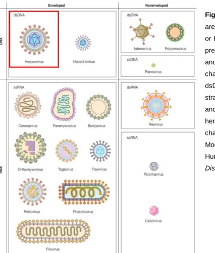
Viruses are highly complex, obligate intracellular microorganisms that rely on living cells to reproduce and propagate. They are generally classified into seven groups based on the genomic nature (RNA or DNA, double-stranded or single-stranded, and linear, circular or segmented), the mode of genome replication, and the morphological features (enveloped vs. non-enveloped) (Fig.
2.1)1. Of note, they are nearly found in all organisms and can persist lifelong through longtime acquired co-evolution with their respective hosts over millennia. In doing so, they are responsible for multiple human diseases that can be life threatening in immunocompromised individuals, which are unable to mount an effective antiviral immune response. The Acquired Immune Deficiency Syndrome (AIDS) caused by human immunodeficiency virus (HIV), or infectious mononucleosis provoked by Epstein-Barr virus (EBV), are well-known examples of persistent chronic and latent infections causing severe clinical outcomes. Unfortunately, the therapeutic efficacy of antiviral drugs targeting viruses such as HIV, herpesviruses including EBV, hepatitis B and C, or the novel 2019 emerging severe acute respiratory syndrome coronavirus 2 (SARS-CoV-2)2, is often limited due to viral latency, a high mutation rate, or a missing precise molecular understanding between virus-host interactions, illustrating further a fundamental epidemiological relevance. However, in contrast to chronic/ latent viral infections that persist throughout the lifetime of a person, human being can cope with most acute productive viral infections by triggering an appropriate, prompt immune response, thus inducing virus elimination and control within a few days after exposure.
Generally, the life cycle of a virus consists of three stages: Entry, genome replication, and exit. In this regard, acute and chronic viral infections demand the full life cycle for continuous production of progeny viruses through either lytic or non-lytic properties, thereby representing productive infections. Unlike, latent infections are types of nonproductive viral infections that are established after incomplete clearance of primary infection, and are characterized by an intracellular “dormant”
or “silent” state of the viral genome with little to no transcriptional and translational activities. This type of persistence is a prerequisite and responsible for recurrent active herpesvirus infections in immunosuppressed HIV patients, or in bone marrow (BM) and solid organ transplant recipients receiving immunosuppressive drugs3,4.
Persistent viral infections are a serious public health problem, as they are not fully eliminated after primary infection, and hence consistently prove the body’s own defense system. Although they are responsible for diverse clinical manifestations, their mode of infection provides crucial insights
7
into not yet resolved mandatory mechanisms to subvert otherwise functional virus-directed immune responses, allowing the coexistence with their hosts over prolonged times.
Figure 2.1. Classification of viruses. Viruses are classified by the nature of their genome (DNA or RNA, ss or ds), their mode of replication (i.e.
presence reverse transcriptase in retroviruses and hepadnaviruses), and their morphological characteristics (enveloped or non-enveloped).
dsDNA; double-stranded DNA, ssDNA; single- stranded DNA, dsRNA; double-stranded RNA and ssRNA; single-stranded RNA. Red framed herpesvirus will be explained in the following chapters.
Modified Figure from book “Molecular Virology of Human Pathogenic Viruses”, page 11, chapter 1:
Discovery and Classification.
2.2 Herpesviruses
The herpesviridae family is exemplary for the generation of persistent latent viral infections after resolution of the primary infection. Although the majority of the human population becomes exposed with a least one herpesvirus family member at one moment during their life span, immunocompetent, healthy individuals normally cope well, showing rarely symptomatic features.
Nevertheless, besides proceeding mostly clinically silent, herpesvirus infections are also associated with clearly visible symptoms, such as oral or genital herpes caused by herpes simplex virus 1 (HSV-1) – and 2 (HSV-2), respectively. Even more important, HSV-1 and HSV-2, and varicella-zoster virus (VZV), are human neurotropic viruses, and thus causative agents for multiple brain-related disorders such as encephalitis and corneal blindness5.
Herpesviruses are large (typically 100 - 200 nm big), enveloped, linear double-stranded DNA viruses possessing a genome size of up to 240 kbp. Thus, they are capable of encoding over 200
8
open reading frames (ORFs), allowing the synthesis of 200 potentially antigenic proteins in case of cytomegaloviruses (CMVs)6–9. The genomic DNA is densely packed within an icosahedral capsid, surrounded by a proteinaceous layer called tegument, which in turn is bounded by the outermost lipid bilayer that typically consists of glycosylated proteins10,11. According to these impressive features, herpesviruses are among the largest and most complex mammalian viruses that exist to date, with a genome size ranging between 124 kbp (simian varicella virus12) and 241 kbp (chimpanzee cytomegalovirus13). Their viral genome replication is temporally regulated and proceeds in three sequential phases of gene expression: Immediate early (IE) gene, early (E) gene, and late (L) gene expression. IE and E gene expression occurs within three to four, or five to seven hours post infection (hpi), respectively, and include minor structural and nonstructural proteins. L genes are expressed until 12 hpi and contain major structural proteins14.
Based on cell tropism, genetic organization, and replication properties, the current human herpesviridae family is divided into three genera: Alpha (), beta (), and gamma () herpesviruses. Whereas -herpesviruses (i.e. HSV-1, HSV-2 and VZV) are fast-growing and cytolytic viruses, establishing preferentially latent infections in neurons, (human CMV (HCMV), human herpesvirus 6 –and 7) -and -herpesviruses (EBV and Kaposi’s sarcoma-associated herpesvirus (KSHV)) are rather slow-growing viruses, targeting non-neuronal lymphocytes, myeloid lineage and epithelial cells for latency. EBV and KSHV are further referred to as tumor viruses due to their ability to transform latently infected cells, and hence being associated with various tumors1.
2.3 Epidemiological relevance of CMV infection
CMV, a prototypic member of the -herpesvirus family, is a ubiquitous and opportunistic pathogen that infects the majority of the world’s population. Depending on the socioeconomic status, the geographical location, and the population density, 60 – 90% of the human population carry the virus15,16. Through a highly fine balanced co-evolution with their mammalian hosts, CMV acquired strategies allowing them to persist lifelong following primary infection in susceptible hosts.
Although CMV infection is usually of subclinical nature in healthy and immunocompetent individuals, it can lead to serious consequences in immunocompromised or immunosuppressed people such as HIV patients, BM and solid organ transplant recipients. Furthermore, congenital CMV (cCMV) infection is one of the leading viral causes of birth defects and fetal maldevelopment17,18. In this regard, transplacental infection of the fetus can provoke major implications and be a clinical risk of developing congenital abnormalities such as sensorineural hearing loss, cerebral palsy, mental retardation, and visual impairments19,20. Further HCMV
9
caused diseases, even in apparently immunocompetent individuals, include encephalitis, hepatitis, pneumonitis, and a mononucleosis syndrome resembling the EBV induced infectious mononucleosis21–23. In short, HCMV infection increases morbidity and mortality. Therefore, CMV pathogenesis is of particular epidemiological relevance and subject to various research activities worldwide. Despite the significant global health impact, there are until now no licensed vaccines for antiviral prevention24–26. One major obstacle in the development of an efficient vaccine against HCMV is the strict species specificity, a hallmark of all -herpesviruses, which precludes crucial preclinical in vivo studies of HCMV infection in animals27. However, various rodent cytomegaloviruses (e.g. mouse7, rat28 and guinea pig CMV29) served in the last decades as valuable animal infection models to study the complex host-pathogen interactions27,30. Moreover, the genetically more closely to HCMV related rhesus and chimpanzee CMV are advantageous for a better understanding of CMV pathogenesis in rhesus monkeys and chimpanzees, respectively13,31,32. Another promising tool for preclinical animal testing of potential vaccine candidates are humanized mice (i.e. immune deficient mice that are engrafted with human tissue), which enable the analysis of HCMV infection directly in mice33,34. These humanized mouse models are commonly used for other human-restricted viruses such as EBV or HIV30,35,36. Of importance, as viral evolution has paralleled mammalian speciation and hence acquired unique species- specific properties, CMV relatedness corresponds to species relatedness37. Thus, mice might not be ideal from a phylogenetic point of view. Nevertheless, as infection of mice with murine CMV (MCMV) is a well-studied and widely used animal infection model, it is currently the gold standard for the investigation of in vivo CMV pathogenesis.
2.4 MCMV infection and in vivo Pathogenesis
HCMV naturally enters the human body via the epithelium of the upper alimentary, respiratory or genitourinary tracts, and in less undesirable circumstances via blood transfusion or during solid organ transplantation. The latter medical interventions are of particular importance as they facilitate recurrent reactivation events of latent CMV in these immunosuppressed conditions, which can become problematic for the recipients. Viral spreading of HCMV mostly occurs via contact with primarily mucosal secretions containing infectious viruses. These include saliva, semen, vaginal and cervical secretions, and breast milk. Depending on the dose of infection, the route of inoculation, and the genetic background and health status of mice, the course of disease during MCMV infection varies substantially in mice38. Besides, the selection of the MCMV mutant and the source of the virus (tissue cultured vs. salivary gland-derived virus) strongly influences
10
virulence and associated viral dissemination properties to peripheral organs such as the salivary glands (SGs), and consequently the corresponding immune response39–41.
2.4.1 MCMV as mouse model for CMV infection
The above-mentioned high degree of species specificity by millions of years of co-evolution with individual hosts, demands an appropriate animal model that greatly recapitulates the human setting. Not least through the nowadays rather easy genetic manipulation of mice, or the various recombinant MCMV mutants, together with the large availability of diverse reporter mouse lines, infection of mice with MCMV is the preferred choice as it enables addressing fundamental biological questions regarding viral pathogenicity and associated antiviral immune responses30. Moreover, HCMV and MCMV share common features: Both behave nonpathogenic upon primary infection in their respective immunocompetent hosts, establish lifelong latency with spontaneously occurring reactivation events42, and can cause extensive viral histopathology with lethal outcome in immunosuppressed conditions43–45. Therefore, mice of BALB/c or C57BL/6 (B6) strain are suitable mouse models. Whereas BALB/c mice are referred to as “susceptible” strain, the C57BL/6 mice are considered “resistant”. The reason lies within the in innate immune recognition properties of the virally-encoded m157 protein by natural killer (NK) cells38.
2.4.2 Cell tropism & viral dissemination
As previously mentioned, primary infection of humans with HCMV can occur at different stages of life; Prenatal (transplacental or intrauterine transfusion), perinatal (cervical secretions), or postnatal (body fluids such as urine, saliva or breast milk, and iatrogenic (blood transfusions and organ transplantations))18. However, the exact route is difficult to identify in epidemiological studies and thus remains mostly uncertain. Nevertheless, based on the low prevalence of cCMV (around one percent), most individuals acquire primary CMV infection postnatal, presumably during infancy and adolescence20.
CMV shows a remarkably broad cell tropism by infecting many cell types. In this regard, myeloid cells such as monocytes, macrophages and dendritic cells (DCs), and non-hematopoietic smooth muscle, stromal, endothelial or epithelial cells, are common targets. In case of HCMV, cells of the monocyte/ macrophage lineage preferentially contain viral DNA in their genome, and thus provide an appropriate cellular reservoir for the virus46–48. However, if the virus actively replicates within these cells, or if they rather provide a latent niche, cannot be explained by this observation.
Earlier studies focused on the analysis of blood samples from seropositive, healthy donors. In doing so, they frequently detected HCMV DNA in peripheral blood mononuclear cells (PBMCs),
11
however, with little or no accompanying lytic gene transcription (i.e. IE gene transcription)49. Furthermore, only a limited number of isolated PBMCs from healthy carriers were expressing IE genes following in vitro HCMV exposure, but distinct more upon the differentiation of monocytes to macrophages (= monocyte-derived macrophages). This highly suggests that monocytes rarely support infectious virus production and that only differentiated macrophages show increased permissiveness to HCMV infection with subsequent viral replication in vitro50,51. Consistent with this hypothesis, ex vivo differentiation of myeloid DC progenitors into mature DCs is associated with increased reactivation events of the infectious virus52. In this regard, immature DC progenitors are mostly occupied by latent HCMV during natural infection, whereas enhanced IE promoter activation of the virus is directly linked to the differentiation state of these cells. Next, the fact that monocytes are only transiently in circulation and that free virus particles in blood of healthy people are usually not observed, argues that the acquisition of HCMV already happens in BM progenitor cells53,54. In this regard, CD34+ hematopoietic progenitor cells are likely targeted by HCMV in immunosuppressed patients, whereas these cells were not identified as major viral reservoir in healthy, HCMV-seropositive individuals55. Of importance, polymorphonuclear leukocytes (also known as granuloytes) do not harbor persistent HCMV in asymptomatic hosts as PBMCs do56. Moreover, a rarely detectable portion of OKT4+ and OKT8+ lymphocytes (nowadays known as CD4+ and CD8+ T cells, respectively) provide latent viral niches of HCMV in PBMCs of naturally infected individuals47. Generally, infectious virus cannot be isolated from the blood of healthy seropositive carriers51. Moreover, these observations indicate that BM-derived mononuclear cells such as monocytes, macrophages or dendritic cells are major cellular sites permissive to viral entry in vivo, and that the establishment of viral latency in various PBMCs depends on the immune state of the host. Regarding the clinical aspect, leukocyte depletion is known to dramatically reduce the risk of occurring reactivation events of HCMV during blood transfusions in immunosuppressed recipients, highlighting the importance of latent viral genome in PBMCs as potent source for horizontal transmission57,58.
Similar to the human setting, various peripheral blood leukocytes are implicated in viremia of MCMV in mice59,60. Thus, MCMV not only targets various myeloid cells for viral dissemination to peripheral organs, but also establishes latency within these cells61. Notably, CX3CR1hi patrolling monocytes (PMs) provide immune-privileged vehicles for MCMV dissemination to distal sites62. Interestingly, the necessity of PMs was only required upon local footpad (f.p.) administration, as systemic intraperitoneal (i.p.) inoculation bypassed the CX3CR1-dependent process, and cell-free virus accessed many organs within the first hours after entrance into the peritoneal cavity. CD169+ subcapsular sinus macrophages in the mediastinal lymph nodes (LNs), ER-TR7+CD29+ reticular fibroblasts in the spleen, and hepatocytes in the liver, are general first cellular targets of MCMV
12
upon i.p. inoculation63. However, if CX3CR1 is a specific lineage marker for PMs, remains questionable64.
Although i.p. and f.d. inoculations are widely-accepted routes of administration, they do not reflect the natural transmission portal. On the contrary, entrance of MCMV via the olfactory epithelium is a more likely occurring infection process. Hence, olfactory neurons are reasonable initial targets by MCMV upon nasal infection. In fact, the nasal route provides an ideal entrance, with rapid systemic spread and long-term persistence in the SGs65. In this regard, Farrell et al. have recently shown that MCMV disseminates sequentially via CD11c+ DCs from infected lungs to the LNs, blood, and finally to the SGs. Moreover, dispersion to the SGs was greatly facilitated by the viral M33 chemokine receptor, thus identifying a possible target to limit CMV spread66. In summary, route of inoculation dictates which cell type gets targeted by the virus and how the virus is spread within the organism67.
2.4.3 NK cell response in BALB/c vs. C57BL/6 mice
Most mouse studies are performed in the BALB/c and B6 mouse strain. Hence, both are approved as convenient mouse models to study CMV infection a nonhuman setting68.
One major difference between these two mouse strains is the ability to mount an early, effective NK cell response. These cells belong to the innate cell compartment and are the first responders among lymphocytes to viral threats, tumors and stressed cells. Rather than expressing rearranged antigen receptors, NK cells and express a set of activating and inhibitory receptors on their cell surface. Whether a NK cell will attack or tolerate a virally infected, malignant, or unhealthy cell, depends on the net balance of signals they are receiving through these receptors69,70. Therefore, this fine-tuned system regulates NK cell response. NK cells are highly implicated in the susceptibility to CMV infection. Although an impaired NK cell functionality, a NK cell deficiency, or even a complete lack of NK cells are quite rare in humans, affected individuals unusually suffer severely71,72. In this line, NK cell-depleted mice, or mice harboring nonfunctional NK cells with reduced interferon gamma (IFN production and cytotoxic properties, are more susceptible to MCMV infection and reveal significantly increased viral titers73–76. Furthermore, adoptive reconstitution of NK-cell deficient mice with NK-enriched leukocytes demonstrated a potent antiviral role for NK cells during MCMV infection77. Interestingly, the susceptibility to lymphocytic choriomeningitis virus (LCMV) infection remained unchanged upon these interventions, assuming a rather virus-specific protection capacity than a general effect. Nevertheless, these observations indicate a central role of NK cell-mediate immunity against various pathogenic triggers. Especially the production of IL-12 and IFN is crucial against various infectious viruses78–80.
13
The Ly49 receptor family encodes for activating and inhibitory NK cell receptors, which typically bind to MHC class I molecules81. Ly49H, a member of the Ly49 family, is an activating NK cell receptor, recognizing the m157 glycoprotein on MCMV-infected cells in inbred B6 mice82. In doing so, they can directly target and eliminate virus harboring cells and thus contribute to crucial MCMV control83,84. Interestingly, NK cells can exert certain immunological pressure on MCMV, in the way that the virus undergoes rapid and specific mutation within the m157 gene locus to efficiently evade NK cell recognition by not engaging with the Ly49H receptor (escape mutants)85. However, the Ly49 receptor family is polygenic and polymorphic, revealing considerable variabilities in the different mouse strains86. In this regard, 129/J and BALB/c mice lack the Ly49H molecule. Instead, the virally-encoded m157 protein interacts with another allelic version in 129/J mice, with the inhibitory Ly49I receptor, whereas BALB/c mice do not even recognize the expression of m157 on MCMV infected cells83. Therefore, based on the absence of Ly49H positive NK cells, the two latter mouse strains are referred to as “MCMV susceptible” strains. In contrast, the B6 mice are
“resistant” to MCMV infection, as they do not express the Ly49I allele but provide Cmv1-mediated resistance87.
2.5 Viral pathogenicity & lifelong survival strategies
Along with the previously described evolutionary loss of a functional m157 glycoprotein in most wild type (WT) MCMV strains, which enables the virus to evade specifically NK cell recognition and NK cell-mediated elimination, MCMV has acquired additional beneficial characteristics that facilitate lifelong coexistence with their hosts.
First, the establishment of a latent state where the full genome of the virus remains present within the susceptible cell, however, the virus does not actively replicate and hence produce new virus progenies (= virions), is one prerequisite not to evoke a presumably virus-clearing immune response. Next, the colonization of specific “privileged” sites that not only provide perfect conditions for viral replication and have a less striking immune surveillance (i.e. general low MHC class I expression on epithelial cells and rarely present immune cells), but also possess ideal shedding options of infectious viruses into body fluids, is mandatory for a long-term settlement and transmission of this -herpesvirus family member. Lastly, MCMV impairs and manipulates the host’s immune system by interfering with cellular processes that allow the pathogen to bypass or silence otherwise potent antiviral immune defense mechanisms. Therefore, it is not by accident that CMV successfully established several strategies to simultaneously increase virulence and limit immune control, which permit the virus to persist as lifelong companion of the mammalian population.
14
2.5.1 CC chemokine homolog MCK-2
Chemokines are chemoattractant molecules or signaling proteins secreted by a wide variety of different cell types. Their main function is to guide the migration of cells. Some of these chemotactic cytokines are released during homeostatic conditions, such as during tissue maintenance, immune surveillance, and development, whereas others are known to be produced upon pathogenic triggers such as during viral or bacterial infections, or cancer88,89. These latter are known as pro-inflammatory chemokines and are of particular relevance in recruiting immune cells to site of infection. In short, they represent a sophisticated communication system between cells.
CMV carries several genes that are homologous to genes of the host organism. These genes, including virally encoded chemokine homologs with almost identical functions, are hypothesized to be hijacked from the host genome during millions of years well-adapted co-evolution90,91. The MCMV ORF m131/129 encodes for a 31.4 kDa protein with homology to mammalian - chemokines (also known as CC chemokines), and hence is referred to as MCMV-encoded chemokine 2 (MCK-2)92–96. MCK-2 is a potent virulence factor that facilitates viral dissemination to the SGs, the preferred mucosal niche for sustained viral replication and persistence, and the major site of horizontal transmission using the saliva as vehicle. Although acute infection with genetically manipulated MCMV strains equipped with a disrupted m131/m129 locus (referred to as MCK mutants) was rarely impaired in the spleen and liver compared to WT MCMV strain, infection was faster cleared due to a more vigorous immune response. Furthermore, these MCK mutants revealed reduced replication in the SGs, highlighting a crucial role in organ-specific viral spread40,94. Moreover, MCK-2 promotes the recruitment of mononuclear leukocytes, most likely monocytes and macrophages, to the initial site of infection, in order to enable monocyte- associated dissemination of MCMV within the host82,97. In this regard, efficient viral spread was mainly associated with immature, BM-derived myelomonocytic cells, and was independent of CCR2 and MCP-1, suggesting a negligible role of mature macrophages98.
Along with the enhanced migration of leukocytes to the initial site of infection, MCK-2 increases simultaneously inflammation83. More recently, MCK-2 was implicated in enhanced viremia by facilitating the infection of CX3CR1+ patrolling monocytes62 and promoted SG colonization upon nasal entry65. In addition, MCK-2 was shown to highly influence cell tropism99.
Nevertheless, the contribution of MCK-2, CCL2 and CX3CR1 in MCMV dissemination is currently challenged100.
15
2.5.2 MHC class I immune evasion
In addition to the virally encoded CC chemokine homolog MCK-2, a key characteristic of MCMV is the huge repertoire of viral genes that interfere with antigen presentation (VIPRs) in MCMV infected cells. To date, three immune evasion genes are known that successfully dampen the MHC class I pathway, namely m04, m06 and m152101–103. The expression of these VIPRs represents a highly sophisticated mechanism that allows MCMV to bypass specifically the antiviral CD8+ T cell response. Since intracellular pathogens, including viruses, are prone to be detected and processed into peptides by the immunoproteasome, which are then presented to these cytotoxic T cells in the context of MHC class I molecules, the prevention of viral peptide-loaded MHC class I complexes on the cell surface of infected cells represents an efficient process to remain hidden. Whereas m06 redirects MHC class I molecules to the lysosome for degradation103, m152 retains them in the ER-Golgi intermediate compartment104. In contrast to m06 and m152, m04 does not lead to reduced MHC class I surface expression but presumably prevents antigen recognition105,106. So far, it is still not fully understood how these immune evasion genes interact with each other on a functional level, and if all three are required for complete inhibition of CD8+ T cell-mediated lysis. In respect thereof, it was not only shown in vitro that VIPRs act synergistically as well as antagonistically, but also that the extent of the respective effect depended on the MHC class I haplotype107,108. Regarding the in vivo situation, MCMV mutants lacking only the m152 gene were more susceptible to CD8+ T cell control, indicating that even the deletion of a single immune evasion gene impacts the fitness of CMV109. Interestingly, the m152 gene product gp40 also downregulates H-60, a high-affinity ligand for NKG2D receptors, thereby diminishing NK cell response in BALB/c mice110. Consequently, the viral gp40 protein possesses a dual function in inhibiting the innate and adaptive arm of the immune system.
However, the decreased expression or complete absence of MHC class I molecules on virally infected cells would render them quite vulnerable to NK cell-mediated lysis. Since MHC class I molecules on the surface of cells present inhibitory signals to NK cells by interacting with inhibitory receptors, diminished expression of these molecules due to a reduced transport to the cell surface would induce a “missing self” signal to the NK cells, which subsequently eliminate these cells111. Therefore, MCMV encodes for a MHC class I homolog, known as m144, which mimics the host version and hence potentially acts as a decoy for NK cells by engaging with inhibitory NK cell receptors112. In doing so, m144 confers protection to NK cell-mediated elimination. Deletion of the m144 gene was associated with reduced viral growth during acute infection in vivo. Moreover, deletion of NK cells restored virulence of the m144 deletion mutant, indicating that this gene interferes with NK-cell mediated clearance of MCMV infected cells.
16
2.6 Adaptive immunity against CMV
MCMV infection proceeds in three distinct phases: A) acute infection in most visceral organs (e.g.
spleen, liver, lung or kidney), b) persistent infection, which mainly occurs at mucosal sites (preferentially in the SGs), and c) latent infection in most organs, whereby the virus remains primarily “dormant”, but sporadically reactivates42,113–115.
The acute phase is characterized by a strong innate NK cell and adaptive CD8+ T cell response, whereby the virus peaks between day 3 and 5 pi, and is controlled within one to two weeks in most organs with the exception of the SGs. Although being under control, CMV is incompletely eradicated during this early stage of infection, and switches from a productive into a nonproductive
“silent” state, which is well-known as latency. However, ongoing high viral loads are still observed in the SGs, which represents the preferred glandular tissue of sustained viral replication and persistence, and an ideal mucosal niche for horizontal transmission by long-term virus shedding into the saliva116. Here, it takes several weeks for the immune system to confer protection and hence inhibit continuous infectious virus production and viral spread.
2.6.1 CD8
+T cell response to CMV
Despite the fact that cytotoxic CD8+ T cells are of particular relevance in fighting infectious intracellular pathogens and neoplastic cells, they possess an unusual dispensable role during MCMV infection in the SGs117–119. Nevertheless, their requirement for protective immunity during MCMV infection was shown in various reports. In this context, CD8+ T cells mediated antiviral protection against the major IE antigens of MCMV, pp89, during acute lethal CMV disease, thus interfering with viral replication at the initial phase of its replication cycle120–122. Furthermore, vaccination of mice with a recombinant vaccinia virus expressing the MCMV-specific pp89 protein induced long-lasting protective immunity against otherwise lethal MCMV infection in BALB/c mice.
This antiviral immune response was assigned to CD8+ T cells, as depletion of these cells before MCMV challenge extensively abolished the protective immunity. In addition, adoptive transfer of MCMV-primed CD8+ T cells into sublethally irradiated mice limited viral replication, whereas CD4+ T cells had no impact on virus control123. Moreover, adoptive transfer of sensitized CD8+ T cells into -irradiated mice developing interstitial pneumonia after CMV infection resulted in substantial antiviral properties, even after establishment of the disease in the lung124.
Regarding the more relevant clinical setting, adoptive transfer of ex vivo stimulated pp65-specific T lymphocytes (equivalent to the pp89-specific T cells in mice) from CMV seropositive donors, into
17
allogeneic stem cell transplantation patients, which are highly immunosuppressed and refractory to antiviral treatment, protected against CMV reactivation events and reduced viral burden significantly125. A similar effect was observed in immunodeficient BM transplant recipients that received ex vivo propagated CMV-specific cytotoxic CD8+ T cell clones126–128. This implies that the adoptive transfer of CMV sensitized CD8+ T cells is an effective way to reconstitute cellular immunity against CMV in BM transplant recipients129. Nevertheless, CD4+ T helper cells are crucial assistants of effective CD8+ T cell responses. They are necessary for maintenance memory CD8+ T cells and enable heightened recall responses of this T cell subset to various viral and bacterial infections. Therefore, CD4+ T cells are key components of a functional protective immunity130,131. In case of MCMV, CD4+ T cells possess even a superior role in limiting infectious virus production in the SGs. Cellular and humoral immunity by CD8+ T cells and antibodies, respectively, are dispensable for terminating lytic viral replication during primary MCMV infection132,133. In this regard, it was shown that single deletion of one T cell subset (CD4+ or CD8+ T cells) resulted in a slight increase of recurrent MCMV infection, whereas the ablation of both populations induced enhanced reactivation events of MCMV, assuming a functional redundancy of both T cell populations. Although not of prime importance during primary infection, reactivation from viral latency upon immunosuppressive treatment resulted in higher viral titers in B-deficient mice compared to WT B6 mice134. In summary, CD8+ T cells and B cells are dispensable during acute MCMV infection, but are essential upon recurrent infection events in latently MCMV infected mice.
2.6.2 CD4
+T cell response to CMV
While the HCMV-specific CD8+ T cell responses have been extensively studied in the last decades, the relevant contribution of CD4+ T cells was mostly neglected, and hence remained less well understood. However, it is meanwhile accredited that their numbers (especially of those secreting IFN) inversely correlate with various CMV-associated diseases, highlighting a crucial antiviral component of the adaptive immunity135–137. Although originally thought to have rather a supportive function, recent studies revealed their direct antiviral potential and further highlighted their clinical importance138. In this regard, it was shown in vitro that HCMV-specific CD4+ T cells displayed a polyfunctional phenotype, responding to various immunodominant HCMV epitopes with a striking IFN response. Moreover, they produced a range of cytotoxic and secretory effector functions when co-cultured together with HCMV infected dendritic cells and prevented viral spread, thus revealing direct antiviral immunity139. Furthermore, blood frequencies of HCMV- reactive CD4+ T cells are a prognostic indicator for the likelihood of viral reactivation events and the presence of clinical symptoms during the first months after renal transplantation140.
18
During MCMV infection, CD4+ T helper cells are essential for virus control in the SGs. Studies conducted by Jonjić et al. showed that the depletion of the CD8+ T cell subset had no influence of overall virus kinetics. In fact, CD8+ T cell-deficient mice cleared MCMV as good as WT B6 mice, most likely because the CD4+ T cell subset replaced their function and provided virus control in these mice132. In contrast, they convincingly demonstrated that the depletion of the CD4+ T cell subset had remarkable selective consequences on virus control in the SGs. While CD8+ T cells compensated for the loss of CD4+ T helper cells in most other organs, the SGs were very susceptible to infectious virus production, and thus revealed increased viral burden over prolonged periods compared to MCMV infected WT B6 mice141. Lučin et al. further showed that in vivo neutralization of the pro-inflammatory cytokine IFN abolished the crucial contribution of type-1 helper (Th1) CD4+ T cells in effective antiviral immune control, primarily in the SGs, indicating a predominant role of this cytokine in viral clearance142. However, since recombinant IFNalone did not replace Th1 function in vivo, it was hypothesized that this pro-inflammatory cytokine is a crucial component, but not the only determining factor. Interestingly, the absence of IFNeffector functions did not negatively impact CD8+ T cell-mediated virus control in spleens and lungs, assuming that its requirement is specifically SG related. Therefore, a subsequent study by the same group identified tumor necrosis factor alpha (TNF) as another important signaling molecule that in combination with IFN revealed synergistic antiviral functions on MCMV replication in vitro143. In this regard, in vivo neutralization of TNFalso reduced severely the protective capacity of CD4+ T cells in the SGs, comparable to a CD4+ depletion144. Of note, this synergistic effect of IFNandTNFin inhibiting infectious virus production is also observed during other DNA virus infections, such as during HSV145,146 or adenovirus infections147.
More recently, our lab demonstrated a direct antiviral effect of IFN producing CD4+ T cells on - irradiation resistant non-hematopoietic cells, by using mixed bone marrow chimeras. In doing so, IFN induced IFN receptor (IFNR) signaling on acinar glandular epithelial cells (AGECs), endothelial cells, and stromal cells, conferred increased protection in the SGs against MCMV compared to IFNR signaling on -irradiation susceptible hematopoietic cells148. This implies that mainly cells of the non-hematopoietic lineage are crucial responders to IFNsecretion.
Furthermore, the absence of cross-presenting antigen presenting cells (APCs) and the extensive MHC class I downregulation on AGECs by immune evasion genes render the CD8+ T cells inefficient in virus control in this peripheral glandular tissue149 (Fig. 2.2). In summary, IFNproductingCD4+ T cells are required for long-term antiviral protection in the SGs.
19
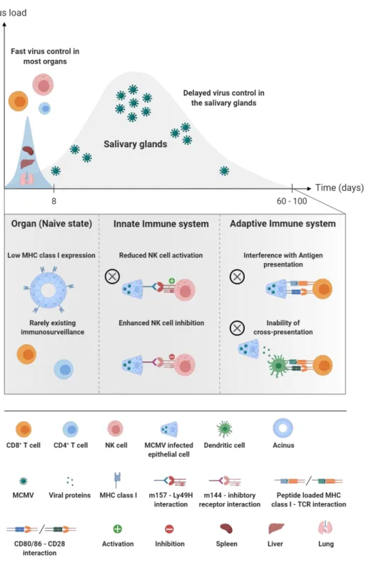
Fig. 2.2. Organ-specific virus kinetics & reasons for a delayed immune control in the salivary glands. Whereas MCMV is controlled in most organs (e.g. spleen, lung and liver) within one to two weeks, the salivary glands represent the preferred mucosal site with sustained high viral loads over several weeks (up to ten weeks). Low immune surveillance in naïve state, or the avoidance of NK and CD8+ T cell recognition, are key survival strategies of MCMV.
Created with BioRender.com.
20
3. Aims
The salivary glands (SGs) are a unique mucosal site that is highly afflicted with replicating cytomegaloviruses (CMVs) over several weeks upon primary exposure. Although the infection of mice with murine CMV (MCMV) induces the prompt recruitment of antigen-experienced, virus- sensitized T cells to this peripheral tissue, a long-lasting heightened viral burden is still present at times, where the immune system has already coped with the infection in most other organs.
Of note, a pivotal role has been assigned to CD4+ T cells that exert direct antiviral effector functions by secreting large amounts of the key pro-inflammatory cytokines IFN and TNF These cytokines are further able to induce an antiviral state in neighboring cellsIn this regard, it has already been shown decades ago that the presence of a functional, effector CD4+ T cell pool in the SGs corresponds directly to increased protection capabilities, and hence faster virus control.
Nevertheless, until now, it is not fully understood how exactly CD4+ T cells mediate crucial antiviral protection. Furthermore, it is still an enigma why it takes months to finally cease viral replication into this organ, despite substantial T cell infiltration. Finally, and of particular relevance, a precise spatiotemporal understanding of critical steps in the antiviral activities, which might explain the delayed protective immune response, was absent so far.
In order to fill all these knowledge gaps, we set up a broad spectrum of various experimental approaches. In doing so, the usage of various genetically modified reporter mouse strains, the application of multicolor flow cytometry and advanced, cutting-edge imaging procedures, and the incorporation of an elaborate mathematic modelling, allowed us to address the following aspects of the CD4+ T cell-mediated immune response in MCMV infected SGs:
I. Characterization of M25-specific CD4+ T cells o Kinetics
o Phenotype o Effector functions o Spatial localization
II. Spatiotemporal resolution of MCMV infection and antiviral effector functions o T cell receptor signaling and IFN production
o Chemokine-mediated recruitment of SG-infiltrating T cells III. Mathematical modeling of the CD4+ T cell-mediated immune control
o Simulation of viral replication, viral spread and the CD4+ T cell response
21
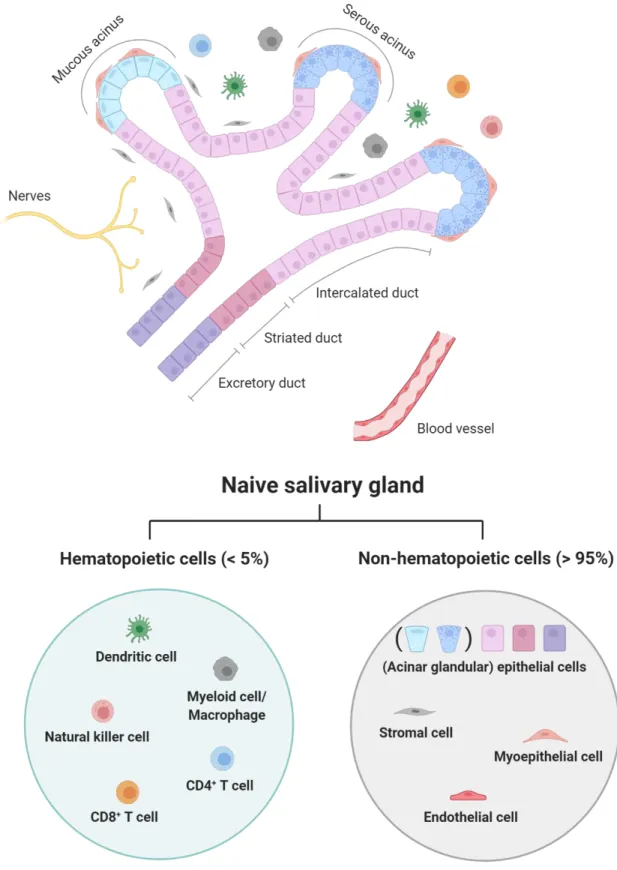
Fig. 2.3. Structure of the duct system & cellular composition of a naïve salivary gland.
Created with BioRender.com
22
4. Results
Locally confined IFN production by CD4
+T cells provides niches for murine cytomegalovirus replication in the salivary gland
Josua Oderbolz1, Nathan Zangger1*, Lea Zimmermann2*, Ioana Sandu1,3, Frederik Graw2 &
Annette Oxenius1
1Institute of Microbiology, ETH Zurich, 8093 Zurich, Switzerland
2BioQuant - Center for Quantitative Biology, Heidelberg University, 69120 Heidelberg, Germany
3Institute of Molecular Systems Biology, ETH Zurich, 8093 Zurich, Switzerland
J.O. and A.O. designed the experiments. J.O, N.Z. and L.Z. performed the experiments. J.O., N.Z.
and L.Z. analyzed the data. I.S. provided essential tools. J.O. and A.O. wrote the manuscript.
* contributed equally to this work
23
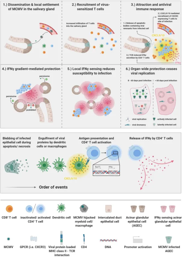
Fig. 4.1. Graphical abstract of the manuscript Locally confined IFN production by CD4+ T cells provides niches for murine cytomegalovirus replication in the salivary gland.
24
4.1 Abstract
Cytomegalovirus (CMV) has evolved a unique virus-host relationship in the salivary glands (SGs) to sustain prolonged viral replication and hence chances for horizontal transmission. Previous reports have established a decisive role for IFN producing CD4+ T cells to control murine CMV (MCMV) infection in the SGs; however, micro-anatomical information regarding their mode of action is largely missing. Here, we provide a spatiotemporal analysis of defined antiviral immune actions that eventually culminate in control of lytic MCMV replication in this preferred mucosal niche. CXCR3-mediated guidance of CD4+ T cells towards CXCL9 and CXCL10 expressing cells resulted in discrete clusters close to infection foci where they reported TCR engagement and produced IFN. Of note, these clusters occasionally contained virus-associated remnants, most likely apoptotic bodies derived from previously infected cells, in CD11c+ cells, enabling antigen presentation to CD4+ T cells. The induced IFN production within these CD4+ T cell accumulations triggered IFNR signaling in a confined perimeter, thereby inducing local, but not organ-wide protection, and allowing MCMV replication to continue at not yet protected sites. Combining our data with a mathematical modeling of the spatiotemporal dynamics of infection and CD4+ T cell dynamics, reveals a scenario, in which ultimate MCMV control is achieved through accumulating sites of regionally-confined tissue protection.
Key words: MCMV, salivary glands, CD4+ T cells, IFN