Original Paper
Pathobiology 2015;82:53–57 DOI: 10.1159/000381903
Frequency of TERT Promoter Mutations in Prostate Cancer
Robert Stoehr
a
Helge Taubert
b
Ulrike Zinnall
a
Johannes Giedl
a
Nadine T. Gaisa
c
Maximilian Burger
d
Petra Ruemmele
e
Carolyn D. Hurst
f
Margaret A. Knowles
f
Bernd Wullich
b
Arndt Hartmann
a
a Institute of Pathology and b Department of Urology, University Hospital Erlangen, Friedrich-Alexander-University Erlangen-Nuremberg, Erlangen , c Institute of Pathology, RWTH Aachen University, Aachen , d Department of Urology, Caritas St. Josef Medical Center and e Institute of Pathology, University of Regensburg, Regensburg , Germany;
f Section of Experimental Oncology, Leeds Institute of Cancer and Pathology, University of Leeds, St. James’s University Hospital, Leeds , UK
the TERT core promoter (–260 to +60) was analyzed by direct Sanger sequencing or SNaPshot analysis. Results: All cases were analyzed successfully. Mutations within the core pro- moter of the TERT gene were not detected in any of the cas- es with all tumors exhibiting a wild-type sequence. Conclu- sion: TERT core promoter mutations reported from several other malignancies were not detected in our unselected co- hort of PrCa. These data indicate that alterations within the core promoter of the TERT gene do not play an important role in prostate carcinogenesis. © 2015 S. Karger AG, Basel
Introduction
The search for recurrent mutations in single genes within tumor genomes is driven by the hope of uncover- ing important driver alterations that lead to a better un- derstanding of carcinogenesis. The availability of new and sensitive high-throughput sequencing technologies sped up this search and led to the identification of new mutation hotspots in a variety of cancer entities. Al- though a new definition of a recurrent gene mutation was Key Words
TERT · Promoter · Mutation · SNaPshot analysis · Sanger sequencing · Prostate cancer
Abstract
Objective: Recently, recurrent mutations within the core promoter of the human telomerase reverse transcriptase (TERT) gene generating consensus binding sites for ETS tran- scription factor family members were described in melano- mas and other malignancies (e.g. bladder cancer, hepatocel- lular carcinoma). These mutations were discussed as early drivers for malignant transformation. In prostate cancer (PrCa) TERT expression has been associated with a poor prognosis and higher risk for disease recurrence. The under- lying mechanisms for high TERT expression in PrCa have still not been clarified. To date, data on TERT promoter mutation analysis in PrCa are sparse. Therefore, we performed se- quence analysis of the core promoter region of the TERT gene in an unselected cohort of prostate tumors. Methods:
Sections from 167 formalin-fixed, paraffin-embedded and cryopreserved prostate tumors were microdissected and used for DNA isolation. The mutation hotspot region within
Received: January 12, 2015
Accepted after revision: March 25, 2015 Published online: May 21, 2015
PD Dr. Robert Stoehr, PhD © 2015 S. Karger AG, Basel
suggested in terms of combining recurrent alterations on the scale of a pathway rather than only one single gene, discovering new recurrent gene mutations is still a worth- while approach in cancer research [1] .
Recently, mutations within the promoter of the telom- erase reverse transcriptase (TERT) gene that encodes the catalytic subunit of the telomerase were identified in fa- milial and sporadic melanoma with a high frequency [2] . The immortality of cells is still a classical hallmark of tu- mors and reactivation of telomerase leading to telomere maintenance remains a fundamental process in carcino- genesis. Alterations within the coding region of the TERT gene are a rare event in cancers. Therefore, the identifica- tion of recurrent mutations within the core promoter of the TERT gene leading to new binding motifs for tran- scription factors of the ETS family attracted great interest among the cancer research field [3] . The consequences of these mutations are still not completely understood but they lead to a 2- to 4-fold increased transcriptional activ- ity in vitro [4] . Subsequently, these mutations were found in several other malignancies, e.g. bladder carcinoma, thyroid cancer or cancers of the nervous system, and were discussed as early drivers for malignant transformation [3] .
Expression and reactivation of telomerase has also been described as an important feature of prostate cancer (PrCa). Telomerase activity was found in up to 100% of analyzed PrCa cases [5] . Interestingly, high expression of telomerase components does not always result in manda- tory telomerase activity [6] . In addition, a significant as- sociation between TERT expression and aggressive be- havior of prostate tumors has been reported [7] . Recently, promising in vitro data were published showing telomer- ase as an important target of an antiandrogen therapy for PrCa, and the usefulness of boron derivatives as a telom- erase inhibitor in PrCa cells [8, 9] . These data suggest telomerase inhibition as a reasonable therapeutic ap- proach for the treatment of PrCa. The molecular and cel- lular pathways involved in telomerase reactivation in PrCa are still not clear. Expression of TERT and the activ- ity of telomerase were shown to be regulated by androgen receptor (AR) signaling whereas exogenous expression of AR surprisingly led to inhibition of TERT transcription in PrCa cells [10, 11] . The genomic region of the TERT gene (chromosome 5p15.33) was not described as a re- gion containing copy number alterations in prostate tu- mors, making gene amplification as a mechanism for TERT expression in PrCa unlikely [12] . Less is known about TERT promoter mutations in PrCa. To date only three studies with a combined number of 49 prostate tu-
mors have reported sequence analysis of the TERT pro- moter and found no evidence for involvement of TERT promoter mutations in PrCa [13–15] . These data already indicate that the cellular mechanisms of telomerase reac- tivation in PrCa are only poorly understood and further clarification is needed. As TERT promoter mutations are a potential mechanism for a possible telomerase reactiva- tion we wanted to further the discussion of this topic for PrCa. We therefore analyzed the core promoter region of the TERT gene containing the reported mutation hotspots in the largest series of PrCa examined to date.
Materials and Methods
Patients and Tissue Samples
Overall, 167 unselected, archival prostate tumors (formalin- fixed and paraffin-embedded tissue samples, n = 119; snap-frozen tissue samples, n = 48) were investigated. All patients were Cauca- sians. The tumors were diagnosed according to the WHO classifi- cation of prostate tumors and staged according to the TNM system [16, 17] . The characteristics of the study participants are shown in table 1 . Prior institutional review board (University Hospital Erlangen) approval was obtained for molecular analysis on archi- val material.
Tissue Microdissection and DNA Isolation
DNA was extracted from prostate tumors after precise manual microdissection (purity of tumor cells >85%) of serial sections (5 μm) using the High Pure PCR Template Preparation Kit (Roche GmbH, Mannheim, Germany) according to the manufacturer’s instructions. DNA quality and quantity was determined using the Synergy 2 Multi-Detection Reader (BioTek, Bad Friedrichshall, Germany) according to the manufacturer’s instructions.
Table 1. Characteristics of the study patients PrCa cases, n
Age, years Range Median
167 46–87 66
Mean 64.9±6.7
Stage, n
Organ-confined disease Non-organ-confined disease No data available
76 89 2 Gleason score
Range Median
3–10 7 Gleason sum, n
<7 7
>7
No data available
52 54 54 7
TERT Promoter Analysis Using Sanger Sequencing
A region of the core promoter (–260 to +60) of the TERT gene containing the described mutation hotspots was amplified by PCR using primers (sense: 5 ′ - att cgc ggg cac aga cgc -3 ′ ; anti- sense: 5 ′ - tcg cgg tag tgg ctg cgc -3 ′ ) obtained from Metabion (Martinsried, Germany) in a total volume of 25 μl containing ap- proximately 150 ng DNA, 0.2 m M dNTP (Promega, Mannheim, Germany), 0.18 μ M primers, 5% DMSO and 0.0025 U/μl GoTaq (Promega). The thermal cycling conditions were as follows: ini- tial denaturation for 3 min at 95 ° C, 45 cycles of denaturation at 94 ° C for 1 min, annealing at 69.3 ° C for 1 min, elongation at 72 ° C for 1 min and final primer extension at 72 ° C for 10 min. Gradi- ent PCR was used for the optimization of cycling conditions. Af- ter amplification, PCR products (size 335 bp) were purified using the Qiagen Dye Ex 2.0 TM Spin Kit according to the manufacturer’s conditions. Sequence analysis was performed with PCR primers using a Big Dye Terminator v.1.1 Cycle Sequencing Kit and an ABI 3500 Genetic Analyzer (both Applied Biosystems, Foster City, Calif., USA).
TERT Promoter Analysis Using SNaPshot Analysis
A previously reported SNaPshot assay (Life Technologies Corp., Carlsbad, Calif., USA) was used for the detection of hotspot mutations at positions –57, –124 and –146. Capillary electropho- resis and detection of fluorescence-labeled products were per- formed using an Applied Biosystems ABI 3500 Genetic Analyz- er. A detailed description of the method can be found elsewhere [18] .
Cell Lines Used as Positive Controls for TERT Mutation Analysis
The malignant melanoma cell line SK-MEL28 derived form a 51-year-old male patient was kindly donated by Prof. Dr. A.
Bosserhoff (Institute of Biochemistry and Molecular Medicine, FAU Erlangen-Nuremberg, Germany). SK-MEL28 cells showed a –57 A → C mutation of the TERT promoter. The urothelial car- cinoma cell line RT112 derived from a female patient with a transitional cell carcinoma of the urinary bladder was pur- chased from the German collection of microorganisms and cell cultures (Leibniz Institute DMSZ, Braunschweig, Germany).
RT112 cells harbor a –124 C → T mutation of the TERT promot- er [4] .
Results
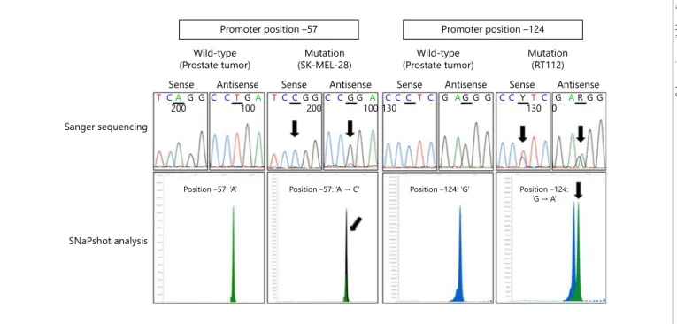
Analysis of previously reported mutation hotspots within the core promoter of the TERT gene was success- fully performed in all available cases. Mutation analysis using Sanger sequencing could be performed in 108/167 cases ( fig. 1 ). In 59/167 cases the core promoter region could not be amplified due to insufficient DNA quality (e.g. DNA degradation, low DNA concentration). In these cases SNaPshot analysis of mutation sites at –57,
Fig. 1. Representative examples for Sanger sequencing and SNaPshot analysis of the promoter mutation hotspots at –57 and –124. Upper lane: Sanger sequencing of DNA from a prostate tumor showing a wild-type sequence for TERT promoter posi- tion –57 (A) and position –124 (G). DNA from the melanoma
cell line SK-MEL-28 showing ‘A → C’ mutation at position –57.
DNA from the bladder cancer cell line RT112 showing ‘G → A’
mutation at position –124. Lower lane: corresponding SNaPshot analyses. Arrows indicate mutations in the promoter sequence.
Color version available online
Sanger sequencing
SNaPshot analysis
Sense Antisense Sense Antisense Sense Antisense Sense Antisense Wild-type
(Prostate tumor) Mutation
(SK-MEL-28) Wild-type
(Prostate tumor) Mutation (RT112)
Promoter position –57 Promoter position –124
Position –57: ‘A’
TC200A G G C CT GA TC CG G C CG G A C C C T C G AG G G C C Y T C G AR G G
200 130 130 0
100 100
3RVLWLRQ²¶$ମ&· Position –124: ‘G’ Position –124:
¶*ମ$·
–124 and –146 was performed ( fig. 1 ). Promoter muta- tions were not detected in any of the samples with all cas- es exhibiting a wild-type sequence.
Discussion
In the present study we performed sequence analysis of the core promoter region of the TERT gene in a cohort of 167 PrCa samples. The results of our study suggest that TERT promoter mutations are not involved in the devel- opment of PrCa as no mutations were detected in any of the investigated cases. These data are in line with previ- ously published studies on only very small cohorts and corroborate the minor importance of TERT promoter al- terations in PrCa [13–15] . Meanwhile, a study investigat- ing the whole genomes of 57 PrCa cases has been pub- lished [19] . Apart from already known data from exome analyses, this study displayed the spectrum of whole-ge- nome alterations in prostate tumors. Here only one TERT missense mutation was detected (p.R819C) but no pro- moter mutations were reported. This study also strength- ens our findings and should, together with our data, final- ize the discussion on TERT mutations in PrCa.
There are several lines of evidence that genomic vari- ations but not mutation might influence TERT expres- sion and disease risk in PrCa. In a large case-control study an intronic single nucleotide polymorphism (SNP) in the TERT gene (rs2242652, C 8992 T) was found that was strongly associated with increased PrCa risk. Be- cause of this strong correlation it was suggested that the SNP might have a functional relevance. Indeed, further evaluation showed increased TERT expression associat- ed with the presence of SNP variants in benign prostate tissue from patients who underwent radical prostatec- tomy [20, 21] . This increased TERT expression might provide a possible predisposition for PrCa. Another in- fluence on TERT expression might be length polymor- phisms in variable number tandem repeats (VNTRs).
Recently, it was shown that the TERT gene contains five VNTRs that are located within introns 2 and 6. A large case-control study found a significantly higher PrCa risk for individuals carrying rare VNTR2-2nd alleles than for individuals with common alleles. These VNTRs were also discussed as having an enhancer function for gene transcription. In vitro studies on PrCa cell lines analyz- ing the activity of the TERT promoter in combination with different VNTR variants clearly showed an in- creased luciferase activity for the VNTR2-2nd variants [22, 23] . These effects might also be expected for TERT
expression and could also increase the individual risk for PrCa.
Besides these genomic influences, TERT expression is also regulated by several cellular processes in PrCa. Mat- sumura et al. [24] analyzed the impact of the phosphory- lation status of Fas-associated death domain-containing protein (FADD) on TERT expression in PrCa. FADD has a crucial role in the formation of the death-inducing sig- naling complex and is also involved in cell cycle regula- tion. The phosphorylated form of FADD was highly ex- pressed in PrCa with a lower Gleason score and was in- versely associated with a shorter recurrence-free survival after prostatectomy. In parallel, cases with high levels of phosphorylated FADD also showed only low TERT ex- pression, suggesting a direct influence of FADD phos- phorylation on TERT expression. Shimada et al. [25]
found significant differences between FADD phosphory- lation levels and clinicopathological outcomes for Glea- son scores 3 + 4 and 4 + 3. These data indicate that Glea- son 4 + 3 tumors should be considered as high-risk tu- mors and stimulating agents that drive transition from nonphosphorylated to phosphorylated FADD (e.g. pacli- taxel) might be considered as therapeutic options. High TERT expression correlates with aggressive PrCa levels of TERT and nonphosphorylated FADD, which might rep- resent potent biomarkers for the biological behavior of PrCa. Furthermore, different factors can regulate TERT expression positively or negatively. Several transcription factors (e.g. SP1), hormones (e.g. androgen) and the PI3K/Akt and MAP kinase pathways can upregulate TERT transcription [reviewed in 26 ]. In addition, the downregulation of TERT by six microRNAs (let-7g * , miR-133a, miR-138–5p, miR-342–5p, miR-491–5p and miR-541–3p) has recently been reported [27] .
In summary, high expression of TERT is unlikely to be caused by promoter mutations or other genomic altera- tions in PrCa. TERT expression is more likely influenced by diverse cellular pathways resulting in increased cell cycle activity and proliferation.
Acknowledgements
The authors thank Karina Dresel, Stefanie Herlein, Claudia Knoll, Nina Oks, Verena Popp and Katrin Weigelt for excellent technical assistance.
Disclosure Statement
The authors have no conflict of interest.
References
1 Babaei S, Hulsman M, Reinders M, de Ridder J: Detecting recurrent gene mutation in inter- action network context using multi-scale graph diffusion. BMC Bioinformatics 2013;
14: 29.
2 Horn S, Figl A, Rachakonda PS, Fischer C, Sucker A, Gast A, Kadel S, Moll I, Nagore E, Hemminki K, Schadendorf D, Kumar R:
TERT promoter mutations in familial and sporadic melanoma. Science 2013; 339: 959–
961.
3 Vinagre J, Pinto V, Celestino R, Reis M, Po- pulo H, Boaventura P, Melo M, Catarino T, Lima J, Lopes JM, Maximo V, Sobrinho-Si- moes M, Soares P: Telomerase promoter mu- tations in cancer: an emerging molecular bio- marker? Virchows Arch 2014; 465: 119–133.
4 Huang FW, Hodis E, Xu MJ, Kryukov GV, Chin L, Garraway LA: Highly recurrent TERT promoter mutations in human melanoma.
Science 2013; 339: 957–959.
5 Meeker AK: Telomeres and telomerase in prostatic intraepithelial neoplasia and pros- tate cancer biology. Urol Oncol 2006; 24: 122–
130.
6 Kamradt J, Drosse C, Kalkbrenner S, Rohde V, Lensch R, Lehmann J, Fixemer T, Bonkhoff H, Stoeckle M, Wullich B: Telomerase activity and telomerase subunit gene expression levels are not related in prostate cancer: a real-time quantification and in situ hybridization study.
Lab Invest 2003; 83: 623–633.
7 de Kok JB, Verhaegh GW, Roelofs RW, Hes- sels D, Kiemeney LA, Aalders TW, Swinkels DW, Schalken JA: DD3 PCA3 , a very sensitive and specific marker to detect prostate tumors.
Cancer Res 2002; 62: 2695–2698.
8 Korkmaz M, Avci CB, Gunduz C, Aygunes D, Erbaykent-Tepedelen B: Disodium pentabo- rate decahydrate (DPD) induced apoptosis by decreasing hTERT enzyme activity and disrupting F-actin organization of prostate cancer cells. Tumour Biol 2014; 35: 1531–
1538.
9 Liu S, Qi Y, Ge Y, Duplessis T, Rowan BG, Ip C, Cheng H, Rennie PS, Horikawa I, Lustig AJ, Yu Q, Zhang H, Dong Y: Telomerase as an important target of androgen signaling block-
ade for prostate cancer treatment. Mol Cancer Ther 2010; 9: 2016–2025.
10 Guo C, Armbruster BN, Price DT, Counter CM: In vivo regulation of hTERT expression and telomerase activity by androgen. J Urol 2003; 170: 615–618.
11 Moehren U, Papaioannou M, Reeb CA, Gras- selli A, Nanni S, Asim M, Roell D, Prade I, Farsetti A, Baniahmad A: Wild-type but not mutant androgen receptor inhibits expres- sion of the hTERT telomerase subunit: a nov- el role of AR mutation for prostate cancer de- velopment. FASEB J 2008; 22: 1258–1267.
12 Williams JL, Greer PA, Squire JA: Recurrent copy number alterations in prostate cancer:
an in silico meta-analysis of publicly available genomic data. Cancer Genet 2014; 207: 478–
488.
13 Killela PJ, Reitman ZJ, Jiao Y, et al: TERT pro- moter mutations occur frequently in gliomas and a subset of tumors derived from cells with low rates of self-renewal. Proc Natl Acad Sci U S A 2013; 110: 6021–6026.
14 Wu S, Huang P, Li C, Huang Y, Li X, Wang Y, Chen C, Lv Z, Tang A, Sun X, Lu J, Li W, Zhou J, Gui Y, Zhou F, Wang D, Cai Z: Telomerase reverse transcriptase gene promoter muta- tions help discern the origin of urogenital tu- mors: a genomic and molecular study. Eur Urol 2014; 65: 274–277.
15 Zheng X, Zhuge J, Bezerra SM, Faraj SF, Mu- nari E, Fallon JT 3rd, Yang XJ, Argani P, Net- to GJ, Zhong M: High frequency of TERT pro- moter mutation in small cell carcinoma of bladder, but not in small cell carcinoma of other origins. J Hematol Oncol 2014; 7: 47.
16 Epstein JI AF, Allsbrook WC, et al: Tumours of the prostate; in Eble JN Sauter G, Epstein JI, Sesterhenn IA: (eds): World Health Orga- nization Classification of Tumours: Patholo- gy and Genetics of Tumours of the Urinary System and Male Genital Organs, ed 1. Lyon, IARC, 2004.
17 Sobin LH WC: TNM Classification of Malig- nant Tumors, ed 6. New York, John Wiley and Sons, 2002.
18 Hurst CD, Platt FM, Knowles MA: Compre- hensive mutation analysis of the TERT pro-
moter in bladder cancer and detection of mu- tations in voided urine. Eur Urol 2014; 65:
367–369.
19 Baca SC, Prandi D, Lawrence MS, et al: Punc- tuated evolution of prostate cancer genomes.
Cell 2013; 153: 666–677.
20 Kote-Jarai Z, Saunders EJ, Leongamornlert DA, et al: Fine-mapping identifies multiple prostate cancer risk loci at 5p15, one of which associates with TERT expression. Hum Mol Genet 2013; 22: 2520–2528.
21 Kote-Jarai Z, Olama AA, Giles GG, et al:
Seven prostate cancer susceptibility loci iden- tified by a multi-stage genome-wide associa- tion study. Nat Genet 2011; 43: 785–791.
22 Yoon SL, Jung SI, Do EJ, Lee SR, Lee SY, Chu IS, Kim WJ, Jung J, Kim CS, Cheon SH, Leem SH: Short rare hTERT -VNTR2–2 nd alleles are associated with prostate cancer susceptibility and influence gene expression. BMC Cancer 2010; 10: 393.
23 Leem SH, Londono-Vallejo JA, Kim JH, Bui H, Tubacher E, Solomon G, Park JE, Horika- wa I, Kouprina N, Barrett JC, Larionov V: The human telomerase gene: complete genomic sequence and analysis of tandem repeat poly- morphisms in intronic regions. Oncogene 2002; 21: 769–777.
24 Matsumura Y, Shimada K, Tanaka N, Fuji- moto K, Hirao Y, Konishi N: Phosphoryla- tion status of Fas-associated death domain- containing protein regulates telomerase ac- tivity and strongly correlates with prostate cancer outcomes. Pathobiology 2009; 76: 293–
302.
25 Shimada K, Matsuyoshi S, Nakamura M, Ishi- da E, Konishi N: Phosphorylation status of Fas-associated death domain-containing pro- tein (FADD) is associated with prostate can- cer progression. J Pathol 2005; 206: 423–432.
26 Daniel M, Peek GW, Tollefsbol TO: Regula- tion of the human catalytic subunit of telom- erase (hTERT). Gene 2012; 498: 135–146.
27 Hrdlickova R, Nehyba J, Bargmann W, Bose HR Jr: Multiple tumor suppressor microRNAs regulate telomerase and TCF7, an important transcriptional regulator of the Wnt pathway.
PloS One 2014; 9:e86990.