in normal and disease conditions
INAUGURAL-DISSERTATION zur
Erlangung des Doktorgrades
der Mathematisch-Naturwissenschaftlichen Fakultät der Universität zu Köln
vorgelegt von Charles-Peter Xavier
aus Bangalore, Indien
2008
II Vorsitzender: Frau Universitätsprofessorin Dr. rer. nat. Karin Schnetz
1. Berichterstatter: Frau Universitätsprofessorin Dr. rer. nat. Angelika A. Noegel
2. Berichterstatter: Herr Universitätsprofessor Dr. rer. nat. Siegfried Roth
Tag der mündlichen Prüfung: 16.02.2009
Erklärung
Ich erkläre hiermit, dass ich die vorliegende Arbeit ohne unzulässige Hilfe Dritter und ohne Benutzung anderer als der angegebenen Hilfsmittel angefertigt habe; die aus fremden Quellen direkt oder indirekt übernommenen Gedanken sind als solche kenntlich gemacht.
Die in dieser Arbeit beschriebenen Experimente sind von mir selbst durchgeführt worden.
Anleitungen oder Anregungen zu Experimenten habe ich von Frau Prof. Dr. Noegel, Herrn PD Dr. Clemen und den Mitarbeitern des Institutes für Biochemie I erhalten. Bei verschiedenen Experimenten wurde ich bei der Versuchsdurchführung von technischem Personal unterstützt.
Des Weiteren habe ich nicht die Hilfe eines Promotionsberaters in Anspruch genommen.
Dritte haben weder unmittelbar noch mittelbar geldwerte Leistungen für Arbeiten erhalten, die im Zusammenhang mit dem Inhalt der vorliegenden Dissertation stehen.
Die Arbeit wurde von mir bisher weder im Inland noch im Ausland in gleicher oder ähnlicher Form einer anderen Prüfungsbehörde vorgelegt und ist auch noch nicht veröffentlicht.
Köln, den 05.12.2008 __________________
(Charles-Peter Xavier)
III
Acknowledgement
First and foremost I would like to express my heartiest gratitude to Prof. Angelika Noegel for giving me an opportunity to work in her supervision. Her positive attitude, constant encouragement, promptness and sustained interest in my project proved essential for successful completion of my PhD thesis.
I convey my sincere heartfelt gratitude to PD Dr. Christoph Clemen, our group leader, for providing valuable guidance, excellent advice with my work. His constant encouragement, immense optimism and friendly nature are highly acknowledged. It was a privilege to work with him.
Very special thanks to Ms. Maria Stumpf who helped me a lot with my experiments throughout the course of my Ph.D and also for creating a friendly environment with more fun.
I take this opportunity to thank all investigators who extended their support in form of collaborative work. I thank Prof. Dietmar Thal, PD Dr. Stefan Linder, Prof. Andreas Hofmann, Prof. Rolf Schröder and Prof. Micheal Schleicher for successful collaborative efforts. I also thank Dr. André Rosentreter (with whom I shared part of my work), Prof. Reginald O. Morgan, Prof.
Maria-Pilar Fernandez and PD Dr. Ludwig Eichinger for their help.
I thank Mr. Rolf Müller for his good technical support in gel filtration assays and cloning. I also thank Mr. Berthold Gassen especially for yummy cakes, antibodies and cell culture work.
I thank Vivek, Subhanjan, Deen, Vel, Sabari, Palani, Sharada, Yogikala, Sam and Channa for their help and good company. I like to sincerely thank Pastor David, Annie, Bro. Samy, Pastor Nathan, Bro. Robert, Dr. Pradheep and Andrew.
I also thank all members of the lab, Raphael, Mary, Karthik, Kalle, Christian, Surayya, Rashmi Rosi, Hua, Sudhir, Tanya, Anne, Margit, Georgia, Eva, Sascha, Ria, Budi, Francisco and Akis for their good company, help and support. I also thank Ms. Dörte Püsche for her help and co- operation with the administrative work that made my life easy. I take this opportunity to thank Gudrun for her prompt help anytime needed. I could also like to thank my friends Veena (special thanks), Fareed, Sam, Deepak, Selva, Vinayaga, Ram, Vinod and Raja for their wonderful company.
I express my heartfelt gratitude to my brother Vincent, parents and family members for their patience, tolerance, sacrifice and encouragement throughout my life. I also thank Dr. Betty Daniel and Dr. Villoo Patel for their support and encouragement.
My heartfelt sincere gratitude to Suja for her company, patience and encouragement.
I also thank the Deutsche Forschungsgemeinschaft for financial assistance during my Ph.D.
Cologne, 5th Dec 2008 Charles-Peter Xavier
IV
Contents
I. INTRODUCTION 1
1.1 Coronins 1
1.2 Coronin Structure 4
1.2.1 N-terminal domain (β-propeller structure) 4
1.2.2 Unique region 4
1.2.3 Coiled coil domain 5
1.3 Oligomerisation 5
1.4 Interactions with Arp2/3 complex 6
1.5 Coronin regulation by phosphorylation 6
1.6 Coronins in disease 8
1.7 CRN2 (coronin-1C, coronin-3) 8
1.8 Aim of the work 10
1.9 Preliminary publications 11
II. RESULTS 12
2.0 Role of CRN2 in cellular processes 12
2.1 CRN2 interacts with the F-actin cytoskeleton in vivo 12 2.2 Absence of CRN2 or lack of single domains inhibits fibroblast migration 13 2.3 Expression of the WD-repeat domain of CRN2 inhibits cell proliferation 15 2.4 CRN2 participates in the formation of cellular protrusions 16 2.5 Full-length CRN2 is essential for the process of fluid phase pinocytosis 17 2.6 CRN2 forms a complex with the Arp2/3-complex and with cofilin 18 2.7 Structural properties of CRN2 deduced from the CRN4 crystal structure 19
3.0 Expression of CRN2 in diffuse gliomas is related to malignancy 21
3.1 CRN2 is expressed in a subset of human brain tumours and in reactive astrocytes 21
3.2 CRN2 expression correlates with the grade of malignancy in diffuse gliomas 25
V 3.3 CRN2 knockdown inhibits cellular functions related to tumour malignancy 27
4.0 Structural and functional diversity of novel CRN2 isoforms. 32 4.1 Identification of three CRN2 proteins encoded by two mRNA species 32
4.2 Evolutionary insight into CRN2 regulation 34
4.3 CRN2i3 is part of the myogenic differentiation program 36 4.4 Different oligomerization state of the CRN2 isoforms 37
4.5 Structural models of CRN2 isoforms 38
4.6 GFP-fusion proteins of CRN2 isoforms localize to F-actin structures 40
4.7 CRN2 is a novel component of podosomes 41
4.8 CRN2i3 is a structural component of neuromuscular junctions and myofibrils 43
5.0 Influence of phosphorylation on structure and function of CRN2 46
5.1 Posttranslational modifications of CRN2 46
5.2 CRN2 interacts with actin cytoskeleton regulator CK2α kinase 46
5.3 In vivo phosphorylation of CRN2 47
5.4 In vitro phosphorlyation of CRN2 by CK2α kinase 48 5.5 Phosphorylation of Ser463 regulates oligomerisation of CRN2 49 5.6 Phosphomimetic S463D mutant CRN2 inhibits actin polymerisation 50 5.7 Phosphomimetic S463D mutant of CRN2 suppresses migration 51 5.8 Phosphomimetic S463D mutant CRN2 hinders formation of cellular protrusions 52
III. DISCUSSION 53
6.0 Role of CRN2 in cellular processes 53
6.1 Structural properties of CRN2 53
6.2 CRN2 regulates F-actin processes 53
6.3 Influence of phosphorylation on structure and function of CRN2 55 6.4 Structural and functional diversity of novel CRN2 isoforms 56
7.0 Expression of CRN2 in diffuse gliomas is related to malignancy 59
IV. MATERIALS AND METHODS 61
1.0 Kits and Regeants 61
1.2 Enzymes, antibodies, radioactive probes, antibiotics, inhibitors 61
1.2.1 Enzymes for molecular biology 61
VI
1.2.2 Inhibitors 62
1.2.3 Antibodies 62
1.2.4 Antibiotics 62
1.2.5 Radiolabelled nucleotide 63
1.3 Reagents 63
1.4 Plasmids 64
1.4.1 Oligonucleotides 65
1.5 Bacterial host strains 65
1.6 Insect cell lines 65
1.7 Mammalian cell lines 65
1.8 Media and buffers 65
1.8.1 Media for E. coli culture 66
1.8.2 Media for Mammalian cell culture 66
1.8.3 Media for Insect Cell Culture 66
1.9 Instruments and Equipments 66
1.10 Immunoblotting and antibodies 66
1.11 Mammalian cell culture 66
1.12 2D-gel electrophoresis 67
1.13 Statistical methods 67
2.0 Role of CRN2 in cellular processes 67
2.1 Generation of EGFP–fusion constructs of CRN2 domains 67
2.2 Subcellular fractionation 68
2.3 RNAi silencing 68
2.4 Immunofluorescence, confocal microscopy, and life cell imaging 68 2.5 In vitro wound healing, proliferation, cytokinesis, and cell activity assays 68
2.6 Co-immunoprecipitation 69
3.0. Expression of CRN2 in diffuse gliomas is related to malignancy 70
3.1 Neuropathology 70
3.2 Visualization of invadopodia 70
3.3 Lentiviral transduction of shRNA vectors 70
VII
3.4 Determinations of cell based assays. 71
4.0 Structural and functional diversity of novel CRN2 isoforms 71
4.1 Immunofluorescence and immunohistochemistry 71
4.2 Mammalian cell culture 71
4.3 RNA purification, northern blotting, 5’-RACE, 5’-RLM-RACE, and RT-PCR 72 4.4 Differential centrifugation, subcellular fractionation, and gel filtration 72
4.5 Plasmids for expression of CRN2 isoforms 73
4.6 Bioinformatic analysis 73
5.0 Influence of Phosphorylation on Structure and function of CRN2 73 5.1 Expression of wild-type and mutant CRN2 coiled coil peptides. 73 5.2 In vitro wound healing and single protrusion assay 74
5.3 In vitro phosphorylation assay 74
5.4 Actin polymerization assays 74
5.5 Co-immunoprecipitation 75
5.6 CRN2 pull down experiments 75
IV. BIBILIOGRAPHY 77
VI. APPENDIX 85
1. Abbreviations 85
I. Introduction
Dynamic remodelling of the actin cytoskeleton is indispensable for many physiological processes, which comprise cell migration, intracellular trafficking of vesicles and organelles, cell polarity, and signal transduction. Cell motility defines critical role in various disease states like cancer and autoimmunity. Cell motility is a highly coordinated process involving dynamic re-organization of the actin cytoskeleton. Actin dynamics comprising polymerization and depolymerisation of actin filaments is a tightly regulated process engaging various actin binding proteins. The precise molecular mechanism underlying this regulation has been one of the domains of intense study for decades in actin biology (Uetrecht and Bear, 2006). At the heart of this process of actin regulation is the evolutionarily conserved seven-subunit containing Arp2/3 complex, which brings about actin nucleation and branching. Arp2/3 by itself is relatively inactive and requires necessary interaction and activation by the WA/VCA domain of a nucleation-promoting factor (NPF), such as the SCAR/WASp family of proteins (Rodal et al., 2005). The Arp2/3 mediated formation of the F-actin network is a crucial process and demands a tight regulation of Arp2/3 complex. One of the protein family that seems to contribute to this regulation is the coronin family of proteins (Uetrecht and Bear, 2006).
1.1 Coronins
The coronin family of proteins is prominent amongst the WD-repeat-actin binding proteins (Fig.1). They are evolutionarily conserved across different organisms, with origin from simple eukaryotes to higher vertebrates. Coronin was first discovered from actin-myosin preparations in Dictyostelium discoideum. The term coronin was coined, due to its co localization with the actin rich crown-shaped cellular projections (de Hostos et al., 1991). Even though coronin protein involvement in actin-dependent processes appears primordial, the individual members of the coronin protein family contribute to largely different cellular functions like like signal transduction, transcriptional regulation, remodelling of the cytoskeleton, and regulation of vesicular trafficking (for an extensive review on coronin proteins see (Clemen et al., 2008)).
Phylogenetic analyses revealed twelve subfamilies of coronin proteins, comprising seven
vertebrate paralogs and five subfamilies in nonvertebrate metazoa, fungi and protozoa, some
of them unclassified so far. Delineation of these twelve subfamily clades has been used to
propose a comprehensive revision of the coronin protein nomenclature (Fig. 2) (Morgan and
Fernandez, 2008). Currently, two nomenclature systems are used for coronin proteins. Both
do not cover all coronin subfamilies and moreover, do not allow unambiguous labelling of
gene duplications and isoforms. These disadvantages will be overcome using the revised coronin protein nomenclature which is based on evolutionary history, structural change and functional adaptation. The coronin subfamilies were united and re-numerated, and ‘CRN’ has been proposed as the new symbol for coronin proteins.
Figure 1. Coronin proteins involved in cytoskeletal F-actin binding and cross-linking, the red circle indicates dimerized coronin cross-linking two F-actin filaments. Taken from www.bio.brandeis.edu/faculty.
Figure 2. Molecular phylogeny of the coronin family of proteins across different organisms using the proposed new nomenclature: current official symbols in brackets. According to (Morgan and Fernandez, 2008), taken from (Clemen et al., 2008)
On the basis of amino acid content, mammalian coronins can be distinguished into two subfamilies (Rybakin and Clemen, 2005). Short coronins CRN1-6 display three different structural domains. The long coronin CRN7 contains the short coronin topology in duplicate.
Table. 1 illustrates the seven coronins present in mammals (Xavier et al., 2008).
Table.1 Summary of all mammalian coronin proteins with their corresponding molecular weight,
amino acid content, percentage identity with Dictyostelium short coronin CRN12, expression and function.
1.2 Coronin Structure
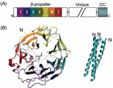
Short coronins exhibit a characteristic three-domain organisation, consisting of an N-terminal domain containing a WD repeat-β-propeller structure, an unique C-terminal extension and a C-terminal coiled coil domain. (Fig 3).
1.2.1 N-terminal domain (β-propeller structure)
The WD40 repeat region is the signature domain of coronin family of proteins (de Hostos, 1999). The crystal structure of murine CRN4 (synonyms: coronin-1A, coronin-1) lacking its coiled coil domain, disclosed a seven-bladed β-propeller structure assembled from five canonical WD repeats and two non-canonical repeats (Appleton et al., 2006). The secondary structure motif of WD repeat sequence forms four consecutive β-strands connected by loops and turns, together defined as β-propeller. Within the propeller, each blade is built up of four anti-parallel β-strands extending from the centre to the periphery and the blades are arranged in a circular fashion around a central axis. Structurally the innermost β-strands A, B, and C of a blade and the outermost strand D are contributed by the second, third and fourth strands of one and the first strand of the next WD sequence repeat, respectively. results in stability Additionally, two tandem stretches of conserved residues located in the C-terminal extension tightly pack against the bottom side of the propeller, which possibly provides further structural integrity (McArdle and Hofmann, 2008). Even though β-propeller structures are in principle thought to serve as a stable platform for protein-protein interaction, so far only F- actin has been identified as a potent binding partner of this region in Dictyostelium, yeast and mouse CRN1. Therefore, thorough evaluation of this domain may reveal new binding partners (Gandhi and Goode, 2008).
1.2.2 Unique region
A region in the coronin structure that remains poorly understood is the unique C-terminal region. It highly varies in length, sequence and functions (Gandhi and Goode, 2008a).
Notably this region in S. cerevisiae CRN11 (synonym: Crn1p) and D.melanogaster CRN7
(synonym: Dpod1) showed sequence homology with the microtubule-binding region of
mammalian MAP1B. Correspondingly, purified coronin protein of these organisms was
shown to crosslink microtubules and actin filaments in vitro. In Drosophila, CRN7 is required
for proper axonal guidance. (Goode et al., 1999; Rothenberg et al., 2003)
Figure 3. Coronin domain organisation and protein structure. A. Schematic of coronin domain architecture B. Crystral structure of mouse CRN4 β-propeller domain (Appleton et al., 2006) and coiled-coil domain (Kammerer et al., 2005). Taken from (Gandhi and Goode, 2008).
1.2.3 Coiled coil domain
The very C-terminus of short coronin proteins is composed of a moderately conserved coiled coil domain (~35-50 residues, 4-7 heptad repeats) which in CRN4 is identified to posses a leucine zipper motif. Eventhough, it is the smallest functional domain of coronin structure, it mediates at least three crucial functions like homo-oligomerisation and interaction with F- actin and Arp2/3 complex (Gandhi and Goode, 2008a). Coiled coil motifs are found to display a characteristic hepta-peptide repeat (abcdefg)n, where positions a and b are predominately hydrophobic residues and positions e and g are charged residues (McArdle and Hofmann, 2008). In case of CRN4 threefold repeat of such a characteristic hepta-peptide sequence is determined and the motif R-L/I/V-X-X-L/V-E (450-455 in CRN4) has been recognised as a mediator of trimerisation in coiled coil (Kammerer et al., 2005).
1.3 Oligomerisation
All mammalian short coronins reported so far exhibit a coiled coil domain that has been implicated in oligomerisation (Uetrecht and Bear, 2006).A number of studies have shown that oligomerization of coronins is exclusively homotypic (Cai et al., 2005; Gatfield et al., 2005;
Oku et al., 2005). A functional significance of coronin oligomerisation is to execute actin filament bundling (Goode et al., 1999). Therefore this observation leads to one possible model in which multimerization of the coronin β-propeller would establish actin bundling. Deletion of the coiled-coil domain weakened the actin binding affinity of coronin (Cai et al., 2005).
Another study reports, that the ability of the coiled-coil domain is to increase the actin binding
affinity through multimerization of the β-propeller domain (Cai et al., 2007). This gives rise to the second possibility, in which individual (non-oligomerised) coronin molecules might cross-link actin filaments using two separate actin binding sites, one in the β-propeller and another in coiled-coil domain (Gandhi and Goode, 2008).
1.4 Interactions with Arp2/3 complex
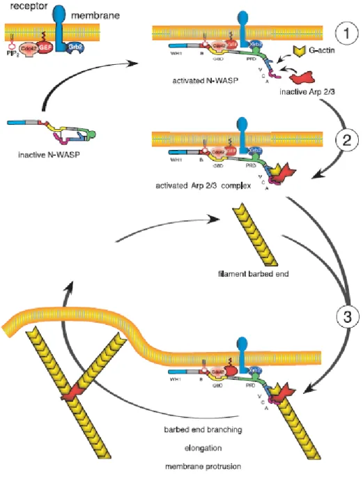
The diversity of F-actin structures and associated cellular functions depends on many actin- binding proteins (Cvrckova et al., 2004; Sutherland and Witke, 1999). The submembranous area of a cell is enriched in branched F-actin filaments and the Arp2/3 complex (Goley and Welch, 2006). Prime examples of a cellular function involving branched F-actin are the formation of lamellipodia and cellular motility. The Arp2/3 complex is enriched in the periphery of lamellipodia and plays a key role in forming a short-branched F-actin network that generates the protruding force depending on a locally well-organized assembly and disassembly of F-actin (Fig. 4) (Nabi, 1999; Pantaloni et al., 2001; Ponti et al., 2005; Rybakin and Clemen, 2005). The Arp2/3 complex consists of seven subunits. p34 (Arc35, ARPC2) together with ARPC4 forms the center of the Arp2/3 complex which initiates site-directed branching and elongation of actin filaments and nucleates new actin filaments (Cooper et al., 2001; Gournier et al., 2001; Pollard and Beltzner, 2002; Stradal and Scita, 2006).
In addition to F-actin binding the unique C-terminal region and the coiled-coil domain of yeast CRN11 was shown to bind with Arp2/3 complex (Humphries et al., 2002; Machesky et al., 1997). Further, purified yeast CRN11 was demonstrated to execute inhibitory effect on Arp2/3 driven F-actin polymerisation. The inhibitory influence is due to maintenance of Arp2/3 complex in an inactive or open confirmation which becomes unavailable for WASp activation which results in Arp2/3 mediated actin polymerisation. CRN11 constructs lacking the coiled-coil domain were inefficient to interact with Arp2/3 complex (Rodal et al., 2005).
Colocalization studies of CRN1 (synonyms: coronin-1B, coronin-2) with Arp2/3 complex was also reported in a variety of cells. In addition, mouse CRN4 was shown to control steady- state F-actin levels via an Arp2/3 complex driven mechanism in T-lymphocyctes (Foger et al., 2006).
1.5 Coronin regulation by phosphorylation
A number of evidence demonstrates mammalian coronins to be regulated by phosphorylation
(Uetrecht and Bear, 2006). CRN4 was shown to be phosphorylated in vitro by purified PKC,
which is stimulated in vivo by PMA treatment (Itoh et al., 2002). In addition, PKC inhibitor
celerythrine was shown to block both phosphorylation and dissociation of CRN4 from
phagosomes (Gatfield et al., 2005). Similar studies pertaining to PKC stimulation with phorbol ester PMA and down regulation using PKC inhibitors were shown to be responsible for phosphorylation of CRN1 (Cai et al., 2005). Further, CRN7 was shown to get phosphorylated on tyrosine residues (Rybakin et al., 2004).
Figure 4. Arp2/3 mediated actin polmerisation at the leading edge. N-WASP is activated and targeted to the membrane by signaling molecules (circled 1). G-actin and the Arp2/3 complex together bind to N-WASP (VCA), forming a branching complex (circled 2), binding of the branching complex with a filament leads to the formation of a branch (circled 3). Both the branches grow at equal rates. N-WASP catalyzes several consecutive cycles of branching. Taken from (Pantaloni et al., 2001).
1.6 Coronins in disease
Since regulation of actin dynamics is an essential part of many disease states, actin binding coronin proteins could play a role in diseases. A role of CRN4 in pathogenic bacterial infection can be considered as one of the best-characterized examples (Ferrari et al., 1999). A well established mechanism to destroy engulfed bacteria by macrophages are through controlled membrane fusion of phagosomes with lysosomes, wherein delivery of lytic enzymes acidify phagolysosomes resulting in effective destruction of bacteria (Anes et al., 2003; Vergne et al., 2004). Actin polymerisation around nascent phagosomes plays a potential role in driving these critical vesicle fusion events, emphasizing importance of actin dependent process in immune response to combat bacterial infections (Anes et al., 2003). Recent studies reveal that pathogenic Mycobacterium tuberculosis are able to circumvent the normal immune response by macrophages through retaining CRN4 function to alter the actin dynamics on the phagosome, thereby preventing the maturation of phagosomes and permitting bacterial propagation within the macrophage (Ferrari et al., 1999). Further CRN1 was linked with neurite formation and axon regeneration with subsequent spinal cord injury (Di Giovanni et al., 2005).
1.7 CRN2 (coronin-1C, coronin-3)
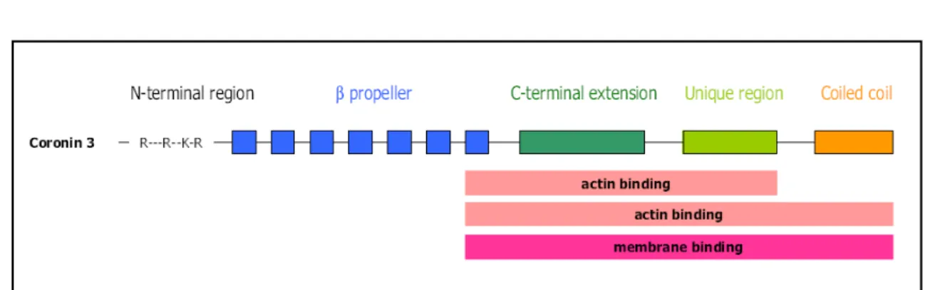
CRN2 (current official symbol: CORO1C, current official name: coronin-1C, most common
synonym: coronin-3) is a short coronin protein (Rybakin and Clemen, 2005) of 474 amino
acids with a calculated molecular mass of 53 kDa in human and murine cells; the apparent
molecular mass from SDS-PAGE analysis is approximately 57 kDa (Fig.5). Based on the
crystal structure of CRN4 a structural homology model of CRN2 was proposed (Fig.6)
(McArdle and Hofmann, 2008)
.At the subcellular level CRN2 exists in two different pools,
an actin cytoskeleton associated non-phosphorylated pool and a diffusely distributed
phosphorylated cytosolic pool and represented as multiple spots on 2D gels (Spoerl et al.,
2002). CRN2 has been identified as an actin filament-crosslinking and bundling protein
(Hasse et al., 2005; Spoerl et al., 2002) involved in distinct cellular functions like
proliferation, migration, formation of cellular protrusions, endocytosis and secretion
(Rosentreter et al., 2007). Recent studies indicate CRN2 as an important factor in the
development of the murine brain (Hasse et al., 2005) as well as a potential marker for
melanoma progression (Roadcap et al., 2008).
Figure 5. Human CRN2 domain organisation. Mapped membrane and actin binding sites are indicated. Taken from (McArdle and Hofmann, 2008).
Figure 6. Putative topology of CRN2 based on structural information of CRN4 showing the N-terminal 7- blades of the β propeller structure and the C-terminal extension and coiled coil domain. Taken from (McArdle and Hofmann, 2008).
1.8 Aim of the work
Coronins are one the most versatile family of proteins evolutionarily well conserved amongst eukaryotes and mammals including Dictyostelium, Drosophila, mouse and humans. With its origin as a single gene in simple eukaryotes, the mammalian coronin gene family has shown expansion to include seven members. Human CRN2 is one of the ubiquitously expressed short coronin proteins with F-actin binding and cross-linking properties. In this thesis work we attempt to characterize human CRN2 with respect to its biochemical and cellular functions.
•
We investigate the role of CRN2 in cellular processes in association with actin cytoskeleton through over expression of coronin domains, down-regulation of CRN2 levels using the RNAi technique and co-immunoprecipitation studies.
•
We analyse the expression of CRN2 in normal as well as various types of malignant and benign human brain tumours. Further we pursue in vitro functional studies using CRN2 knockdown in glioblastoma cells and investigate the role of CRN2 in regulating cell proliferation, migration and invasion.
•
We study functional and structural diversity of three human CRN2 isoforms, designated as CRN2i1, CRN2i2 and CRN2i3.
•
We investigate the regulation of the cellular activity of CRN2.
Note: In some of the figures CRN2 is specified as coronin-3 or coronin-1C which are
synonyms of CRN2.
1.9 Preliminary publications
Significant parts of this work have been published as research articles:
•
Xavier C-P, Rosentreter A, Reimann J, Cornfine S, Linder S, Hofmann A, Morgan RO, Fernandez MP, Stumpf M, Müller R, Jungbauer T, Schröder R, Noegel AA, Clemen CS. Coronin-1C isoforms are novel components of podosomes and sarcomeres. Submitted for publication.
•
Xavier C-P, Eichinger L, Fernandez MP, Morgan RO, Clemen CS. Evolutionary and Functional Diversity of Coronin Proteins. In: The Coronin Family of Proteins. Vol. 48.
Eds.: Clemen CS, Eichinger L, Rybakin V. Landes Bioscience & Springer. 2008.
ISBN 978-0-387-09594-3. Open access at http://www.eurekah.com/chapter/3808.
•
Thal DR, Xavier C-P, Rosentreter A, Waha A, Pietsch T, Stumpf M, Noegel AA, Clemen CS. Expression of coronin-3 (coronin-1C) in diffuse gliomas is related to malignancy. J Pathol 2008 214:415-24. Equal contributors.
•
Rosentreter A, Hofmann A, Xavier C-P, Stumpf M, Noegel AA, Clemen CS. Coronin
3 involvement in F-actin dependent processes at the cell cortex. Exp Cell Res 2007
313:878-95.
II. Results
2.0 Role of CRN2 in cellular processes
2.1 CRN2 interacts with the F-actin cytoskeleton in vivo
In HaCaT cells endogenous CRN2 shows a characteristic however not continuous localization to the submembranous area. This localization is particularly obvious in cells, which exhibit a strong accumulation of actin filaments underneath the plasmamembrane. CRN2 additionally was detected at punctate structures in the cytoplasm, with a perinuclear accumulation of these spots (Fig. 7A, upper panel). To confirm the subcortical enrichment of CRN2, HaCaT cells were retrovirally transduced to express GFP–CRN2. The distribution of GFP–CRN2 (Fig. 7B, left panel) and endogenous CRN2 (Fig. 7B, right panel; this is a higher magnification of the upper left image of Fig. 7A) are congruent, however, the GFP–signal of CRN2 more clearly shows the protein's cortical localization.
Figure 7. CRN2 shows an F-actin-dependent distribution in HaCaT cells. A. HaCaT cells treated with or without latrunculin B were first fixed with 4% paraformaldehyde and then permeabilized with 0.2% Triton X- 100 (data shown). CRN2 was detected using mAb K6-444 and goat anti-mouse Cy3 as secondary antibody; F- actin was visualized using FITC–phalloidin
.
This cortical localization of endogenous CRN2 is maintained after Triton X-100
permeabilization prior to fixation (data not shown), indicating a stable association of coronin3
and F-actin in vivo. By contrast, HaCaT cells treated with latrunculin B prior to fixation never
showed any accumulation of CRN2 underneath the plasmamembrane, but CRN2 appears in
spots clearly distinguishable from the remaining F-actin staining (Fig. 7A, lower panel). The
latter Factin structures represent a pool that is resistant to the treatment with latrunculin B (Ammar et al., 2001). HaCaT cells treated with latrunculin B and Triton X-100 prior to fixation neither showed a structured staining of CRN2 nor of F-actin (data not shown); most of both proteins disappeared and only a diffuse residual staining remained.
Figure 7, continued. B Higher magnifications of HaCaT cells expressing GFP–CRN2(left; same expression vector is used in HEK293 cells, Fig. 7B) and of antibody-stained endogenous CRN2 (right; higher magnification of the upper left image from A) demonstrate a submembranous localization of CRN2. C. Corresponding to A, cells were incubated with or without latrunculin B, washed with Triton X-100, and lysed cells were separated by differential centrifugation. Samples of the Triton X-100 soluble fraction (Triton wash) and the centrifugation steps were analyzed by SDS–PAGE followed by Western blotting using antibodies against CRN2 (mAb K6- 444) and β-actin S, supernatant, P. pellet fractions.
These observations were confirmed by differential centrifugation and immunoblotting (Fig.
7C). The relative distribution of CRN2 within different fractions of each experiment can be compared. In untreated HaCaT cells CRN2 is most abundant in the 100,000 g pellet; minor amounts are detectable in the 10,000 g pellet and in the 100,000 g supernatant. CRN2 as well is detected in the Triton X-100 soluble fraction (Triton X-100 wash). Note that the 10,000 g pellet contains the crude cytoskeletal fraction (highly crosslinked actin filaments) and the pellet of 100,000 g includes cytoskeletal elements associated with membranes (free or loosely crosslinked actin filaments) (Fox, 1985; Lehtonen et al., 2002; Tohyama et al., 1994). After treatment with latrunculin B the amount of CRN2 in the 100,000 g pellet is markedly reduced and instead appears in the 100,000 g supernatant. The data from immunofluorescence analysis and differential centrifugation confirmed a binding of CRN2 to F-actin as previously described (Spoerl et al., 2002) and clearly demonstrate a localization of CRN2 that depended strongly on F-actin in vitro as well as in vivo.
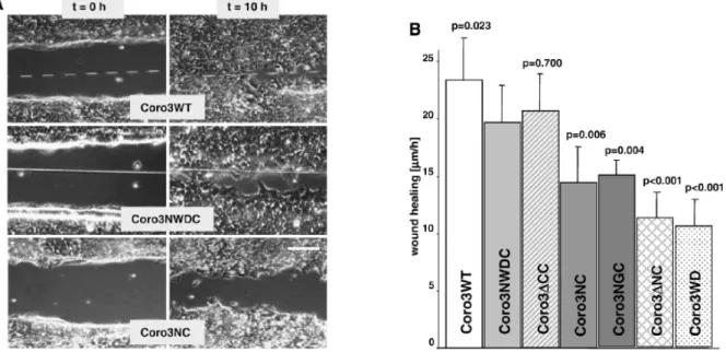
2.2 Absence of CRN2 or lack of single domains inhibits fibroblast migration
To determine the contribution of CRN2 in cellular processes, we investigated wound healing
of confluent layers of HEK293 cells stably expressing CRN2 proteins. Wound healing was
monitored over 10 h (Figs.8A, B). We observed a statistically significantly reduced velocity of wound closure in case of all CRN2 polypeptides compared to WT cells, which exhibited the fastest wound closure with an average velocity of 23 µm/h. Comparing CRN2-NWDC (20 µm/h) with the other CRN2 polypeptides demonstrated that except for CRN2-∆CC (21 µm/h;
F-actin binding regions are present in this fusion protein) all proteins led to statistically significantly slower wound closure velocities. Cells, which express CRN2-NC and CRN2- NGC, lacking the central WD40 repeats or carrying a glycine loop instead, showed reduced velocities of 14 µm/h and 15 µm/h respectively. The strongest impairment in wound healing was noted for CRN2-∆NC (11 µm/h) and CRN2-WD (11 µm/h).
Figure 8. CRN2 knockdown using siRNA as well as expression of EGFP–CRN2 fusion proteins affect wound healing. A. Column on the left presents the wounds immediately after wounding a confluent HEK293 cell layer with a needle, the column on the right presents the wounds after 10 h. Cells expressing CRN2-WT (endogenous CRN2), CRN2-NWDC, and CRN2-NC are shown. Scale bar: 100 µm. B. Effect of coronin fusion proteins on wound healing. Error bars indicate the standard deviation from three independent experiments, each with three determinations of the width of the wound. p-values refer to the population expressing CRN2-NWDC and were calculated by Student's t-test; p <0.05 is significant. CRN2-∆CC displayed least effect in wound closure velocity with p=0.700 in comparison to CRN2-NC (p=0.006), CRN2-NGC (p=0-004). Both CRN2-∆NC and CRN2-WD (p <0.001) exhibited highest inhibitory effect in wound closure.
To further support the involvement of CRN2 in wound healing we performed wound healing
assays with NIH3T3 fibroblasts in which the level of CRN2 was significantly decreased by
RNAi silencing. 48 h after two treatments with a mixture of four different siRNA duplexes,
confluent monolayers of wild-type NIH3T3 and siRNA transfected NIH3T3 cells were
wounded. RNAi-treated cells showed a significantly delayed wound closure with an averaged
velocity of 25 µm/h compared to untransfected wild-type cells (33 µm/h). Control cells treated only with luciferase-specific siRNA or with transfection reagent showed no impairment.
Immunofluorescence images of CRN2 siRNA-treated cells did not reveal any obvious change and behaved like wild-type in the F-actin staining, although the CRN2-specific signal intensity was reduced in virtually all cells. The reduction in CRN2 expression calculated from Western blotting was approximately 90% (Fig. 8C).
Figure 8, continued. C. Representation of the wound healing behaviour of wt and siRNA-treated NIH3T3 cells.
Error bars indicate the standard deviation from three independent experiments, each with five determinations of the width of the wound. Western blot shows a ~90% reduction of the CRN2 expression, immunofluorescence imaging as well indicate a decreased expression level of CRN2, but no obvious change in the F-actin cytoskeleton. The p-value was calculated with single factor ANOVA; p <0.05 is significant.
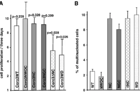
2.3 Expression of the WD-repeat domain of CRN2 inhibits cell proliferation
We addressed the effects of CRN2 on cell proliferation and cytokinesis. Wild-type HEK293 and the stably transfected cell populations, which exhibited differences compared to CRN2- NWDC in the wound healing assay, were seeded in low concentrations and counted again after 3 days to calculate cell proliferation. CRN2-NWDC, CRN2-NC, CRN2-NGC, and wild- type cells showed only slight and insignificant differences in cell proliferation. A statistically significant inhibition of proliferation was detected for cells expressing CRN2-∆NC and CRN2-WD (Fig.9A).
To verify that the reduced cell proliferation rate is not due to defects in cytokinesis, we
repeated the experiment and determined the number of multinucleated cells after 3 days of
cultivation (Fig. 9B). Here, only CRN2-NWDC and wild-type HEK293 cells showed similar
low levels of multinucleated cells, but cells transfected with any of the CRN2 domain
constructs showed approximately five-fold increased levels of multinuclear cells. An
evaluation of the number of cells harboring two, three, or four and more nuclei detected no further differences. Note that CRN2-NC and CRN2-NGC induce defects in cytokinesis though no change in proliferation rate is detected. Only CRN2-∆NC and CRN2-WD exhibit both, a reduced cell proliferation rate and an increased number in multinucleated cells. Cell proliferation and cytokinesis independently are changed by the presence of CRN2 peptides.
Figure 9. EGFP–CRN2 fusion proteins affect cell proliferation and cytokinesis. A. Proliferation rates of HEK293 cells expressing the indicated fusion proteins after 3 days of cultivation. The error bars represent the standard deviation of three independent experiments. p-values refer to the CRN2-NWDC cell population and were calculated by Student's t-test; p <0.05 is significant. B. Bars indicate the number of cells having more than one nucleus per cell for different HEK293 cell populations. The values are from two independent experiments with approximately 185 cells analyzed per cell line
.
A significantly high percentage of multinucleated cells were seen in CRN2-WD, ∆NC, NC, and CRN2-NGC in comparison to CRN2-WT (endogenous coronin) and CRN2- NWDC, where lowest percentage was measured.2.4 CRN2 participates in the formation of cellular protrusions
One major aspect of cell migration is the formation of cellular protrusions. Therefore, the
activity of single wild-type HEK293 cells or cells expressing CRN2 domains were recorded
over 30 min (Fig.10A). Figure 10 is representative of several experiments. Wild-type
HEK293 cells could not be distinguished from the CRN2-∆CC, CRN2-NC, and CRN2-NGC
populations with respect to the number of protrusions formed. CRN2-NWDC cells exhibit a
slightly increased number of protrusions, which was also detected by immunofluorescence
images of fixed cells. A markedly reduced number of lamellipodia as well as filopodia were
detected in HEK293 cell populations expressing CRN2-∆NC and CRN2-WD. Further they
showed the lowest frequencies of changes in the number of forming or retracting protrusions
(2.5 and 1.0, respectively). Although HEK293 populations expressing CRN2-NC do not exhibit a change in the number of protrusions, the frequency of their formation is reduced (Fig.10B). The presence of the glycine loop between the N- and C-terminus (CRN2-NGC) ameliorates the defect.
Figure 10. CRN2 fusion proteins alter the activity of HEK293 cells. The changes in the number of cellular protrusions including lamellipodia and filopodia followed over 30 min for the wt and cell populations expressing the CRN2 polypeptides indicated are given. A. Graph is representative for several independent experiments.
B. Mean frequency of changes in the number of protrusions of each cell population over the time. A significantly reduced number of lamellipoidia or filopodia was demonstrated by CRN2∆NC and CRN2-WD
.
Unlike CRN2-WD, all other constructs (CRN2-WT, NWDC, ∆CC, NC, NGC) displayed almost similar number of cellular protrusions without notable difference.
2.5 Full-length CRN2 is essential for the process of fluid phase pinocytosis
To investigate further possible roles of CRN2 at the submembranous cytoskeleton, the uptake
of material was investigated. Phagocytosis is restricted to specialized mammalian cells and
was not studied. Fluorescent transferring uptake was used to quantify the receptor-mediated
and clathrin-associated endocytosis. This process was found to be unchanged in all HEK293
cell populations compared to the wild-type (data not shown). In contrast, pinocytosis of the
fluid phase marker HRP was significantly affected (Fig.11). CRN2-∆NC and CRN2-WD
caused a distinctly reduced pinocytotic activity, while CRN2-NC and CRN2-NGC exhibited
an intermediate decrease of pinocytosis.
Figure 11. CRN2 proteins influence fluid phase pinocytosis. HEK293 cells expressing CRN2 fusion proteins were harvested and lysed after exposing them for 60 min to HRP-containing medium (2 mg/ml). The experiments were carried out three times in duplicate. The results were normalized to the total protein amount. p- values calculated by single-factor ANOVA refer to the population of CRN2-NWDC; p <0.05 is significant. A statistically considerable reduction in pinocytotic activity was measured in case of CRN2-∆NC (p=0.021) and CRN2-WD (p=0.033). Intermediate pinocytotic activity was displayed by CRN2-NGC (p=0.043) and CRN2- NC,p=0.063) with no difference in case of CRN2-NWDC and CRN2-WT (p=0.948).
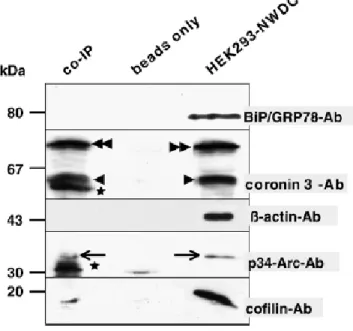
2.6 CRN2 forms a complex with the Arp2/3-complex and with cofilin
Previous studies had reported an interaction between yeast CRN11 and mammalian CRN1
and the Arp2/3 complex (Humphries et al., 2002) (Cai et al., 2005; Rodal et al., 2005). Here
we tested complex formation with CRN2. HEK293 cells expressing CRN2-NWDC were used
for several independent co-immunoprecipitation assays employing a monoclonal GFP-
antibody (Fig.12). Prior to harvest, the cells were treated with latrunculin B to prevent
unspecific co-precipitation of proteins bound to and tied together by F-actin. Precipitated
proteins were analyzed by mAb K6-444 detecting the endogenous CRN2 as well as the
EGFP–fusion protein. As expected, the endogenous CRN2 binds to and co-precipitates with
EGFP–CRN2, most likely in a heterotrimeric complex (Spoerl et al., 2002). Using antibodies
specifically recognizing cofilin and the p34 (arc35) subunit of the Arp2/3 complex, we
detected these proteins in the immunoprecipitate. For control we used BiP/GRP78- and β-
actin-specific antibodies excluding unspecific binding of the cell lysate or actin to the protein
G sepharose beads.
Figure 12. Endogenous CRN2, p34-Arc, and cofilin co-immunoprecipitate with EGFP–CRN2. HEK293 cells expressing EGFP–CRN2 were treated with latrunculin B and used for co-immunoprecipitations with an anti-GFPmAb. Samples were analyzed by immunoblotting using antibodies specifically recognizing CRN2, p34- Arc, and cofilin (co-IP). For control the experiment was carried out with cell lysate (beads only) or with anti- GFP mAb only, and the blot was probed with antibodies directed against BiP/GRP78 and β-actin. Arrowhead, endogenous CRN2; double-arrowhead, EGFP–CRN2; arrow, p34 (arc35); asterisk, unspecific bands of the GFP–
antibody solution. The signals for BiP/GRP78, CRN2, and p34-Arc are from the same membrane probed serially; signals for β-actin and cofilin are from parallel Western blots.
2.7 Structural properties of CRN2 deduced from the CRN4 crystal structure
Originally, CRN2, like all short coronin proteins, was thought to consist of an N-terminal
domain (aa 1–71), a core region containing five WD40-repeats (aa 72–299), and a C-terminal
domain (aa 300–474) (de Hostos, 1999; Rybakin and Clemen, 2005). Based on a manual
amino acid alignment, we have proposed a CRN2 structure similar to the seven-bladed
propellers known from other WD40 proteins [Hofmann and Clemen, unpublished]. The two
additional WD40-repeats of CRN2 are located in the N-terminal domain (aa 1–71) and the
conserved part of the C-terminus (aa 300–404) (Fig.13); aa 1–11, the coronin-specific
signature motif, and 348–404 do not contribute to the respective WD40-repeats. This
hypothesis was confirmed by the crystal structure of truncated CRN4 (aa 10–aa 400) which
revealed the presence of seven WD40-repeat domains (Appleton et al., 2006) which had also
been predicted by (Gatfield et al., 2005).Based on these data of CRN4, we investigated the
overall structure as well as intramolecular interactions of CRN2. It seems highly likely that
CRN2 adopts a similar N-terminal fold as CRN4 sharing the characteristic feature of the first
N-terminal beta strand completing the last propeller blade of the WD40-repeat domain
constituted by amino acids 13–348 (Fig.13). As identified in CRN4 intramolecular interactions might stabilize the C-terminal extension by anchoring it to the WD40 beta- propeller. The main contributors to these interactions are hydrogen bonds of Tyr362 to the backbone carbonyl groups of Val327 and Glu331, and the side chain interactions of the N- terminal Lys9 with Arg352 and Asp360. There is also a hydrophobic interface being formed between residues His139, Ile157, Trp182 and Ile189 from the WD40-repeat domain and Trp377 and Phe378 from the C-terminal extension. Since there is a high degree of conservation between CRN4 and CRN2 in these regions, the significance of these interactions concluded from the CRN2 homology model seems quite likely. Analyses of structure and electrostatic potential of the surface of CRN4 resulted in the identification of potential F-actin binding sites. They comprise a region on the top of propellers 1, 6, 7, and a second region on the bottom side of propellers 6 and 7 (Appleton et al., 2006). Similar regions can be assigned to CRN2. The F-actin binding site in CRN4 also includes a stretch of positively charged amino acids (400–416 (Gatfield et al., 2005) in the C-terminal extension, which is mirrored in CRN2 (aa 398–aa 419; KNRDLKVVKKNILDSKPTANKK).
Figure 13. Homology model of CRN2 (aa 7–400). The model has been generated with the CRN4 crystal structure (Appleton et al., 2006) [PDB accession number 2aq5] as template using MODELLER (Sali and Blundell, 1993) and a manual alignment of amino acid sequences. The seven-blade propellers of the N-terminal WD40-repeat domain are colored individually and annotated by their sequential order. The C-terminal extension is shown in brown and polar interactions stabilizing its fold are drawn explicitly. The yellow dot surface maps a hydrophobic interface formed by residues His139, Ile157, Trp182, Ile189, Pro203, Trp377, and Phe378. Figure prepared with PyMOL (W.L. DeLano, 2002). Taken from (Rosentreter et al., 2007).
3.0 Expression of CRN2 in diffuse gliomas is related to malignancy
3.1 CRN2 is expressed in a subset of human brain tumours and in reactive astrocytes
The expression of CRN2 in normal human brain and in brain tumours was determined by histopathological analysis and immunoblotting. Normal human brain tissue in general exhibited virtually no CRN2 protein (data not shown). The white matter did not show CRN2 immunoreactive cells. Cortical neurons, on the other hand, were faintly labelled. Other groups of neuronal cells in the hippocampus (CA1-4, dentate gyrus), the neocortex and the cerebellum (Purkinje cells), as well as subpial astrocytes in neocortical layer I were also faintly labelled (Table 2).
Table 2. Expression of CRN2 in the human brain and in different types of human brain tumours. +, expression of CRN2; (+), weak expression of CRN2; −, no expression of CRN2; n, number of cases; IHC, immunohistochemistry; WB, western blotting; , autopsy cases from which different brain regions, i.e. cerebral cortex, hippocampal formation and cerebellum, were observed.
Figure 14. Expression of CRN2 in diffuse gliomas. A. Low-grade diffuse gliomas exhibited less prominent expression of CRN2 than high-grade gliomas. The diffuse astrocytoma showed CRN2 expression in no more than 50% of the tumour cells. Oligodendrogliomas often failed to express CRN2 (shown here) or were only weakly stained (refer to Table 2). In benign oligoastrocytomas CRN2 was mainly expressed in the astroglial component (arrowheads), while the oligodendroglial component was either not or only weakly stained (arrows).
B. In contrast, expression of CRN2 in an increased number of tumour cells was noted in high-grade diffuse gliomas. Anaplastic astrocytomas and anaplastic oligodendrogliomas exhibited CRN2 immunoreactivity in
>50% of the tumour cells. In anaplastic oligoastrocytomas, CRN2 was expressed in the malignant tumour component. In the presented case, CRN2 expression was mainly found in the astroglial component (arrowheads), whereas the oligodendroglial component appeared well differentiated and did not stain for CRN2(arrows). In anaplastic oligoastrocytomas with a malignant transformation of the oligodendroglial component, this component also started to express CRN2. In glioblastomas, nearly all tumour cells showed CRN2 expression.
The expression of CRN2 in tumours varied among different types of brain tumour. No
expression of CRN2 was detectable in tumours of neuronal origin, viz in the desmoplastic
medulloblastomas and a neuroblastoma (Table 2). On the other hand, two benign tumour entities, benign meningiomas and pilocytic astrocytomas, exhibited a high number of tumour cells expressing CRN2 (Table 2). Low-grade diffuse astrocytomas, oligodendrogliomas and oligoastrocytomas, however, showed a limited number of CRN2-positive tumour cells (Fig.14A and Table 2; note that oligodendrogliomas showed either no or a small number of tumour cells expressing CRN2), while highgrade anaplastic astrocytomas, anaplastic oligodendrogliomas, anaplastic oligoastrocytomas and glioblastomas exhibited high numbers of CRN2-positive tumour cells (Fig. 14B and Table 2).
Figure 15. Expression of CRN2 in the astroglial and oligodendroglial component of anaplastic oligoastrocytomas. A: Most of the astroglial tumour cells expressed CRN2 (arrowheads). The oligodendroglial component with anaplastic features such as mitoses (asterisk) showed mild to moderate levels of CRN2 (arrows).
B: Double label immunofluorescence of another anaplastic oligoastrocytoma showed no expression of CRN2 in the oligodendroglial component. This component is characterized by a perinuclear staining pattern of the tumour cells with anti-MAP2. C: The astroglial tumour component of this tumour exhibited a co-localization of CRN2 with MAP2. This expression of MAP2 in a pattern showing cell processes indicated the astroglial tumour cell nature of these cells.
Figure 16. Expression of CRN2 in different regions of a glioblastoma multiforme. A: In a glioblastoma multiforme nearly all tumour cells expressed CRN2. B: The most prominent expression, however, was detected in areas of vital tumour with microvascular proliferation. C: Surviving tumour cells near areas of palisading necroses were only weakly labeled with anti-CRN2. The boxes in A indicate the areas enlarged in B and C.
Within mixed gliomas, i.e. oligoastrocytomas and anaplastic oligoastrocytomas, different expression patterns of CRN2 within both tumour components were detected: the astroglial tumour cells more often expressed CRN2 than those of the oligodendroglial component (Fig.15). Double-label immunofluorescence of CRN2 and MAP2 confirmed the tumour cell nature of the CRN2-positive astroglial cells in oligoastrocytomas. However, in some cases the oligodendroglial compartment also showed a prominent CRN2 expression, mostly occurring in anaplastic oligoastrocytomas with features of malignancy of the oligodendroglial component, such as mitotic figures.
Moreover, particularly in glioblastomas that expressed CRN2 in nearly all tumour cells, a variation in the expression of CRN2 within the tumour was seen (Fig.16). Tumour cells adjacent to proliferating microvessels in the tumour periphery (Fig. 16B) exhibited a stronger CRN2 staining than those adjacent to areas of necrosis (Fig.16C).
The matrix of brain tumours contains a varying number of reactive astrocytes, non-malignant
but activated and migrating cells that also expressed CRN2 (Fig.17F-H). These CRN2-
positive reactive astrocytes, however, are not restricted to tumours. In the vicinity of other
brain lesions, ie infarcts and traumatic lesions, expression of CRN2 was also detectable within
reactive astrocytes (Fig.17A). In contrast, non-specific astroglial activation in the absence of a
circumscribed lesion, e.g. in the course of epilepsy, showed only a faint expression of CRN2 in astrocytes (Fig. 17C-E).
Figure 17. Expression of CRN2 in reactive astrocytes and microglial cells. A: Activated astrocytes highly exhibiting CRN2 were present in the brain close to an infarct lesion at the stage at which gliosis was beginning.
B: In an infarct lesion in the stage of resorption macrophages showing foam cell morphology were labelled by anti-CRN2. C-E: Gliosis in a case of chronic epilepsy showed weak CRN2 expression in reactive astrocytes (C, arrows), which also exhibited glial fibrillary acidic protein (GFAP) (D, arrows), and in perivascular macrophages (C, arrowheads) identified by their CD-68 expression (E, arrowheads). F-H: CRN2 was detected in tumour- associated reactive astrocytes. Reactive astrocytes were identified comparing the staining of CRN2, GFAP, and MAP2. The latter specifically marked tumour cells (Rosentreter et al., 2007) (arrowheads), whereas GFAP stained both tumour cells (arrowheads) and reactive astrocytes (arrows). Based on the different morphology of tumour cells and reactive astrocytes, CRN2-positive cells in this low-grade astrocytoma were predominantly classified as reactive astrocytes (arrows), while most tumour cells failed to stain for CRN2 (arrowheads).
3.2 CRN2 expression correlates with the grade of malignancy in diffuse gliomas
As our data showed that diffuse gliomas, i.e. astrocytomas, oligodendrogliomas and
oligoastrocytomas, exhibited increasing numbers of tumour cells expressing CRN2 with
increasing WHO grade, which indicates the malignant potential of a tumour, we focused on
these tumours (Fig.18A). A semiquantitative analysis of vital tumour expressing CRN2
demonstrated that oligodendrogliomas, diffuse astrocytomas and oligoastrocytomas contained
only a small number of tumour cells positive for CRN2 (Fig.18A). In contrast, usually more than 50% of the tumour cells were stained by mAb K6-444 anti-CRN2 antibodies in malignant gliomas (Fig.18A; p < 0.01). Accordingly, western blots of gliomas showed increased levels of CRN2 compared to normal tissue of grey and white matter (Fig.18B,C; p
< 0.05). Although Western blotting in general confirmed the increased expression of CRN2 in diffuse gliomas with their grade of malignancy, the analysis of tissue homogenates showed varying expression levels. This was particularly visible in low- and high-grade astrocytomas (Fig.18B, C). The reason behind this is that the area of tumour tissue used for immunoblotting is not exactly defined. Therefore, two factors will interfere with the determination of the CRN2 expression by immunoblotting, but not by immunostaining. First, the expression levels of CRN2 vary in tumour cells within different regions of a certain tumour (Fig.19); and second, the tumour matrix contains a varying number of reactive astrocytes that also express CRN2 (Figs.18A; 17F-H).
Figure 18 Quantification of the expression of CRN2 in tumour cells and tumour tissues.A. Tumour cell expression in relation to malignancy was determined by quantification of the immunohistochemical expression of CRN2 in diffuse gliomas. With increasing malignancy, as indicated by the WHO grade, the number of tumour cells expressing CRN2 increased. Mean and standard deviation are presented; *** indicates p < 0.01, as calculated by the Kruskal–Wallis H-test and the trend-test. Tumourcell expression in different types of gliomas was analysed by quantification of the immunohistochemical staining pattern of CRN2 within the different types of gliomas. The highest numbers of tumour cells expressing CRN2 were present in anaplastic astrocytomas, anaplastic oligoastrocytomas and in glioblastomas. * Indicates p < 0.05, as determined by the Mann–Whitney U- test to compare the different types of tumours. p values were corrected for multiple testing. Expression in tumour-associated reactive glial cells in diffuse gliomas was observed by the semiquantitative assessment of the number of reactive astrocytes expressing CRN2associated with diffuse gliomas. Oligodendrogliomas, anaplastic oligodendrogliomas, astrocytomas and anaplastic astrocytomas were frequently associated with reactive glial cells that also exhibited CRN2. Oligoastrocytomas, anaplastic oligoastrocytomas and glioblastomas appeared to be less frequently associated with a reactive gliosis. However, there was a wide range of the degree of gliosis in
each tumour entity. Therefore, the trend seen in the diagram did not reach significance (Poisson regression analysis, p = 0.8254; deviance, 301 812).
Figure 18, continued. B. Western blot analysis of the different types of diffuse gliomas exhibited weak to moderate CRN2 expression levels in tissue samples of astrocytomas (AII), oligodendrogliomas (OII), oligoastrocytomas (OAII) and anaplastic astrocytomas (AAIII). Two anaplastic astrocytomas (AAIII), the anaplastic oligodendroglioma (AOIII) and the glioblastomas (GBMIV) showed a strong expression of CRN2.
Pilocytic astrocytomas (pA), loaded as positive controls, exhibited moderate CRN2 expression levels. Normal human brain tissue of grey (GM) and white matter (WM) did not express more than traces of CRN2; β-actin is given as a loading control.
Figure 18, continued. C. CRN2 expression in tumour tissue observed by quantitative western blotting. Tumour tissue expression in relation to malignancy showed an increase from benign diffuse gliomas to high-grade glioblastomas (*, Kruskal–Wallis H-test, Trend-test, p < 0.05). Tumour tissue expression in different types of gliomas exhibited a nearly similar pattern as the immunohistochemical analysis but failed significance because of the possible interference of reactive glial cells in this small sample.
3.3 CRN2 knockdown inhibits cellular functions related to tumour malignancy
U373 and A172 glioblastoma cells, commonly used to study glioma-related cellular functions,
express high levels of CRN2. To address the question of whether the expression of CRN2 in
gliomas has an impact on tumour cell proliferation, migration, and invasion, we employed
lentivirus mediated shRNA-dependent knockdown of CRN2 in U373 and A172 glioblastoma
cells, which resulted in a ~95% CRN2 protein reduction (Fig. 19A).
Figure 19. A Effective shRNA-mediated knockdown of CRN2. Western blotting confirms a strong reduction (~95%) of the expression level of CRN2 in transduced U373 and A172 glioblastoma cell lines. Wild-type cells (U373 only), cells transduced with empty vector or a non-target shRNA-cassette (U373 and A172), as well as β- actin staining,are given as controls. Immunofluorescence imaging of U373 glioblastoma cells fixed with 4%
paraformaldehyde also indicates an overall decreased expression level of CRN2 in knockdown cells. The F-actin cytoskeleton seems not to be affected in CRN2 knockdown cells. The images showing lamellipodia in higher magnification are not enlarged areas from the images above, but from a different cell. CRN2 was detected by mAb K6-444, F-actin was stained with TRITC-phalloidin. Note the colour-coded fluorescence intensity signals (images with asterisk). Bars, 5 µm.
Figure 19, continued. B. CRN2 knockdown inhibits invadopodia formation. Immunofluorescence imaging of invadopodia of a U373 glioblastoma cell. Glioblastoma cells grown on cover slips coated with FITC-gelatin
were fixed with 4% paraformaldehyde and stained with TRITC-phalloidin. One representative cell each is shown for knockdown and non-target control cells. Invadopodia at the ventral surface of the cell are defined by the presence of F-actin and absence of the matrix FITC-signal (arrows). Bars, 10 µm
Immunofluorescence images of the CRN2 knockdown cells did not reveal any obvious changes in the F-actin staining, localization of the Arp2/3 complex and the morphology of lamellipodia. These structures appeared as in normal and non-target controls, although the CRN2-specific signal intensity (images with asterisk; note the colour code, yellow pixels versus dark red pixels) was reduced in virtually all knockdown cells (Fig 19A and data not shown). However, there were still low amounts of CRN2 in the knockdown cells, which exhibited a similar distribution as in non-target glioblastoma cells (Fig 19A). The signal intensity of cortical F-actin staining varied in both non-target and knockdown cells, and the F- actin content remained unchanged in the CRN2 knockdown glioblastoma cells (data not shown).
Figure 20. Knockdown of CRN2 inhibits cellular functions related to malignancy. A. CRN2 knockdown inhibits cell proliferation, but not cell cycle state. MTT-assay based proliferation rates of U373 and A172 glioblastoma cells lacking CRN2 expression are compared to control cells; 100 independent experiments in 96- well format each, Student’s t-test;U373, ∆OD595 0.001 versus 0.045, p = 2.7 × 10⁻¹²; A172, ∆OD595 0.14 versus
0.20, p = 1.0 × 10⁻¹⁰. Trypsination/replating based proliferation rates of U373 and A172 glioblastoma cells lacking CRN2 expression are compared to control cells; a certain number of cells were seeded and counted again after 2 days; four independent experiments each. Student’s t-test; U373, factor 2.2 versus 3.4, p = 0.005; A172, factor 1.4 versus 2.1, p = 0.012. PI-FACS analysis of cell cycle state of U373 and A172 CRN2 knockdown cells.
Fixed cells were stained with propidium iodide and 10,000 single cells from three independent experiments were analysed by fluorescence-activated cell sorting (PI-FACS). The histograms demonstrate the fractions of cells in G0/1, G2/M and S phase; A, apoptosis. For control, cells treated for 16 h with a cdk4-inhibitor (Calbiochem, 219476) are given.
In contrast, multiple malignancy-related cellular functions were inhibited. Glioblastoma cells with reduced CRN2 levels exhibited an inhibition of the cell proliferation rate, as determined by two different assays, cell viability measurements (U373, −98%, p < 10
−11; A172, −30%, p
= 10
−10) as well as trypsination/replating experiments (U373, −35%, p = 0.005; A172, −33%, p = 0.012) (Fig 20A). An analysis of the cell cycle state by PI-FACS did not indicate an inhibition of the cell cycle (Fig 20A). Monitoring single CRN2 knockdown glioblastoma cells, we determined a reduction of their ability to migrate compared to non-target shRNA control cells (U373, −62%, p < 10
−6; A172, −54%, p < 10
−8) (Fig 20B).
Figure 20, continued. B. CRN2 knockdown inhibits cell migration and invasion. The single cell motility chart indicates the values from a total of 40 cells, each derived from several independent experiments; Student’s t-test:
U373, velocity 5.36 µm/70 min versus 14.3 µm/70 min, p = 6.4 × 10⁻⁷; A172, velocity 5.0 µm/70 min versus 10.9 µm/70 min, p = 9.3 × 10⁻⁹. The extracellular matrix (ECM) invasion rate graph shows one representative experiment with mean and standard deviation of 40 and 20 measurements of U373 and A172 glioblastoma cells, respectively; Student’s t-test: U373, 0.6 AFU versus 0.7 AFU, p = 0.016; A172, 0.7 AFU versus 0.9 AFU, p = 6.9 × 10⁻⁵. Controls with 15 µM latrunculin B added to the cells in order to depolymerize the cellular F-actin demonstrate the maximum reduction of the invasion. Controls lacking the fluorescent dye showed background values of 0.2 AFU. For ECM invasion, see also Fig 19B.
In addition, we tested the ability of CRN2 knockdown glioblastoma cells to degrade and invade extracellular matrix (ECM). ECM invasion of CRN2 knockdown glioblastoma cells also exhibited a reduction (U373, −14%, p = 0.016; A172, −22%, p < 0.0001) (Fig 17B). To investigate the effects of the CRN2 knockdown on ECM invasion in more detail, we determined the overall activity of matrix metalloproteinases (MMPs). A172 cells lacking CRN2 exhibited a reduced activity of MMPs (−12%, p < 10
−10), however, no difference was detectable for U373 cells (Fig 20C).
Figure 20, continued. C. CRN2 knockdown inhibits matrix metalloproteinase activity. A172 glioblastoma cells lacking CRN2 exhibit a reduced activity of secreted matrix metalloproteinases (MMP); however, such a difference was not detectable in U373 cells. Thirty measurements from six independent experiments each.
Student’s t-test: U373, 14.4 AFU versus 14.5 AFU, p = 0.66; A172, 10.7 AFU versus 12.2 AFU, p = 1.4 × 10⁻¹¹. Controls lacking the fluorescent substrate showed background values of 7 AFU
We also visualized the formation of invadopodia in vitro (Fig 19B). Invadopodia are defined
by co-localization of invadopodia markers, ie typical F-actin structures, with the degradation
of fluorescently labelled extracellular matrix (Weaver, 2006). U373 non-target cells
demonstrated a strong activity of matrix degradation on gelatin matrices, whereas in U373
knockdown cells virtually no invadopodia were detected. For A172 cells, no invadopodia
formation was found under our experimental conditions. Only two (gelatin, fibronectin) out of
various components of the extracellular matrix have been used in the invadopodium assay,
while the matrix metalloproteinase assay detected the overall activity of various, but not all
known enzymatic activities. Thus, in both U373 and A172 knockdown, cell matrix
degradation activities, although to different degrees, are inhibited upon reduction of CRN2
levels.
4.0 Structural and functional diversity of novel CRN2 isoforms.
4.1 Identification of three CRN2 proteins encoded by two mRNA species
CRN2 usually migrates as a diffuse band of approximately 57 kDa. Immunoblotting of HEK293 and undifferentiated C2F3 cell lysates with the CRN2 specific monoclonal antibody K6-444 (Spoerl et al., 2002) revealed that the diffuse band can be separated into two closely migrating bands of 57/58 kDa (later referred to as CRN2i1/CRN2i2 (coronin-1C isoform 1/isoform 2) (Fig.21A). Moreover, a second slower migrating protein of 60 kDa (later referred to as CRN2i3 (coronin-1C isoform 3) can be detected in murine skeletal muscle and differentiating C2F3 myoblasts as well as in brain and heart tissue (Fig.21A; figure 2B in reference (Spoerl et al., 2002). This protein is the only CRN2 isoform present in mature murine, bovine and human skeletal muscle tissue.
Figure 21. A slower migrating isoform of CRN2 appears during myogenesis. A, lysates of HEK293 cells, murine skeletal muscle tissue, C2F3 myoblasts, and C2F3 myotubes (diff.) were analyzed by SDS-PAGE followed by western blotting using mAb K6-444 specific for CRN2 (Spoerl et al., 2002). B, 20 µg of total RNA from C2F3 myoblasts and 50 µg of total RNA from myotubes (diff.) were subjected to gel electrophoresis under denaturing conditions and analyzed by northern blotting using a full-length CRN2 cDNA as probe; for control, the ethidium bromide stained RNA-gel is shown.


![Figure 13. Homology model of CRN2 (aa 7–400). The model has been generated with the CRN4 crystal structure (Appleton et al., 2006) [PDB accession number 2aq5] as template using MODELLER (Sali and Blundell, 1993) and a manual alignment of amino acid sequ](https://thumb-eu.123doks.com/thumbv2/1library_info/3709155.1506314/27.892.278.658.594.951/homology-generated-structure-appleton-accession-modeller-blundell-alignment.webp)
