Evaluation of geno- and phenotypic alterations in Luminal B breast cancer using tumor mice
(TM) and humanized tumor mice (HTM)
Dissertation
zur Erlangung des Doktorgrades der Biomedizinischen Wissenschaften
(Dr. rer. physiol.)
der
Fakultät für Medizin der Universität Regensburg
vorgelegt von Eva-Maria Rom-Jurek
aus Weiden i.d.Opf.
im Jahr 2020
Dekan: Prof. Dr. Dr. Torsten E. Reichert
Betreuer: Prof. Dr. med. vet. Anja-Katrin Wege
Tag der mündlichen Prüfung:
Für meine Eltern Elisabeth und Werner Rom, meine Kinder
Jonathan und Nicolas und meinen Mann Ben
Parts of this work have already been published in peer-reviewed journals in an open- access format:
Rom-Jurek, E.-M., Kirchhammer, N., Ugocsai, P., Ortmann, O., Wege, A.K. and Brockhoff, G. (2018), “Regulation of Programmed Death Ligand 1 (PD-L1) Expression in Breast Cancer Cell Lines In Vitro and in Immunodeficient and Humanized Tumor Mice”, International journal of molecular sciences, Vol. 19 No. 2.
The content of this thesis is unpublished but in preparation:
Rom-Jurek E.-M., Wege, Brockhoff, Denkert, Jank, Trumpp, Klein, Irlbeck, Pfarr, Weichert, et al: “MDM2 gene amplification in Luminal B tumors is associated with increased aggressiveness and metastatic potential in humanized tumor mice (HTM).” (in preparation)
2 Table of content
Zusammenfassung ... 6
Summary ... 9
1. Introduction ...11
1.1 From breast physiology to the pathophysiology of Luminal B breast cancer ...11
1.1.1 Different cell types involved in breast (cancer) physiology ...11
1.1.2 Breast cancer etiology ...12
1.1.3 Classification of breast cancer and breast cancer subentities ...13
1.1.4 Luminal breast cancer ...14
1.2 Breast cancer metastases ...15
1.2.1 Metastatic sites in breast cancer ...15
1.2.2 EMT and MET in breast cancer metastases ...16
1.2.3 Stem cells traits and their relevance in EMT-MET and breast cancer metastases 17 1.2.4 Disseminated tumor cells (DTCs) ...20
1.3 The role of genetic aberrations in breast cancer ...20
1.4 TILs and immune checkpoints in breast cancer ...21
1.5 PDX models and humanized PDX models in breast cancer research ...22
1.6 Aim of the thesis ...26
2. Material ...27
2.1 Consumables ...27
2.2 Buffers and Solutions ...27
2.3 Buffers ...28
2.4 Culture Media ...29
2.5 Cell lines ...29
2.6 Anesthesia ...29
2.7 Antibodies Flow Cytometry ...30
2.8 Antibody Immunohistochemical Staining ...31
2.9 Kits ...31
2.10 siRNA ...32
2.11 Reagents and chemicals ...32
2.12 Devices ...33
2.13 Software ...33
3 Methods ...34
3.1 Human sample preparation ...34
3.1.1 Isolation of tumor cells from serous effusions ...34
3.1.2 Primary tumor tissue ...35
3
3.1.3 Isolation of hematopoietic stem cells from cord blood ...35
3.2 Animal Experiments ...37
3.2.1 Experimental Design ...37
3.2.2 Generation of tumor mice (TM) ...39
3.2.3 Generation of humanized tumor mice (HTM) ...41
3.2.4 Preparation of blood, organs and tumor tissue ...42
3.3 Flow cytometry ...44
3.3.1 Phenotypic Analysis ...44
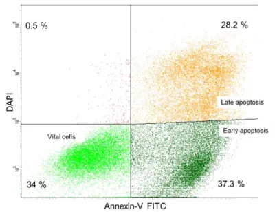
3.3.2 Apoptosis ...45
3.3.3 S-phase fraction ...46
3.4 Human -Mouse PCR ...46
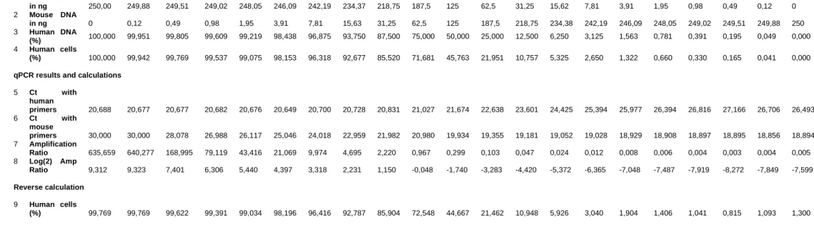
3.4.1 DNA extraction and quantitative real-time PCR ...46
3.5 Immunohistochemical Staining ...50
3.6 Analysis of Lung metastases ...51
3.7 Fluorescence in-situ-hybridisation (FISH) ...51
3.8 Cell culture and cryopreservation ...52
3.9 Protein biochemical analysis ...53
3.9.1 Protein isolation ...53
3.9.2.BCA Assay ...53
3.9.3. Western Blotting ...54
3.10 Breast cancer cell line treatments and assays ...55
3.10.1 Experimental Design ...55
3.10.2 MDM2 siRNA Knockdown ...56
3.10.3 AMG232 treatment ...57
3.10.4 Wound Healing Assay ...57
3.11 Bone Marrow DTC (disseminated cancer cell) single-cell analysis (Cooperation with Christoph Klein and Christoph Irlbeck UKR) ...58
3.11.1 PDX BM staining for DTC detection ...58
3.11.2 Single cells isolation and whole genome amplification (WGA) performed by Christoph Irlbeck ...58
3.11.3 Control and quality PCR of WGA product ...59
3.11.4 Gel electrophoresis ...60
3.11.5 Reamplification ...61
3.11.6 Low pass-sequencing for copy number alteration profiling (performed by Christoph Irlbeck) ...61
3.11.7 Preparation of Ref Sequ files and analysis by progenetix software ...62
3.12 Panel Sequencing (Performed by Nicole Pfarr and Wilko Weichert TUM) ...62
3.12.1 DNA Isolation of FFPE tissue ...62
3.12.2 Sequencing Panels ...63
4
3.12.3 Massive parallel sequencing ...63
3.12.4 Data analysis and prediction of copy number variations ...64
3.13 Statistical analyses ...64
4 Results ...66
4.1 Primary tumor patient samples ...66
4.2 Characterization of the primary tumor specimen ...70
4.2.1 Phenotypical characterization of patient Luminal A and B tumor cells using (stem cell FACS) SCF panel ...70
4.2.2 Immune cell infiltration in primary Luminal patient tumors ...72
4.2.3 T cell phenotyping of TILs in the primary tumors of Luminal patients ...74
4.2.4 Tumor cell phenotyping and immune cell characterization in solid tumors compared to effusions (metastases) of Luminal B patients. ...76
4.2.5 Primary tumor culturing ...78
4.3 Characterization of all Luminal B tumor mice (TM) ...79
4.3.1 Engraftment success of primary tumors in TM (tumor mice) ...79
4.3.2 Total engraftment numbers of tumors in TM and HTM ...80
4.3.3 Characterization of human origin in PDX tumors ...81
4.3.4 The immunohistochemical phenotype of the PDX tumor is congruent with the patients’ primary tumor ...81
4.4 MDM2 /TP53 /MDM4 alterations in Luminal B breast cancer PDX ...84
4.4.1 Uncovered alterations in Luminal B breast cancer by Panel Sequencing ...84
4.4.2 Classification of PDX models according to chromosomal aberrations of MDM2 and p53 ...87
4.4.3 Classification of PDX models according to the MDM2 and p53 protein levels ...88
4.4.4 MDM2 amplification increased tumor weight and tumor volume over time, decreased the disease-free survival but did not alter the overall survival ...89
4.4.5 Phenotypic differences and alterations in the MIC population (CD44+/cMET+/CD47+) between TM MDM2 amplified tumors and TM MDM2 WT tumors ...90
4.4.6 MDM2 amplified tumors promote lung metastases and differ phenotypically from the corresponding tumor...93
4.4.7 MDM2 amplification decreases T-cell infiltration in HTM ...94
4.4.8 Effect of MDM2 amplification in Luminal B humanized PDX mice ...96
4.4.9 HTM MDM2 amplified tumors promote lung metastases that differ phenotypically from the corresponding tumor ...97
4.4.11 Copy number variation low pass-sequencing of the Luminal B HTM P and TM P PDX model ...99
4.4.12 Metastatic potential of generated PDX and the genetic aberrations of HTM and TM tumors ... 102
4.5 The relevance of immune checkpoints (PD-L1) in Luminal breast cancer ... 102
4.6 Targeting MDM2 in ZR-75-1 breast cancer cell line in vitro ... 105
5
4.6.1 Effect of MDM2 knockdown on apoptosis, S-phase fraction, and cell number .... 105
4.6.2 Effect of AMG232 inhibition on apoptosis, S-phase fraction, and cell number... 108
4.6.3 MDM2 knockdown increases apoptosis via the p53 pathway ... 110
5. Discussion ... 112
5.1. Luminal B breast cancer PDX models and Luminal B primary tumor cell culturing ... 112
5.2. Genomic and phenotypic markers that could identify a high-risk Luminal B tumor ... 117
5.2.1 Genomic markers ... 117
5.2.2 Phenotypic markers ... 120
5.3 Immune cell interactions and immune cell checkpoint relevance in Luminal B breast cancer ... 122
5.4 Genotypic differences between the primary tumor and bone marrow DTCs ... 125
5.5 Organotropism in Luminal B breast cancer due to genetic alterations ... 127
5.6 Conclusion ... 128
References ... 129
List of abbreviations ... 144
List of tables ... 146
List of figures ... 147
Acknowledgements ... 149
Selbständigkeitserklärung ... 151
Curriculum vitae ... 152
6 Zusammenfassung
Luminal B Brustkrebs tritt bei 20 % aller diagnostizierten Mammakarzinome auf.
Jedoch haben Patientinnen bei dieser Diagnose nur eine 50 prozentige Überlebenswahrscheinlichkeit für die nächsten 5 Jahre. Hierbei zählt vor allem ein Rückfall oder die spätere Metastasierung nach wie vor zu den häufigsten Todesursachen, auch wenn es zwischenzeitlich einige Fortschritte in deren Behandlung gibt. Dennoch stellt vor allem die Risikoeinschätzung, die ein potentielles Rezidiv und/oder die Manifestierung von Fernmetastasen vorhersagt, ein Problem dar.
Die gängigen Marker die hierzu verwendet werden, wie der Hormonrezeptor Status, die Proliferationsindizes, die Zelldifferenzierung und genetischen „Assays“ reichen nicht aus, um Luminal B Patientinnen in „Hoch und „Niedrig-Risikogruppen“
einzuteilen. Deshalb war die Zielsetzung dieses Projekts Marker zu identifizieren, die eine Einteilung der hoch aggressiven Luminal B Tumoren ermöglichen und Tumorzelldisseminierung und Metastasierung vorhersagen. Diese sollen es erleichtern eine Therapieentscheidung zu treffen und zusätzlich die Frage beantworten, ob eine Luminal B Brustkrebspatientin Chemotherapie benötigt oder nicht.
Um diese Fragen zu beantworten, wurden die Primärtumore der Luminal B Patientinnen phänotypisch untersucht. Gleichzeitig wurden aus den Primärtumoren sogenannte Luminal B Xenotransplantations-Modelle generiert (Tumormäuse TM) und analysiert. Zudem sollte auch der Einfluss des humanen Immunsystems auf Luminal B Tumoren analysiert werden, was durch das Generieren der humanisierten Tumormäuse (HTM) ermöglicht wurde. Phänotypisch zeigte sich, dass man Luminal B Tumoren anhand der erhöhten CD24 Expression im Vergleich zu Luminal A ermitteln kann. Zudem zeigte sich bei den primären Luminal B Hoch-Risikotumoren (definiert durch Todesfall der Patientin, Rückfall oder Metastasierung, und durch das Anwachsen eines Luminal B Tumors als Xenotransplantations-Model) ein erhöhtes Vorkommen der Co-expression von CD44/cMET/CD47 der sogenannten Metastasierungs-initiierenden Zellpopulation im Vergleich zu Luminal B Niedrig- Risikotumoren (alle Patientinnen, die überlebt haben; keinen Rückfall erlitten und bei welchen der Tumor nicht im Xenograftmodel angewachsen ist). Zusätzlich konnte beim phänotypischen Vergleich von Luminal B Primärtumoren und der zugehörigen Metastase eine erhöhte Expression von cMET und CD44 nachgewiesen werden. Dies
7
zeigte sich sowohl beim Vergleich von TM oder HTM Tumoren mit der dazugehörigen Lungenmetastase, als auch bei den Primärtumoren der Patientinnen die mit Luminal B Aszites Präparaten oder Pleuraergüssen verglichen wurden. Es konnte auch ein erhöhtes CD4/CD8 Verhältnis auf Immunzellen, die den Tumor infiltrieren, in Luminal B Hoch-Risikotumoren nachgewiesen werden. Eine der wichtigsten Entdeckungen dieser Arbeit stellte dabei das vermehrte Auftreten einer MDM2 Amplifikation in Luminalen Tumoren dar. Diese zeigte sich mit erhöhter Tumoraggressivität und einer hohen Metastasierungswahrscheinlichkeit in Luminal B TM und HTM. In TM und HTM wiesen MDM2 amplifizierte Tumore zudem häufig Lungenmetastasen und disseminierte Zellen im Knochenmark auf. Diese Ergebnisse wurden in Zellkulturversuchen durch eine Herunterregulierung von MDM2 validiert und zeigten dabei einen p53 abhängigen Mechanismus, der die Proliferation und Apoptose der Tumorzellen steuert. Im Einklang mit diesen Ergebnissen stand auch die Behandlung der Zellen mit einem MDM2 Inhibitor (AMG232) und liefert dadurch einen klinisch relevanten Ansatz zur Therapie von MDM2 amplifizierten Tumoren. Zudem wurden genetische TP53 Mutationen in Luminal B Tumoren mit erhöhter Disseminierung von Tumorzellen ins Knochenmark in Verbindung gebracht. Die Amplifikation von MDM4 in Luminal B Tumoren zeigte in der TM hingegen sogar eine Metastasierung des Tumors in multiple Organe wie der Lunge, der Leber, des Gehirns, und des Knochenmarks. Interessanterweise regulieren sich alle entdeckten genomischen Aberrationen (MDM2/P53/MDM4) gegenseitig und gehören zum gleichen Signalweg.
Das wiederum deutet auf eine wichtige Rolle von MDM2, p53 und MDM4 beim aggressiven Luminal B Mammakarzinom hin. Auffallend hierbei sind nicht nur die Aggressivität der Tumore, sondern auch die aberrationsabhängige Metastasierung in bestimmte Organe. Ein weiterer Teil dieser Arbeit beschäftigte sich mit den genetischen Unterschieden zwischen Einzelzellen aus dem Primärtumor und disseminierten Tumorzellen im Knochenmark in der TM und HTM. Hierbei konnte gezeigt werden, dass die Tumorzellen aus dem Primärtumor (TM und HTM) und die disseminierten Tumorzellen aus dem Knochenmark unterschiedliche Cluster bilden und damit unterschiedliche genetische Modifikationen aufweisen. Interessanterweise differenzierten sich zudem disseminierte Tumorzellen aus dem Knochenmark von HTM von allen anderen Tumorzellen und auch den disseminierten Zellen aus der TM.
Dies deutet auf einen möglichen Selektionsdruck auf Zellen mit bestimmter genetischer Ausstattung hin, der in der Peripherie von humanen Immunzellen
8
verursacht wird. Zudem kann dies aber auch auf Knochenmarksnischen-bedingte Selektion, die durch humane Immunzellen verändert wird, zurückgeführt werden.
Insgesamt zeigte sich in diesen Experimenten die generell geringe Immunogenität von Luminal B Tumoren sowohl auf Patientenebene als auch in der HTM. Somit spiegelt das HTM Xenotransplantations-Modell die Situation im Luminal B Patienten erfolgreich wider und ist damit auch ein Modell für zukünftige Therapiestudien für dieses Patientenkollektiv.
Zusammenfassend zeigte sich, dass Luminal B Xenotransplantations-Modelle und humanisierte Luminal B Xenotransplantations-Modelle ein geeignetes System zur Identifizierung von phäno- und genotypischen Veränderungen sind, die mit einem erhöhten Metastasierungspotential und einer erhöhten Aggressivität einhergehen.
Zurzeit werden MDM2 Amplifikationen und Expressionen im Zusammenhang mit dem (tumorfreien) Überleben an einem größeren Patientenkollektiv in unserem Labor untersucht. Weitere klinische Studien könnten dann zeigen, ob Luminal B Brustkrebspatientinnen mit Tumoren, die eine MDM2/MDM4/TP53 Veränderungen aufweisen, zusätzlich von einer Chemotherapie oder eventuell von einer zielgerichteten Inhibition von MDM2 profitieren würden.
9 Summary
The breast cancer subtype Luminal B is diagnosed in 20% of all breast cancer cases whereas only 50 % of the patients are still alive 5 years after the first diagnosis. Despite the advances in treatment, patients suffering from Luminal B breast cancer frequently experience a relapse or develop distant metastases. Besides the current strategy of hormone-receptor-positivity, proliferation indices, grading, and gene signature assays to categorize the Luminal breast cancer patients into high and low-risk groups, there is still a lack of appropriate markers that reliably predict events of recurrence. Overall, this thesis aims to identify biomarkers that are associated with aggressivity, cell dissemination and/or metastases formation. Importantly these markers might contribute to the therapy decision if Luminal B breast cancer patients need a chemotherapeutic intervention or not.
Therefore, the primary Luminal B patient samples were analyzed and PDX models were generated by the transplantation of primary Luminal B patient samples into NSG mice, the so-called tumor mouse (TM). Additionally, humanized Luminal B tumor mice (HTM) were generated and assessed under the influence of the human immune system. The phenotypic analysis of the primary patient samples revealed that a high expression of CD24 in Luminal B breast cancer patients differs from Luminal A breast cancer patients. The occurrence of MICs (CD44+/cMET+/CD47+) in the high-risk Luminal B tumors (patients that died, suffered from a relapse, or when the PDX model was successful) compared with low-risk Luminal B tumors (patients that are alive, without a relapse, and where the PDX model failed) could serve as a marker for the identification of high-risk Luminal B breast cancer patients. Remarkably, tumor cells of lung metastases differed phenotypically to those of the primary tumor, showing an increased CD44 and cMET expression in the TM, as well as in the patient metastases (e.g. pleural effusion and ascites). Enhanced expression of cMET and CD44 in Luminal B metastases were determined to be independent of the absence or the presence of a human immune system. Moreover, an increased CD4/CD8 ratio was determined as an indicator of a high-risk Luminal B tumor. However, the most important finding was the dependence of MDM2 amplification to form highly aggressive tumors accompanied by the high probability for metastatic spread in Luminal B TM and HTM. When MDM2 was amplified in tumors, the metastases preferentially were found in the lung of the PDX model, and DTCs in the bone marrow. This means that an amplification of MDM2 in
10
Luminal breast cancer characterizes the patients as high-risk patients. These findings were confirmed in vitro by a MDM2 knockdown experiment, showing a p53 mediated mechanism of apoptosis and cell proliferation. Targeting MDM2 by AMG232 inhibition revealed increased apoptosis and reduced proliferation, which demonstrated the potential clinical relevance. TP53 mutation was also detected as a high-risk marker in Luminal B TM as this alteration in the primary tumor promoted BM DTCs. MDM4 amplification was verified to promote metastatic spread into various organs, such as the lung, the liver, the brain, and the BM, and subclassifies the tumor as a high-risk tumor. All the determined genomic alterations of MDM2, p53, and MDM4 regulate each other, which shows the importance of the pathway for high-risk Luminal B breast cancer.
Single cell sequencing revealed one cluster formation of primary tumor with specific genomic losses and gains and another cluster mainly formed by DTCs. The differences in copy number profiles were preferentially shown by DTCs that derived from HTM PDX but not from TM, implicating a selection pressure in the periphery potentially evoked by human immune cells. Moreover, a selection determined by the bone marrow niche, which is altered by human immune cells in the HTM, could enable DTCs with a special genetic profile to colonize. The low immunogenicity of Luminal B tumors was demonstrated in primary patient samples and in the HTM, rendering the Luminal B HTM PDX as an adequate model to analyze Luminal B breast cancer. These models could be useful for preclinical immune-modulatory studies in Luminal B breast cancer in the future.
In summary, we showed the suitability of Luminal B PDX and humanized PDX models that are able to identify geno- and phenotypic markers that predict a high potential for metastatic spread and aggressiveness of the tumor. However, prospectively further studies on MDM2 amplification and MDM2 expression in Luminal B breast cancer have to be validated in large patient cohorts. Further clinical studies should determine if breast cancer patients with genetic MDM2/MDM4/TP53 predisposition might additionally benefit from cytotoxic intervention or from specific MDM2 targeting (e.g., by MDM2 inhibitors).
11 1. Introduction
Suffering from breast cancer metastases is the final and fatal step in the progression of Luminal B breast cancer. Despite the stratification of hormone receptor-positive Luminal B tumors by means of molecular intrinsic marker like the proliferation index (>
14%), the grading, and molecular assays for risk assessment, there is still a lack of appropriate markers to identify the high-risk Luminal B tumors, that might metastasize.
While Luminal A (low-risk) tumors can be treated efficiently, Luminal B high-risk tumors have an unfavorable outcome of disease. The worse prognosis is mainly determined by therapy resistance and the development of distant metastases after a long latency.
Therefore, it is important to understand the biology of Luminal B breast cancer as well as the metastatic driver molecules. Moreover, the influence of human immune cells should be taken into account to detect appropriate markers for the better identification of Luminal B high-risk breast cancer.
1.1 From breast physiology to the pathophysiology of Luminal B breast cancer 1.1.1 Different cell types involved in breast (cancer) physiology
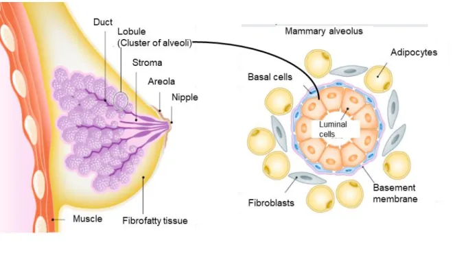
Before birth, until puberty, in the reproductive phase, during pregnancy and after menopause, the mammary gland is subjected to continuous remodeling processes that are due to the hormonal changes (Macias and Hinck, 2012). The adult female mammary gland consists of branching trees of ducts that radially extend from the nipple and terminate in the lobules that comprise clusters of alveoli. The mammary alveolus is built up of the basement membrane, containing the basal cells referred to as myoepithelial cells, and the inner layer composed of luminal cells (Figure 1). The multipotent stem cells give rise to luminal epithelial stem cells and basal stem cells that further divide into luminal and basal progenitor cells. However, the luminal progenitor cells differentiate into two types of hormone receptor-negative cells and one hormone receptor-positive cell type (Cristea and Polyak, 2018). The basal layer is embedded in breast stroma containing adipocytes, fibroblasts, and immune cells (macrophages and lymphocytes), blood and lymph vessels (Pellacani et al., 2019).
12
Figure 1: Anatomy of the human mammary gland
Each lactiferous duct in the mammary gland originates from the nipple and branches into ducts that end in the alveoli. Those mammary alveoli comprise the basal cells on the outside and an inner layer of luminal cells. The alveoli are embedded in the mammary stroma containing fibroblasts and adipocytes. The picture is adapted and modified from Pellacani 2019.
During the reproductive phase of a female, the mammary gland is susceptible to restructure the tissue due to the monthly menstrual cycle that is orchestrated by the uterus and pituitary hormones like estrogen and progesterone resulting in the continuous reorganization of the mammary gland (Ramakrishnan et al., 2002). In the case of pregnancy, the luminal epithelial layer is able to produce and secrete milk upon hormonal stimulus. Furthermore, the luminal layer is characterized by its high epithelial cell adhesion molecule (EpCAM), cytokeratin (CK) 18, and CK 8 expression. In contrast, the basal cells express EpCAM very low and could be identified by CK 14 staining. In normal mammary tissue, approximately 7 % of the epithelial cells in the mammary gland are hormone receptor-positive, whereas 87 % of the cells are luminal epithelial cells or occupied an intermediate position in the duct wall (Petersen et al., 1987). These distinct expression patterns are not only helpful to identify the different cell types in healthy mammary tissue but are also responsible for the different entities in breast malignancies.
1.1.2 Breast cancer etiology
In the mammary gland, the cells grow and divide and are strictly controlled through homeostatic regulation between proliferation and death. However, an imbalance between proliferation and cell death could lead to the development of breast cancer,
13
whereas the reasons for carcinogenesis are not fully understood. There are several risk factors that promote the formation of breast malignancies like the genetic predisposition, race or ethnicity, childlessness, non-breastfeeding, hormone receptor replacement therapy after menopause, excessive alcohol consumption, smoking, or obesity (Feng et al., 2018). Besides the so-called acquired risk factors for breast cancer, only 5-10 % of breast cancer malignancies are due to inherited reasons like BRCA1/2 mutations or TP53 mutations (Duda and Schulz-Wendtland, 2017; Feng et al., 2018).
1.1.3 Classification of breast cancer and breast cancer subentities
One of the most common types of breast cancer is the non-invasive or pre-invasive intraductal carcinoma in situ (DCIS) which develops inside the normal ducts. DCIS itself is not invasive but in situ carcinomas have a high potential to become invasive.
In contrast, invasive breast cancer invades and spread outside the normal breast lobules and ducts and grows into the surrounding tissues. The invasive breast cancer cells derive either from the epithelial cells of the mammary ducts (ductal) or from the mammary lobules (lobular). The invasive ductal carcinoma (70-80 %), as well as the invasive lobular carcinoma (10 %), represents the highest proportion of mammary malignancies and derives from early lesions (carcinoma in situ). About 90 % of breast cancer cases are invasive (Feng et al., 2018). Besides this histological classification, the classification of breast cancer is performed according to the pTNM staging. This staging considers the size of the primary tumor (T), the lymph node involvement (N), and the presence of distant metastases (M), as it represents important information for therapy and prognosis of the carcinoma (Duda and Schulz-Wendtland, 2017). The grading system which is scaled into three stages (G1-G3), provides additional information on the degree of malignancy, thereby including the formation of tubular gland structures, the nuclear atypia, and the frequency of mitosis (Elston and Ellis, 1991). The higher the grading the worse is the prognosis. Accordingly, Grade 1 proliferates slowly and is well-differentiated, Grade 2 is moderately differentiated, and Grade 3 propagates fast and is poorly differentiated (Klöppel et al., 2013). However, an additional important low prognostic but highly predictive factor is the hormone receptor status. The nuclear estrogen receptor alpha (ER) expression and the nuclear progesterone receptor expression (PR) expression is determined by immunohistochemical staining and calculated according to the Remmele Score (Remmele and Stegner, 1987). The score comprises the staining intensity and the
14
percentage of the stained nuclei which are summarized in an immune reactive score (0-12). The chief markers to subcategorize breast cancer are not only ER and PR but also the oncogene human epidermal growth factor receptor 2 (Her2). The combination of these markers allows the assignment of individual cases to specific categories, namely Luminal breast cancer ER+ (ER+/HER2–), HER2+ breast cancer (ER–/HER2+), triple-negative breast cancer (TNBC; ER–/PR–/HER2–), and Luminal B /Her2+ breast cancer (ER+/PR(+/-)/HER2+) (Bertos and Park, 2011).
1.1.4 Luminal breast cancer
Luminal breast cancer is the predominant type of breast cancer with an incidence of 70-80%. This tumor entity can further be subdivided into Luminal A (low-risk) and Luminal B (high-risk) tumors (Sørlie et al., 2001; Perou et al., 2000). According to St.
Gallen conference 2011, the proliferation capacity of these tumors is characterized by Ki67 < 14 % and Ki67 > 14 %, respectively (Goldhirsch et al., 2011; Cheang et al., 2009).This classification is a guidance value for therapy decisions. In matters of a high proliferation capacity, the decision for Luminal B breast cancer patients is in favor of chemotherapy treatment, whereas this therapy approach is disputable when the proliferation threshold is close to 14 %. Luminal A tumors with a low proliferation capacity do not necessarily benefit from cytostatic drugs and receive only endocrine therapy in most cases. It is known that the response of an anti-hormonal therapy is more efficient in Luminal A breast cancer patients compared to Luminal B breast cancer patients (Rouzier et al., 2005; Hayes et al., 2007; Goldhirsch et al., 2011).
However, the differentiation between high and low-risk Liminal B tumors remains crucial. Another step towards risk stratification in Luminal breast cancer are the gene expression tests that are able to predict a risk assessment for recurrence or the development of distant metastases and therefore help to estimate the need for chemotherapeutic intervention. Oncotype DX (21 gene assay) and EndoPredict (11 gene assay) were both prognostic for the risk of distant recurrence (Narain and Adcock, 2017). Mammaprint (Agendia) assesses the risk of recurrence through the determination of 70 genes. The Prosigna (PAM50; 55 gene assay) assay has been validated as a prognosticator in clinically low-risk, postmenopausal patients with ER+ early-stage breast cancer treated with endocrine therapy (Narain and Adcock, 2017).
The test separates the high and low-risk Luminal B patients by a risk of recurrence score. However, the prediction of variable gene expression tests is still unsatisfying as the tests are not interchangeable and reveal different results for the same patients
15
(Alvarado et al., 2015). Luminal B tumors also tend to metastasize into various organs including the bone marrow and are therefore associated with a poorer prognosis.
However, if Luminal B breast cancer patients profit from chemotherapy is still disputed (Goldhirsch et al., 2011; Lønning, 2012).
1.2 Breast cancer metastases
One of the major problems of suffering from breast cancer is still not the primary tumor but the development of distant metastases. Although approximately 6 % of the newly diagnoses patients harbor metastases, about 30 % of the women with breast cancer will develop distant metastases (O'Shaughnessy, 2005). The frequency did not change in the last decades, which is referable to the fact that the biology of metastatic processes and adequate prevention is less understood.
1.2.1 Metastatic sites in breast cancer
Breast cancer metastases are frequently found in the distant lymph nodes, the liver, the lung, the bone marrow, and the brain (Wu et al., 2017). However, there are differences between breast cancer subentities and metastatic sites. Luminal B tumors show preferentially metastases in the liver, the lung, and the distant lymph nodes. If the Luminal B tumors additionally overexpress Her2, the brain, and the bone marrow are frequent sites of metastatic colonization (Chen et al., 2018). Still, 70 % of the metastases are determined in the bone and is, therefore, the most prominent target site in breast cancer (Weilbaecher et al., 2011). This so-called organotropism was shown to be driven by the different breast cancer subentities, different gene signatures, and different signaling pathways of metastatic tumor cells, and the crosstalk with the host (immune) microenvironment, (Chen et al., 2018). This phenomenon is supported by the hypothesis of “seed and soil” that was claimed by Paget decades ago. The tumor cells (seed) can only grow in a distant organ (soil) if it is “planted” in the appropriate microenvironment (Paget, 1889). The chemical attraction is one of the key modulators to successfully colonize at distant organs. Multiple factors like cytokines, bone sialoprotein, or osteopontin expression in the microenvironment are implicated to play a major role in metastases formation (Ibrahim et al., 2000). Nevertheless, the distinct drivers for breast cancer metastasis organotropism are still not fully understood.
16
1.2.2 EMT and MET in breast cancer metastases
Metastases development is a multifactorial process that requires several factors to enable the cancer cell to spread. In each step, the cancer cell could be eliminated by the failure of adaption or due to immune cell eradication. Therefore, only a few cells that are adjusted will succeed in the colonization of distant organs (Fidler, 2003;
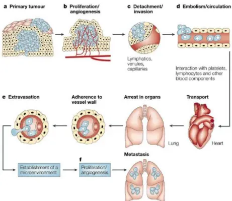
Valastyan and Weinberg, 2011). The stepwise cascade from the primary tumor to the adaptation to foreign tissue microenvironments comprises (1) the local invasion of primary tumor cells through surrounding extracellular matrix (ECM) and stromal cell layers accompanied by the intravasation of the tumor cell into the blood vessels, (2) the survival as a circulating tumor cell (CTC) in the vasculature periphery, (3) the arresting at distant organ sites, (4) the adherence to the vessel wall and the extravasation into the distant tissues, (5) the persistence in the foreign microenvironment, and (6) the proliferation to form micrometastases in the distant organ (Valastyan and Weinberg, 2011; Fidler, 2003; Bill and Christofori, 2015).
Figure 2: Schematic description of the development of distant metastases
A) The initial step requires progressive tumor growth. B) The tumor needs extensive vascularisation to allow the tumor cells to C) detach and invade the blood vessels. D) The tumor cells are then circulating in the peripheral blood where they have to survive. Next, the cells arrest in an organ by the adherence to the vessel wall. E) The extravasation is followed by the adaptation to the new microenvironment. F) The proliferation and the angiogenesis completes the metastatic process to form solid metastases.This figure is adapted and modified from Fidler 2003.
17
These multiple processes require an adaptation of the tumor cell to the physiology of a certain location. Epithelial to mesenchymal transition (EMT) is a reversible process that attains the tumor cells a mesenchymal phenotype in order to exit the primary tumor site and allows them to metastasize to distant organs (Kotiyal and Bhattacharya, 2014).
In contrast, mesenchymal to epithelial transition (MET) is required after colonization of an organ to build a new malignant tumor growth the so-called metastasis. In terms of EMT, the TGFbeta and RTK/ Ras signaling, EMT transcription factors, and pathways such as Wnt, Notch, and Hedgehog are known to contribute to that process (Bill and Christofori, 2015; Felipe Lima et al., 2016). The reduced expression of E-cadherin, as a cell-cell adhesion molecule, regulated by the transcription factors Snail and Twist seems to be one of the crucial steps that drive EMT (Huber et al., 2005). Cancer stemness has been associated with an enhanced capacity for EMT. Breast cancer stem cells exhibit cellular plasticity as they are able to reversibly transit between the mesenchymal and the epithelial state. This tumor cell plasticity and the evolvement of breast cancer stem cells (CSC) are associated with EMT that typically goes along with altered expression or activity of cytokeratin, vimentin, CD24, Claudin, ALDH, SLUG, and SNAIL and consequently with an increased capacity of self-renewal, tumor initiation, and recurrence (Al-Hajj et al., 2003; Liu and Wicha, 2010).
1.2.3 Stem cells traits and their relevance in EMT-MET and breast cancer metastases
To date, it remains still challenging to detect all tumor cells in the periphery e.g. as DTCs in the BM or circulating tumor cells (CTCs) in the peripheral blood. The DTC and CTCs are a prognostic factor for patient survival and metastatic spread (Braun et al., 2005). The established method for CTC detection in the blood is the CellSearch®
system that is able to quantify the tumor cells in seven ml blood due to the expression of EpCAM on the surface of the tumor cells. EpCAM as epithelial cell adhesion molecule is only expressed on epithelial cells and thereby this method excludes the mesenchymal and hematopoietic cells (Gires and Stoecklein, 2014). EpCAM plays a major role in embryonic development and is located at the basolateral membrane in normal epithelial tissue (Gires, 2011). Besides the expression in normal tissue EpCAM is expressed in a variety of malignancies including breast cancer. Preferentially EpCAM is found on luminal cells (Visvader and Stingl, 2014). However, its regulation is rather dynamic. While EpCAM is highly expressed in the primary tumor, the expression of EpCAM can be downregulated in the CTC (Gires and Stoecklein, 2014).
18
This fact shows that CTC capturing through EpCAM expression might not detect all tumor cells. EpCAM is highly expressed especially in Her2+ Luminal B breast cancer and associated with a rather unfavorable prognosis (Soysal et al., 2013). Additionally, EpCAM expression in the primary breast cancer tumor is associated with increased bone marrow metastases and increased stem cell capacity of the tumor cells (Hiraga et al., 2016; Huber et al., 2015). Not only EpCAM as a cell adhesion molecule but also CD44 and CD24 play a pivotal role in breast cancer stemness and metastases. CD44 is necessary for the communication and adhesion between adjacent cells and between cells and the extracellular matrix and was shown to contribute to metastasis formation (Naor et al., 2002). It can interact with a variety of effectors such as the hyaluronic acid – an abundant compound of the extracellular matrix (Toole, 2009; Louderbough and Schroeder, 2011) and is expressed by a multitude of carcinoma cells especially on cancer-initiating ones (Zöller, 2011; Wang et al., 2018b). Moreover, it plays a major role in cell adhesion, cell proliferation, migration, invasiveness, chemoresistance and metastasis initiation (Baccelli et al., 2013; Zöller, 2011; Naor et al., 2008; Williams et al., 2013). CD24 is a heavily glycosylated mucin-like glycosylphosphatidyl-inositol- linked cell surface protein and is expressed in a wide variety of human malignancies (Jaggupilli and Elkord, 2012). The high CD24 expression levels are associated with enhanced proliferation (Baumann et al., 2005), clonogenicity in vitro (Smith et al., 2006), and metastases (Friederichs et al., 2000). However, the expression of CD44 high /CD24 low was determined previously to be the tumor-initiating phenotype for breast cancer stem cells whereas the multitude of cells within the same tumor exhibit CD44
low / CD24 high (Al-Hajj et al., 2003; Mani et al., 2008). It was also shown that the CD44
high /CD24 low phenotype is frequently expressed in highly aggressive TNBC and Her2+ breast cancer (Honeth et al., 2008). CD47 is an integrin associated transmembrane protein and known for its interaction with the SIRP alpha receptor to prevent phagocytosis by macrophages or dendritic cells. The expression of CD47 as “Don’t eat me signal”, therefore, enables the cancer cell to be eradicated by immune cells (Nagahara et al., 2010). High expression of CD47 in CTCs and DTCs was associated with decreased DFS in breast cancer patients (Nagahara et al., 2010). Additionally, high CD47 and CD44 co-expression were shown previously to be a prognosticator for limited survival in Luminal breast cancer (Baccelli et al., 2014). Another important biomarker is cMET also called HGFR (hepatocyte growth factor receptor). This receptor tyrosine kinase activates, upon hepatocyte growth factor (HGF) binding,
19
diverse cellular functions that play an important role in organ development and cancer progression (Trusolino et al., 2010). HGF is one of the factors that promote invasive tumor growth, metastases formation and induction of EMT (Christofori, 2006). By means of its role in metastasis initiation (Baccelli et al., 2013) a high cMET expression has been shown to be associated with reduced survival and an aggressive phenotype in breast cancer patients (Ho-Yen et al., 2015). In addition, cMET has also been determined to inversely correlate with tumor size in breast cancer (Ho-Yen et al., 2014).
Interestingly, the HGF induced activation of cMET stimulated the CD44 signal transduction and stabilized the androgen receptor functions in prostate cancer (Ghatak et al., 2010). However, the co-expression of EpCAM+/ CD44+/ CD47+/cMET+ was demonstrated by Baccelli to be the prominent phenotype in CTCs to initiate metastases (metastases initiating cells (MIC)) in breast cancer (Baccelli et al., 2013) further showing the potential of CD47 and cMET as CSC biomarkers. Another important oncogene in breast cancer is Her2. Her2 as receptor tyrosine kinase is overexpressed in 20 - 25 % of invasive breast cancer and predicts a poor clinical outcome (Slamon et al., 1987). The overexpression of Her2 is in most cases due to the amplification of the Her2 gene. The constitutive kinase activity in Her2+ breast cancer, therefore, promotes increased proliferation and invasion of the tumor cells (Olayioye, 2001). Despite the prognostic and predictive value of Her2 in breast cancer, it is also implicated as a driver for breast cancer stemness as Her2+ breast cancer cells showed an increased mammosphere formation due to increased clonogenicity (Korkaya et al., 2008;
Korkaya and Wicha, 2009; Magnifico et al., 2009). Other researchers reported the detection of Her2+ DTCs that arose from Her2- breast cancer tumor, suggesting a small subpopulation of Her2 overexpressing tumor cells in the primary tumor that might be missed by routine diagnostic of the primary tumor (Pantel and Alix-Panabières, 2014).
The prognosis of Her2 breast cancer is poor as those tumors often tend to generate distant metastases. However, it is possible to treat Her2+ breast cancer adequately with the monoclonal antibody Trastuzumab (Herceptin) in combination with chemotherapy. This therapeutic intervention targets Her2 and therefore efficiently diminish the proliferation and increases disease-free survival (Piccart-Gebhart et al., 2005). Even though several biomarkers are known to be regulated in breast cancer and breast cancer metastases, it still remains to be elucidated which additional factor might play a pivotal role in breast cancer stemness and metastases formation.
20 1.2.4 Disseminated tumor cells (DTCs)
During the way of the tumor cells through the periphery (e.g. the blood or the lymph vessels) the cells referred to CTC, whereas after extravasation and colonization the tumor cells are termed as disseminated tumor cells (DTC). The DTCs can be detected in the mesenchymal tissue due to their epithelial origin thereby expressing cytokeratins such as CK8, CK18, and CK19 (Braun et al., 2005). The occurrence of DTCs in the bone marrow of breast cancer patients is a risk factor for the development of distant metastases (Wiedswang et al., 2003). Moreover, the DTC persistence correlates with diminished disease-free survival (DFS) and reduced overall survival (OS) (Janni et al., 2011). However, the time at which dissemination takes place is still disputed. There are two well-known theories when dissemination occurs. The first one is that the cancer cells were shown to disseminate at late stages (Koscielny et al., 1984), and the second one is the well-accepted theory of parallel progression of tumor cell dissemination and tumor growth (Klein, 2009). Moreover, tumor cell dissemination can occur even in the absence of a detectable tumor as so-called cancer of unknown primary(van de Wouw et al., 2002). Surprisingly, epidemiologic studies revealed that metastases could be initiated already five to seven years before the primary tumor is diagnosed (Engel et al., 2003). Supporting the early dissemination hypothesis it was shown that DTCs from breast cancer patients harbor fewer aberrations than the primary tumor cells at an advanced state, indicating a slow progression of DTCs (Schmidt-Kittler et al., 2003).
However, there are some studies that demonstrated a genomic congruency of DTCs and primary tumor cells in breast cancer (Mathiesen et al., 2012; Stoecklein et al., 2008). Interestingly, the recurrence and metastases in breast cancer take a long period of time after primary tumor detection indicating that DTCs could somehow transform/switch in a state of dormancy. To date, there are two types of dormancy known. One state of dormancy is defined by the potential of the cancer cell to stay in an arrested cell cycle phase (G0/G1) that is reversible (Hayat, 2013) and the other state is called tumor mass dormancy where the equilibrium of cell death and self- renewal of the cancer cell (Kareva, 2016). However, the fate of the disseminated cell is supposed to be triggered by several intrinsic and extrinsic factors that contribute to dormancy or active proliferation (Osisami and Keller, 2013).
1.3 The role of genetic aberrations in breast cancer
Despite Her2, there are several other aberrations that are frequently altered in breast cancer. Inherited BRCA1 and BRCA2 mutations or mutations in the TP53 gene are
21
detected frequently. The latter is preferentially found in very aggressive tumor types such as TNBC and Her2+ breast cancer (Abubakar et al., 2019) and is associated with poor clinical outcome. P53 as a product of the TP53 tumor suppressor gene is not expressed in healthy cells or rather to a low degree. If stressors like DNA damages, hypoxia, or activation of oncogenes occur in healthy cells, p53 turns on transcriptional target genes to send the cell in cell cycle arrest in order to prevent DNA lesions.
However, if the DNA damages are irreparable the p53 upregulation promotes apoptosis (Shi and Gu, 2012). P53 also induces the transcription of mouse double minute protein 2 (MDM2), an E3-ligase, which serves as a negative feedback regulator.
It ubiquitinylates p53 for its degradation on the proteasome. MDM2 deregulations frequently occur in Luminal B breast cancer (31%) and in Her2+ breast cancer (30%) (Network, 2012) and were shown to promote invasiveness, EMT, and metastases in breast cancer (Haupt et al., 2017). In contrast, TP53 mutations are predominantly detected in TNBC and Her2+ breast cancer.
1.4 TILs and immune checkpoints in breast cancer
The evaluation of tumor-infiltrating lymphocytes (TIL) is gaining more and more clinical relevance in breast cancer as they were shown to have a potential prognostic and predictive value (Salgado et al., 2015). The most TILs can be found in in TNBC with an average of 20 % infiltration (range: 4 % - 37 %) and in Her2+ breast cancer with an average of 16 % (range: 11 % - 24 %). In both subtypes/entities, increased infiltration is associated with a survival benefit (Stanton et al., 2016). Furthermore, all breast cancer subtypes with TILs predicted response to neoadjuvant chemotherapeutic intervention. The same study described increased immune cell infiltration as an adverse prognostic factor for the outcome in Luminal-HER2- breast cancer patients, (Denkert et al., 2018). Even though the TNBC and Her2+ subtypes are immunogenic, Luminal breast cancer is rather escaping immunosurveillance and is only low infiltrated with an average of 6 % (range: 3 % - 12 %) (Stanton et al., 2016). Nevertheless, Luminal B Her2+ breast cancer is infiltrated with TILs to a greater extent (9 %) compared to Luminal A/B Her2- breast cancer (1%) (Pruneri et al., 2017). The CD8 infiltration inversely correlated with the ER-alpha and PR expression whereas the presence of CD8+ immune cells predicted a favorable outcome for the patient (Mahmoud et al., 2011). The infiltration of CD4+ cells was shown to be increased preferentially in aggressive tumor types such as TNBC and Her2+ breast cancer (Meng et al., 2018). However, CD8+ and CD4+ immune cells and various other immune cells
22
like T regs or B cells play a prognostic role in different stages of the mamma carcinoma (early breast cancer, neoadjuvant or adjuvant situation, metastasized) (DeNardo and Coussens, 2007; Emens, 2012). Not only the immune cell infiltration but also the tumor- immune cell interactions play a pivotal role. This interaction involves tumor cell elimination, tumor-immune cell equilibrium, but also the escape of tumor cells from the immunological defense. This could be achieved, besides other mechanisms, by the expression of inhibitory immune checkpoint molecules (Mittal et al., 2014). Therapeutic strategies designed to stimulate the patient’s inherent immunological tumor defense e.g., by targeting immune checkpoints are considered to enhance conventional treatment regimens. A prominent target that contributes to tumor cell evasion is the immune checkpoint programmed death-ligand 1 (PD-L1). If expressed by tumor cells (Blank et al., 2005), the interaction of PD-L1 with its counterpart, the programmed death receptor 1 (PD-1) leads to T-cell anergy or apoptosis (Keir et al., 2008). Triple- negative breast cancer (TNBC) specimens showed the highest level of PD-L1 expression, followed by Her2 overexpressing subtypes, and lastly the Luminal (Luminal A and Luminal B) entities (Ali et al., 2015; Ghebeh et al., 2006). However, PD-L1 expression on tumor cells has repeatedly been associated with a worse outcome (Wang et al., 2016). Still, the relevance of PD-L1 and PD-1 expression in breast cancer is discussed controversially (Stovgaard et al., 2019).
1.5 PDX models and humanized PDX models in breast cancer research
Mice are still the most common animals for usage as a model organism because of high genetic homology, easy genetic manipulation, a fully sequenced genome, low cost in breeding and quick reproduction cycles (Perlman, 2016). The understanding of the genomic landscape, the metastatic spread and the biology of breast cancer still remains challenging. The development of patient-derived xenografts (PDX) opened up new possibilities, thus, the breast cancer tumor engrafted in mice properly, reflected the heterogeneity of the primary tumor, the tumor behavior, and also the metastatic properties. Moreover, it was possible to detect new breast cancer targets and the PDX model was susceptible to predict treatment response which was a big step towards personalized medicine (Whittle et al., 2015; Landis et al., 2013). Different ways of primary tumor transplantations were used for the generation of breast cancer PDX models. In most cases, the tumors were transplanted orthotopic, hence, into the mammary fat pad of the mice (Al-Hajj et al., 2003; DeRose et al., 2011; Zhang and Lewis, 2013; Kabos et al., 2012). Other methods like subcutaneous, under the
23
subrenal capsule, intraductal or interscapular transplantations were also successful (Marangoni et al., 2009; Cottu et al., 2012; Reyal et al., 2012; Fiche et al., 2019; Eirew et al., 2015; Bergamaschi et al., 2009). It was shown that successful engraftment is also determined by breast cancer subentity and their molecular traits. TNBC and Her2+ breast cancer and metastatic breast cancer showed higher engraftment rates compared with the Luminal hormone receptor-positive entities (Landis et al., 2013;
Baccelli et al., 2013). However, several improvements contributed to increased PDX take rates like the supplementation of estradiol as a subcutaneous transplanted pellet in hormone receptor-positive tumors (Al-Hajj et al., 2003; Marangoni et al., 2007), the use of Matrigel (Fleming et al., 2010; Kabos et al., 2012), the supplementations of human mesenchymal stem cell, (DeRose et al., 2011) or the use of highly immunosuppressed mice (Oakes et al., 2012; Zhang et al., 2013; Kabos et al., 2012).
Highly immunosuppressed mice like NOD-SCID IL2Rγnull (NSG) mice have no T, B, or natural killer cells and a reduced myeloid cell function (Shultz et al., 2005). The enhanced life span of these mice of over 1.5 years is an advantage due to the prolonged engraftment duration of a primary breast cancer tumor of approximately six months up to one year. Additionally, the NSG mouse strain exhibited the highest engraftment rates (Carreno et al., 2009). A list of the most important breast cancer PDX studies, different breast cancer entities, mouse strains, transplantation sites and the overall success rate of engraftment is provided in Table 1. Various studies with breast cancer PDX models were shown to display the patients tumor heterogeneity, the metastatic behavior of the tumor, the patients disease outcome (DeRose et al., 2011; Valdez et al., 2011), and the concordance of drug response in patients (Marangoni et al., 2007; Cottu et al., 2012; Cottu et al., 2014). The EurOPDX consortium was founded to enable translational knowledge in oncology by providing access to a multitude of PDX models. Hence, the EurOPDX harnesses the clinically relevant models in cancer, avoids the duplication of research efforts and improves drug development processes through interchanging data with other researchers.
Nevertheless, the influence of the human immune system cannot be elucidated in these PDX models. The severe combined immune deficiency (SCID) mutation of the NSG mice additionally provides the opportunity to engraft human immune cells. This can be performed by the isolation of hematopoietic stem cells (CD34+) from the human peripheral blood, the cord blood, the fetal liver cells from an abort, or the bone marrow, and can then be injected intravenously, intracardially, intraperitoneally, intrafemorally,
24
or intrahepatically using neonatal or adult immunodeficient mice (Wege, 2018). The humanized PDX model is another option of the humanized mouse model whereby the transplantation of primary breast cancer cells follows 12 weeks after humanization.
These mouse models have been used in preclinical checkpoint inhibitor studies of TNBC humanized PDX mice that were treated with anti-PD-1 therapy. This resulted in a reduction of the tumor size whereby this reduction could be traced back to the inhibition of PD-1/CD8+ cells (Wang et al., 2018a). However, to date humanized PDX models in breast cancer research are used only rarely, which might be due to the fact that several factors like the availability of newborn mice, the immune system engraftment, primary tissue availability and toleration, and long latencies until tumor outgrowth that has to be combined in one experiment. Nevertheless, humanized breast cancer PDX models are a step towards a detailed understanding of breast cancer biology, metastasizing, and dissemination, under the influence of a human immune system. In addition, these mice can be used for preclinical studies in the area of immunomodulation.
Table 1: Overview of generated breast cancer PDX models
Study Breast cancer
subtype
Mouse Strain Transplantation site tissue/ single cells supplementation
Success rate overall (Visonneau et al.,
1998)
N.A. SCID s.c.
tumor fragments
50 %
(Beckhove et al., 2003)
N.A. NOD/SCID im
tumor fragment matrigel
90 %
(Ma et al., 2012) TNBC NOD/SCID mfp
single cells fibroblasts
N.A.
(Liu et al., 2010; Al- Hajj et al., 2003)
4 TNBC 2 Her2+ 2 ER+
NOD/SCID thoracic mfp tissue fragments estradiol
N.A.
(Marangoni et al., 2007; Cottu et al., 2012; Reyal et al., 2012)
15 TNBC,2 Her2+, 1 ER+, (Marangoni et al., 2007), 22 TNBC, 8 ER+ (Cottu et al., 2012)
Swiss nude interscapular fat pad tumor fragments estradiol
12.5 %
(Bergamaschi et al., 2009)
1 TNBC, 1 ER+ SCID s.c. in the back pocket tumor fragments
20 %
25
matrigel (DeRose et al., 2011) 5 TNBC, 2 Her2+, 2
ER+,
NOD/SCID cleared mfp
tissue fragments, metastastic single cells estradiol
27 %
(Zhang et al., 2013) 21 TNBC, 3 Her2+, 3 ER+
SCID beige, NSG
cleared mfp tumor fragments
+/- fibroblasts, estradiol 30 %
(Kabos et al., 2012) 1 TNBC, 5 ER+ NOD/SCID, NSG
abdominal mfp tumor fragments matrigel
42 %
(Petrillo et al., 2012) 4 TNBC, 1 Her2+, 0 ER+
NOD/SCID s.c. dorsal flank tumor fragments estradiol
25 %
(Fleming et al., 2010) 2 Her2+, 2 ER+ NOD/SCID abdominal mfp pleural effusion cells estradiol, matrigel
N.A.
(Vaillant et al., 2013;
Oakes et al., 2012)
17 TNBC, 13 ER+, 2 ER-PR+, 5 Her2+,
NSG inguinal mfp
tumor fragments estradiol
23 %
(Eirew et al., 2015) 6 TNBC, 5 ER+, 4 Her2+
NSG, NRG mfp, s.c., sr N.A.
(Fiche et al., 2019) 21 ER+ NSG intraductal mfp single cells
N.A.
TNBC Triple-negative breast cancer, ER estrogen receptor, Her2 human epidermal growth factor receptor, NOD/SCID non-obese diabetic / severe combined immune deficiency, NSG NOD-SCID IL2Rγnull, NRG NOD.Cg- Rag1tm1Mom IL2rgtm1Wjl, mfp mammary fat pad, s.c. subcutaneous, id intraductal, sr subrenal capsule, im intramuscular, N.A: not available
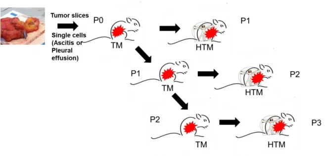
26 1.6 Aim of the thesis
The aim of this thesis, as part of the Deutsche Krebshilfe founded Luminal B consortium, was to determine markers for the heterogeneous Luminal B group to differentiate them into high and low-risk patients. Hence, the therapy decision could be facilitated if a Luminal B breast cancer patient might benefit from chemotherapeutic intervention or not. This goal should be realized by the generation and the analysis of patient-derived Luminal B xenografts in the so-called tumor mice (TM) or humanized tumor mice (HTM). These mice were generated by orthotopic transplantation and the subsequent expansion of primary tumor material from Luminal B patients in immunodeficient NSG mice. Finally, the expanded tumor material from these mice was further transplanted into mice which were neonatally reconstituted with human hematopoietic stem cells (HSC) derived from human cord blood. These mice developed a human immune system together with human tumor growth and allowed studies under human-like conditions. The main questions which should be addressed are:
(1.) the phenotypic differences of human high- and low-risk Luminal B
(2.) the evaluation of the capacity of Luminal B breast cancer to disseminate and to form metastases in TM and HTM (i.e., in the presence of a human immune system),
(3.) the identification of geno- and phenotypic patterns in primary Luminal B tumors that are associated with tumor outgrowth and cell dissemination (metastasis formation) and EMT / basal cell-like traits,
(4.) the analysis of phenotypical changes between the primary tumor and the corresponding metastases in the TM and HTM model, and
(5.) the assessment of human immune cell activity and invasion in correlation to tumor outgrowth, dissemination, and metastases.
Overall, the main goal of this thesis was to identify biomarkers that are associated with tumor outgrowth, cell dissemination and/or metastases formation using humanized PDX mice
27 2. Material
2.1 Consumables
12 % Mini Criterion TGX Stain-free gels BioRad Laboratories, Munich, Germany
15 ml tube Greiner Bio-One Bioscience, Frickenhausen,
Germany
17ß-Estradiol pellets 0,18mg/90days Innovative Research of America, Florida, USA
40 µm cell strainer Corning, NY, USA
50 ml tube Greiner Bio-One Bioscience, Frickenhausen,
Germany
Adhesion slides Carl Roth, Karlsruhe, Germany
BD Discardit II 2 ml, 5 ml, 10 ml, 20 ml (syringes)
BD Biosciences, Heidelberg, Germany BD Microlance 3 20G, 22G, 27G
(cannulas)
BD Biosciences, Heidelberg, Germany BD SafetyGlide Insulin syringe BD Biosciences, Heidelberg, Germany
Cell Scraper Greiner, Solingen, Germany
Cord blood collection bag Macopharma, Langen, Germany Culture-Insert 2 Well in µ-Dish 35 mm Ibidi, Gräfelfing, Germany
Cyro tubes Greiner, Solingen, Germany
MACS separation LD columns Miltenyi Biotech, Bergisch Gladbach,Germany MACS separation MS columns Miltenyi Biotech, Bergisch Gladbach,Germany Medicon Einsatze unsteril BD Biosciences, Heidelberg, Germany
Medimachine Medicon sterile BD Biosciences, Heidelberg, Germany Petri dish sterile Sarstedt AG & Co., Nümbrecht, Germany Round Bottom Polystyrene Test Tube
(5ml)
Falcon, Heidelberg, Germany
SuperFrost Plus Slides Menzel GmbH, Braunschweig, Germany
T25 tissue flask Greiner Bio-One, Frickenhausen, Germany
Trimming blades pfm medical AG, Cologne, Germany
Vicryl surgical sutures Johnson & Johnson, New Brunswick, New Jersey, USA
2.2 Buffers and Solutions
AB serum, human BioRad Laboratories, Munich, Germany
Accutase 100 ml Sigma-Aldrich Chemie GmbH, Deisenhofen,
Germany
Amphotericin B solution Sigma-Aldrich Chemie GmbH, Deisenhofen, Germany
B27 supplement 50x minus Vitamin A Life Technologies, Carlsbad, CA, USA
BSA 10x 10ml BioRad Laboratories, Munich, Germany
Cell Lysis Buffer Cell Signaling Technology, Inc., Beverly, MA, USA
Cholera Toxin 5ml Sigma-Aldrich Chemie GmbH, Deisenhofen,
Germany