Article
Comparative Genomics of Marine Bacteria from a Historically Defined Plastic Biodegradation Consortium with the Capacity to Biodegrade Polyhydroxyalkanoates
Fons A. de Vogel1,2 , Cathleen Schlundt3,†, Robert E. Stote4, Jo Ann Ratto4and Linda A. Amaral-Zettler1,3,5,*
Citation: de Vogel, F.A.; Schlundt, C.;
Stote, R.E.; Ratto, J.A.; Amaral-Zettler, L.A. Comparative Genomics of Marine Bacteria from a Historically Defined Plastic Biodegradation Consortium with the Capacity to Biodegrade Polyhydroxyalkanoates.
Microorganisms2021,9, 186.
https://doi.org/10.3390/
microorganisms9010186
Received: 30 November 2020 Accepted: 7 January 2021 Published: 16 January 2021
Publisher’s Note:MDPI stays neutral with regard to jurisdictional claims in published maps and institutional affil- iations.
Copyright: © 2021 by the authors.
Licensee MDPI, Basel, Switzerland.
This article is an open access article distributed under the terms and conditions of the Creative Commons Attribution (CC BY) license (https://
creativecommons.org/licenses/by/
4.0/).
1 Department of Marine Microbiology and Biogeochemistry, NIOZ Royal Netherlands Institute for Sea Research, P.O. Box 59, 1790 AB Den Burg, The Netherlands; fons.de.vogel@nioz.nl
2 Faculty of Geosciences, Department of Earth Sciences, Utrecht University, P.O. Box 80.115, 3508 TC Utrecht, The Netherlands
3 Josephine Bay Paul Center for Comparative Molecular Biology and Evolution, Marine Biological Laboratory, Woods Hole, MA 02543, USA; cschlundt@geomar.de
4 U.S. Army Combat Capabilities Development Command Soldier Center, 10 General Greene Avenue, Natick, MA 01760, USA; robert.e.stote.civ@mail.mil (R.E.S.); joann.rattoross.civ@mail.mil (J.A.R.)
5 Department of Freshwater and Marine Ecology, Institute for Biodiversity and Ecosystem Dynamics, University of Amsterdam, P.O. Box 94240, 1090 GE Amsterdam, The Netherlands
* Correspondence: linda.amaral-zettler@nioz.nl; Tel.: +31-(0)222-369-482
† Current address: GEOMAR, Helmholtz Center for Ocean Research, Kiel, Düsternbrooker Weg 20, 24105 Kiel, Germany.
Abstract: Biodegradable and compostable plastics are getting more attention as the environmen- tal impacts of fossil-fuel-based plastics are revealed. Microbes can consume these plastics and biodegrade them within weeks to months under the proper conditions. The biobased polyhydrox- yalkanoate (PHA) polymer family is an attractive alternative due to its physicochemical properties and biodegradability in soil, aquatic, and composting environments. Standard test methods are available for biodegradation that employ either natural inocula or defined communities, the latter being preferred for standardization and comparability. The original marine biodegradation standard test method ASTM D6691 employed such a defined consortium for testing PHA biodegradation.
However, the taxonomic composition and metabolic potential of this consortium have never been confirmed using DNA sequencing technologies. To this end, we revived available members of this consortium and determined their phylogenetic placement, genomic sequence content, and metabolic potential. The revived members belonged to theBacillaceae,Rhodobacteraceae, andVibrionaceaefamilies.
Using a comparative genomics approach, we found all the necessary enzymes for both PHA pro- duction and utilization in most of the members. In a clearing-zone assay, three isolates also showed extracellular depolymerase activity. However, we did not find classical PHA depolymerases, but identified two potentially new extracellular depolymerases that resemble triacylglycerol lipases.
Keywords:biodegradation standard test methods; plastic biodegradation; polyhydroxyalkanoate (PHA) cycle; PHA depolymerases; comparative genomics; plastisphere
1. Introduction
Each year, an estimated 5 to 13 million metric tons of plastic waste flows into the ocean from land, a figure that is expected to only increase in the future [1]. Due to its durability, plastic waste is accumulating and becoming more visible, increasing its ecological and economic impacts [2]. Governments, industry, academia, and consumers are therefore looking for plastic alternatives and ways of reducing plastic use and waste. This has already prompted the International Convention for the Prevention of Pollution from Ships (MARPOL) [3], a plan of action for preventing waste and marine litter by the United Nations in their Sustainable Development Goals [4], and by the European Union (EU) in setting up
Microorganisms2021,9, 186. https://doi.org/10.3390/microorganisms9010186 https://www.mdpi.com/journal/microorganisms
Microorganisms2021,9, 186 2 of 26
a strategy on plastic waste reduction [5]. The EU is currently taking a leading role in bans on oxo-(bio)degradable plastics (petroleum-based plastics designed to fragment faster) and single-use plastics for which alternatives exist (i.e., cotton bud sticks, tableware, and expanded polystyrene packaging material). Next to passing legislation to reduce plastic use, the EU is also implementing extended producer responsibility for litter clean-up (i.e., tobacco filters and fishing gear), and improving waste management and recycling [6,7].
Replacing certain conventional plastics with biodegradable alternatives offers an op- portunity to reduce plastic waste accumulation, a concept put forth already decades ago [8]
and now seeing renewed interest. Although durability is still a desirable property for some plastics, single-use plastics could be effective targets. While all plastics are fragmented due to weathering by ultraviolet radiation and mechanical action [9], microbes can metaboli- cally utilize biodegradable and compostable plastics and fully degrade them within weeks to months instead of multiple years to even centuries [10]. The organic building blocks of the plastic are used to gain energy and to form new cellular biomass under the forma- tion of carbon dioxide and water in aerobic conditions and also to methane in anaerobic conditions [11]. Biodegradable plastics can, however, be made from both renewable or petroleum-based resources and these plastics are not biodegraded in all environments.
Specific conditions are often needed for biodegradation, for instance, industrial composting conditions, where pH, moisture, and the microbial community are controlled, and the temperature reaches over 50◦C for prolonged periods of time. In contrast to industrial composting, natural environments vary widely in microbial concentrations and community composition, temperature, oxygen, sunlight, humidity, nutrient limitations, and in the case of aquatic environments, also hydrostatic pressure [12]. Marine environments further sub- ject plastic debris to both horizontal and vertical transport where they encounter changing conditions and not just a static environment as a final destination [13]. Biodegradability is always connected to a specific environment and this makes it extremely challenging to design, control, and ensure significant biodegradation [11,14].
In order to estimate biodegradation under natural conditions, standard test methods have been developed which provide a way to compare the fates of different forms of plastic materials within a reasonable cost and timeframe [15]. These tests are available from ASTM International, CEN (European Committee for Standardization) and ISO (International Organization for Standardization) for biodegradability of plastics in composting environ- ments (e.g., ASTM D6400 and D6868, EN13432, EN 14995, ISO 17088, and ISO 18606), soil (e.g., ASTM D5988, EN 17033, and ISO 17556) and marine environments (e.g., ASTM D6691 and D7991). The standards require an amount of carbon content being converted to CO2within a specific timeframe, as measured by respirometry. This is >90% within 180 days in an industrial composting facility, >90% after 2 years in soil, and at least 60%
within 180 days at 30 ◦C for marine environments. The tests employ either a natural inoculum or a defined microbial community. A natural inoculum, however, might not be representative of environmental biodegradation, especially if there are no specifications for the natural inoculum source [16]. For optimal standardization, comparability, and repro- ducibility, a defined microbial community is preferred. However, microbial representatives derived from relevant natural environments are then required. It is a challenge to define a representative consortium, since it is still largely unknown if there is a group of core members colonizing plastics in nature. The plastic-associated microbes seem not only to be dependent on the environment but also geographic location, time, and substrate [17].
A relevant defined consortium should probably contain representatives of all domains of life, including eukaryotes like fungi, which are known colonizers of plastics, in addition to bacteria.
A major pathway in (bio)degradation of polymers, before microbial utilization, is hydrolysis, which leads to chain scission and molecular weight decrease [18]. In natural environments, hydrolysis can occur both abiotically and biotically, the latter via secreted enzymes of microorganisms catalyzing this reaction. Hydrolysable polymers with sus- ceptible chemical bonds include polyanhydrides, polyamides, polyethers, polyesters, and
polysaccharides [18], with the best-known examples of biodegradable plastics falling into the latter two categories. Examples of these plastics are: PBAT (polybutylene adipate terephthalate), PBS (polybutylene succinate), PCL (polycaprolactone), PHAs (polyhydrox- yalkanoates), PLA (polylactic acid) and starch and cellulose based (co-)polymers. Among the best suitable alternatives for conventional plastics are the PHA-derived plastics, since they show biodegradability in both soil and aquatic environments, and in composting conditions, recently reviewed in [19–21]. This in contrast to PLA and PBS that seem to biodegrade poorly in seawater [22,23].
PHAs are not only biodegradable, but also biobased polyesters, that have physico- chemical properties comparable to conventional plastics [24,25]. With over 150 described monomers, they are classified according to the number of carbons in their monomers, the position of the hydroxyl group, and the presence of functional groups in their side chains (e.g., phenoxy, phenyl and acetoxy groups) [26]. PHAs that consist of monomers of 3–5 carbon atoms are referred to as short-chain length PHAs or PHAscl, while those comprised of 6–15 carbon atoms are called medium chain length PHAs or PHAmcl. Applications of PHAs are found in packaging and agriculture and their biodegradation capability also makes them interesting for medical and therapeutic applications [27–29]. Currently, their production cost is relatively high and tuning of the monomeric composition and the molec- ular weight is often required to get suitable thermal and mechanical properties, limiting a wider application of the polymer [30]. However, the production of this plastic is predicted to reach over 900,000 tons/year in 2020 [31].
Many bacteria accumulate PHAs as storage compounds of both carbon and energy in response to carbon excess and/or nitrogen or phosphate stress [32]. With the first PHAs already being described in 1926 inBacillus megaterium[33], it is now known that both Gram-positive and Gram-negative bacteria have PHA production capabilities, including members of the generaAeromonas,Alcaligenes,Bacillus,Cupriavidus, andPseudomonas[26].
These various microorganisms can be and are currently utilized and optimized to produce PHAs for the biobased plastics industry [34]. PHAs differ in their biophysiological state, based on whether they occur inside or outside the cell. Intracellular PHA is referred to as native PHA (nPHA) and these granules are amorphous, consisting of a polymer core with a surface layer of structural and functional proteins [35]. Extracellular PHA granules, which are released after cell death and cell lysis, consist of a denatured form of PHA (dPHA), which is semi-crystalline in form and lacks this surface layer. These dPHAs can be scavenged by microorganisms from the environment for utilization [36].
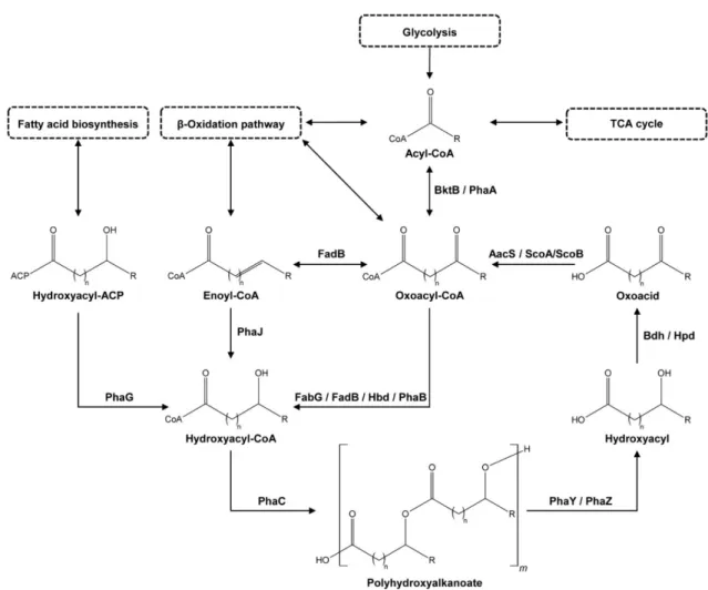
There are numerous metabolic pathways known for PHA biosynthesis, most-recently reviewed in Choi et al. [26], which result in various PHA (co-)polymers. A simplified overview of the PHA biosynthetic and degradation pathways, the PHA cycle, is depicted in Figure1. The cycle starts with an acyl-CoA (coenzyme A) molecule. This acyl-CoA can be generated from unrelated carbon sources via natural biosynthetic pathways. It is, for instance, a product of glycolysis and an intermediate in the TCA (tricarboxylic acid) cycle and theβ-oxidation pathway. PhaA (Acetyl-CoA acetyltransferase) andβ-ketothiolases like BktB (EC 2.3.1.9/ 2.3.1.16) can convert the acyl-CoA to an oxoacyl-CoA. PhaB (Acetoacetyl- CoA reductase, EC 1.1.1.36) or Hbd (3-hydroxybutyryl-CoA dehydrogenase, EC 1.1.1.157) then hydrolyze the oxoacyl-CoA to a hydroxyacyl-CoA. This step can also be performed by multifunctional enzymes like FabG, a 3-ketoacyl-[acyl-carrier-protein] (ACP) reductase (EC 1.1.1.100) that can also catalyze 3-ketoacyl-CoA to 3-hydroxyacyl-CoA, or FadB, which can act as a hydroxyacyl-CoA dehydrogenase (EC 1.1.1.35) and an enoyl-CoA hydratase (EC 4.2.1.17). The hydroxyacyl-CoA can also be produced from other compounds like enoyl- CoA, also present in theβ-oxidation pathway, by PhaJ ((R)-specific enoyl-CoA hydratase, EC 4.2.1.119) or from a hydroxyacyl-[acyl carrier protein] complex, a compound made in fatty acid biosynthesis, by PhaG (hydroxyacyl-CoA-[acyl-carrier-protein] transferase, EC 2.4.1.-). PhaC (PHA synthase, also called PHA polymerase, EC 2.3.1.-), polymerizes the hydroxyacyl-CoA into PHA, the last step in the PHA biosynthesis [26].
Microorganisms2021,9, 186 4 of 26
Microorganisms 2021, 9, x FOR PEER REVIEW 4 of 27
specific enoyl-CoA hydratase, EC 4.2.1.119) or from a hydroxyacyl-[acyl carrier protein]
complex, a compound made in fatty acid biosynthesis, by PhaG (hydroxyacyl-CoA-[acyl- carrier-protein] transferase, EC 2.4.1.-). PhaC (PHA synthase, also called PHA polymer- ase, EC 2.3.1.-), polymerizes the hydroxyacyl-CoA into PHA, the last step in the PHA bi- osynthesis [26].
Figure 1. General overview of the polyhydroxyalkanoate (PHA) cycle with examples of enzymes catalyzing the reactions.
Enzymes shown are: AacS—acetoacetyl-CoA synthetase, Bdh—3-hydroxybutyrate dehydrogenase, BktB—β-ketothiolase, FabG—3-oxoacyl-[acyl-carrier-protein] reductase, FadB—multifunctional enoyl-CoA hydratase and hydroxyacyl-CoA de- hydrogenase, Hbd—3-hydroxybutyryl-CoA dehydrogenase, Hpd—3-hydroxypropionate dehydrogenase, PhaA—acetyl- CoA acetyltransferase, PhaB—acetoacetyl-CoA reductase, PhaC—PHA synthase, PhaG—hydroxyacyl-CoA-[acyl-carrier- protein] transferase, PhaJ—(R)-specific enoyl-CoA hydratase, PhaY—PHA oligomer hydrolase, PhaZ—PHA depolymer- ase, and ScoA/ScoB—3-oxoacid CoA-transferase subunit A and B, with CoA = coenzyme A and ACP = acyl carrier protein.
In the PHA degradation pathway (see Figure 1), PHA is first depolymerized to mon- omers by PHA depolymerases (PhaZ, EC 3.1.1.75 and EC 3.1.1.76) and PHA oligomer hy- drolases (PhaY, EC 3.1.1.22) to hydroxyacyls [37–39]. PhaZs are part of the alpha/beta- hydrolase family, together with enzymes like cutinases, esterases, and lipases [40], and belong to the group of carboxylic ester hydrolases (EC 3.1.1.-) [41]. However, unlike these other enzymes, they have a high diversity in amino acid sequence composition [42]. Both intracellular and extracellular PhaZs exist, with the latter type being required for biodeg- radation of commercial PHAs and allowing microorganisms to scavenge dPHAs [42].
Similar to PhaZ, PhaY proteins can also be intracellular and/or extracellular, however, they show a higher affinity towards PHA oligomers than polymers [43–46]. After depol- ymerization, dehydrogenases like Bdh (3-hydroxybutyrate dehydrogenase, EC 1.1.1.30) [47,48] or Hpd (3-hydroxypropionate dehydrogenase, EC 1.1.1.59) oxidize the hydroxy- acyl [49,50]. Coenzyme A synthetases, like AacS (Acetoacetyl-CoA synthetase, EC Figure 1.General overview of the polyhydroxyalkanoate (PHA) cycle with examples of enzymes catalyzing the reactions.
Enzymes shown are: AacS—acetoacetyl-CoA synthetase, Bdh—3-hydroxybutyrate dehydrogenase, BktB—β-ketothiolase, FabG—3-oxoacyl-[acyl-carrier-protein] reductase, FadB—multifunctional enoyl-CoA hydratase and hydroxyacyl-CoA dehydrogenase, Hbd—3-hydroxybutyryl-CoA dehydrogenase, Hpd—3-hydroxypropionate dehydrogenase, PhaA—acetyl- CoA acetyltransferase, PhaB—acetoacetyl-CoA reductase, PhaC—PHA synthase, PhaG—hydroxyacyl-CoA-[acyl-carrier- protein] transferase, PhaJ—(R)-specific enoyl-CoA hydratase, PhaY—PHA oligomer hydrolase, PhaZ—PHA depolymerase, and ScoA/ScoB—3-oxoacid CoA-transferase subunit A and B, with CoA = coenzyme A and ACP = acyl carrier protein.
In the PHA degradation pathway (see Figure 1), PHA is first depolymerized to monomers by PHA depolymerases (PhaZ, EC 3.1.1.75 and EC 3.1.1.76) and PHA oligomer hydrolases (PhaY, EC 3.1.1.22) to hydroxyacyls [37–39]. PhaZs are part of the alpha/beta- hydrolase family, together with enzymes like cutinases, esterases, and lipases [40], and belong to the group of carboxylic ester hydrolases (EC 3.1.1.-) [41]. However, unlike these other enzymes, they have a high diversity in amino acid sequence composition [42]. Both intracellular and extracellular PhaZs exist, with the latter type being required for biodegra- dation of commercial PHAs and allowing microorganisms to scavenge dPHAs [42]. Similar to PhaZ, PhaY proteins can also be intracellular and/or extracellular, however, they show a higher affinity towards PHA oligomers than polymers [43–46]. After depolymerization, dehydrogenases like Bdh (3-hydroxybutyrate dehydrogenase, EC 1.1.1.30) [47,48] or Hpd (3-hydroxypropionate dehydrogenase, EC 1.1.1.59) oxidize the hydroxyacyl [49,50]. Coen- zyme A synthetases, like AacS (Acetoacetyl-CoA synthetase, EC 6.2.1.16), or 3-oxoacid CoA-transferases like ScoA/ScoB (EC 2.8.3.5) then close the PHA cycle, converting the oxoacid intermediate back to an oxoacyl-CoA molecule [51,52].
In this study, we revisited the historically defined bacterial consortium from the orig- inal ASTM marine biodegradation standard test method ASTM D6691: “Standard Test Method for Determining Aerobic Biodegradation of Plastic Materials in the Marine Envi-
ronment by a Defined Microbial Consortium” [53]. This consortium was developed in the 1990s as a starting point for standardizing inocula used in plastic biodegradation testing.
The members of this consortium were selected based on their individual biodegradation ca- pacity of one or multiple biodegradable plastics. This capacity was determined by clearing- zone assays and/or the ability to grow in medium with a specific polymer as the sole carbon source. The tested plastics included different formulations of the PHA co-polymer PHBV (poly(3-hydroxybutyrate-co-3-hydroxyvalerate)), PCL, PVOH (polyvinyl alcohol), cellulose, starch and other polysaccharides [54,55]. This resulted in fourteen suitable iso- lates for the biodegradation consortium. Marine biodegradation experiments combining eleven members of the consortium, showed positive results for the biodegradation of PHAs, and protein and polysaccharide-based polymers [56,57]. A combination of thirteen of the isolates was used in testing the biodegradation of PHB (poly(hydroxybutyrate)) and PHBV [15,58], and other PHAs of various composition, form, and, crystallinity. In order to identify the members of the consortium, the Biolog Substrate Metabolism System (Biolog, Inc., Hayward, CA, USA), in combination with biochemical methods (i.e., Gram stains) and microscopy, was used originally [54,55]. DNA-sequencing based approaches have hitherto never confirmed the taxonomic composition and metabolic potential of this consortium. This experimentally-confirmed biodegrading consortium offers much-needed insights into the process of biodegradation from a community perspective since our knowl- edge of plastic biodegradation and the enzymes responsible for doing so is underexplored.
We hypothesized that the genomes of several, if not all members, contain genes of the PHA cycle in order to utilize PHA monomers, and that at least one of the consortium members contains genes encoding extracellular hydrolases and/or depolymerases in order to scavenge and break down commercial PHAs. To this end, we revived available members of the original consortium and tested the individual isolates for extracellular depolymerase activity. Furthermore, we determined the phylogenetic placement of the isolates, and performed comparative genomic analysis on six of the consortium members. Four isolate genomes are presented for the first time in this paper. Comparative genomics allowed us to identify PHA cycle genes and assess the biodegradability potential of part of the biodegradation consortium. We identified putative enzyme candidates closely related to extracellular depolymerases and triacylglycerol lipases in the sequenced genomes as likely contributors to PHA degradation.
2. Materials and Methods
2.1. Microbe Culture Origins and Revival
The original biodegradation consortium, to be employed for ASTM D6691 [53], con- sisted of up to fourteen members (see Table1). Eight of the members, NTK009, NTK016B, NTK060, NTK071, NTK072, NTK073, NTK074B, and NTK_Randy, were isolated from experiments in which polymers were exposed to sediment and water collected from Wingaersheek Beach, Gloucester, MA, USA. More specifically, NTK009 was isolated from a PHBV strip exposed to sediment and NTK016B was isolated from sediment during a PCL biodegradation experiment. NTK060, NTK071, NTK072, NTK073 and NTK074B were all isolated from a PCL surface that was exposed to seawater for 8 weeks and NTK_Randy was isolated from a water sample. Five consortium members, isolates NTK029, NTK034, NTK039, NTK045, and NTK049, were isolated from water samples from the Pacific Ocean near Hawaii (USA). Lastly, NTK074Act was isolated from EVOH (ethylene vinyl alco- hol) powder evaluated during a respirometry experiment. Unfortunately, seven of the isolates from the consortium were lost since then. The isolates originally identified as Vibrio proteolyticus(NTK045) andVibrio alginolyticus(NTK049) were therefore replaced with commercially available strains in 2009. These includedV. proteolyticusATCC 15338 = NBRC 13287 [59] andV. alginolyticusATCC 33787 [60] respectively. The other four isolates, NTK009, NTK039, NTK060 and NTK074Act, were not replaced or further retrieved. All the remaining and replaced isolates were provided in 2017 by the U.S. Army Combat Capabili-
Microorganisms2021,9, 186 6 of 26
ties Development Command Soldier Center (Natick, MA, USA) for analysis undertaken in the present study.
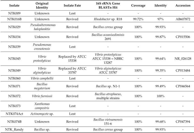
Table 1.Identities of the biodegradation community isolates, as determined by the original identification methods, isolate fate since original biodegradation testing, and the isolate identity as determined by the sequenced 16S rRNA gene sequence.
Isolate Original
Identity Isolate Fate 16S rRNA Gene
BLASTn Hit Coverage Identity Accession
NTK009 Unknown Lost - - - -
NTK016B Unknown Revived Rhodobactersp. R18 99.72% 97% AB607872
NTK029 Pseudoalteromonas
haloplanktis Revived Bacillus cereusgroup 100% 99.93% -
NTK034 Unknown Revived Bacillus oceanisediminis
2691 100% 99.87% CP015506
NTK039 Pseudomonas
creosotensis Lost - - - -
NTK045 Vibrio
proteolyticus
Replaced by ATCC 15338
Vibrio proteolyticus ATCC 15338 = NBRC
13287
100% 99.64% NR_026128
NTK049 Vibrio
alginolyticus
Replaced by ATCC 33787
Vibrio alginolyticus
ATCC 33787 100% 99.35% CP013484
NTK060 Vibrio campbellii Lost - - - -
NTK071 Bacillus
megaterium Revived Bacillussp. N1-1 100% 99.49% CP046564
NTK072 Vibrio furnissii Revived Bacillus atrophaeus,
multiple strains 100% 100% -
NTK073 Xanthomas
campestris Lost - - - -
NTK074Act Actinomycetesp. Lost - - - -
NTK074B Unknown Revived Bacillus vietnamensis
151-6 100% 99.68% CP047394
NTK_Randy Bacillussp. Revived Bacillus cereusgroup 100% 99.93% -
We revived all available isolates from frozen stocks by inoculation on TSY-agar plates (10 g tryptone, 5 g yeast extract, and 15 g agar in 1 L of 75% seawater (salinity ≈ 30, Vineyard Sound, MA, USA) and 25% MilliQ water) and incubation at 30◦C. We then grew liquid cultures from single colonies and both cryopreserved these in 10% DMSO for future work and performed DNA extractions on the harvested cultures.
2.2. DNA Extraction and Sanger Sequencing of 16S rRNA Genes
Genomic DNA from each culture was extracted and purified using the Gentra Pure- gene Yeast/Bacteria Kit (Qiagen, Hilden, Germany), following the manufacturer’s protocols for Gram-positive and/or Gram-negative bacteria when applicable. Genomic DNA was amplified using modified bacterial specific 16S ribosomal RNA (rRNA) gene primers 8F (50-GTTTGATCCTGGCTCAG-30) and 1492R (50-TACCTTGTTACGACTT-30) [61,62] and directly sequenced at the University of Chicago’s Comprehensive Cancer Center’s DNA Sequencing and Genotyping Facility using their methods. We assembled the resulting forward and reverse sequence reads and manually edited these in Geneious Prime build 29 November 2019 [63]. The resulting consensus sequences were subsequently queried against the nr database at NCBI (National Center for Biotechnology Information), using a BLASTn search [64] to retrieve the sequence and GenBank numbers of the most closely related taxa for phylogenetic placement of NTK sequences.
2.3. Phylogenetic Analysis
We searched for additional nearest neighbors of the NTK strains in the SILVA SSU database release 138 Ref NR 99 [65] and aligned them using SINA aligner v1.2.11 [66].
The NTK strain sequences and sequences from the NCBI top BLASTn results not already in the SILVA reference database, were aligned to the database using the command-line version of SINA 1.6.0. We then imported the aligned, arb-formatted sequences into the 138 Ref NR 99 SSU database using ARB v6.0.6 [67], where we selected additional 16S rRNA gene sequences to complete the backbone of the tree. We then exported the resulting alignment and adjusted it manually in Geneious Prime. The shortest sequences were removed, resulting in 70 taxa for phylogenetic reconstruction. The remaining sequences were trimmed and sites containing any gaps were stripped, leaving 1279 phylogenetically informative positions. We then used IQ-TREE (multicore version 1.6.7 for Linux 64-bit, built 23 August 2018) [68] with the ModelFinder flag [69], which selected the K2P + I + G4 model as the best-fit evolutionary model. We choseBacillus subtilisDSM10 as an outgroup and ascertained the confidence of the branching in the tree topology via 1000 bootstrap iterations. The resulting tree was visualized using the Interactive Tree Of Life tool (iTOL, v5.5.1) [70] and further refined in Adobe Illustrator 23.1 (Adobe Systems Inc., San Jose, CA, USA).
2.4. Isolate Growth on PHAs and Screening for Extracellular PHA Depolymerase Activity Growth of individual isolates on PHBV was checked after isolation in the 1990s [54,55].
Next to that, a screening was performed with a non-specific compositional form of PHAs (i.e., a PHA polymer consisting of multiple different hydroxyalkanoates) [unpublished study]. For this screening, liquid cultures were grown in the presence of 0.2w/v% PHA film (Imperial Chemical Industries) or 0.2w/v% of grounded polymer powder, which was added to a carbon-free mineral salts medium. The medium consisted of 1 g NH4Cl, 0.8 g MgSO4•7H2O, 0.45 g K2SO4, 12 mL of 1.1 M phosphoric acid, 0.015 g Fe2(SO4)3•7H2O and 24 mL trace element solution added to 964 mL distilled water. The trace elements solution consisted of 0.02 g CuSO4•5H2O, 0.1 g ZnSO4•6H2O, 0.1 g MnSO4•4H2O and 2.6 g CaCl2•2H2O in 1 L distilled water. Two of the isolates, NTK009, and NTK016B, would not grow on the above media and were rescreened along with others using a marine- specific defined media consisting of 2 g NH4Cl, 2 g MgSO4•7H2O, 0.05 g K2SO4, 0.5 g KNO3, 500 mL synthetic seawater and 500 mL distilled water. Presence of growth was determined visually (powder cultures) and by measuring weight loss of the films.
Here, we tested extracellular PHA depolymerase activity for the individual isolates on 25 mm Petri plates with f/2-silicate medium [71], made with low-nutrient seawater with 10 g/L agar added. A thin PHA layer was added on top of the agar, by pouring 10 mg of PHA (Goodfellow—PH326300—3 mm granules—Extrusion Grade) dissolved in 3 mL chloroform. The chloroform was then evaporated, while plates were rocking at 10 rev/min on a rocking shaker, leaving a solid PHA layer. We placed the cultures in Marine Broth 2216 (BD—Difco) from cryopreserved stocks, and grew them overnight, shaking at 200 rpm in a shaking incubator at 30◦C. From the revived cultures, 20µL of the liquid cultures were spread onto the PHA-plates and left to air dry, before being incubated at 30
◦C. After growth was confirmed, plates were stored at 4◦C and checked at regular intervals for clearing zones.
2.5. Whole Genome Sequencing, Assembly, and Annotation
We selected four NTK strains for whole-genome sequencing: NTK016B, NTK071, NTK072, and NTK074B. The genomic DNA (68 ng from NTK074B and 100 ng from the remaining cultures) was sheared to 275 bp on a Covaris S220 focused-ultrasonicator (Co- varis, Inc., Woburn, MA, USA). We purified sheared samples using Agencourt AMPure XP magnetic beads (Beckman Coulter, Inc., Danvers, MA, USA). The genomic libraries were constructed using the Ovation Ultralow DR Multiplex System V2 1–8 library con- struction kit (NuGEN Technologies Inc., San Carlos, CA, USA). The libraries were pooled
Microorganisms2021,9, 186 8 of 26
equimolarly and size-selected using a Pippin Prep (Sage Science, Inc., Beverly, MA, USA), targeting a size of 390 bp. We purified the size-selected pool again using AMPure magnetic beads in a 1:1 sample to bead ratio. Size, quantitation, and quality were confirmed using a 2100 Bioanalyzer DNA High Sensitivity chip (Agilent Technologies, Santa Clara, CA, USA).
The genomic library pool was further verified through qPCR using KAPA SYBR-FAST for Illumina platforms (Kapa Biosystems, Inc., Wilmington, MA, USA). An Illumina NextSeq V2 Mid Output Sequencing kit (300 cycles) was used, and based on the qPCR results, the genomic library pool was diluted to 2 pM, denatured, and clustered according to the Illumina NextSeq protocol. PhiX DNA was added at 1% for quality control purposes. The samples were then run on an Illumina NextSeq 500 instrument at the Marine Biological Laboratory’s W.M. Keck Sequencing Facility.
We checked the quality of the obtained raw reads using FastQC v0.11.3 (Babraham Bioinformatics, Babraham Institute, Cambridge, United Kingdom). Reads were paired after low-quality bases were trimmed using Trimmomatic v0.35 [72], with settings HEADCROP:6 LEADING:28 TRAILING:28 SLIDINGWINDOW:4:20 MINLEN:40. Genomes were then assembled from the FastQ files, using SPAdes v3.11.1 [73] with the “careful” flag included.
Both paired reads and unpaired reads were used as input and maximum k-mer length iterations were chosen resulting in an average coverage above 50. For NTK016B these parameters were: 21, 33, and 55; for NTK074B these were: 21, 33, 55, and 77; and for NTK034 and NTK071 these were: 21, 33, 55, 77, and 99.
We retrieved genome sequences forV. proteolyticusATCC 15338 = NBRC 13287 (Ac- cession no. BATJ01000000: BATJ01000001-BATJ01000050) andV. alginolyticusATCC 33787 (Accession no. chromosome 1: CP013484, chromosome 2: CP013485, plasmid pMBL96:
CP013488, plasmid pMBL128: CP013486 and plasmid pMBL287: CP013487) from NCBI.
All genomes were annotated using the RASTtk pipeline, which makes use of the FIGfams database [74–76].
2.6. Pangenomic Analysis
We performed comparative genomics analysis using the pangenomics workflow in Anvi’o v6.1, “esther” [77,78]. We chose close relatives of the NTK strains based on our 16S rRNA gene phylogenetic analyses, genome completion level, type strain, and environmental source (e.g., marine) where feasible. As with the NTK strains, gene functions were annotated using RASTtk and imported into Anvi’o. Searching for amino acid sequence similarity was performed by Anvi’o using NCBI BLASTp [64]. Genome completeness was calculated with the anvi-run-hmms module of Anvi’o, which is based on curated single- copy core genes. Figures were made with the Anvi’o interactive display module and further adjusted in Adobe Illustrator 23.1.
2.7. Analysis of Metabolic Potential
We used Anvi’o to search our annotated genomes for the presence of genes encoding key enzymes in the PHA biosynthetic and degradation pathways to predict the PHA metabolic potential of the NTK community members. We also used SignalP-4.1 [79] for Gram-negative or Gram-positive bacteria, when applicable, to search for signal peptides in PHA hydrolases and depolymerases we found to ascertain whether the proteins were secreted. We further refined our search strategy for the two strains that showed clearing zones on the PHA plates, V. proteolyticus ATCC 15338 = NBRC 13287 andBacillus sp.
NTK074B.
For the refined search, we performed a functional analysis of protein domains using InterProScan 5.20–59.0 [80], with InterPro member databases CATH-Gene3D 3.5.0 [81], Pfam-30.0 [82], PRINTS-42.0 [83] and SUPERFAMILY-1.75 [84]. Together with InterProScan, we ran SignalP-4.1 again, to confirm secretion. A shortlist of extracellular depolymerase candidates was made based on the proteins having both a predicted signal peptide and alpha/beta hydrolase fold domain, indicative of the protein belonging to the alpha/beta hydrolase superfamily, to which the PHA depolymerases belong. We then performed a
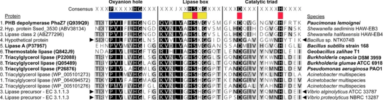
motif-based search in Geneious Prime for the conserved features of extracellular PHA depolymerases. These are the pentapeptide lipase box (amino acid sequence: GXSXG, AHSMGV, A[I,T]S[S,T]G or AHSXG), the oxyanion pocket (PXXXXHG or HGC), and the amino acids of the catalytic triad, serine (part of the lipase box), aspartic acid and histidine [38]. We then checked for the presence of these candidates in other NTK strains against the calculated gene clusters from Anvi’o.
We constructed alignments of the conserved features in the protein candidates with re- lated extracellular PHA depolymerases from the PHA Depolymerase Engineering Database [42], including an experimentally validated PhaZ [85]. Closely related secreted lipases as deter- mined by Oh et al. [41] and recently described extracellular lipases fromAcinetobacterspp., that can potentially depolymerize both scl- and mcl-PHAs [86], were also included in the alignment. These alignments were made manually in Geneious Prime and further refined in Adobe Illustrator 23.1.
3. Results
3.1. Isolate Identification
The original ASTM D6691 biodegradation consortium was identified using the Biolog Substrate Metabolism System (Biolog, Inc., Hayward, CA, USA), along with biochemical methods (i.e., Gram stains) and microscopy [54,55]. This resulted in the identification of the isolates to the genus and sometimes to the species level, see Table1. The identities of the strains were, however, never confirmed with marker gene sequencing. We revived nine isolates from the consortium to perform this. The original identities of the revived strains included:Pseudoalteromonas haloplanktis(NTK029),V. furnissii(NTK072), and two Bacillusspecies (NTK071 and NTK_Randy), with one identified as aB. megaterium. For three other revived strains (NTK016B, NTK034, and NTK074B), the original identities were unknown. Two replacement strains were also revived. These strains replaced the original isolates NTK045 and NTK049, already at the U.S. Army Combat Capabilities Development Command Soldier Center in Natick. These were the isolatesV. proteolyticusATCC 15338 = NBRC 13287 andV. alginolyticusATCC 33787 respectively.
We determined the identity of the revived isolates by sequencing of the nearly com- plete 16S rRNA gene (see Table 1). Isolates were assigned to three bacterial families:
Bacillaceae,Rhodobacteraceae, andVibrionaceae. After a BLASTn search, isolate NTK016B showed 97% identity (99.72% coverage) with the roseobacter bacteriumRhodobactersp.
R18. This closely relatedRhodobacterstrain was isolated from aNannochloropsis oculataalgal culture and can inhibit the growth ofVibrio anguillarum, a fish pathogen [87]. NTK029 and NTK_Randy returned hits with equal coverage and identity scores to various members from theBacillus cereusgroup, includingB. anthracis,B. albus,B. cereus,B. nitratireducens,B.
paranthracis,B. thuringiensis,B. tropicus, andB. wiedmannii. While the taxonomic assignment ofBacillusmembers in this group based on the 16S rRNA marker gene is challenging, the identity of NTK029 did not correspond to the original one. Originally it was identified as Pseudoalteromonas haloplanktis, a Gram-negative proteobacterium and not a Gram-positive firmicute. NTK034 shared the highest sequence similarity toBacillus oceanisediminis2691, a strain isolated from an intertidal marine sediment on the Yellow Sea coast of South Korea that contains a high amount of heavy metal resistance genes [88]. The obtained 16S rRNA gene sequences of theV. proteolyticusandV. alginolyticusstrains were not 100% identical to the purchased ATCC strains because they contained ambiguous nucleotides. This is likely the result of microheterogeneities in their gene copies. Vibrios have 10 copies of the 16S rRNA gene on average, according to the ribosomal RNA operon database [89]. The ambiguous nucleotides of our sequences corresponded to the heterogeneity of the 16S rRNA gene sequences deposited in NCBI. The 16S rRNA gene sequence of NTK071 had the highest similarity toBacillussp. N1-1, which is capable of degradingκ-selenocarrageenan, a selenium polysaccharide, and was isolated from a deep sea cold seep marine sediment in the South China Sea [90]. NTK072 had equal top hits to tens of strains ofBacillus atrophaeus, all with a 100% coverage and identity, but not withV. furnissii, as originally assigned
Microorganisms2021,9, 186 10 of 26
based on non-molecular approaches. The top BLASTn hit of NTK074B was a cadmium tolerantBacillus vietnamensis151-6, isolated from cadmium-contaminated soil from a former industrial site in China [91]. The 16S rRNA gene sequences are deposited under GenBank accession numbers MW435594-MW435596 and in CPXXXXXXX-CPXXXXX. Associated metadata are specified in Supplementary Tables S9 and S10.
3.2. Phylogenetic Placement of NTK Sequences
The phylogenetic placement of the NTK consortium isolates can be seen in Figure2.
Additional metadata associated with the microorganisms are also shown: type strain designation, isolation source, genome availability in NCBI with its assembly level, and an indication of whether it is a representative genome for the species. These data are further specified in Supplementary Tables S1–S3, S9 and S10.
Microorganisms 2021, 9, x FOR PEER REVIEW 11 of 27
4 °C and checked again after 2 months. Clearing zones of several millimeters around the colonies were then observed for V. proteolyticus ATCC 15338 = NBRC 13287 and for NTK072 and NTK074B, two Bacillus strains, see Table 2.
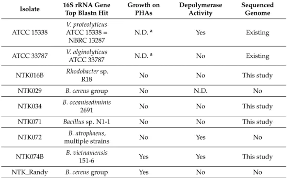
Table 2. Growth results of the NTK isolates in liquid medium with PHAs as the sole carbon source and determination of depolymerase activity, as assessed by clearing zones formed after 2 months on PHA covered culture plates. Genome availability is indicated in the last column. N.D. = not determined.
Isolate 16S rRNA Gene Top Blastn
Hit Growth on PHAs Depolymerase
Activity Sequenced Genome ATCC 15338 V. proteolyticus ATCC 15338 =
NBRC 13287 N.D.a Yes Existing
ATCC 33787 V. alginolyticus ATCC 33787 N.D.a No Existing NTK016B Rhodobacter sp. R18 No No This study
NTK029 B. cereus group No N.D. No
NTK034 B. oceanisediminis 2691 No No This study NTK071 Bacillus sp. N1-1 No No This study
NTK072 B. atrophaeus, multiple strains No Yes No NTK074B B. vietnamensis 151-6 Yes Yes This study
NTK_Randy B. cereus group Yes No No
a The original NTK isolates NTK045 and NTK049, did not show growth on PHAs.
Figure 2.Maximum Likelihood inferred phylogenetic analysis of the NTK consortium isolates and close relatives, based on the 16S rRNA marker gene sequence. Bootstrap support values above 50% are shown on the nodes and GenBank accession numbers are noted next to the strains. Information on the right provides the following: type strain information, isolation source (marine versus non-marine), genome assembly level available, and representative genome status for a given taxon.
The evolutionary distance is indicated by the scale bar.
The NTK isolates fell into three bacterial families:Bacillaceae,Rhodobacteraceae, and Vibrionaceae. TheBacillaceaeisolates branched among five differentBacillusgroups in our phylogeny: NTK_Randy and NTK029 branched among theB. cereusgroup, while NTK034 shared most recent common ancestry withB. oceanisediminisstrains and members of the B. firmusgroup.Bacillussp. NTK071 was most closely related toB. hwajinpoensisstrains andAnaerobacillus macyae, while NTK072 showed most recent common ancestry withB.
atrophaeusstrains (bootstrap support 74 and 88). The branching of NTK074B was not well- resolved betweenB. marisflavi,B. oryzaecorticisandB. vietnamensisstrains.Rhodobactersp.
NTK016B displayed high branch support for placement with the aforementionedRhodobac- tersp. R18 strain and formed part of a larger well-supported cluster withPararhodobacter sp. strain CIC4N-9 (bootstrap support 94), a bacterium isolated from deep-sea water in the Indian Ocean [92]. V. alginolyticusATCC 33787 branched with the type strain ofV.
alginolyticus, NBRC 15630, but the position ofV. proteolyticusNBRC 13287 was poorly resolved among other vibrios owing a lack of resolving power among 16S rRNA gene sequences to differentiate betweenVibriospecies in general.
3.3. Isolate Growth on PHAs and Screening for Extracellular Depolymerase Activity
The isolates NTK074B and NTK_Randy showed positive growth when grown in liquid culture with PHAs as the only carbon source, see Table2, indicating a full metabolic potential to utilize PHAs. After we incubated the NTK isolates on PHA-covered agar culture plates for 4 days at 30◦C, colonies started to become visible for all strains except NTK029, which was not tested. The number of colonies ranged from two to more than 100 colonies. However, no clearing zones were observed in the 4-day timeframe, which would indicate high activity of extracellular depolymerases. The plates were therefore stored at 4◦C and checked again after 2 months. Clearing zones of several millimeters around the colonies were then observed forV. proteolyticusATCC 15338 = NBRC 13287 and for NTK072 and NTK074B, twoBacillusstrains, see Table2.
Table 2.Growth results of the NTK isolates in liquid medium with PHAs as the sole carbon source and determination of depolymerase activity, as assessed by clearing zones formed after 2 months on PHA covered culture plates. Genome availability is indicated in the last column. N.D. = not determined.
Isolate 16S rRNA Gene Top Blastn Hit
Growth on PHAs
Depolymerase Activity
Sequenced Genome ATCC 15338
V. proteolyticus ATCC 15338 = NBRC 13287
N.D.a Yes Existing
ATCC 33787 V. alginolyticus
ATCC 33787 N.D.a No Existing
NTK016B Rhodobactersp.
R18 No No This study
NTK029 B. cereusgroup No N.D. No
NTK034 B. oceanisediminis
2691 No No This study
NTK071 Bacillussp. N1-1 No No This study
NTK072 B. atrophaeus,
multiple strains No Yes No
NTK074B B. vietnamensis
151-6 Yes Yes This study
NTK_Randy B. cereusgroup Yes No No
aThe original NTK isolates NTK045 and NTK049, did not show growth on PHAs.
Microorganisms2021,9, 186 12 of 26
3.4. Whole-Genome Sequencing
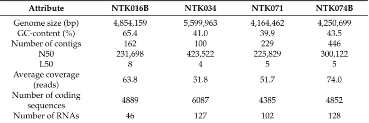
While the twoVibriostrains were already sequenced, four NTK strains were selected for whole-genome sequencing: Rhodobacter sp. NTK016B and Bacillus spp. NTK034, NTK071 and NTK074B (Table2). Table3summarizes the genome features after assem- bly and annotation in RASTtk. Genome sizes varied from 4,164,462 bp (NTK071) to 5,599,963 bp (NTK034), with a GC-content of around 40% for theBacillusgenomes and of about 65% for theRhodobactergenome. The number of contigs varied between 100 (NTK034) to 446 (NTK074B) in total, with half of the genome length covered by four to eight contigs (L50 value). The N50 value was between 225,829 for the smallest genome, to 423,522 for the largest genome and the average coverage of all genomes was above 50 reads. Using the RASTtk pipeline, 4385 to 6087 genes and 46–128 RNAs were annotated per genome. The RASTtk annotated genomes are available onlineviahttps://rast.nmpdr.org/
by logging in as guest and can be found under ID numbers 6666666.521526 (Rhodobactersp.
NTK016B), 6666666.521528 (Bacillussp. NTK034), 6666666.521530 (Bacillussp. NTK071) and 6666666.521531 (Bacillussp. NTK074B). The raw sequence reads are deposited in the NCBI Sequence Read Archive under BioProject PRJNA649735 with sample accession numbers SRR12354198 to SRR12354201. Annotated genomes are also deposited in INSDC under ref- erences CPXXXXXX-CPXXXXXX, and associated metadata are specified in Supplementary Table S10.
Table 3.Genome features of sequenced NTK isolates.
Attribute NTK016B NTK034 NTK071 NTK074B
Genome size (bp) 4,854,159 5,599,963 4,164,462 4,250,699
GC-content (%) 65.4 41.0 39.9 43.5
Number of contigs 162 100 229 446
N50 231,698 423,522 225,829 300,122
L50 8 4 5 5
Average coverage
(reads) 63.8 51.8 51.7 74.0
Number of coding
sequences 4889 6087 4385 4852
Number of RNAs 46 127 102 128
3.5. Pangenomic Analysis
The genome relatedness of the sequenced NTK isolates with close neighbors was exam- ined by a pangenomic analysis. Figures3–5show the genomic alignment of the genomes, based on gene clustering, with the genome order based on gene cluster presence/absence.
Genome accession information can be found in Supplementary Table S3. The estimated completeness of the genomes from all the NTK consortium members was 100%, as pre- dicted from the presence of single-copy genes by Anvi’o. Exact numbers for the genome properties can be found in Supplementary Tables S4–S6. The whole-genome comparisons of the three sequencedBacillusspp. genomes (NTK034, NTK071 and NTK074B), with 12 nearest neighbors are shown in Figure3. As in our 16S rRNA phylogenetic reconstruction, the isolates and nearest neighbors clustered into three groups, based on the gene cluster presence/absence tree.
Microorganisms2021,9, 186 13 of 26
close relationship of NTK016B with the Pararhodobacter sp. strains CICN4N-9 and CCB- MM2.
The genome sequences of the two NTK consortium vibrios, V. proteolyticus NBRC 13287 and V. alginolyticus ATCC 33787, were already available in public databases. Figure 5 shows the whole-genome comparison of the Vibrio strains with 10 close neighbors with sequenced genomes. The Anvi’o display shows a large portion of shared gene clusters among all the chosen Vibrio species. The gene cluster presence/absence tree topology is similar to that of the 16S rRNA gene tree.
Figure 3. Genome comparison of the three sequenced NTK Bacillus spp. genomes (NTK034, NTK071 and NTK074B) in blue, with closely related neighbors in grey. The semicircles show gene presence (dark color) and absence (light color). Alignment of the genomes is based on gene clus- tering, with the genome order based on the gene cluster presence/absence tree, shown in the upper right corner. The dendrogram in the center represents the hierarchy in gene clustering using Eu- clidean distance and Ward linkage. Genome properties shown: “Number gene clusters” represents the total number of gene clusters found in the genome; “Singleton gene clusters” represents the number of genes found in only one genome, “Completion” in (%) is calculated based on single- copy genes, “GC-content” shows the average guanine and cytosine nucleotide content and “Total length” is the genome length in base pairs.
Figure 3.Genome comparison of the three sequenced NTKBacillusspp. genomes (NTK034, NTK071 and NTK074B) in blue, with closely related neighbors in grey. The semicircles show gene presence (dark color) and absence (light color). Alignment of the genomes is based on gene clustering, with the genome order based on the gene cluster presence/absence tree, shown in the upper right corner. The dendrogram in the center represents the hierarchy in gene clustering using Euclidean distance and Ward linkage. Genome properties shown: “Number gene clusters” represents the total number of gene clusters found in the genome; “Singleton gene clusters” represents the number of genes found in only one genome, “Completion” in (%) is calculated based on single-copy genes, “GC-content”
shows the average guanine and cytosine nucleotide content and “Total length” is the genome length in base pairs.
close relationship of NTK016B with the Pararhodobacter sp. strains CICN4N-9 and CCB- MM2.
The genome sequences of the two NTK consortium vibrios, V. proteolyticus NBRC 13287 and V. alginolyticus ATCC 33787, were already available in public databases. Figure 5 shows the whole-genome comparison of the Vibrio strains with 10 close neighbors with sequenced genomes. The Anvi’o display shows a large portion of shared gene clusters among all the chosen Vibrio species. The gene cluster presence/absence tree topology is similar to that of the 16S rRNA gene tree.
Figure 3. Genome comparison of the three sequenced NTK Bacillus spp. genomes (NTK034, NTK071 and NTK074B) in blue, with closely related neighbors in grey. The semicircles show gene presence (dark color) and absence (light color). Alignment of the genomes is based on gene clus- tering, with the genome order based on the gene cluster presence/absence tree, shown in the upper right corner. The dendrogram in the center represents the hierarchy in gene clustering using Eu- clidean distance and Ward linkage. Genome properties shown: “Number gene clusters” represents the total number of gene clusters found in the genome; “Singleton gene clusters” represents the number of genes found in only one genome, “Completion” in (%) is calculated based on single- copy genes, “GC-content” shows the average guanine and cytosine nucleotide content and “Total length” is the genome length in base pairs.
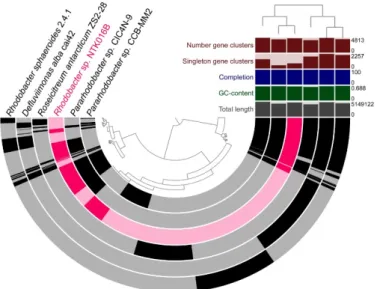
Figure 4. Genome comparison of isolate Rhodobactersp. NTK016B, highlighted in pink, with Rhodobacteraceaeneighbors with sequenced genomes in grey. The semicircles show gene presence (dark color) and absence (light color). Gene clustering and genome alignment, order, and properties are presented as in Figure3.
Microorganisms2021,9, 186 14 of 26
Microorganisms 2021, 9, x FOR PEER REVIEW 14 of 27
Figure 4. Genome comparison of isolate Rhodobacter sp. NTK016B, highlighted in pink, with Rhodo- bacteraceae neighbors with sequenced genomes in grey. The semicircles show gene presence (dark color) and absence (light color). Gene clustering and genome alignment, order, and properties are presented as in Figure 3.
Figure 5. Genome comparison of V. proteolyticus NBRC 13287 and V. alginolyticus ATCC 33787 (in green), with closely related neighbors (in grey). The semicircles show gene presence (dark color) and absence (light color). Gene clustering and genome alignment, order, and properties are pre- sented as in Figure 3.
3.6. Analysis of Metabolic Potential
The PHA metabolic potential of the NTK consortium members was assessed by searching for the presence of genes encoding key enzymes in the PHA biosynthetic and degradation pathways, which are depicted in Figure 1. The results of this search, together with the existence of these genes in closely related species, are shown in Table 4. We found genes encoding for a full PHA biosynthetic capacity in all NTK strain genomes, going from an acyl-CoA to PHA, via an oxoacyl-CoA (by PhaA or BktB) and a hydroxyacyl-CoA (by FabG, FadB, Hbd or PhaB). The only exception to this was strain NTK071, which seemed to be missing the PHA synthase gene (phaC). This gene was also missing in the close relatives Anaerobacillus macyae, the B. hwajinpoensis strains, and Bacillus sp. N-1-, the latter with a closed genome. The genes phaJ (Hydroxyacyl-CoA-acyl carrier protein trans- ferase) and phaG ((R)-specific enoyl-CoA hydratase), which provide an alternative for syn- thesizing the PHA monomer, were not detected in any of the genomes.
Table 4. Presence (✓) and absence (-) overview of PHA biosynthesis- and degradation-related genes in the annotated genomes, based on the functional annotation.
Function 1 2 3 4 5 6 7 8 9 10 11 12
Species Gene phaA/b
ktB fabG fadB hbd phaB phaC (e) phaY (e) phaZ (i) phaZ bdh aacS scoA
and scoB Bacillus sp.
NTK034 ◄ ✓ ✓ ✓ ✓ - ✓ - - - ✓ - ✓
Bacillus oceanisedi-
minis 2691 ✓ ✓ ✓ ✓ - ✓ - - - ✓ - ✓
Bacillus oceanisedi-
minis H2 ✓ ✓ ✓ ✓ - ✓ - - - ✓ - ✓
Bacillus infantis
NRRL B-14911 ✓ ✓ ✓ ✓ - ✓ - ✓ ✓ ✓ - ✓
Bacillus firmus
NCTC 10335 ✓ ✓ ✓ ✓ - ✓ - - - ✓ - ✓
Figure 5.Genome comparison ofV. proteolyticusNBRC 13287 andV. alginolyticusATCC 33787 (in green), with closely related neighbors (in grey). The semicircles show gene presence (dark color) and absence (light color). Gene clustering and genome alignment, order, and properties are presented as in Figure3.
Figure4shows the whole-genome comparison of NTK016B (Rhodobactersp.) with five closely related neighbors that were chosen based on phylogenetic analysis of the 16S rRNA gene. As with the phylogenetic placement, comparative genomics confirmed the close relationship of NTK016B with thePararhodobactersp. strains CICN4N-9 and CCB-MM2.
The genome sequences of the two NTK consortium vibrios,V. proteolyticusNBRC 13287 andV. alginolyticusATCC 33787, were already available in public databases. Figure5 shows the whole-genome comparison of theVibriostrains with 10 close neighbors with sequenced genomes. The Anvi’o display shows a large portion of shared gene clusters among all the chosenVibriospecies. The gene cluster presence/absence tree topology is similar to that of the 16S rRNA gene tree.
3.6. Analysis of Metabolic Potential
The PHA metabolic potential of the NTK consortium members was assessed by searching for the presence of genes encoding key enzymes in the PHA biosynthetic and degradation pathways, which are depicted in Figure1. The results of this search, together with the existence of these genes in closely related species, are shown in Table4. We found genes encoding for a full PHA biosynthetic capacity in all NTK strain genomes, going from an acyl-CoA to PHA, via an oxoacyl-CoA (by PhaA or BktB) and a hydroxyacyl-CoA (by FabG, FadB, Hbd or PhaB). The only exception to this was strain NTK071, which seemed to be missing the PHA synthase gene (phaC). This gene was also missing in the close relatives Anaerobacillus macyae, theB. hwajinpoensisstrains, andBacillussp. N-1-, the latter with a closed genome. The genesphaJ(Hydroxyacyl-CoA-acyl carrier protein transferase) and phaG((R)-specific enoyl-CoA hydratase), which provide an alternative for synthesizing the PHA monomer, were not detected in any of the genomes.
Table 4.Presence (X) and absence (-) overview of PHA biosynthesis- and degradation-related genes in the annotated genomes, based on the functional annotation.
Function 1 2 3 4 5 6 7 8 9 10 11 12
Species Gene phaA/bktB fabG fadB hbd phaB phaC (e)phaY (e)phaZ (i)phaZ bdh aacS scoAandscoB
Bacillussp. NTK034J X X X X - X - - - X - X
Bacillus oceanisediminis2691 X X X X - X - - - X - X
Bacillus oceanisediminisH2 X X X X - X - - - X - X
Bacillus infantisNRRL B-14911 X X X X - X - X X X - X
Bacillus firmusNCTC 10335 X X X X - X - - - X - X
Bacillus vietnamensisNBRC 101237 X X X X - X - - X X - X
Bacillus vietnamensis151-6 X X X X - X - - X X - X
Bacillussp. NTK074BJ X X X X - X - - - X - X
Bacillus aquimarisTF-12 X X X X - - - - X X - X
Bacillus marisflaviTF-11 X X X X - X - - - X - X
Bacillussp. NTK071J X X X X - - - - - X - X
Bacillus hwajinpoensisY2 X X X X - - - - - X - X
Anaerobacillus macyaeDSM 16346 X X X X - - - X X X - X
Bacillussp. N1-1 X X X X - - - - - X - X
Bacillus hwajinpoensis22506_14_FS X X X X - - - - - X - X
Rhodobacter sphaeroides2.4.1 X X X X X X - Xa X X - X
Defluviimonas albacai42 X X X X X X - - X X - X
Roseicitreum antarcticumZS2-28 X X X X X X - - X X - X
Rhodobactersp. NTK016BJ X X X X X X - - X X X X
Pararhodobactersp. CIC4N-9 X X X X X X - - X X X X
Pararhodobactersp. CCB-MM2 X X X X X X - - X X X X
Vibrio furnissiiATCC 35016 X X X - X X - - - - X -
Vibrio proteolyticusNBRC 13287J X X X - X X - - - - X -
Vibrio tubiashiiATCC 19109 X X X - X X - - - - X -
Vibrio atypicusHHS02 X X X X X X - - - - X -
Vibrio parahaemolyticusATCC 17802 X X X - X X - - - - X -
Vibrio diabolicusFDAARGOS_105 X X X - X X - - - - X -
Vibrio alginolyticusNBRC 15630 X X X - X X - - - - X -
Vibrio alginolyticusATCC 33787J X X X - X X - - - - X -
Vibrio natriegensNBRC 15636 X X X - X X X - - - X -
Vibrio rotiferianusB64D1 X X X - X X - - - - X -
Vibrio harveyiFDAARGOS_107 X X X - X X - - - - X -
Vibrio campbelliiCAIM 519 X X X - X X - - - - X -
Function:
1 Acetyl-CoA acetyltransferase/β-ketothiolase
(EC 2.3.1.9 and EC 2.3.1.16) 7 (extracellular) PHA oligomer hydrolase
(EC 3.1.1.22)
2 3-oxoacyl-[acyl-carrier-protein] reductase
(EC 1.1.1.100 with EC 1.1.1.36 capacity) 8 (extracellular) PHA depolymerase (EC 3.1.1.75 and EC 3.1.1.76)
3 Enoyl-CoA hydratase (EC 4.2.1.17)/
Hydroxyacyl-CoA dehydrogenase (EC 1.1.1.35) 9 (intracellular) PHA depolymerase (EC 3.1.1.75 and EC 3.1.1.76)
4 3-hydroxybutyryl-CoA dehydrogenase (EC 1.1.1.157) 10 3-hydroxybutyrate dehydrogenase (EC 1.1.1.30)
5 Acetoacetyl-CoA reductase (EC 1.1.1.36) 11 Acetoacetyl-CoA synthetase (EC 6.2.1.16)
6 PHA synthase (EC 2.3.1.-) 12 3-oxoacid CoA-transferase subunit A and B (EC 2.8.3.5)
JNTK biodegradation consortium member.aDeposited in the PHA Depolymerase Engineering Database [42] as an extracellular depolymerase, but contains no signal peptide, according to SignalP 4.1.