OXIDATIVE STRESS-INDUCED EPIGENETIC TRANSCRIPTIONAL MEMORY AS A MECHANISM OF PROGRAMMED ENDOTHELIAL DYSFUNCTION IN LARGE FOR GESTATIONAL AGE NEWBORN OF
WOMEN WITH OBESITY
DISSERTATION ZUR ERLANGUNG DES DOKTORGRADES DER NATURWISSENSCHAFTEN (DR. RER. NAT)
DER FAKULTÄT FÜR BIOLOGIE UND VORKLINISCHE MEDIZIN DER UNIVERSITÄT REGENSBURG
Vorgelegt von
Ivo Carrasco Wong
aus
Iquique – CHILE
im Jahr
2019
Das Promotionsgesuch wurde eingereicht am: 10.01.19
Die Arbeit wurde angeleitet von:
Prof. Dr. Paola Casanello, Pontificia Universidad Católica, Santiago, Chile
Prof. Dr. Gernot Längst, Universität Regensburg
Unterschrift:
1
Die vorliegende Arbeit mit dem Titel
OXIDATIVE STRESS-INDUCED EPIGENETIC TRANSCRIPTIONAL MEMORY AS A MECHANISM
OF PROGRAMMED ENDOTHELIAL
DYSFUNCTION IN LARGE FOR GESTATIONAL AGE NEWBORN OF WOMEN WITH OBESITY
vorgelegt von Ivo Carrasco Wong
entstand unter der gemeinsamen Betreuung der Universität Regensburg
und
der Pontificia Universidad Católica de Chile
im Rahmen des internationalen Promotionsprogramms iP UR als Doppelpromotion
2 ACKNOWLEDGEMENTS
My deepest gratitude to all those who supported me in this process.
FUNDING
This thesis was funded by the Chilean National Fund for Scientific and Technological Development, FONDECYT #1171406 and #11209281
3 TABLE OF CONTENTS
ACKNOWLEDGEMENTS 2
FUNDING 2
TABLE OF CONTENTS 3
FIGURES INDEX 6
TABLE INDEX 7
ABBREVIATIONS 8
RESUMEN 12
ABSTRACT 14
CHAPTER I 15
THE MOLECULAR BASES OF VASCULAR PROGRAMMING IN LARGE FOR GESTATIONAL AGE NEWBORN FROM WOMEN WITH PREGESTATIONAL OBESITY
Introduction 16
Programming of vascular dysfunction 17
Oxidative stress 18
NRF2, the oxidative stress sensor at cellular level 19
Epigenetic regulation of gene expression 20
DNA methylation 22
Post-translational histone modification 22
Histones variants 23
ATP-dependent chromatin remodelling complexes 23
Non-coding RNAs (ncRNAs) 24
Epigenetic Transcriptional Memory (ETM) 24
Mechanisms that regulate gene transcription 25
Transcription initiation 25
Carboxy terminal domain (CTD) RNAPII phosphorylation 27
RNAPII pausing 27
Enhancers in gene transcription regulation 27
Hypothesis 28
General aim 29
Specific aims 29
References 31
CHAPTER II 39
HUMAN UMBILICAL ARTERY ENDOTHELIAL CELLS FROM LARGE-FOR-
GESTATIONAL-AGE NEWBORN HAVE INCREASED ANTIOXIDANT
EFFICIENCY AND GENE EXPRESSION.
ACKNOWLEDGEMENTS 41
ABSTRACT 42
INTRODUCTION 43
MATERIALS AND METHODS 44
Samples 44
Cell isolation and culture 45
HUAEC infection with HyPer probe 45
4 Microscopy and imaging of HyPer biosensor 46
Dihydroethidium (DHE) assay 47
GSH:GSSG assay 47
Determination of superoxide dismutase activity (SOD) 48
Western blotting 48
Immunohistochemistry 49
Isolation of total RNA and reverse transcription 50
Quantitative PCR 50
DNAse Hypersensitivity assay 51
Statistical analysis 52
RESULTS 52
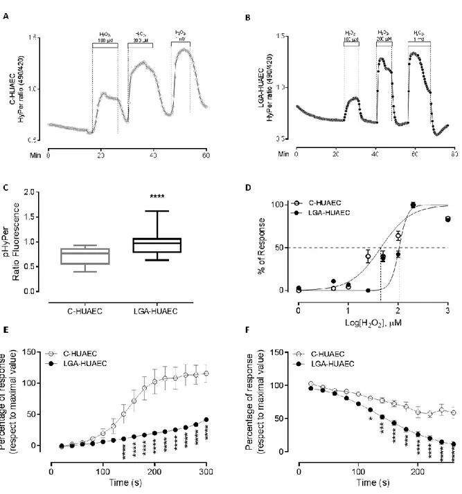
LGA-HUAEC have an altered redox status and an increased exogenous
H2O2 buffering capacity 52
LGA-HUAEC have reduced superoxide anion level and increased SOD
activity 54
LGA-HUAEC have reduced GSH:GSSG ratio 55
LGA-HUAEC do not present changes in pro- or antioxidant proteins
expression 55
Umbilical arteries endothelium from LGA newborn form women with pregestational obesity (LGA-OM) show altered levels of pro- and anti-
oxidant enzymes 55
LGA-HUAEC have altered NRF2 and HMOX1 gene expression 56 LGA-HUAEC overexpress the GPX1 gene when exposed to a pro-oxidant
stimulus 56
GPX1 gene transcription start site has an open state of chromatin in LGA-
HUAEC 58
DISCUSSION 58
CONCLUSIONS 64
REFERENCES 66
FIGURE LEGENDS 79
CHAPTER III 91
NRF2 PRE-RECRUITMENT AT ENHANCER2 IS A HALLMARK OF H2O2-
INDUCED EPIGENETIC TRANSCRIPTIONAL MEMORY IN HMOX1 IN HUMAN ENDOTHELIAL CELLS.
ABSTRACT 93
INTRODUCTION 94
RESULTS 96
H2O2-priming induces an epigenetic transcriptional memory-like
response in primary HUAEC 96
H2O2-priming induces an epigenetic transcriptional memory-like
response in EA.hy926 cells 97
mRNA accumulation in P/T cells is not caused by an alteration in mRNA
decay 97
H2O2-primed cells have an open state of chromatin in the HMOX1
regulatory regions 98
5 Phospho-S5-RNAPII at TSS and NRF2 at E2 are enriched in H2O2-
primed cells 99
pS5-RNAPII and NRF2 enrichment is not different between UP/T and P/T cells 99
DISCUSSION 99
MATERIAL AND METHODS 104
Samples 104
Cell isolation and culture 105
Treatments 106
Isolation of total RNA and reverse transcription 106
Relative Quantitative of mRNA 106
mRNA decay assay 106
DNase Hypersensitivity assay 107
Chromatin immune precipitation (ChIP) assays 107
Statistical analysis 108
ACKNOWLEDGEMENTS 108
REFERENCES 109
CHAPTER IV 126
GENERAL DISCUSSION. CHRONIC AND ACUTE OXIDATIVE STRESS INDUCE DIFFERENTIAL ADAPTATIONS IN ENDOTHELIAL CELLS Chapter 2: Human umbilical artery endothelial cells from Large-for-Gestational- Age newborn from women with pregestational obesity have increased antioxidant efficiency and gene expression 127
Antioxidant gene programming by the altered maternal metabolic and pro- oxidant environment 130
Chapter 3. Nrf2 pre-recruitment at enhancer2 is a hallmark of H2O2-induced epigenetic transcriptional memory in HMOX1 in human endothelial cells 131
Epigenetic transcriptional memory mechanism 131
Reductive stress could be part of endothelial dysfunction in LGA-HUAEC 134
Conclusion and perspectives 136
REFERENCES 139
6 FIGURESINDEX
CHAPTER I
Figure 1. Schematic model of the NRF2–KEAP1 signaling pathway 21 Figure 2. Proposed model of OxS-ETM acquisition in the HMOX1 gene 26
CHAPTER II
Figure 1. Indirect H2O2 measurement in C-HUAEC and LGA-HUAEC by HyPer
under basal and oxidative stress conditions 84
Figure 2. Basal superoxide anion level, SODs activity and quantitative measurement
of GSH and GSSG in C-HUAEC and LGA-HUAEC 85
Figure 3. Basal Protein level of antioxidant and pro-oxidant enzymes in C-HUAEC
and LGA-HUAEC 86
Figure 4. Umbilical artery endothelium in situ levels of H2O2 producing and
reducing enzymes in Control and LGA-MO umbilical cords 87 Figure 5. Basal mRNA level of genes involved in the antioxidant and pro-oxidant
response in C-HUAEC and LGA-HUAEC 88
Figure 6.Transcript levels of antioxidant and pro-oxidant genes after H2O2
treatment and its chromatin accessibility in C-HUAEC and LGA-HUAEC 89 Figure 7. HUAEC from LGA-MO newborn show a pro-oxidant status and a higher
antioxidant performance 90
CHAPTER III
Figure 1. NRF2, HMOX1 and GCLM mRNA level induced by double-hit-
protocol in Control-HUAEC 113
Figure 2. HMOX1 mRNA level induced by double-hit-protocol in EA.hy926 114 Figure 3. HMOX1 mRNA half-life after double-hit-protocol in EA.hy926 cells 115 Figure 4. DNase I chromatin accessibility at HMOX1 gene regulatory regions
in Unprimed and Primed EA.hy926 116
Figure 5. Enrichment of RNAPII-total, RNAPII-pS5 and NRF2 at HMOX1 regulatory regions of unprimed or primed EA.hy926 cells in basal or induced
transcriptional state 117
Supplemental figure 1. Effect of tBHQ double-hit-protocol in antioxidant
proteins mRNA level in Control-HUAEC 120
7 Supplemental figure 2. NRF2, GPX1 and GCLM mRNA level induced by
double-hit-protocol in EA.hy926 121
Supplemental figure 3. HMOX1 and GCLM mRNA level induced by double-hit
protocol in HeLa cells 122
Supplemental figure 4. NRF2 and NQO1 mRNA level induced by double-hit-
protocol in HeLa cell 123
Supplemental figure 5. H2O2 priming effect on cellular proliferation and on H2O2 high concentration resistance 124 Supplemental figure 6. CpGs and NRF2 binding sites at HMOX1 regulatory
regions 125
CHAPTER IV
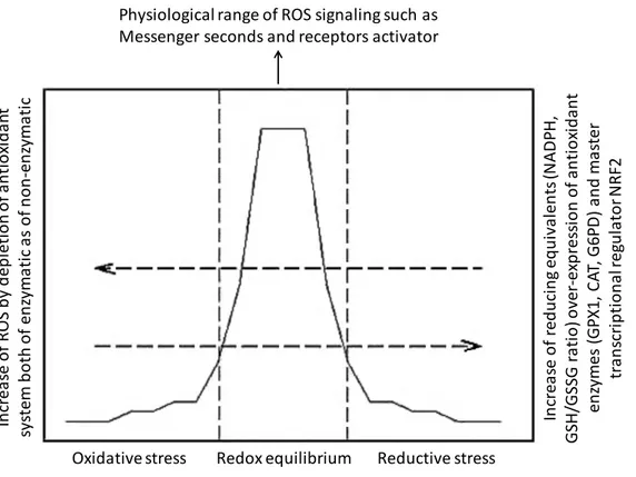
Figure 1. The redox equilibrium is essential for cellular homeostasis 135 Figure 2. Summarize of the results showed in this thesis 138
TABLE-INDEX CHAPTER II
Table 1. Baseline characteristics of the study population 75 Table 2. Primers sequences and standardized amplification conditionsfor mRNA
RT-qPCR 76
Table 3. Primers sequences and standardized amplification conditions
for gDNA qPCR 77
Table 4. Parameters of HyPer fluorescence kinetics in C-HUAEC and LGA-
HUAEC 78
CHAPTER III
Supplementary Table 1. Primers specifications 118 Supplementary Table 2. Predicted NRF2-binding sites 119
8 ABBREVIATIONS
5hmC 5mC ARE ATP5F AUC BACH Brd4 CBP CDK9 C-HUAEC CpG CRMs CTCF CTD DHFR DNMT DOHaD DSIF E1 E2 EC50
ECs eNOS eRNAs ETM FOXO3A GCLM GPX1
5-hydroxymethylcytosine 5-methylcitosine
Antioxidant response element
ATP synthase peripheral stalk-membrane subunit b Area under the curve
BTB domain and CNC homolog Bromodomain-containing protein 4 CREB Binding Protein
Cyclin-dependent kinase 9
HUAEC isolated from control newborn Dinucleotide CG
cis-regulatory modules
CCCTC DNA element binding factor RNAPII carboxy-terminal domain dihydrofolate reductase
DNA methyl transferase
Developmental origin of health and disease DRB-Sensitivity-Inducing factor
Enhancer 1 Enhancer 2
Half-maximal effective concentration Endothelial cells
Endothelial nitric oxide synthase RNA transcribed at enhancer regions Epigenetic transcriptional memory Forkhead Box O3
Glutamate-Cysteine Ligase Modifier Subunit Glutathione peroxidase 1
9 GSH
H2O2 H3K27me3 H3K4me1 H3K4me2 H3K4me3 H3K9ac HB2 HB4 HDAC1 HMOX1 HO•
HUAEC IL2 IUGR
Keap1-Cul3/Rbx1 complex LGA
LGA-HUAEC lncRNA MAF MBD MeCPs MEF cells MLL ncRNA Neh domain NELF NF-E2 p45 NO NOX
Glutathione
Hydrogen peroxide
Tri-methylation of lysine 27 of Histone 3 Mono-methylation of lysine 4 of Histone 3 Di-methylation of lysine 4 of Histone 3 Tri-methylation of lysine 4 of Histone 3 Acetylation of lysine 9 of Histone 3 Dihydrobiopterin
Tetrahydrobiopterin Histone Deacetylase 1 Heme oxygenase 1 Hydroxyl anion
Human umbilical artery endothelial cell Interleukin 2
Intrauterine growth restriction NRF2 ubiquitin ligase complex Large for gestational age
HUAEC isolated from LGA newborn long non-coding RNA
Avian Musculoaponeurotic Fibrosarcoma methyl-CpG-binding domain
methyl-CpG-binding proteins Mouse Embryonic Fibroblasts mixed lineage leukemia non-coding RNA
Nrf2-ECH homology domain Negative elongation factor
Nuclear Factor, Erythroid-Derived 2 45 KDa Nitric oxide
NAD(P)H oxidase
10 NQO1
NRF1 NRF2 NRF3
NURD/Mi-2/CHD O2•−
OM ONOO− ORAC OxS OxS-ETM P/T P/UT P P300 p66shc p90 PIC PMA/I PRDX3 PRDX6 pS5-RNAPII P-TEFb RISC RNAPII RNS RO•
RO2•
ROS
NAD(P)H Quinone Dehydrogenase 1 Nuclear Factor, Erythroid 2 Like 1 Nuclear Factor, Erythroid 2 Like 2 Nuclear Factor, Erythroid 2 Like 3
Nucleosome Remodeling Deacetylase complex Superoxide anion
Obese mother Peroxynitrite
Oxygen radical absorbance capacity Oxidative stress
ETM induced by oxidative stress Primed and Treated
Primed and Untreated Primed
E1A-Binding Protein, 300kD SHC Adaptor Protein 1 percentile 90th
Pre-initiation complex
Phorbol-Myristate-Acetate and Ionomycin Peroxiredoxin 3
Peroxiredoxin 6
RNAPII phosphorylated at serine 5 of CTD Positive transcription elongation factor-b RNA-induced silencing complex
RNA polymerase II Reactive nitric species Alkoxyl anion
Peroxyl anion
Reactive oxygen species
11 RPLP0
RPLP2 RUNX2 S.E.M SEC SOD tBHP tBHQ TBP TET TF TFII TSS TXNDR UP/T UP/UT UP y.o.
Ribosomal Protein Lateral Stalk Subunit P0 Ribosomal Protein Lateral Stalk Subunit P2 Runt Related Transcription Factor 2
Standard error of the mean Super elongation complex Superoxide dismutase tert-Butyl Hydroperoxide tert-Butyl Hydroquinone Tata binding protein ten-eleven translocation Transcription factor
General transcription factors Transcription start site Thioredoxin reductase Unprimed and Treated Unprimed and Untreated Unprimed
Years old
12 RESUMEN
La obesidad es un problema de salud pública mundial. Contrariamente a la idea general de que los resultados de un estado de obesidad solo afectan al mismo individuo, ahora está claro que existe una asociación directa entre la obesidad materna y un aumento de los riesgos cardiometabólicos en la progenie. En varias enfermedades cardiovasculares, la disfunción endotelial suele ser el evento más temprano que se presenta, mucho antes de que se pueda evidenciar clínicamente la hipertensión. En las arterias placentarias y umbilicales de recién nacidos grandes para su edad gestacional y de madres obesas (LGA-OM, su acrónimo en inglés), se ha descrito una disfunción endotelial relacionada con una alteración en la respuesta antioxidante y niveles elevados de marcadores oxidativos circulantes. Sin embargo, no hay evidencia, hasta el momento, que muestre el posible efecto del ambiente pro-oxidante inducido sobre la maquinaria antioxidante de células endoteliales umbilicales de recién nacidos LGA-OM; incluyendo el desconocimiento de cómo la alterada fisiología perinatal presente a lo largo del desarrollo fetal, puede alterar el rendimiento vascular. En esta tesis, se probó la hipótesis de que el estrés oxidativo crónico inducido por la obesidad materna puede modular la fisiología vascular fetal mediante una modificación en el rendimiento de la maquinaria antioxidante. Al usar una sonda fluorescente sensible al H2O2, se evidenció una mayor capacidad antioxidante cuando se indujo un desafío oxidativo exógeno en células endoteliales de la arteria umbilical humana (HUAEC) aisladas de recién nacidos LGA-OM.
Esta condición no solo estaba relacionada con una expresión basal del gen HMOX1 elevada;
sino también, con una transcripción exacerbada del gen GPX1; asociado a un estado abierto de la cromatina en el promotor, de este último, sugiriendo la participación de mecanismos epigenéticos. Por lo tanto, propusimos que posiblemente una activación constante del gen GPX1 debido al estrés oxidativo ambiental podría inducir el rendimiento de respuesta exacerbada mediante la adquisición de una memoria transcripcional epigenética (ETM, su acrónimo en inglés). Para evaluar esta idea, aplicamos un protocolo de “doble impacto” a las células de control HUAEC y EA.hy926. Al contrario de lo que se esperaba, solo el gen HMOX1 mostró una respuesta similar a la ETM. En dicho gen, el primer desafío oxidativo indujo un estado abierto de cromatina tanto en su promotor proximal como en el enhancer 2 (ubicado a 9 kilobases río arriba); los que se asociaron, respectivamente, a un
13 enriquecimiento de RNAPII pausada y al factor de transcripción NRF2. Todos los datos obtenidos en esta tesis indican que el estrés oxidativo, por sí mismo, podría actuar como un factor que puede modular la futura respuesta del gen HMOX1; pero, que no es suficiente para modular otros genes antioxidantes como GPX1. Como perspectiva, proponemos que ETM podría ser parte del mecanismo involucrado en la programación fetal de la disfunción endotelial in vivo; Aunque, aún es necesario encontrar la señalización específica.
14 ABSTRACT
Obesity is a worldwide public health problem. Contrary to the general idea that the outcomes from an obesity state only affect to the individual, it is now clear that a direct association between maternal obesity and an increased cardiometabolic risks in the progeny. In several cardiovascular diseases, endothelial dysfunction is usually the earliest event present, much before hypertension can be clinically evidenced. In placental and umbilical arteries from large for gestational age newborn from obese mothers (LGA-OM), endothelial dysfunction has been described, related to an alteration in the antioxidant response and to elevated levels of circulating oxidative markers. However, there is no evidence at the time showing the possible effect of the pro-oxidant environment induced in LGA-OM on the antioxidant machinery in umbilical endothelial cells; including how the altered perinatal physiology present throughout fetal development, can alter the vascular performance. In this thesis, the hypothesis that maternal obesity-induced chronic oxidative stress can modulate the fetal vascular physiology by a modification on the antioxidant machinery performance was tested.
Using an H2O2-sensitive fluorescent probe an increased antioxidant capacity when exposed to exogenous oxidative challenge was evidenced in human umbilical artery endothelial cells (HUAEC) isolated from LGA-OM. This condition was not only related to an elevated basal HMOX1 gene expression but also to an over-responsive GPX1 gene transcription; associated to an open state of the chromatin in the promoter, suggesting the involvement of epigenetic mechanisms. Thus, we proposed that possibly a constant activation of the GPX1 gene due to the environmental oxidative stress could induce the over-responsive performance by the acquisition of an epigenetic transcriptional memory (ETM). To evaluate this idea we applied a double hit protocol to control-HUAEC and EA.hy926 cells. Contrary to what was expected only HMOX1 gene showed an ETM-like response. However, when induced by the first oxidative challenge, an open state of chromatin in both its proximal promoter and enhancer 2 (located 9 kilo base upstream) were associated to an enrichment of paused-RNAPII and NRF2 transcription factor, respectively. All the data indicate that oxidative stress, by itself, could act as a factor that can modulate the future response of HMOX1 gene, but it is not sufficient to modulate other antioxidant genes and GPX1. As perspective, we propose that ETM could be part of the mechanism involved in the fetal programming of endothelial dysfunction in vivo; although, still is necessary to find the specific signaling.
15 CHAPTER 1
GENERAL INTRODUCTION
THE MOLECULAR BASES OF VASCULAR PROGRAMMING IN LARGE FOR GESTATIONAL AGE NEWBORN FROM WOMEN WITH PREGESTATIONAL OBESITY
16 GENERAL INTRODUCTION
THE MOLECULAR BASES OF VASCULAR PROGRAMMING IN LARGE FOR GESTATIONAL AGE NEWBORN FROM WOMEN WITH PREGESTATIONAL OBESITY
Introduction
Developmental origin of health and diseases (DOHaD), also known as fetal programming, is the field of medical sciences that studies how certain conditions in susceptible periods of development can modulate the physiology of an individual at birth and throughout life. Many epidemiological reports have described that maternal nutritional status, as well as some obstetrical conditions, can increase cardiometabolic risk in the offspring.
Obesity is a global epidemic1, and an important public health issue due to the elevated risk of developing chronic, non-communicable diseases2 such as type 2 diabetes, obesity, hypertension, among others. Chile is not far from this situation and the last national health survey showed that 31,2% of the adult populations presents obesity3. Near 31% of women in reproductive age begin their pregnancy with obesity3. Maternal obesity is one of the leading causes of infant obesity and an elevated risk of cardiometabolic disease4.
When neonatal weight is greater than p90 for a determined gestational age, that newborn is classified as large for gestational age (LGA)and macrosomic, when the birth weight is ≥ 4 kg.5–7. The elevated cardio-metabolic risk in individuals born LGA from women with pregestational obesity has been described through all ages: as early as the neonatal period8, childhood9,10, adolescence9, and adulthood11.
17 Big efforts have been done to try to understand how vascular physiology changes during the intrauterine period. In humans, the earliest time window to obtain fetal vascular samples are the placenta and umbilical cord collected at delivery. In placental chorionic arteries, isolated from LGA newborn, endothelial dysfunction was described 8,12. These findings indicate that the altered nutritional status of the mother influences the setting of vascular physiology of the offspring actually begins during fetal life.
The main aim of this thesis is to find the molecular/epigenetic mechanisms that underlie the altered antioxidant defense associated with endothelial dysfunction in large for gestational age newborn (LGA) from obese mothers. Primary cell culture of human artery endothelial cell (HUAEC) isolated from control and LGA newborn was used as a model to find basal differences, and those unveiled after an acute oxidant stimulus, on the antioxidant capacity at the cellular and molecular level. Additionally, EA.hy926 endothelial cell line was used to evaluate if pro-oxidant agents can induce epigenetic transcriptional memory (ETM).
In order to offer a better understanding of the results and discussion showed in the following chapter, the general topics of endothelial cell physiology, epigenetic and gene expression regulatory mechanism are briefly reviewed.
Programming of vascular dysfunction
The relationship between birthweight and cardiovascular performance during life has been previously indicated. Maternal obesity has been associated with cardiovascular-related diseases such as angina, stroke and peripheral vascular disease in the offspring in adulthood13. Endothelial cells (ECs) are central in vascular function,14 modulating smooth muscle tone,15 immune cell adhesion, and activation and its altered function is an early event in
18 arteriosclerosis16, hypertension17 and diabetes18. Endothelial dysfunction is defined as “an imbalance between vasodilating and vasoconstricting substances produced by (or acting on) endothelial cells”19, and vascular programming has been broadly studied during fetal life. In 150 days old mice progeny from dams fed a high-fat diet, showed an altered endothelium- dependent relaxation, induced by low levels of nitric oxide (NO), Endothelium-Derived Hyperpolarizing Factor (EDHF) or prostacyclin PGI2, together with increased systolic pressure20. It has been evidenced that the setting of vascular performance starts during development, in utero. Schneider et al. evidenced endothelial dysfunction in chorionic arteries isolated from IUGR and LGA newborn and suggested that oxidative stress was at least one of the components of the abnormal vascular tone and NO-dependent relaxation observed8.
Oxidative stress (OxS)
Oxidative stress occurs when reactive oxygen species (ROS) and reactive nitrogen species (RNS) surpass the antioxidant defense capacity in a cell or organism21. The term ROS describes O2-derived free radicals such as superoxide anion (O2•−), hydroxyl (HO•), peroxyl (RO2•) and alkoxyl (RO•); as well, O2-derived free nonradicals such as hydrogen peroxide (H2O2)22. In endothelial cells, high superoxide and NO production induce the formation of peroxynitrite (ONOO−)27, which nitrates tyrosine residues to form nitrotyrosine in proteins28. In general, ROS and their derivates can attack most of the functional molecules in the cell including lipids29, proteins30, and DNA31.
To deal with pathophysiological levels of ROS/RNS, cells possess complex interconnected antioxidant enzymatic and non-enzymatic systems. Members of the enzymatic system are SOD, catalases, glutathione peroxidases (GPx), thioredoxin and peroxiredoxin32. The non-
19 enzymatic system includes glutathione (GSH), vitamin C, bilirubin, vitamin E, carotenoids and flavonoids33. In parallel, several oxidative stress-induced transcription factors (TF) like SP134, Oct-135, FOXO36, PPARγ37, HIF1α38, and NRF239 transactivate antioxidant enzymes coding genes.
The influence of maternal obesogenic diet on redox status and antioxidant coding-protein gene expression in the offspring has been already demonstrated. Wistar rat pups from cafeteria-diet-fed dams showed a reduced oxygen radical absorbance capacity (ORAC) and catalase activity in blood together with an elevated plasma hydroperoxide, carbonyl protein levels, and SOD activity40. Reduced gene expression of hepatic Sod1, Sod2, Gpx1 was reported in the offspring of dams fed with high-fat diet during gestation and lactation41. In humans, high levels of catalase and SOD activities were found in cord blood of macrosomic newborn from obese mothers, together with elevated levels of oxidative stress markers in both peripheral bloods of mother and fetus42.
NRF2, the oxidative stress sensor at a cellular level
NF-E2 related- factor 2 (NRF2) has been named a master transcription factor since its ability to transactivate a plethora of antioxidant- and anti-xenobiotic-protein-coding genes43. NRF2 is a leucine zipper transcription factor and, together with NRF1, NRF3, NF-E2 p45, and BACH belong to the Cap’n’Collar transcription factor family44.
The transactivation of the gene targets of NRF2 occurs mainly by its stabilization45. In a steady-state cell, NRF2 is constantly polyubiquitinated by the KEAP1-CUL3/RBX1 complex46; thus, the half-life of NRF2 is between 15 mintutes45 to 3 hours46. A direct attack to cysteine residues in KEAP1 by ROS can interrupt NRF2-KEAP1 interaction inducing NRF2 release47. Similarly, the phosphorylation of serine 40 of NRF2, among other post-
20 translational modifications, has been observed generating the same effect48. Once NRF2 is released, it translocates to the nucleus49; where it heterodimerizes with small MAF protein G or K (sMAF-G/K) that is already bound to antioxidant response elements (ARE) 50 which consensus sequence is 3´-RTKAYnnnGCR- 5´51,52, located upstream of genes that respond to OxS53. For a complete activation, NRF2 is acetylated by CBP/p300 acetyl-transferase54,55. Interestingly, BACH1 also binds to ARE elements but induces repression of the gene expression (Figure 1)56.
Mainly from cancer studies, the suggestion that the NRF2-ARE pathway can be altered by epigenetic mechanisms has emerged. In a human cell model of lung cancer, H3K27 tri- methyltransferase EZH2 binds to NRF2 promoter inducing its repression57. Likewise, changes in TET-dependent DNA methylation pattern in NRF2, and the upregulation of NRF2 and HMOX1 mRNA were associated with an increase in tumor resistance to the chemotherapeutic agent, 5-FU58. Those reports suggest that antioxidant coding genes transcribed by NRF2 could be controlled by epigenetic mechanisms in pathologies as obesity.
Epigenetic regulation of gene expression
Currently the term epigenetic refers to two different fields of study: 1) To explain how diverse phenotypes are inherited without a change in DNA sequence; and 2) To study how multiple cellular differentiation occurs from a single cell during development59. Almost all the epigenetic mechanisms that control the expression of genes occurs in the chromatin, which is the multimeric structure formed by nucleosomes. It has been described that nucleosomes are 145-147 bp of DNA wrapping histones, an octamer protein complex60.
21 Figure 1. Schematic model of the NRF2–KEAP1 signalling pathway. Under unstressed conditions, NRF2 is degraded in a KEAP1-dependent manner via the ubiquitin (Ub)–
proteasome pathway. The KEAP1 homodimer binds to single NRF2 molecules by means of two-site binding utilizing DLGex and ETGE motifs. Both motifs individually bind to a pocket in the DC (double glycine repeat and C-terminal) domain of KEAP1. Lysine (K) residues that reside between the two motifs are the targets of ubiquitination. In response to oxidative and electrophilic stresses, NRF2 inducers inactivate KEAP1 via the modification of cysteine residues (Cys), and NRF2 is stabilized, and de novo synthesized NRF2 translocates into the nucleus. NRF2 heterodimerizes with small Maf proteins (sMaf) and activates target genes through binding to the antioxidant response element (ARE), exerting cytoprotective effects against various noxious insults61.
22 DNA methylation
Methylation refers to a transference of a methyl group from an S-adenosyl-L-methionine, SAM, (donor) to the position 5´of a cytosine which is located beside guanine (dinucleotide called CpG ), generating a 5-methylcytosine (5mC)62. CpG methylation is associated with gene repression, which is inherited through cell division63. Mainly by a reduction of transcription factors biding when regulatory elements in DNA include a methylable CpG64 or by recruitment of gene repressor protein complex65–68.
Three methyltransferases in mammalian cells, DNMT1, DNMT3a and DNMT3b62 have been described. From those, DNMT1 is the only one that reproduces a CpG methylation pattern in a new DNA strand during cell division69. Both DNMT3a and DNMT3b generate de novo methylation, thus leading to a new methylation pattern during gametogenesis, embryo development, and cellular differentiation70,71.
The finding that the methyl group from cytosine can be transformed and, thus, eliminated changed all the paradigm of CpG methylation as a permanent modification. The elimination Ten-eleven translocation (TET) family enzymes are crucial enzymes for the remotion of the methyl group in cytosine because they can oxidize 5mC producing 5-hydroxymethylcytosine (5hmC)72.
Post-translational histone modification
The nucleosome-forming histones, named H2A, H2B, H3, and H473, have a C-terminal globular domain and an N-terminal flexible tail, by which they bind to each other and interact actively with DNA, respectively64. The post-translational modification of the histone tails includes arginine (R) methylation; lysines (K) methylation, acetylation, ubiquitination and sumoylation; and serine (S) and threonine (T) phosphorylation74,75. Although the histone modifications act in a complex manner a “histone code” model has been proposed, describing
23 that a specific histone modification pattern is related to a chromatin state induced by recruitment of non-histone proteins76. Of them, the histone acetylation occurs in lysine residues and is associated with gene expression77,78. Acetylated lysine residues can be deacetylated by histone deacetylase (HDAC) 78. The histone deacetylation is related to gene repression; even more, methylated DNA recruits HDAC and methyl-CpG-binding proteins (MeCPs)65,79,80. The methylation in K4, K36, and K79 of H3 and K20 of H4 are associated with gene expression; meanwhile, histone 3 K9 and K27 methylations are related to gene repression64. Also, the methyl group added to histone tails can be removed by the action of histones demethylases (HDM) 81.
Histones variants
Histone variants differ in a small number of amino acids in comparison with canonical histones82. Using the D. melanogaster model, H3.3 variant was found to be incorporated into chromatin in a DNA-replication-independent manner, showing a preferential enrichment in actively transcribed chromatin83. H2A.X variant is enriched in transcription start site of genes and present a negative correlation with 5mC; even more, its phosphorylation strongly marks DNA double-strand breaks in DNA82. It has been evidenced that dedicated machinery that recognizes select histone variant is able to induce a rapid exchange of histone variant in chromatin, as the H2A.Z variant exchange by SWR1 chromatin remodeling complex84. ATP-dependent chromatin remodeling complexes
Those complexes are energy-driven multiprotein machineries, that allow the access to specific histones or DNA regions by altering nucleosome positions85. It has been proposed that ATPase-remodeling complexes act first by a partial releasing of DNA from histones (Looping) allowing histone movement (Sliding)86. Other authors have indicated that complete eviction of histones can also occur87. At the moment 4 families of ATPase-
24 remodeling complex have been identified: SWI/SNF88, ISWI89, NURD/Mi-2/CHD90, and INO8091.
Non-coding RNAs (ncRNAs)
In human cells, the most studied short ncRNA are miRNA; which, after several processes from its genesis, end up as RNAs of 21 nucleotides of length that are loaded in RNA-induced silencing complex (RISC) and used as a guide to repress mRNA translation92. In the other hand long-non-codings RNAs (lncRNAs) basically, seem to have 3 main functions: 1) To shape nuclear organization93–95, 2) to capture miRNAs96 and 3) to modify gene expression.
For the last, an example is the lncRNA RMST that regulates the correct localization SOX2 transcription factor binding in neurogenesis97.
Epigenetic mechanisms have been defined as crucial during development. In that period critical processes such as dosage compensation98, genes imprinting99, and protection against transposable elements ocurrs100. However, epigenetic mechanisms also participate in the inheritable fine-tuning of gene expression, such as epigenetic transcriptional memory.
Epigenetic Transcriptional Memory (ETM)
It has been described that some inducible genes are expressed faster and stronger when they are previously transcribed, several cell cycles before. The fact that the first gene transactivation is “remembered” by the chromatin has been called epigenetic transcriptional memory (ETM).101,102,103,104,105. The events and mechanisms that control ETM have been studied mainly in eukaryotic cells, in both yeast and human cells. This memory can be maintained for four or five cell divisions, without any re-stimulation in the meantime103,105,106. The features that characterize ETM can be summarized in 1) a second gene induction is faster and stronger compared to non-treated cells101; 2) the “memory” is maintained for four to five cell divisions105; 3) the locus of genes that acquire ETM are
25 recruited to nuclear boundary during gene transcription and they stay there for 4 to 12 hours after gene expression101; 4) an open state of the chromatin at the gene proximal promoter103; 5) a long-range interaction between gene promoter and cis-regulatory modules (CRMs) sustained by cohesines107, forming a chromatin loop; 6) incorporation of histone variants H2A.Z to proximal-promoter chromatin regions101,104; 7) enrichment of H3K4me2 histone modification and poised-RNAPII during the “resting period” after first activation102,106 (Figure 2).
Mechanisms that regulate gene transcription
For better understanding, some of the mechanisms of gene transcription regulation are exposed bellow, with special focus in paused-RNAPII.
Transcription initiation
Classically, gene transcription cycle has been divided into three phases; initiation, elongation and termination108,109. Initiation of transcription is a highly regulated process, which includes sequential events. First, the binding of specific activators, as transcription factors (TF), to specific sequences induces the recruitment of chromatin remodeling complexes in order to make the core promoter accessible to transcriptional machinery110,111. Then, the pre-initiation complex (PIC) is formed by the assembly of general transcription factors (TFII—A, -B, -D, -E, -F and –H) and RNAPII112. TFII-H generates the TSS DNA melting and one strand is loaded into RNAPII cleft, forming the open promoter complex113. TFII-B helps RNAPII find the TSS starting the productive elongation phase, called promoter clearance114. Initiation factors stay at the core promoter, forming the promoter-bound scaffolds, which facilitate the re-initiation of another RNAPII108.
26 Figure 2. Proposed model of oxidative stress-ETM acquisition in the HMOX1 gene. A) In an unstimulated state, Enhancer 2 has a closed-state of the chromatin (represented by ARE elements found in the nucleosome wrapping DNA), and BACH1 co-binding with sMAF proteins on non-canonical ARE sites; CpG could be methylated favoring closed chromatin in that region. Although the gene has a basal activity, the proximal promoter is represented as closed, with completely unmethylated CpG Islands. B) In a stimulated state, with gene expression, at Enhancer 2 the chromatin structure becomes more relaxed; BACH1 is released allowing NRF2 binding to sMAF protein on ARE; CBP/P300 acetyltransferase (not showed) acetylate histones nearby and NRF2 for full activation. Relaxed chromatin at the proximal promoter allows the binding of general transcription factors (GTF) and RNAPII. During the initiation step, serine 5 of RNAPII is phosphorylated by TFIIH kinase (not showed); the productive elongation step is induced by recruitment of P-ELFb (not showed), which phosphorylates serine 2. C) In the OxS-ETM state, Enhancer 2 maintains an open state of chromatin where NRF2 is enriched with sMAF co-activator protein. CpG demethylation in non-canonical ARE sites could facilitate NRF2 enrichment. At proximal promoter paused- RNAPII is enriched, maintained at pausing region (+20-60 pb) by NELF and unphosphorylated DSIF; H3K4dimethylation (ETM hallmark in pro-inflammatory ETM model) could be present; CpG island pattern remains without changes.
ARE ARE ARE
CpG BACH1 sMAF
ARE ARE ARE
ARE ARE
CpG BACH1
NRF2 sMAF
ace ace
ace
ARE ARE ARE
ARE ARE
CpG
NRF2 sMAF
?
?
CpG ARE
CpG
ARE +20 - 60
RNAPII
m7G
GTF
phopho
CpG
ARE +20 - 60
RNAPII
NELF DSIF
pho
GTF
me2 me2
Proposed model of OxS-ETM acquisition in HMOX1 gene
OxS-ETM state
Enhancer 2 Core promoter
Unstimulated state
Stimulated state A
B
C
27 Carboxy-terminal domain (CTD) RNAPII phosphorylation
RNAPII holoenzyme is formed by a core complex of 12 subunits and a sub-complex of two subunits115. In human, Carboxy-Terminal Domain (CTD) of subunit 1 (Rpb1) is formed by a 52 tandem repetitions of a heptapeptide sequence Tyr1-Ser2-Pro3-Thr4-Ser5-Pro6-Ser7 (YSPTSPS)116. Different phosphorylation of serine residues into CTD has been associated with the different phases of the transcription cycle, so-called CTD code117. Thus, phosphorylation of serine 5 (pS5) by cyclin-dependent kinase 7 (CDK7) happened at the initiation phase, and its level decreases as RNAPII moves into the gene body118–120. Serine 2 phosphorylation by ctk1121 occurs during productive elongation phase, and it is de- phosphorylated at the end of transcription associated with RNAPII re-cycling118,122,123. Serine 7 phosphorylation occurs at the beginning of the transcription and is maintained until the end124.
RNAPII pausing
It has been indicated that high accumulation of RNAPII at the proximal promoter region (i.e.
30-60 nucleotides downstream of TSS), occurs in both developmental and inducible genes125,126. After initiation pS5-RNAPII reach stalling site, pausing is induced by binding of negative elongation factor (NELF) and DRB-Sensitivity-Inducing factor (DSIF) to RNAPII127,128. After the stimulus, positive transcription elongation factor-b (P-TEFb) complex, that includes cyclin T1 and Cyclin-dependent kinase 9 (CDK9), phosphorylates Ser2 at CTD of RNAPII, NELF (inducing its release) and DSIF, transforming it into a positive elongation factor125,126,129, starting productive elongation phase of RNAPII.
Enhancers in gene transcription regulation
28 It is known that core promoter of genes is sufficient to recruit the RNAPII machinery;
however, the robustness of gene transcription can be increased by genomic regions located far away from the transcription start site (TSS). These elements are termed enhancers or cis- regulatory modules (CRMs)130. A big amount of TF binding motifs are located there, allowing that the recruitment of activating or repressive TF and co-regulators modulate the activity of the enhancers130. Enhancers have been classified as inactive, poised, primed and active131. Inactive enhancers have compacted chromatin, with the absence of TF131 and, mainly, H3K4me1 and H3K4me2 modifications132. Additionally, H3K27me3, a repression- associated mark, indicates a poised enhancer133. Active enhancers are featured by H3K27ac modification133 and the presence of an active RNAPII134. Just in the middle, primed enhancers contain H3K4me1 and H3K4me2 modifications but lack histone acetylations133. They are posed closely bound to transcription factors, maintaining a nucleosome-free region135 which is hypersensitive to DNase I136.
Since the enhancer can be located at several hundred kilobases from gene promoter, a long range of interactions are maintained by mediator complexes137 and cohesines138. It has been showed that for gene transcription, the recruitment of RNAPII not only occurs at the TSS but also at enhancers themselves inducing the active expression of Enhancer RNAs (eRNAs)139. The experimental evidence suggests that eRNAs play a pivotal role in RNAPII promoter escape140.
Hypothesis
Considering the background set in the introduction, the hypothesis of this thesis is that the previous exposure to oxidative stress induces an epigenetic transcriptional memory in the
29 HMOX1 gene in HUAEC from large-for-gestational-age (LGA) offspring of women with pre- gestational obesity, which can be mimicked in vitro in an endothelial cell line.
General and Specific Aims
General aim. To determine the presence of an epigenetic transcriptional memory (ETM) in the HMOX1 gene associated with a previous exposure to oxidative stress in LGA-HUAEC, and in a human endothelial cell line.
Specific aim 1. To characterize the potentiated HMOX1 mRNA expression by pro-oxidant stimuli in LGA-HUAEC.
Specific aim 1.1. To determine if the pro-oxidant priming induces changes in the levels and timing of the HMOX1 mRNA in LGA-HUAEC.
Specific aim 1.2. To determine if the changes in the HMOX1 mRNA levels are due to the stability of HMOX1 mRNA in LGA-HUAEC.
Specific aim 1.3. To determine if the treatment with a pro-oxidant agent induces changes in the methylation pattern at the HMOX1 proximal promoter from LGA-HUAEC.
Specific aim 2. To determine the acquisition of ETM in the HMOX1 gene by pro-oxidant stimuli, in the endothelial cell line, EA.hy926.
Specific aim 2.1. To standardize a model of ETM induced by a pro-oxidant stimulus in the HMOX1 gene in EA.hy926.
Specific aim 2.2. To determine the presence of a poised-RNAPII at the HMOX1 gene proximal promoter in an ETM cell model in EA.hy926.
30 Specific aim 2.3. To evaluate the state of the chromatin at the proximal promoter of the HMOX1 gene proximal promoter in an ETM cell model in EA.hy926.
Specific aim 2.4. To characterize the kinetics of the post-transcriptional modifications of histones during the basal, gene activation and post-activation cycle in an ETM cell model in EA.hy926.
In the second chapter of this thesis, the mechanisms that define how endothelial antioxidant machinery from LGA newborn works under exogenous oxidative challenge are shown. In summary, the results show that LGA-HUAEC has a pro-oxidant environment, characterized by a reduced GSH:GSSG ratio and a high H2O2 tone. The latter was associated with a diminished level of superoxide anion and an increased SODs activity. In response to a new H2O2 challenge, LGA-HUAEC showed a robust global antioxidant machinery; tolerating higher concentrations of exogenous H2O2 and showing least efficient oxidation kinetics and a faster reduction rate of an H2O2-induced fluorescent probe. Although candidate antioxidant proteins levels in cell culture were not different compared to control cells, the in situ protein levels of NOX4 and SOD1 were increased. The basal mRNA level of NRF2 and HMOX1 were reduced and elevated, respectively, in the primary cell culture model. Interestingly, when LGA-HUAEC cells were stimulated with H2O2 for 3 hours, GPX1 mRNA was overexpressed. A concurrence of an open chromatin state at GPX1 core promoter pointed to plausible epigenetic mechanisms.
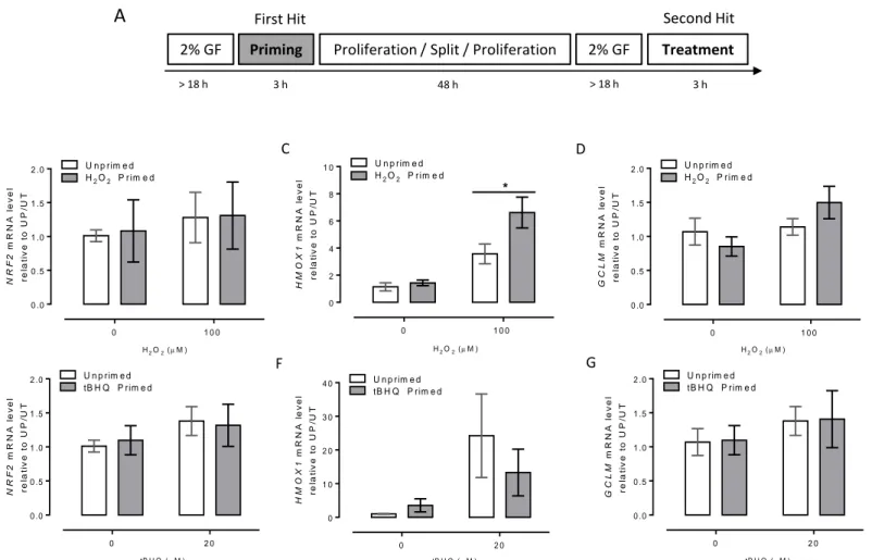
Considering the original hypothesis, an ETM mechanism could answer the question left by chapter two. ETM implies epigenetic modifications at the promoter of inducible genes, left in the promoter by a first stimulus, called gene priming. This priming favors an over- expression of the gene when cells are stimulated for a second time141. The in vivo oxidative
31 stress in LGA newborn of obese mothers could act as a first priming factor in the GPX1 gene in HUAEC, being unmasked by a second in vitro challenge.
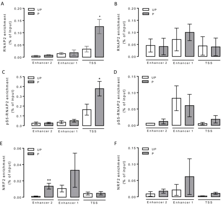
In the third chapter of this thesis, the question if oxidative stress itself is necessary and sufficient to lead to epigenetic modifications in antioxidant coding-protein genes in an ETM manner. Using a pro-oxidant double hit protocol in primary control-HUAEC, EA.hy926 or HeLa cells, among several antioxidant-coding genes, HMOX1 gene acquired an ETM state.
The ETM was characterized by an open chromatin state at the core promoter and enhancer 2 (located at -9 kb from TSS) of HMOX1, together with a paused RNAPII and NRF2 enrichment at the core promoter and enhancer 2, respectively.
The fourth chapter comprises is a general discussion of redox status alterations on endothelial cell physiology; as well as the possible molecular/epigenetic mechanisms that could be involved in the programming of vascular function.
This thesis shows the first evidence that maternal obesity is associated with changes in the antioxidant capacity of endothelial cells of LGA-offspring and proposes epigenetic transcriptional memory in the HMOX1 gene as a specific mechanism.
References
1. Hamann, A. Aktuelles zur Adipositas (mit und ohne Diabetes). Diabetologe 13, 331–
341 (2017).
2. WHO. Global status report on noncommunicable diseases 2014. World Health 176 (2014). doi:ISBN 9789241564854
3. MINSAL. Primeros y segundos resultados de ENS 2016-2017. (2018). at
<http://epi.minsal.cl/resultados-encuestas/>
4. Adamo, K. B., Ferraro, Z. M. & Brett, K. E. Can we modify the intrauterine environment to halt the intergenerational cycle of obesity? International Journal of Environmental Research and Public Health 9, 1263–1307 (2012).
5. Baeten, J. M., Bukusi, E. A. & Lambe, M. Pregnancy complications and outcomes among overweight and obese nulliparous women. Am. J. Public Health 91, 436–440 (2001).
32 6. Ehrenberg, H. M., Mercer, B. M. & Catalano, P. M. The influence of obesity and diabetes on the prevalence of macrosomia. in American Journal of Obstetrics and Gynecology 191, 964–968 (2004).
7. Bhattacharya, S., Campbell, D. M., Liston, W. A. & Bhattacharya, S. Effect of Body Mass Index on pregnancy outcomes in nulliparous women delivering singleton babies.
BMC Public Health 7, (2007).
8. Schneider, D. et al. Oxidative stress as common trait of endothelial dysfunction in chorionic arteries from fetuses with IUGR and LGA. Placenta 36, 552–558 (2015).
9. Chiavaroli, V. et al. Progression of cardio-metabolic risk factors in subjects born small and large for gestational age. PLoS One 9, e104278 (2014).
10. Perng, W., Gillman, M. W., Mantzoros, C. S. & Oken, E. A prospective study of maternal prenatal weight and offspring cardiometabolic health in midchildhood. Ann.
Epidemiol. 24, 793–800 (2014).
11. Curhan, G. C. et al. Birth weight and adult hypertension and obesity in women.
Circulation 94, 1310–1315 (1996).
12. Hayward, C. E. et al. Chorionic plate arterial function is altered in maternal obesity.
Placenta 34, 281–287 (2013).
13. Reynolds, R. M., Allan, K. M. & Edwin, A. Maternal obesity during pregnancy and premature mortality from cardiovascular event in adult offspring : Br. Med. J. 4539, 1–10 (2013).
14. Furchgott, R. F. & Zawadzki, J. V. The obligatory role of endothelial cells in the relaxation of arterial smooth muscle by acetylcholine. Nature 288, 373–6 (1980).
15. Borland, C. Endothelium in control. Br. Heart J. 66, 405 (1991).
16. Ross, R. The pathogenesis of atherosclerosis: a perspective for the 1990s. Nature 362, 801–809 (1993).
17. Panza, J. A., Quyyumi, A. A., Brush, J. E. & Epstein, S. E. Abnormal Endothelium- Dependent Vascular Relaxation in Patients with Essential Hypertension. N. Engl. J.
Med. 323, 22–27 (1990).
18. Hadi, H. A. R. & Suwaidi, J. Al. Endothelial dysfunction in diabetes mellitus. Vasc.
Health Risk Manag. 3, 853–76 (2007).
19. Deanfield, J. et al. Endothelial function and dysfunction. Part I. J. Hypertens. 23, 7–
17 (2005).
20. CHRISTENSEN, H. N. & COOPER, P. F. Glycine and alanine concentrations of body fluids; experimental modification. J. Biol. Chem. 168, 191–196 (1947).
21. Panayiotidis, M. Reactive oxygen species (ROS) in multistage carcinogenesis. Cancer Lett. 266, 3–5 (2008).
22. Halliwell, B. & Cross, C. E. Oxygen-derived species: their relation to human disease and environmental stress. Environ. Health Perspect. 102 Suppl 10, 5–12 (1994).
23. Vásquez-Vivar, J. et al. Superoxide generation by endothelial nitric oxide synthase:
The influence of cofactors. Biochemistry 95, 9220–9225 (1998).
24. Fukai, T. & Ushio-Fukai, M. Superoxide Dismutases: Role in Redox Signaling, Vascular Function, and Diseases. Antioxid. Redox Signal. 15, 1583–1606 (2011).
25. Dworakowski, R., Alom-Ruiz, S. P. & Shah, A. M. NADPH oxidase-derived reactive oxygen species in the regulation of endothelial phenotype. Pharmacol. Rep. 60, 21–8 (2008).
26. Brand, M. D. et al. Mitochondrial superoxide: Production, biological effects, and activation of uncoupling proteins. Free Radical Biology and Medicine 37, 755–767
33 (2004).
27. Huie, R. E. & Padmaja, S. The reaction of no with superoxide. Free Radic. Res.
Commun. 18, 195–9 (1993).
28. Radi, R. Nitric oxide, oxidants, and protein tyrosine nitration. Proc. Natl. Acad. Sci.
U. S. A. 101, 4003–8 (2004).
29. Niki, E., Yoshida, Y., Saito, Y. & Noguchi, N. Lipid peroxidation: Mechanisms, inhibition, and biological effects. Biochem. Biophys. Res. Commun. 338, 668–676 (2005).
30. Poppek, D. & Grune, T. Proteasomal Defense of Oxidative Protein Modifications.
Antioxid. Redox Signal. 8, 173–184 (2006).
31. Hayakawa, H., Taketomi, A., Sakumi, K., Kuwano, M. & Sekiguchi, M. Generation and Elimination of 8-Oxo-7,8-dihydro-2’-deoxyguanosine 5’-Triphosphate, a Mutagenic Substrate for DNA Synthesis, in Human Cells. Biochemistry 34, 89–95 (1995).
32. Birben, E. et al. Oxidative Stress and Antioxidant Defense. WAO J. 5, 9–19 (2012).
33. Panda, S. K. Assay Guided Comparison for Enzymatic and Non-Enzymatic Antioxidant Activities with Special Reference to Medicinal Plants. Intech 381–400 (2012). doi:10.5772/50782
34. Ryu, H. et al. Sp1 and Sp3 are oxidative stress-inducible, antideath transcription factors in cortical neurons. J Neurosci 23, 3597–3606 (2003).
35. Goettsch, C. et al. Arterial flow reduces oxidative stress via an antioxidant response element and Oct-1 binding site within the NADPH oxidase 4 promoter in endothelial cells. Basic Res. Cardiol. 106, 551–561 (2011).
36. Hori, Y. S., Kuno, A., Hosoda, R. & Horio, Y. Regulation of FOXOs and p53 by SIRT1 modulators under oxidative stress. PLoS One 8, e73875 (2013).
37. Polvani, S., Tarocchi, M. & Galli, A. PPARγ and Oxidative Stress: Con(β) Catenating NRF2 and FOXO. PPAR Res. 2012, 641087 (2012).
38. Qutub, A. A. & Popel, A. S. Reactive Oxygen Species Regulate Hypoxia-Inducible Factor 1α Differentially in Cancer and Ischemia▿ †. Mol. Cell. Biol. 28, 5106–5119 (2008).
39. Chen, X.-L. et al. Activation of Nrf2/ARE pathway protects endothelial cells from oxidant injury and inhibits inflammatory gene expression. Am. J. Physiol. Heart Circ.
Physiol. 290, H1862-70 (2006).
40. Bouanane, S. et al. Time course of changes in serum oxidant/antioxidant status in overfed obese rats and their offspring. Clin. Sci. 116, 669–680 (2009).
41. Zhang, X., Strakovsky, R., Zhou, D., Zhang, Y. & Pan, Y.-X. A maternal high-fat diet represses the expression of antioxidant defense genes and induces the cellular senescence pathway in the liver of male offspring rats. J. Nutr. 141, 1254–1259 (2011).
42. Malti, N. et al. Oxidative stress and maternal obesity: Feto-placental unit interaction.
Placenta (2014). doi:10.1016/j.placenta.2014.03.010
43. Ma, Q. Role of nrf2 in oxidative stress and toxicity. Annu. Rev. Pharmacol. Toxicol.
53, 401–26 (2013).
44. Kaspar, J. W., Niture, S. K. & Jaiswal, A. K. Nrf2:INrf2 (Keap1) signaling in oxidative stress. Free Radic. Biol. Med. 47, 1304–9 (2009).
45. Nguyen, T., Sherratt, P. J., Huang, H.-C., Yang, C. S. & Pickett, C. B. Increased protein stability as a mechanism that enhances Nrf2-mediated transcriptional
34 activation of the antioxidant response element. Degradation of Nrf2 by the 26 S proteasome. J. Biol. Chem. 278, 4536–41 (2003).
46. Stewart, D., Killeen, E., Naquin, R., Alam, S. & Alam, J. Degradation of transcription factor Nrf2 via the ubiquitin-proteasome pathway and stabilization by cadmium. J.
Biol. Chem. 278, 2396–402 (2003).
47. Nguyen, T., Nioi, P. & Pickett, C. B. The Nrf2-antioxidant response element signaling pathway and its activation by oxidative stress. J. Biol. Chem. 284, 13291–5 (2009).
48. Niture, S. K., Kaspar, J. W., Shen, J. & Jaiswal, A. K. Nrf2 signaling and cell survival.
Toxicol. Appl. Pharmacol. 244, 37–42 (2010).
49. Jain, A. K., Bloom, D. a & Jaiswal, A. K. Nuclear import and export signals in control of Nrf2. J. Biol. Chem. 280, 29158–68 (2005).
50. Itoh, K. et al. An Nrf2/small Maf heterodimer mediates the induction of phase II detoxifying enzyme genes through antioxidant response elements. Biochem. Biophys.
Res. Commun. 236, 313–22 (1997).
51. Erickson, A. M., Nevarea, Z., Gipp, J. J. & Timothy Mulcahy, R. Identification of a variant antioxidant response element in the promoter of the human glutamate-cysteine ligase modifier subunit gene: Revision of the are consensus sequence. J. Biol. Chem.
277, 30730–30737 (2002).
52. Chorley, B. N. et al. Identification of novel NRF2-regulated genes by ChIP-Seq:
influence on retinoid X receptor alpha. Nucleic Acids Res. 40, 7416–29 (2012).
53. Gene, R., Favreau, L. V, Pickett, C. B. & Chern, C. B. J. B. Transcriptional Regulation of the Rat NAD ( P ) H : Quinone. 268, 19875–19881 (1993).
54. Sun, Z., Chin, Y. E. & Zhang, D. D. Acetylation of Nrf2 by p300/CBP augments promoter-specific DNA binding of Nrf2 during the antioxidant response. Mol. Cell.
Biol. 29, 2658–72 (2009).
55. Kawai, Y., Garduño, L., Theodore, M., Yang, J. & Arinze, I. J. Acetylation- deacetylation of the transcription factor Nrf2 (nuclear factor erythroid 2-related factor 2) regulates its transcriptional activity and nucleocytoplasmic localization. J. Biol.
Chem. 286, 7629–40 (2011).
56. Sun, J. et al. Heme regulates the dynamic exchange of Bach1 and NF-E2-related factors in the Maf transcription factor network. Proc. Natl. Acad. Sci. U. S. A. 101, 1461–6 (2004).
57. Li, Z. et al. The polycomb group protein EZH2 inhibits lung cancer cell growth by repressing the transcription factor Nrf2. FEBS Lett. 588, 3000–3007 (2014).
58. Kang, K. A. et al. Epigenetic modification of Nrf2 in 5-fluorouracil-resistant colon cancer cells: involvement of TET-dependent DNA demethylation. Cell Death Dis. 5, e1183 (2014).
59. Bird, A. Perceptions of epigenetics. Nature 447, 396–398 (2007).
60. Luger, K., Mäder, A. W., Richmond, R. K., Sargent, D. F. & Richmond, T. J. Crystal structure of the nucleosome core particle at 2.8 A resolution. Nature 389, 251–260 (1997).
61. Suzuki, T. & Yamamoto, M. Molecular basis of the Keap1-Nrf2 system. Free Radical Biology and Medicine 88, 93–100 (2015).
62. Goll, M. G. & Bestor, T. H. EUKARYOTIC CYTOSINE METHYLTRANSFERASES. Annu. Rev. Biochem. 74, 481–514 (2005).
63. Klose, R. J. & Bird, A. P. Genomic DNA methylation: the mark and its mediators.
Trends Biochem. Sci. 31, 89–97 (2006).
35 64. Berger, S. L. The complex language of chromatin regulation during transcription.
Nature 447, 407–412 (2007).
65. Fuks, F., Burgers, W. A., Brehm, A., Hughes-Davies, L. & Kouzarides, T. DNA methyltransferase Dnmt1 associates with histone deacetylase activity. Nat. Genet. 24, 88–91 (2000).
66. Esteve, P.-O. et al. Direct interaction between DNMT1 and G9a coordinates DNA and histone methylation during replication. Genes Dev. 20, 3089–3103 (2006).
67. Robertson, A. K., Geiman, T. M., Sankpal, U. T., Hager, G. L. & Robertson, K. D.
Effects of chromatin structure on the enzymatic and DNA binding functions of DNA methyltransferases DNMT1 and Dnmt3a in vitro. Biochem. Biophys. Res. Commun.
322, 110–118 (2004).
68. Viré, E. et al. The Polycomb group protein EZH2 directly controls DNA methylation.
Nature 439, 871–874 (2005).
69. Reik, W. Stability and flexibility of epigenetic gene regulation in mammalian development. Nature 447, 425–432 (2007).
70. Wu, H. & Sun, Y. E. Epigenetic Regulation of Stem Cell Differentiation. Pediatr. Res.
59, 21R–25R (2006).
71. Collas, P., Noer, A. & Timoskainen, S. Programming the genome in embryonic and somatic stem cells. J. Cell. Mol. Med. 11, 602–20 (2007).
72. Tahiliani, M. et al. Conversion of 5-Methylcytosine to 5-Hydroxymethylcytosine in Mammalian DNA by MLL Partner TET1. Science (80-. ). 324, 930–935 (2009).
73. Kimura, A., Matsubara, K. & Horikoshi, M. A Decade of Histone Acetylation:
Marking Eukaryotic Chromosomes with Specific Codes. J. Biochem. 138, 647–662 (2005).
74. Kouzarides, T. Chromatin Modifications and Their Function. Cell 128, 693–705 (2007).
75. Santos-Rosa, H. & Caldas, C. Chromatin modifier enzymes, the histone code and cancer. Eur. J. Cancer 41, 2381–2402 (2005).
76. Cosgrove, M. S. & Wolberger, C. How does the histone code work? Biochem. Cell Biol. 83, 468–476 (2005).
77. Wang, G. G., Allis, C. D. & Chi, P. Chromatin remodeling and cancer, part I: covalent histone modifications. Trends Mol. Med. 13, 363–372 (2007).
78. Thiagalingam, S. et al. Histone deacetylases: unique players in shaping the epigenetic histone code. Ann. N. Y. Acad. Sci. 983, 84–100 (2003).
79. Drewell, R. A., Goddard, C. J., Thomas, J. O. & Surani, M. A. Methylation-dependent silencing at the H19 imprinting control region by MeCP2. Nucleic Acids Res. 30, 1139–44 (2002).
80. Kondo, E., Gu, Z., Horii, A. & Fukushige, S. The Thymine DNA Glycosylase MBD4 Represses Transcription and Is Associated with Methylated p16INK4a and hMLH1 Genes. Mol. Cell. Biol. 25, 4388–4396 (2005).
81. Trewick, S. C., McLaughlin, P. J. & Allshire, R. C. Methylation: lost in hydroxylation?
EMBO Rep. 6, 315–320 (2005).
82. Henikoff, S. & Smith, M. M. Histone variants and epigenetics. Cold Spring Harb.
Perspect. Biol. 7, a019364 (2015).
83. Ahmad, K. & Henikoff, S. The histone variant H3.3 marks active chromatin by replication-independent nucleosome assembly. Mol. Cell 9, 1191–200 (2002).
84. Mizuguchi, G. et al. ATP-Driven Exchange of Histone H2AZ Variant Catalyzed by