R E S E A R C H Open Access
IL-13 signaling via IL-13R α 2 triggers TGF- β 1 -dependent allograft fibrosis
Stefan M Brunner1*, Gabriela Schiechl1, Rebecca Kesselring1, Maria Martin1, Saidou Balam1, Hans J Schlitt1, Edward K Geissler1and Stefan Fichtner-Feigl1,2
Abstract
Background:Allograft fibrosis still remains a critical problem in transplantation, including heart transplantation. The IL-13/TGF-β1interaction has previously been identified as a key pathway orchestrating fibrosis in different
inflammatory immune disorders. Here we investigate if this pathway is also responsible for allograft fibrosis and if interference with the IL-13/TGF-β1interaction prevents allograft fibrosis.
Methods:FVB or control DBA/1 donor hearts were transplanted heterotopically into DBA/1 recipient mice and hearts were explanted at day 60 and 100 post-transplantation. Cardiac tissue was examined by Masson’s trichrome staining and immunohistochemistry for CD4, CD8, CD11b, IL-13, Fas ligand, matrix metalloproteinase (MMP)-1, MMP-13,β2-microglobulin, and Gremlin-1. Graft-infiltrating cells were isolated and analyzed by flow cytometry. IL-13 and TGF-β1levels were determined by enzyme-linked immunosorbent assay (ELISA) and the amount of collagen was quantified using a Sircol assay; IL-13Rα2expression was detected by Western blotting. In some experiments IL-13/ TGF-β1signaling was blocked with specific IL-13Rα2siRNA. Additionally, a PCR array of RNA isolated from the allografts was performed to analyze expression of multiple genes involved in fibrosis.
Results:Both groups survived long-term (>100 days). The allogeneic grafts were infiltrated by significantly increased numbers of CD4+(P<0.0001), CD8+(P<0.0001), and CD11b+cells (P= 0.0065) by day 100. Furthermore, elevated IL-13 levels (P= 0.0003) and numbers of infiltrating IL-13+cells (P= 0.0037), together with an expression of IL-13Rα2, were detected only within allografts. The expression of IL-13 and IL-13Rα2resulted in significantly
increased TGF-β1levels (P<0.0001), higher numbers of CD11bhighGr1intermediate
TGF-β1+
cells, and elevated cardiac collagen deposition (P= 0.0094). The allograft fibrosis found in these experiments was accompanied by
upregulation of multiple profibrotic genes, which was confirmed by immunohistochemical stainings of allograft tissue. Blockage of the IL-13/TGF-β1interaction by IL-13Rα2siRNA led to lower numbers of
CD11bhighGr1intermediate
TGF-β1
+, CD4+, CD8+, and CD11b+cells, and prevented collagen deposition (P= 0.0018) within these allografts.
Conclusions:IL-13 signaling via IL-13Rα2induces TGF-β1and causes allograft fibrosis in a murine model of chronic transplant rejection. Blockage of this IL-13/TGF-β1interaction by IL-13Rα2siRNA prevents cardiac allograft fibrosis.
Thus, IL-13Rα2may be exploitable as a future target to reduce allograft fibrosis in organ transplantation.
Keywords:IL-13, IL-13Rα2, TGF-β1, Allograft fibrosis, Heart transplantation
* Correspondence:stefan.brunner@ukr.de
1Department of Surgery, University Medical Center Regensburg, Franz-Josef-Strauss-Allee 11, Regensburg 93053, Germany Full list of author information is available at the end of the article
© 2013 Brunner et al.; licensee BioMed Central Ltd. This is an open access article distributed under the terms of the Creative Commons Attribution License (http://creativecommons.org/licenses/by/2.0), which permits unrestricted use, distribution, and reproduction in any medium, provided the original work is properly cited.
Background
Heart transplantation is an effective therapy for chronic heart failure [1]. Recent immunosuppressive strategies have reduced acute rejection episodes and improved early cardiac graft survival [2]. However, these improvements did not ameliorate chronic allograft rejection, which re- mains an obstacle for better long-term heart transplant survival [3]. Chronic rejection of an allograft causes an in- timal fibrosis in the vessels that leads to cardiac allograft vasculopathy [4]. Another consequence of chronic rejec- tion and inflammation is cardiac fibrosis accompanied by increased stiffness of the heart and diminished contractil- ity [5]. Ultimately, these fibrotic reactions can result in myocardial infarction or sudden death [4,6].
On a molecular level, fibrosis is associated with a disrup- tion of the extracellular matrix and with deposition of extracellular collagen produced by myofibroblasts [5]. In various studies TGF-β1has been identified as the key cyto- kine orchestrating fibrosis development [7,8]. TGF-β1 is produced by macrophages after stimulation by IL-13 via the IL-13Rα2in the presence of IL-4 or TNF-α[9]. Further studies have shown that this pathway is a key initiation point for a complex fibrotic program in chronic TNBS colitis [10]. Additionally, it has been demonstrated that TGF-β1inhibition ameliorates lung fibrosis, chronic allo- graft nephropathy, and also cardiac allograft fibrosis [11-13]. However, no effective therapy to prevent heart allograft fibrosis has been identified so far, possibly be- cause ideal murine transplant models have been lacking to study potential targets and therapies. A mouse model for the examination of cardiac allograft fibrosis should enable long-term survival of a transplanted allograft and develop cardiac fibrosis in the setting of chronic rejection. Tanaka et al. have developed a transplantation model in which FVB (H-2q) donor hearts were placed into DBA/1 recipi- ents that display a similar major histocompatibility com- plex (MHC) (H-2q), but different non-MHC genes (CD5, CD8a, NK1.1, and Thy-1) [14]. In this model, the heart al- lografts survive for up to more than 100 days without im- munosuppression and developed graft coronary artery disease as the result of chronic rejection.
The present study was performed under the hypoth- esis that the FVB to DBA/1 model is appropriate to examine cardiac graft fibrosis. Further, we hypothesized that TGF-β1 stimulated by IL-13 signaling through IL- 13Rα2 is responsible for this allograft fibrosis and that blockage of the pathway by IL-13Rα2-specific siRNA can ameliorate allograft fibrosis.
Materials and methods
Mice and heterotopic heart transplantation
Female DBA/1 (H-2q), FVB (H-2q), and as controls BALB/c and C57BL/6 mice, 10 to 12 weeks old, were purchased from The Jackson Laboratory (Bar Harbor,
ME, USA) and housed at our local animal care facility.
Animal use adhered to institutional guidelines.
Vascularized cardiac allografts were transplanted into the abdomen using a microsurgical technique as previ- ously described by Corry et al.[15]. Donor hearts were perfused via the abdominal vena cava and additionally via the aortic arch with cold 0.9% saline (3 mL each) containing 500 IE heparin. Graft function was assessed by palpation of the abdomen and rejection was defined as cessation of cardiac contractility. All donor hearts had palpable contractions at the time of recovery (60 or 100 days; acute rejection 8 days).
IL-13Rα2-specific siRNA
IL-13Rα2-specific siRNA and control (scrambled) siRNA for use in gene silencing studies were obtained from Dharmacon (Chicago, IL, USA). The siRNA (100 μg) was encapsulated in HVJ-E and prepared as previously described before administration by intraperitoneal injec- tion (100μL) every other day [10,16]. The sequence used for the siRNA is 5′-GGAATCTAATTTACAAGGA-3′.
Histology and immunohistochemistry
Formalin-fixed and paraffin-embedded samples were pre- pared and sectioned (2 to 3 μm). Tissue sections were stained with Masson’s trichrome. Frozen sections (2 to 3 μm) were blocked with 1% BSA (Biomol, Hamburg, Germany), 10% goat serum (Sigma-Aldrich, St Louis, MO, USA), or an antibody dilution buffer. As primary anti- bodies, rat monoclonal anti-mouse CD11b (557395; BD, Heidelberg, Germany), CD4 (550280; BD), and CD8 antibodies (Ab25478; Abcam, Cambridge, UK), a goat polyclonal anti-mouse IL-13 antibody (AF-413-NA;
R&D Systems, Minneapolis, MN, USA) and a rabbit polyclonal anti-mouse Fas ligand (Ab15285; Abcam), a rabbit polyclonal anti-mouse matrix metalloproteinase (MMP)-1 (orb101432; Biorbyt, Cambridge, UK), a rabbit polyclonal anti-mouse MMP-13 (Ab39012; Abcam), a rabbit polyclonal anti-mouse β2-microglobulin (Ab87483;
Abcam), and a rabbit polyclonal anti-mouse Gremlin-1 antibody (Ab90670; Abcam) were used. After staining with goat anti-rat-Fab2 (sc-3822; Santa Cruz Biotechnology, Heidelberg, Germany), donkey anti-goat-Fab2 (sc-2042;
Santa Cruz), or goat anti-rabbit-Fab2 (Ab64256; Abcam) secondary antibody, sections were incubated with SensiTek HRP (ScyTec Laboratories, Logan, UT, USA) and positive signals were visualized using a 3,3′-diaminobenzidine- tetrahydrochlorhydrate (DAB) kit (Merck, Darmstadt, Germany) or AEC+ High Sensitivity Substrate Chromogen kit (Dako, Hamburg, Germany). Images were captured using an Axio Observer Z1 microscope (Carl Zeiss, Oberkochen, Germany). For quantifying graft-infiltrating leukocytes, three high power fields (HPFs; 20x magnification) were counted per slide by two independent examiners.
Western blot analyses
Cells were lysed with radioimmunoprecipitation assay buf- fer and the whole cell lysates obtained were subjected to SDS-PAGE. The separated proteins obtained were trans- ferred to a nitrocellulose membrane and immunoblotted.
IL-13Rα2 was detected by incubation with a monoclonal rat anti-mouse IL-13Rα2(R&D Systems), followed by in- cubation with horseradish peroxidase-conjugated anti-rat IgG (Invitrogen, Carlsbad, CA, USA). Membranes were developed with SuperSignal West Pico Chemiluminescent Substrate (Pierce Chemical, Dallas, TX, USA) and exposed to X-ray film.
Collagen assay
Heart allografts were harvested on day 60 and day 100 after transplantation, and homogenized in 0.5 mol/L acetic acid containing pepsin (at a concentration of 10 mg tissue/10 mL of acetic acid solution). The resulting mixture was then incubated and stirred for 24 hours at 4°C. Total soluble collagen content of the mixture was then determined with a Sircol Collagen Assay kit (Biocolor, Carrickfergus, UK), as described by the manu- facturer. Acid soluble type I collagen supplied with the kit was used to generate a standard curve.
Cell isolation from cardiac grafts and spleens
Cardiac tissue was minced in 10 mL of RPMI 1640 medium with 10% FCS, 600 U/mL collagenase II (Roche Diagnostics, Mannheim, Germany), and deoxyribonucle- ase I (DNase; Sigma-Aldrich). This mixture was shaken at room temperature for 2 hours and supernatant was flushed through a 100 μm nylon cell strainer (Schubert
& Weiss, Munich, Germany). Remaining tissue was again digested in 5 mL of RPMI-collagenase-DNase so- lution at 37°C and strained through a 100 μm nylon strainer. Splenic tissue was minced and strained through a 100μm nylon strainer. Digested cell suspensions were centrifuged for 5 minutes at 1,500 rpm (4°C). To remove red blood cells, the pellet was treated with ACK lysis buffer (Lonza Walkersville, Walkersville, MD, USA) and incubated for 2 minutes at room temperature. After cen- trifugation, cells were suspended in HBSS medium (Gibco, Grand Island, NY, USA) and counted.
Flow cytometry
Cell isolates were blocked with 1% mouse serum (Dako, Glostrup, Denmark) and stained with appropriate non- overlapping conjugated monoclonal antibodies (anti- Gr1 antibody from Miltenyi Biotec, Bergisch Gladbach, Germany; all other antibodies from eBioscience, San Diego, CA, USA). Intracellular staining was carried out by first fixing and permeabilizing cells with Cytofix/
Cytoperm solution (BD Pharmingen, San Diego, CA, USA). Analyses were performed using a FACSCanto II
flow cytometer (BD Biosciences, San Jose, CA, USA).
Data were obtained using BD CellQuest Pro acquisition software (BD Biosciences) and analyzed via FlowJo soft- ware (Tree Star Inc, Ashland, OR, USA).
ELISA
Heart allografts were harvested at day 60 and day 100, and graft-infiltrating cells were isolated. Isolated graft- infiltrating cells were cultured at 37°C. For IL-13, we cul- tured 1 × 10⁶ cells per 1 mL medium for 48 hours;
for TGF-β1measurements, we cultured 1 × 10⁵cells per 100 μL medium for 24 hours. During the culture period cells were stimulated with plate-bound anti-CD3 antibody (10μg/mL) and soluble anti-CD28 antibody (1μg/mL; BD Biosciences Pharmingen) for measurement of IL-13 (R&D Systems). For determination of TGF-β1levels (Invitrogen) cells were stimulated with plate-bound anti-CD3 antibody (10 μg/mL), soluble anti-CD28 antibody (1 μg/mL), and recombinant murine IL-13 (20 ng/mL; R&D Systems).
Cytokine concentrations were determined in duplicate by enzyme-linked immunosorbent assay (ELISA) according to the manufacturer’s instructions. TGF-β1was measured in medium containing TGF-β1-depleted human serum.
RNA isolation and PCR array
In heart allografts recovered on day 100 after transplant- ation, RNA was extracted using the RNeasy Mini Kit (Qiagen, Hilden, Germany), as described by the manu- facturer. One microgram of total RNA was reverse tran- scribed using the AffinityScript QPCR cDNA Synthesis Kit (Agilent Technologies, Böblingen, Germany). Expres- sion of genes relevant for fibrosis was determined with a Mouse Fibrosis RT2Profiler PCR Array (SA Biosciences, Hilden, Germany) using the LightCycler 480 Real-Time PCR System (Roche).
Statistics
All data, unless otherwise specified, are shown as the mean ± standard error of the mean (SEM), and were compared using a two-tailed Student’s test. The level of significance was set at a probability ofP<0.05.
Results
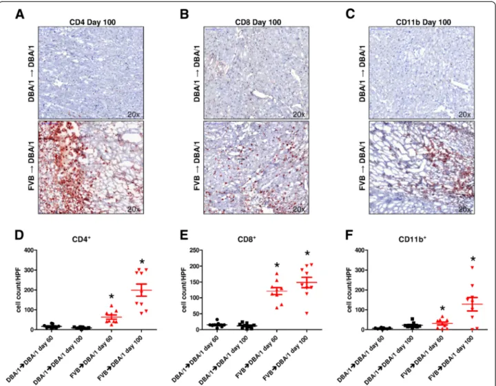
FVB allografts transplanted in DBA/1 recipients showed significantly increased infiltration by CD4+, CD8+, and CD11b+cells
To determine the number of graft-infiltrating cells, heart allografts were harvested on day 60 and day 100 after trans- plantation, and were stained for CD4, CD8, and CD11b. In syngeneic grafts (DBA/1 into DBA/1), low numbers of CD4+(day 60, 16 ± 3 and day 100, 9 ± 1 cells/HPF), CD8+ (day 60, 15 ± 2 and day 100, 12 ± 3 cells/HPF), and CD11b+cells (day 60, 6 ± 1 and day 100, 22 ± 5 cells/HPF) were detected (Figure 1A,B,C,D,E,F). Allogeneic heart grafts
(FVB into DBA/1) at day 60 after transplantation showed significantly higher cell numbers of CD4+ (63 ± 11 cells/
HPF;P = 0.0007), CD8+ (121 ± 11 cells/HPF; P<0.0001), and CD11b+cells (31 ± 7 cells/HPF;P= 0.0045) compared to grafts in the syngeneic group. The numbers of CD4+and CD11b+ cells in the FVB into DBA/1 group increased further by day 100 after transplantation (day 60 versus day 100, CD4+ P = 0.0009; CD11b+ P = 0.0124), whereas the increase in the number of CD8+ cells did not reach statis- tical significance (P = 0.1921). In comparison to control animals at day 100 after transplantation, the allogeneic group showed significantly higher levels of CD4+ (199 ± 31 cells/
HPF; P < 0.0001), CD8+ (149 ± 17 cells/HPF; P <0.0001), and CD11b+cells (128 ± 34 cells/HPF;P= 0.0065).
FVB allografts transplanted into DBA/1 recipients showed significantly increased levels of IL-13, IL-13Rα2, and TGF-β1
To examine if TGF-β1 stimulated by IL-13 signaling is elevated in mice receiving allogeneic transplants, IL-13 levels were measured by ELISA in supernatants of cultured allograft-infiltrating cells. Syngeneic DBA/1 heart grafts showed similar IL-13 concentrations at day 60 (108 ± 13 pg/mL) and at day 100 (112 ± 12 pg/mL) (P = 0.8415). FVB allografts placed in DBA/1 recipients showed significantly elevated IL-13 levels at day 60 (187 ± 10 pg/mL;P= 0.0031) and at day 100 after transplantation (303 ± 23 pg/mL;P= 0.0003) in comparison to allogeneic grafts at the same respective time points (Figure 2A).
Additionally, immunohistochemical staining for IL-13 in
Figure 1Increased infiltration by CD4+, CD8+, and CD11b+cells in allogeneically transplanted grafts. (A,B,C)Representative stainings for CD4, CD8, and CD11b in syngeneic (DBA/1 into DBA/1) or allogeneic (FVB into DBA/1) heart allografts explanted at day 100 after transplantation.
(D)In FVB into DBA/1 transplanted hearts, significantly elevated numbers of CD4+cells were detected (day 60,P= 0.0007 and day 100, P<0.0001; day 60 versus day 100,P= 0.0009) in comparison to DBA/1 into DBA/1 transplanted grafts.(E)FVB allografts transplanted into DBA/1 recipients showed numbers of CD8+cells (day 60,P<0.0001 and day 100,P<0.0001; day 60 versus day 100,P= 0.1921) that were significantly higher than in the syngeneic group.(F)Significantly higher levels of CD11b+cells were found in allogeneic (FVB into DBA/1) grafts (day 60, P= 0.0045 and day 100,P= 0.0065; day 60 versus day 100,P= 0.0124) when compared to DBA/1 to DBA/1 transplanted hearts. The histological score is the mean of 3 HPF (20× magnification); at least five mice per group were analyzed. *P<0.05. HPF, high power field.
FVB allografts transplanted into DBA/1 mice showed sig- nificantly increased numbers of IL-13+cells/HPF at day 60 (16 ± 4 versus 6 ± 2 cells/HPF;P= 0.0228) and at day 100 (96 ± 26 versus 7 ± 2 cells/HPF;P= 0.0037), relative to the syngeneic controls (day 60 versus day 100; P = 0.0083;
Figure 2B,C). Western blot analyses of lysates from allograft- infiltrating cells indicated detectable expression of IL-13Rα2
only in the allogeneic FVB to DBA/1 mice, both at day 60 and at day 100 after heart transplantation (Figure 2D).
As the next step, the effector cytokine TGF-β1was mea- sured by ELISA after culturing and stimulating cells iso- lated from the allografts. In DBA/1 mice grafted with FVB hearts, significantly elevated TGF-β1levels were detected at day 60 (88 ± 5 versus 46 ± 5 pg/mL;P= 0.0010) and at day 100 (133 ± 6 versus 42 ± 7 pg/mL;P< 0.0001) in com- parison to the syngeneic controls, and also versus the FVB control heart transplants (40 ± 8 pg/mL;P = 0.0048 and P = 0.0009, respectively; Figure 2E). In accordance with
Figure 2Activation of IL-13/TGF-β1pathway in allogeneically transplanted grafts. (A)ELISA of supernants of cultured allograft-infiltrating cells detected significantly elevated IL-13 levels in allografts (day 60,P= 0.0031 and day 100,P= 0.0003)compared to syngrafts or FVB control hearts (P= 0.0055 andP= 0.0042).(B)Immunohistochemistry showed significantly higher and over time increasing numbers of IL-13+cells in FVB hearts transplanted into DBA/1 recipients (day 60,P= 0.0228; day 100,P= 0.0037) relative to syngeneic animals (day 60 versus day 100, P= 0.0083).(C)Representative immunohistochemical stainings showed more IL-13+cells in allografts ( FVB into DBA/1) compared to controls (DBA/1 into DBA/1; day 100).(D)Western blot analysis revealed expression of IL-13Rα2only in allograft-infiltrating cells isolated from
allogeneically transplanted hearts (FVB into DBA/1) in contrast to cells isolated from FVB controls or syngrafts (DBA/1 into DBA/1) without IL-13Rα
2expression.(E)Measurement of TGF-β1by ELISA detected significantly elevated TGF-β1levels in cells isolated from DBA/1 mice grafted with FVB hearts at day 60 (88 ± 5 versus 46 ± 5 pg/mL;P= 0.0010) and at day 100 (133 ± 6 versus 42 ± 7 pg/mL;P<0.0001) in comparison to syngrafts, and FVB control hearts (40 ± 8 pg/mL;P= 0.0048 andP= 0.0009, respectively).(F)Flow cytometry of graft-infiltrating cells extracted from allografts showed a higher percentage of CD11bhighGr1intermediate
TGF-β1+cells (7.3%) than in the syngeneic controls (1.2%; day 100).(G)In the flow cytometric analysis (pre-gated for CD45) these CD11bhighcells were the only source of TGF-β1production. The histological score is the mean of 3 HPF (20x magnification); at least five mice per group were analyzed. *P<0.05. ELISA, enzyme-linked immunosorbent assay; HPF, high power field; IL-13, interleukin 13; TGF-β1, transforming growth factor beta 1.
these results, flow cytometry of graft-infiltrating cells extracted from allogeneic grafts at day 100 showed a higher percentage of CD11bhighGr1intermediateTGF-β1+
cells (7.3%) than in the syngeneic controls (1.2%; Figure 2F).
Furthermore, flow cytometry demonstrated that these CD11bhighcells were likely the only source of TGF-β1pro- duction in this transplantation model (Figure 2G).
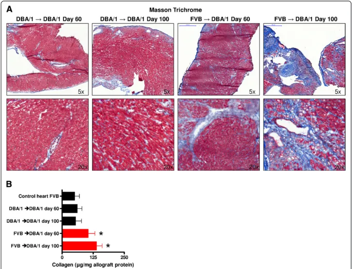
FVB allografts transplanted into DBA/1 recipients showed significantly increased levels of collagen deposition To prove that FVB hearts transplanted in DBA/1 mice develop fibrosis, Masson’s trichrome staining was performed. In these stainings, a strong collagen de- position was found in the allogeneic grafts at day 60, with a further increase in collagen deposition by day 100 after heart transplantation. No such fibrotic
collagen deposition was observed in the syngeneic con- trol mice (Figure 3A).
A Sircol assay was conducted to better quantify collagen levels in heart allografts. With this method, the amount of collagen was found to be significantly greater in FVB hearts placed into DBA/1 recipients at day 60 (105 ± 13 versus 61 ± 9 μg/mg allograft protein; P = 0.0342) and at day 100 after transplantation (139 ± 11 versus 54 ± 12μg/mg allograft protein; P = 0.0022) compared to the DBA/
1-to-DBA/1 mice and also to FVB control hearts (P= 0.0094; Figure 3B).
FVB allografts transplanted into DBA/1 recipients showed upregulation of profibrotic and downregulation of antifibrotic genes
To demonstrate at an mRNA-level that allogeneic grafts have upregulated profibrotic genes, RNA was isolated
Figure 3Increased collagen deposition in allogeneically transplanted grafts. (A)Representative Masson’s trichrome stainings showed increased levels of collagen (blue color) in allogeneically (FVB into DBA/1) compared to syngeneically (DBA/1 into DBA/1) transplanted hearts explanted at day 60 and day 100 after transplantation (5× and 20× magnification).(B)Analysis by Sircol assay detected significantly higher amounts of collagen in FVB hearts placed into DBA/1 recipients at day 60 (P= 0.0342) and at day 100 after transplantation (P= 0.0022) compared to the DBA/1 to DBA/1 mice and also to FVB control hearts (P= 0.0094). At least five mice per group were analyzed. *P< 0.05.
from the grafted tissues and a PCR array was performed.
This PCR array, which profiles the expression of 84 key genes involved in tissue remodeling and fibrosis, revealed an upregulation of IL-13 and TGF-β1(as detected in the previous experiments), but also a strong upregulation of other cytokines relevant for fibrosis, including IL-1α, IL-1β, and TNF-α. Further, chemokines such as Ccl3, Ccl12, Ccr2, and Cxcr4, and genes involved in epithelial- mesenchymal transition or cell adhesion such as Fas ligand, β2-microglobulin, integrin-α2, integrin-β6, MMP-1a, and MMP-13, were upregulated; in contrast, Gremlin-1 and Smad6 were downregulated (Figure 4A,B).
Additionally, immunohistochemical labeling was per- formed for selected targets. According to these PCR array results, FVB allografts placed into DBA/1 recipi- ents showed strong positivity for Fas ligand, MMP-1, MMP-13, and β2-microglobulin at day 100, whereas Gremlin-1 staining was stronger in the syngeneic group (Figure 5). In comparison to acutely rejected grafts (BALB/c to C57BL/6), and also to naive non-transplanted FVB hearts, this expression pattern was unique in fibrosis of FVB allografts transplanted into DBA/1 recipients.
Specific blockage of IL-13Rα2abrogates TGF-β1
production and prevents allograft fibrosis
To investigate if allograft fibrosis depends on TGF-β1
production stimulated by IL-13 secretion, IL-13 signal- ing in DBA/1 recipients grafted with FVB hearts was inhibited by intraperitoneal treatment with specific IL-
13Rα2siRNA or control siRNA. Flow cytometric analysis of graft-infiltrating cells from hearts harvested 100 days after transplantation showed a low percentage of CD11bhighGr1intermediateTGF-β1+
cells (0.3%) in the siRNA- treated group compared to controls (4.2%; Figure 6A).
Furthermore, quantification of collagen by the Sircol assay showed significantly less collagen levels in mice treated with specific IL-13Rα2 siRNA compared to mice treated with control siRNA (122 ± 23 versus 314 ± 28 μg/mg allograft protein; P = 0.0018) (Figure 6B). This was in accordance with Masson’s trichrome staining, in which only small areas of collagen deposition were observed in siRNA-treated hearts, whereas extensive amounts of collagen were detected in control allografts (Figure 6C).
To test whether the differences in TGF-β1 production caused imbalances in CD4+Foxp3+ regulatory T cells (Tregs), a flow cytometric analysis of cells from allografts was performed 100 days after transplantation. However, CD4+Foxp3+ Tregs were found to be present at equal levels in siRNA- and control siRNA-injected animals (15.6% versus 15.2%, respectively; Figure 6D). Immunohis- tochemical staining of allografts after therapy with specific IL-13Rα2 siRNA showed levels of CD4+, CD8+, and CD11b+ cells that were much lower after treatment with control siRNA, but similar to syngeneic transplanted ani- mals (Figure 6E versus Figure 1A,B,C).
In this study we demonstrate for the first time that allograft fibrosis is caused by IL-13 signaling through the receptor IL-13Rα2, which consequently leads to elevated
Figure 4Activation of a‘fibrotic program’in allogeneically transplanted grafts. (A,B)PCR array using RNA extracted from explanted hearts (day 100) of allogeneically (FVB into DBA/1) or syngeneically (DBA/1 into DBA/1) transplanted mice demonstrated an upregulation of pro-fibrotic genes and a downregulation of anti-fibrotic genes in the allogeneic group.
TGF-β1 levels resulting in increased collagen deposition in heart allografts. Additionally, we show that inhibition of this pathway by siRNA specific for IL-13Rα2prevents allograft fibrosis.
The findings presented here that link IL-13 signaling via IL-13Rα2 to allograft fibrosis are based on our previous studies showing that such signaling is essential in the devel- opment of inflammation-associated fibrosis [9,10,17]. These studies showed that IL-13 induces TGF-β1via a two-stage process involving: 1) induction of IL-13Rα2expression by IL-13 (or IL-4) signaling via IL-13Rα1, combined with TNF-αsignaling through its receptor; and 2) IL-13 signal- ing via IL-13Rα2 to induce an AP-1 variant containing c-Jun and Fra-2 that activates the TGF-β1 promoter [9].
The importance of this pathway for development of fibrosis has been shown extensively by our group in bleomycin- induced lung fibrosis and chronic TNBS-induced colitis [10,17]. Thus, these previous studies provided the basis to investigate the importance of IL-13/TGF-β1 signaling in the setting of allograft fibrosis.
The study presented here shows increasing levels of IL- 13 and IL-13+cells within allografts of transplanted mice, in contrast to control mice receiving syngeneic grafts.
Multiple studies have demonstrated that IL-13 is essential for the development of dermal, gastrointestinal, and pul- monary fibrosis, as well as fibro-obliterative lesions found in the bronchiolitis obliterans (BO) syndrome [9,10,17-20].
Consistent with these studies, IL-13Rα2was detected only in the FVB allografts transplanted into DBA/1 recipients in our experiments. This receptor has been shown to link IL-13 signaling with further fibrotic downstream effects [9,21]. In follow-up, we detected elevated levels of TGF-β1
in the mice receiving allogeneic grafts exclusively. Results from previous studies have indicated that TGF-β1is the key cytokine for development of allograft fibrosis in mur- ine models and in humans, and that depletion of TGF-β1
can prevent allograft fibrosis [7,12,22]. Other cytokines such as IL-6 and IL-17 can modulate the TGF-β1-medi- ated fibrotic reactions [7,8]. Additionally, a study by Faust et al. concluded that T cell TGF-βsignaling was required
Figure 5Immunohistochemical analysis of the‘fibrotic program’active in allogeneically transplanted grafts.Representative immunohistochemical stainings show strong positivity for Fas ligand, MMP-1, MMP-13, andβ2-microglobulin in allogeneically (FVB into DBA/1) and for Gremlin-1 in syngeneically (DBA/1 into DBA/1) transplanted hearts explanted at day 100, in comparison to naive non-transplanted FVB hearts and acutely rejected (BALB/c into C57BL/6) allografts (20× magnification). MMP, matrix metalloproteinase.
for the development of allograft fibrosis [8]. In parallel to the elevated levels of TGF-β1, we found increased allograft-infiltration with CD11bhighGr1intermediateTGF-β1+
cells in the DBA/1 mice transplanted with FVB allografts;
it has been shown by our group and others that CD11bhighGr1intermediate
cells are the main source for TGF-β1production [23-25]. In the allogeneic situation of the mouse model we used, activation of the profibrotic IL- 13/TGF-β1 interaction led to allograft fibrosis that was continuously increasing over time after transplantation.
Another important finding from this study is that allograft fibrosis can be prevented by blockage of the IL-13/TGF-β1
interaction through specific IL-13Rα2 siRNA. After
treatment with IL-13Rα2 siRNA, an almost complete re- duction of TGF-β1production by CD11bhighGr1intermediate
cells (the main producers of TGF-β1in this model) was observed [24,25]. The reduction of TGF-β1-producing cells and reduced TGF-β1 levels consequently led to di- minished collagen deposition in heart allografts and there- fore reduced allograft fibrosis. Tregs were also considered as contributors to the TGF-β1effect. While CD4+Foxp3+ Tregs can produce TGF-β1 to mediate their tolerogenic functions and expand induced regulatory T cells (iTregs), there was no difference in their numbers in control versus IL-13Rα2 siRNA-injected mice [26-29]. Notably, after therapy with IL-13Rα2siRNA, CD4+and CD8+cells were
Figure 6Blockage of IL-13Rα2prevents TGF-β1production and fibrosis in allogeneically transplanted grafts. (A)Flow cytometric analysis of graft-infiltrating cells of allogeneically (FVB into DBA/1) transplanted hearts (day 100) detected a strongly diminished percentage of
CD11bhighGr1intermediate
TGF-β1+cells (0.3%) in the siRNA-treated group compared to the control animals (4.2%).(B)Quantification of collagen by Sircol assay showed significantly less collagen levels in mice injected with specific IL-13Rα2siRNA (P= 0.0018) than mice treated with control siRNA. Levels were not significantly different to FVB control hearts (P= 0.1309).(C)Representative Masson’s trichrome stainings showed reduced levels of collagen in allogeneically (FVB into DBA/1) transplanted hearts treated with siRNA compared to mice treated with control peptide (day 100 after transplantation; 20x magnification).(D)In flow cytometric analysis the frequency of CD4+Foxp3+Tregs isolated from allogeneically transplanted grafts (day 100) was similar in siRNA- and control siRNA-injected animals (15.6 versus 15.2%).(E)Representative
immunohistochemical stainings (day 100) showed numbers of CD4+, CD8+, and CD11b+cells in FVB allografts transplanted into DBA/1 mice and treated with siRNA that were lower than mice treated with control siRNA and comparable to syngeneic control grafts (Figure 1 A,B,C). At least five mice per group were analyzed. *P<0.05. Treg, regulatory T cell.
found at levels that were similar to mice receiving syngen- eic grafts, and were much lower than in allotransplanted mice not given IL-13Rα2siRNA treatment.
For our investigations, we used a heterotopic murine heart transplantation model in which FVB hearts were placed in DBA/1 recipients. This chronic rejection model with minor multiple non-MHC mismatches has been used previously to study graft coronary artery disease [14]. We show that the FVB to DBA/1 model can also be used to examine allograft fibrosis. Over time, transplanted allografts are infiltrated by increasing numbers of CD4+, CD8+, and CD11b+ cells, a fact that was also observed by Tanaka et al. in the original description of this transplantation model [14]. We further demonstrated by PCR array that a
‘fibrotic program’ is active in this FVB to DBA/1 model.
Profibrotic factors such as IL-1αand IL-1βthat play a role in liver fibrosis development, and TNF-αwhich is an es- sential cofactor of IL-13 to induce the expression of IL- 13Rα2, were upregulated. Further, Ccl-12 and Cxcr-4 that were both shown to be involved in pulmonary fibrosis, Ccl-3 which has been described to be important in systemic sclerosis, and Ccr-2 that is associated with allograft fibrosis, were overexpressed [9,30-33]. Add- itionally, the PCR array showed upregulation of other influential molecules such as Fas/Fas ligand, which is important in the development of fibrotic lesions associ- ated with adult respiratory distress syndrome (ARDS).
MMP-1 and MMP-13 (involved in remodeling pro- cesses occurring during fibrosis) and β2-microglobuin were also overexpressed in the PCR array and positive in the immunohistochemistry of FVB allografts transplanted into DBA/1 recipients [34-36]. In contrast, genes like Gremlin-1 that may contribute to reversibility of lung fi- brosis in rats, and Smad6 which in complex with Smurf-1 effectively attenuated TGF-β1 signaling, were down- regulated [37,38]. Altogether, these findings support the fact that this FVB to DBA/1 transplantation model is suit- able not only to study graft coronary artery disease, but also to examine organ allograft fibrosis.
Conclusions
In conclusion, this study shows that IL-13 signaling via IL- 13Rα2 induces TGF-β1 and causes allograft fibrosis in a chronic transplant rejection model that is now also est- ablished as a model to study allograft fibrosis. Further, we demonstrate in this study that blockage of this IL-13/TGF- β1interaction by IL-13Rα2siRNA prevents heart allograft fi- brosis. Together, our results indicate that IL-13Rα2may be exploitable as a future target to reduce allograft fibrosis in organ transplantation.
Abbreviations
ACK:Ammonium-chloride-potassium; ARDS: Adult respiratory distress syndrome; BO: Bronchiolitis obliterans; BSA: Bovine serum albumin;
DAB: 3,3'-diaminobenzidine; DNase: Deoxyribonuclease I; ELISA: Enzyme-linked
immunosorbent assay; FCS: Fetal calf serum; HBSS: Hanks’balanced salt solution;
HPF: High power field; HVJ-E: Hemagglutinating virus of Japan envelope;
IgG: Immunoglobulin G; IL-13: Interleukin 13; iTreg: Induced regulatory T cell;
MHC: Major histocompatibility complex; MMP: Matrix metalloproteinase;
PCR: Polymerase chain reaction; RPMI: Roswell park memorial institute;
SEM: Standard error of the mean; siRNA: Small interfering RNA;
TGF-β1: Transforming growth factor beta 1; TNBS: 2,4,6-trinitrobenzene sulfonic acid; TNF: Tumor necrosis factor; Treg: Regulatory T cell.
Competing interests
The authors declare that they have no competing interests.
Authors’contributions
SMB designed the study concept, collected and analyzed data, and wrote the manuscript. GS, RK, SB, and MM collected and analyzed data. HJS and EKG analyzed data and reviewed the manuscript. SFF designed the study concept, collected and analyzed data, and reviewed the manuscript. All authors read and approved the final manuscript.
Acknowledgments
This study was supported by the University of Regensburg, Regensburg, Germany.
Author details
1Department of Surgery, University Medical Center Regensburg, Franz-Josef-Strauss-Allee 11, Regensburg 93053, Germany.2Regensburg Center of Interventional Immunology, University Medical Center Regensburg, Regensburg, Germany.
Received: 30 May 2013 Accepted: 10 October 2013 Published: 22 October 2013
References
1. Booth AJ, Csencsits-Smith K, Wood SC, Lu G, Lipson KE, Bishop DK:
Connective tissue growth factor promotes fibrosis downstream of TGFbeta and IL-6 in chronic cardiac allograft rejection.Am J Transplant 2010,10:220–230.
2. Guethoff S, Meiser BM, Groetzner J, Eifert S, Grinninger C, Ueberfuhr P, Reichart B, Hagl C, Kaczmarek I:Ten-year results of a randomized trial comparing tacrolimus versus cyclosporine a in combination with mycophenolate mofetil after heart transplantation.Transplantation2013, 95:629–634.
3. Savasta M, Lentini S:Immunology insights into cardiac allograft rejection.
Rev Cardiovasc Disord2011,12:e68–e76.
4. Huibers M, De Jonge N, Van Kuik J, Koning ES, Van Wichen D, Dullens H, Schipper M, De Weger R:Intimal fibrosis in human cardiac allograft vasculopathy.Transpl Immunol2011,25:124–132.
5. Pichler M, Rainer PP, Schauer S, Hoefler G:Cardiac fibrosis in human transplanted hearts is mainly driven by cells of intracardiac origin.J Am Coll Cardiol2012,59:1008–1016.
6. Suzuki J, Isobe M, Morishita R, Nagai R:Characteristics of chronic rejection in heart transplantation: important elements of pathogenesis and future treatments.Circ J2010,74:233–239.
7. Booth AJ, Bishop DK:TGF-beta, IL-6, IL-17 and CTGF direct multiple pathologies of chronic cardiac allograft rejection.Immunotherapy 2010,2:511–520.
8. Faust SM, Lu G, Marini BL, Zou W, Gordon D, Iwakura Y, Laouar Y, Bishop DK:
Role of T cell TGFbeta signaling and IL-17 in allograft acceptance and fibrosis associated with chronic rejection.J Immunol2009,183:7297–7306.
9. Fichtner-Feigl S, Strober W, Kawakami K, Puri RK, Kitani A:IL-13 signaling through the IL-13alpha2 receptor is involved in induction of TGF-beta1 production and fibrosis.Nat Med2006,12:99–106.
10. Fichtner-Feigl S, Young CA, Kitani A, Geissler EK, Schlitt HJ, Strober W:IL-13 signaling via IL-13R alpha2 induces major downstream fibrogenic factors mediating fibrosis in chronic TNBS colitis.Gastroenterology2008, 135:2003–2013. 2013 e1–7.
11. Calabrese F, Kipar A, Lunardi F, Balestro E, Perissinotto E, Rossi E, Nannini N, Marulli G, Stewart JP, Rea F:Herpes virus infection is associated with vascular remodeling and pulmonary hypertension in idiopathic pulmonary fibrosis.PLoS One2013,8:e55715.
12. Faust SM, Lu G, Wood SC, Bishop DK:TGFbeta neutralization within cardiac allografts by decorin gene transfer attenuates chronic rejection.
J Immunol2009,183:7307–7313.
13. Harris S, Coupes BM, Roberts SA, Roberts IS, Short CD, Brenchley PE:
TGF-beta1 in chronic allograft nephropathy following renal transplantation.J Nephrol2007,20:177–185.
14. Tanaka M, Zwierzchoniewska M, Mokhtari GK, Terry RD, Balsam LB, Robbins RC, Fedoseyeva EV:Progression of alloresponse and tissue-specific immunity during graft coronary artery disease.Am J Transplant2005,5:1286–1296.
15. Corry RJ, Winn HJ, Russell PS:Primarily vascularized allografts of hearts in mice. The role of H-2D, H-2K, and non-H-2 antigens in rejection.
Transplantation1973,16:343–350.
16. Shimamura M, Morishita R, Endoh M, Oshima K, Aoki M, Waguri S, Uchiyama Y, Kaneda Y:HVJ-envelope vector for gene transfer into central nervous system.Biochem Biophysic Res Comm2003,300:464–471.
17. Fichtner-Feigl S, Fuss IJ, Young CA, Watanabe T, Geissler EK, Schlitt HJ, Kitani A, Strober W:Induction of IL-13 triggers TGF-beta1-dependent tissue fibrosis in chronic 2,4,6-trinitrobenzene sulfonic acid colitis.J Immunol2007, 178:5859–5870.
18. Aliprantis AO, Wang J, Fathman JW, Lemaire R, Dorfman DM, Lafyatis R, Glimcher LH:Transcription factor T-bet regulates skin sclerosis through its function in innate immunity and via IL-13.Proc Natl Acad Sci USA2007, 104:2827–2830.
19. Jakubzick C, Choi ES, Joshi BH, Keane MP, Kunkel SL, Puri RK, Hogaboam CM:
Therapeutic attenuation of pulmonary fibrosis via targeting of IL-4- and IL-13-responsive cells.J Immunol2003,171:2684–2693.
20. Keane MP, Gomperts BN, Weigt S, Xue YY, Burdick MD, Nakamura H, Zisman DA, Ardehali A, Saggar R, Lynch JP 3rd, Hogaboam C, Kunkel SL, Lukacs NW, Ross DJ, Grusby MJ, Strieter RM, Belperio JA:IL-13 is pivotal in the
fibro-obliterative process of bronchiolitis obliterans syndrome.
J Immunol2007,178:511–519.
21. Strober W, Kitani A, Fichtner-Feigl S, Fuss IJ:The signaling function of the IL-13Ralpha2 receptor in the development of gastrointestinal fibrosis and cancer surveillance.Curr Mol Med2009,9:740–750.
22. Rahmutula D, Marcus GM, Wilson EE, Ding CH, Xiao Y, Paquet AC, Barbeau R, Barczak AJ, Erle DJ, Olgin JE:Molecular basis of selective atrial fibrosis due to overexpression of transforming growth factor-beta1.Cardiovasc Res2013, 99:769–779.
23. Yang L, Huang J, Ren X, Gorska AE, Chytil A, Aakre M, Carbone DP, Matrisian Lynn M, Richmond A, Lin PC, Moses HL:Abrogation of TGFβ signaling in mammary carcinomas recruits Gr-1+CD11b+ myeloid cells that promote metastasis.Cancer Cell2008,13:23–35.
24. Fichtner-Feigl S, Terabe M, Kitani A, Young CA, Fuss I, Geissler EK, Schlitt HJ, Berzofsky JA, Strober W:Restoration of tumor immunosurveillance via targeting of interleukin-13 receptor-alpha 2.Cancer Res2008,68:3467–3475.
25. Terabe M, Matsui S, Park J-M, Mamura M, Noben-Trauth N, Donaldson DD, Chen W, Wahl SM, Ledbetter S, Pratt B, Letterio JJ, Paul WE, Berzofsky JA:
Transforming growth factor-βproduction and myeloid cells Are an effector mechanism through which CD1d-restricted T cells block cytotoxic T lymphocyte–mediated tumor immunosurveillance:
abrogation prevents tumor recurrence.J Exp Med2003,198:1741–1752.
26. Fu S, Zhang N, Yopp AC, Chen D, Mao M, Zhang H, Ding Y, Bromberg JS:
TGF-beta induces Foxp3 + T-regulatory cells from CD4 + CD25 - precursors.
Am J Transplant2004,4:1614–1627.
27. Lan Q, Zhou X, Fan H, Chen M, Wang J, Ryffel B, Brand D, Ramalingam R, Kiela PR, Horwitz DA, Liu Z, Zheng SG:Polyclonal CD4 + Foxp3+ Treg cells induce TGFbeta-dependent tolerogenic dendritic cells that suppress the murine lupus-like syndrome.J Mol Cell Biol2012,4:409–419.
28. Tran DQ:TGF-beta: the sword, the wand, and the shield of FOXP3(+) regulatory T cells.J Mol Cell Biol2012,4:29–37.
29. Zheng SG, Wang J, Horwitz DA:Cutting edge: Foxp3+CD4+CD25+
regulatory T cells induced by IL-2 and TGF-beta are resistant to Th17 conversion by IL-6.J Immunol2008,180:7112–7116.
30. Bandinelli F, Del Rosso A, Gabrielli A, Giacomelli R, Bartoli F, Guiducci S, Matucci Cerinic M:CCL2, CCL3 and CCL5 chemokines in systemic sclerosis: the correlation with SSc clinical features and the effect of prostaglandin E1 treatment.Clin Exp Rheumatol2012,30:S44–S49.
31. Kamari Y, Shaish A, Vax E, Shemesh S, Kandel-Kfir M, Arbel Y, Olteanu S, Barshack I, Dotan S, Voronov E, Dinarello CA, Apte RN, Harats D:Lack of interleukin-1alpha or interleukin-1beta inhibits transformation of steatosis to steatohepatitis and liver fibrosis in hypercholesterolemic mice.J Hepatol2011,55:1086–1094.
32. Makino H, Aono Y, Azuma M, Kishi M, Yokota Y, Kinoshita K, Takezaki A, Kishi J, Kawano H, Ogawa H, Uehara H, Izumi K, Sone S, Nishioka Y:
Antifibrotic effects of CXCR4 antagonist in bleomycin-induced pulmonary fibrosis in mice.J Med Invest2013,60:127–137.
33. Nagai T, Tanaka M, Hasui K, Shirahama H, Kitajima S, Yonezawa S, Xu B, Matsuyama T:Effect of an immunotoxin to folate receptor beta on bleomycin-induced experimental pulmonary fibrosis.Clin Exp Immunol 2010,161:348–356.
34. Lopez AD, Avasarala S, Grewal S, Murali AK, London L:Differential role of the Fas/Fas ligand apoptotic pathway in inflammation and lung fibrosis associated with reovirus 1/L-induced bronchiolitis obliterans organizing pneumonia and acute respiratory distress syndrome.J Immunol2009, 183:8244–8257.
35. Sato M, Hwang DM, Guan Z, Yeung JC, Anraku M, Wagnetz D, Hirayama S, Waddell TK, Liu M, Keshavjee S:Regression of allograft airway fibrosis: the role of MMP-dependent tissue remodeling in obliterative bronchiolitis after lung transplantation.Am J Pathol2011,179:1287–1300.
36. Zhao T, Zhao W, Chen Y, Li V, Meng W, Sun Y:Platelet-derived growth factor-D promotes fibrogenesis of cardiac fibroblasts.Am J Physiol Heart Circ Physiol2013,304:H1719–H1726.
37. Farkas L, Farkas D, Gauldie J, Warburton D, Shi W, Kolb M:Transient overexpression of Gremlin results in epithelial activation and reversible fibrosis in rat lungs.Am J Respir Cell Moll Biol2011,44:870–878.
38. Wang S, Sun A, Li L, Zhao G, Jia J, Wang K, Ge J, Zou Y:Up-regulation of BMP-2 antagonizes TGF-beta1/ROCK-enhanced cardiac fibrotic signalling through activation of Smurf1/Smad6 complex.J Cell Mol Med2012, 16:2301–2310.
doi:10.1186/2047-1440-2-16
Cite this article as:Brunneret al.:IL-13 signaling via IL-13Rα2triggers TGF-β1-dependent allograft fibrosis.Transplantation Research20132:16.
Submit your next manuscript to BioMed Central and take full advantage of:
• Convenient online submission
• Thorough peer review
• No space constraints or color figure charges
• Immediate publication on acceptance
• Inclusion in PubMed, CAS, Scopus and Google Scholar
• Research which is freely available for redistribution
Submit your manuscript at www.biomedcentral.com/submit