This journal is © The Royal Society of Chemistry 2014 Chem. Commun.,2014,50, 12665--12668 | 12665 Cite this:Chem. Commun.,2014,
50, 12665
Vesicular aptasensor for the detection of thrombin†
Andreas Mu¨ller and Burkhard Ko¨nig*
Self-assembled phospholipid vesicles are functionalized with thrombin- binding aptamers using a thiol-click reaction. The resulting aptasensors signal the binding of the analyte to the vesicle surface by changes of the emission properties of membrane co-embedded reporter dyes.
The last two decades have seen considerable progress in the design of biopolymer-based receptors for high-affinity and -specificity analyte binding. The first reports on the evolutionary selection of RNA molecules binding to specific ligands in 19901 laid the foundation for the development of numerousness nucleic acid aptamers. A broad variety of ligands were targeted ranging from small organic molecules2ato proteins2band pathogens,2cand the technique was applied in environmental and food analytics.3With an increasing number of available aptamers, suitable methods for transforming the microscopic binding event into a macroscopic signal were established.4Fluorescence-based5aand electrochemical5b detection methods are among the most important ones due to their high sensitivities. They both typically rely on covalent modification of the aptamers with a luminescent or an electroactive moiety. Another commonly used technique is based on hybridization of the aptamers with antisense oligonucleotides whose displacement by the target analytes induces a fluorescent or electrical response.6The require- ment of covalent reporter attachment at the aptamer or the need of accordingly labeled complementary oligonucleotide strands can make such assays laborious and costly. For that reason, label-free strategies were investigated, but their general applicability is often limited as they rely on specific structural characteristics of the applied aptamers such as certain folding modes upon binding to the target. Formation of a double helix domain, for example, allows the intercalation of a suitable dye accompanied by a change of its emission properties.7Previously, our group reported on the modular construction of luminescent chemosensors by means of unilamellar
vesicular membranes as self-assembled supporting frames for metal complex receptors and fluorescent reporter sites.8 This strategy facilitates an effortless sensor preparation based on self-assembly by simple mixing of different functional amphiphiles in aqueous solution. As a consequence, our method allows an easy variation of the sensor composition,i.e., of the binding and signaling units, their ratios and concentration on the surface and the physical properties of the supporting vesicle membranes determined by the nature of the phospholipids.9We now expand the pool of available binding sites to more complex structures based on molecules of biological origin. As a model system, we immobilized the very well established thrombin-binding 15-mer DNA aptamer10on fluorescent phos- pholipid vesicle surfaces. DNA-decorated liposomes have been used as signal amplification tags,11 for controlled liposome adhesion or fusion12and for specific cell targeting with regard to drug delivery.13Moreover, the conjugated polymeric backbone of oligonucleotide-functionalized polydiacetylene vesicles was utilized as colorimetric sensing matrix for target detection.14 These reports prompted us to combine the concept of liposome- anchored aptamers with our strategy of non-covalent receptor and fluorophore co-embedding into phospholipid membranes.
As attachment site for thiol-modified aptamers, we incorporated the amphiphilic maleimideMal-C16(Fig. 1, 5 mol%) into membranes of small unilamellar DSPC vesicles. Amphiphilic pyrenePyr-C18
(-vesiclesV-Pyr) or rhodamine Rho-C1815(- vesiclesV-Rho) were co-embedded as reporter dyes (1–5 mol%). The synthesis of Mal-C16 is described in the ESI,† preparation of Pyr-C18 was performed by in situ click reaction of commercially available N-(1-pyrenyl)maleimide and 1-octadecanethiol. The vesicle prepara- tion was performed in 25 mM HEPES buffer at pH = 7.4 according to previously reported procedures (see ESI†for details).
Next, we functionalized the vesicle membrane covalently with the aptamer by nucleophilic thiol–maleimide addition.
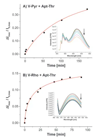
After addition of thiolated Apt-Thr (0.4–0.5 equivalents with respect to the total amount ofMal-C16) toV-Pyr, we observed a time-dependent decrease of the fluorescence signal indicating the reaction progress on the vesicle surface. Phase separation processes in the membrane result in high local concentrations
Institut fu¨r Organische Chemie, Universita¨t Regensburg, 93040 Regensburg, Germany. E-mail: burkhard.koenig@ur.de; Fax:+49 943 1717; Tel:+49 943 4575
†Electronic supplementary information (ESI) available: Experimental details, synthesis of amphiphiles, preparation of functionalized vesicles, emission spectra and binding curves. See DOI: 10.1039/c4cc05221h
Received 7th July 2014, Accepted 4th September 2014 DOI: 10.1039/c4cc05221h www.rsc.org/chemcomm
ChemComm
COMMUNICATION
Open Access Article. Published on 04 September 2014. Downloaded on 21/04/2015 12:47:47. This article is licensed under a Creative Commons Attribution 3.0 Unported Licence.
View Article Online
View Journal | View Issue
12666 | Chem. Commun.,2014,50, 12665--12668 This journal is © The Royal Society of Chemistry 2014 of the embedded functional amphiphiles as it is evidenced by the
presence of the pyrene excimer signal ofV-Pyrat 470 nm (Fig. 2A, inset). We assume that the surface-anchored oligonucleotides strongly impact the physical properties at the membrane inter- face by their high negative charge and steric demand. This local interference results in a partial fluorescence emission quenching of the nearby pyrene aggregates (Fig. 3, top). Moreover, it is well documented that nucleotides are able to quench the fluores- cence of fluorophores, such as pyrene16or rhodamine17deriva- tives, by photoinduced electron transfer. We believe that this mechanism also contributes to the observed effect in our case.
Rhodamine-modified liposomesV-Rhoexhibit a similar behavior during functionalization with the aptamerApt-Thr(Fig. 2B).
The thrombin-binding aptamer folds into a highly compact G-quadruplex structure upon binding to thrombin.18This chair-like conformation is stabilized by a metal cation, particularly potassium cations. Analogously to the sensing mechanism for the membrane functionalization process, we believe that binding of the target to the surface-bound oligonucleotide, accompanied by its conformational change, has a drastic impact on the membrane structure which triggers then the dynamic emission response of the fluorophore molecules in close proximity (Fig. 3, bottom). At last, rather the presence of the ‘‘aptamer bound’’ analyte at the membrane–water interface causes the change of the emission signal than a defined conformation of the immobilized oligonucleotide molecules.
High concentrations of potassium cations are known to induce the transition of the oligonucleotide from random coil to the folded form with a reported binding constant of lgKE2.1.19Therefore, we first tested the suitability of the thrombin aptamer-functionalized vesiclesV-Pyr-Thrfor K+ detection. The fluorescence titration revealed an excimer signal change of almost 60% (Fig. 4, red squares). Due to the relatively low binding affinity of the aptamer Fig. 1 Structures of the functional amphiphiles used for vesicle preparation
and of the immobilized aptamers.
Fig. 2 Fluorescence response monitoring the reaction progress of pyrene- (A) and rhodamine-modified (B) vesicles during surface functionalization with aptamerApt-Thr.
Fig. 3 Principle of vesicle surface functionalization (I) and fluorescence response after analyte binding (II).
Fig. 4 Fluorescence emission titrations of KClversusaptamer-functionalized (red squares) and non-functionalized (blue dots) vesiclesV-Pyr.
Communication ChemComm
Open Access Article. Published on 04 September 2014. Downloaded on 21/04/2015 12:47:47. This article is licensed under a Creative Commons Attribution 3.0 Unported Licence.
View Article Online
This journal is © The Royal Society of Chemistry 2014 Chem. Commun.,2014,50, 12665--12668 | 12667 to potassium cations, KCl concentrations of up to 140 mM were
required to obtain a reasonable binding isotherm. In the same concentration range, non-functionalized vesicles showed a smaller non-specific response with linear relationship (Fig. 4, blue dots). We explain this effect by the huge change of ionic strength in the aqueous buffer. It is well documented that high salt concentrations have a strong influence on the membrane’s physical properties such as surface polarity and hydration and thus can alter the emission of membrane-embedded fluorophores.20Non-linear curve fitting of the obtained binding isotherm resulted in a logarithmic binding con- stant of 1.3 (Table 1). The lower value of the potassium cation binding constant compared to the reported one can be attributed to the interference of specific binding and non-specific salt effects.
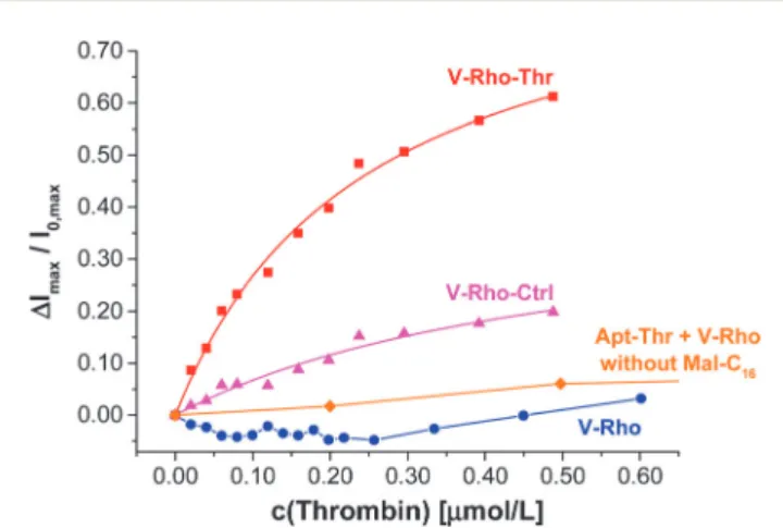
Next, we investigated the detection of thrombin. Pyrene- functionalized vesicles V-Pyr-Thr showed a relatively small fluorescence increase upon addition of thrombin (E20% at a 2.0mM concentration of thrombin, see Fig. S4, ESI†), whereas rhodamine-modified liposomesV-Rho-Threxhibited a considerable luminescence response (Fig. 5, red squares). As expected, non- functionalized vesicles V-Rhodid not significantly alter their emission properties upon the addition of thrombin (Fig. 5, blue dots).
Table 1 summarizes the apparent binding constants of the vesicular aptasensors to different analytes. All calculations were performed by non-linear curve fitting using the equation for 1 : 1 binding.21The affinity constants ofV-Rho-Thrto both bovine and human thrombin are in good agreement with reported
literature values. The limit of detection was determined to be 25 nM (on basis of a signal to noise ratio of 3), while the sensors span a dynamic range of up to 2mM concentration of thrombin (see ESI†for fluorescence titration data). The analytic results are reproducible as shown by repeated measurements. Since thrombin is a crucial enzyme in the blood coagulation cascade, its concentration in circulating blood is low and often undetectable.
During clotting initiation, it becomes significantly elevated at a low nanomolar concentration level and peaks at concentrations in the low micromolar region during the propagation phase.11b,22 Thus, the dynamic recognition range of our sensor matches the physiological concentration range.
To demonstrate that the interaction of thrombin with the covalently membrane-attached aptamer is responsible for the change of the rhodamine emission intensity, we prepared fluores- cent vesicles lacking the amphiphilic binding anchorMal-C16and incubated them with the thiolated thrombin-binding aptamer Apt-Thrleading to no significant response upon analyte addition (Fig. 5, orange diamonds).
We tested the specificity of our aptasensors by immobilizing a non-binding control oligonucleotide Apt-Ctrl (Fig. 1) with comparable length on the vesicle surface. The slight response after thrombin addition can be attributed to non-specific binding (Fig. 5, magenta triangles). Although thrombin has a small negative overall charge in the used buffer system (pH: 7.4, isoelectric point of thrombin: 7.0525), its numerous negatively and positively charged residues are not distributed uniformly over the whole molecule.26They are clustered in a sandwich-like structure of two highly positive poles, whose electrostatic fields spread far into the extramolecular space. We suppose that the electropositive exosites are capable to displace counterions from the negatively charged oligonucleotide backbone by electrostatic attractions.27
Emission titration using the protein elastase, which is besides thrombin a member of the serine proteases enzyme family,versus V-Rho-Thrindicates the aptamer–protein non-specific interaction.
The fluorescence intensity decreases during titration (Fig. 6), which is in contrast to the emission intensity increase observed with thrombin. Elastase has an isoelectric point of 9.5,28 and it is assumed that its high positive net charge possibly results in strong non-specific oligonucleotide–protein interactions,27leading to different fluorescence emission intensity changes.
In conclusion, we have developed a novel type of aptasensor based on self-assembled, luminescent vesicle membranes, which Table 1 Binding affinities of fluorescent, aptamer-functionalized vesicles
to different analytes
Vesicles Analyte lgK Reference
V-Pyr-Thr KCl 1.3 (8%) 2.119
V-Pyr-Thr Bovine thrombin 6.3 (3%) 7.123 V-Rho-Thr Bovine thrombin 7.2 (8%) 7.123 V-Rho-Thr Human thrombin 7.7a 6.7–7.624
aSingle experiment.
Fig. 5 Binding isotherm of thrombin to aptamer-functionalized vesicles V-Rho-Thr(red squares) and fluorescence response of control experiments:
non-functionalized vesiclesV-Rho(blue dots), maleimide-lackingV-Rhovesicles incubated withApt-Thr(orange diamonds) and vesiclesV-Rho-Ctrlfunctiona- lized with non-binding oligonucleotideApt-Ctrl(magenta triangles).
Fig. 6 Fluorescence emission titration of control protein elastase to V-Rho-Thr.
ChemComm Communication
Open Access Article. Published on 04 September 2014. Downloaded on 21/04/2015 12:47:47. This article is licensed under a Creative Commons Attribution 3.0 Unported Licence.
View Article Online
12668 | Chem. Commun.,2014,50, 12665--12668 This journal is © The Royal Society of Chemistry 2014 are functionalized with the thrombin-binding aptamer by nucleo-
philic thiol–maleimide addition. The high-affinity binding of the aptamer to thrombin is indicated by a dynamic response of the co-embedded fluorescent reporter molecules in the physiological concentration range. The obtained binding constants are in good agreement with the reported values. Despite of the observed non-specific interactions in case of mismatched aptamer–protein combinations, the specific binding to thrombin can be clearly distinguished in the fluorescence output. As the reporter group is neither covalently attached nor coordinated to the aptamer, an inexpensive and easy variation of aptamer–fluorophor combina- tions is possible. The signaling mechanism is independent of specific conformational changes of the receptor molecules, which may allow a broader application compared to previously reported label-free aptasensors. An extension to other aptamers, enzymes or proteins and the immobilization of the vesicular aptasensors on solid supports can be readily envisaged.
A.M. thanks the Bayerische Elitefo¨rderung for a research scholarship.
Notes and references
1 (a) C. Tuerk and L. Gold,Science, 1990,249, 505–510; (b) A. D. Ellington and J. W. Szostack,Nature, 1990,346, 818–822.
2 (a) M. McKeague and M. C. DeRosa, J. Nucleic Acids, 2012, 2012, 748913; (b) A. D. Keefe, S. Pai and A. Ellington,Nat. Rev. Drug Discovery, 2010,9, 537–550; (c) E. Torres-Chavolla and E. C. Alocilja, Biosens. Bioelectron., 2009,24, 3175–3182.
3 (a) B. Strehlitz, C. Reinemann, S. Linkorn and R. Stoltenburg,Bioanal.
Rev., 2012,4, 1–30; (b) S. Amaya-Gonza´lez, N. de-los-Santos-A´lvarez, A. J. Miranda-Ordieres and M. J. Lobo-Castan˜o´n,Sensors, 2013,13, 16292–16311.
4 For reviews, see: (a) J. Liu, Z. Cao and Y. Lu,Chem. Rev., 2009,109, 1948–1998; (b) B. Deng, Y. Lin, C. Wang, F. Li, Z. Wang, H. Zhang, X.-F. Li and X. C. Le,Anal. Chim. Acta, 2014,837, 1–15.
5 (a) R. E. Wang, Y. Zhang, J. Cai, W. Cai and T. Gao,Curr. Med. Chem., 2011,18, 4175–4184; (b) I. Willner and M. Zayats,Angew. Chem., Int.
Ed., 2007,46, 6408–6418 (Angew. Chem., 2007,119, 6528–6538).
6 (a) B. Li, H. Wei and S. Dong,Chem. Commun., 2007, 73–75; (b) Z. Xu, K. Morita, Y. Sato, Q. Dai, S. Nishizawa and N. Teramae,Chem.
Commun., 2009, 6445–6447; (c) M. Zayats, Y. Huang, R. Gill, C.-an Ma and I. Willner,J. Am. Chem. Soc., 2006,128, 13666–13667.
7 (a) L. Hu, Z. Bian, H. Li, S. Han, Y. Yuan, L. Gao and G. Xu,Anal.
Chem., 2009,81, 9807–9811; (b) Z. Zhu, C. Yang, X. Zhou and J. Qin, Chem. Commun., 2011,47, 3192–3194.
8 (a) B. Gruber, S. Stadlbauer, A. Spa¨th, S. Weiss, M. Kalinina and B. Ko¨nig,Angew. Chem., Int. Ed., 2010,49, 7125–7128 (Angew. Chem., 2010,122, 7280–7284); (b) S. Banerjee, M. Bhuyan and B. Ko¨nig, Chem. Commun., 2013,49, 5681–5683.
9 For examples on how the modular construction principle can be utilized for the preparation of highly selective chemosensors, see:
(a) B. Gruber, S. Balk, S. Stadlbauer and B. Ko¨nig,Angew. Chem., Int.
Ed., 2012,51, 10060–10063 (Angew. Chem., 2012,124, 10207–10210);
(b) S. Banerjee and B. Ko¨nig,J. Am. Chem. Soc., 2013,135, 2967–2970.
10 L. C. Bock, L. C. Griffin, J. A. Latham, E. H. Vermaas and J. J. Toole, Nature, 1992,355, 564–566.
11 (a) K. A. Edwards and A. J. Baeumner,Anal. Bioanal. Chem., 2006, 386, 1613–1623; (b) K. A. Edwards, Y. Wang and A. J. Baeumner, Anal. Bioanal. Chem., 2010,398, 2645–2654.
12 (a) P. A. Beales and T. K. Vanderlick,J. Phys. Chem. A, 2007,111, 12372–12380; (b) Y.-H. M. Chan, B. van Lengerich and S. G. Boxer, Proc. Natl. Acad. Sci. U. S. A., 2009,106, 979–984.
13 (a) Z. Cao, R. Tong, A. Mishra, W. Xu, G. C. L. Wong, J. Cheng and Y. Lu,Angew. Chem., Int. Ed., 2009,48, 6494–6498 (Angew. Chem., 2009,121, 6616–6620); (b) H. Kang, M. B. O’Donoghue, H. Liu and W. Tan,Chem. Commun., 2010,46, 249–251.
14 (a) C. Wang and Z. Ma,Anal. Bioanal. Chem., 2005,382, 1708–1710;
(b) J. Lee, H.-J. Kim and J. Kim, J. Am. Chem. Soc., 2008, 130, 5010–5011; (c) J. Lee, H. Jun and J. Kim, Adv. Mater., 2009, 21, 3674–3677; (d) Y. K. Jung, T. W. Kim, H. G. Park and H. T. Soh,Adv.
Funct. Mater., 2010,20, 3092–3097; (e) W. Wu, J. Zhang, M. Zheng, Y. Zhong, J. Yang, Y. Zhao, W. Wu, W. Ye, J. Wen, Q. Wang and J. Lu, PLoS One, 2012,7, e48999.
15 P. M. Keller, S. Person and W. Snipes,J. Cell Sci., 1977,28, 167–177.
16 M. Manoharan, K. L. Tivel, M. Zhao, K. Nafisi and T. L. Netzel, J. Phys. Chem., 1995,99, 17461–17472.
17 S. A. E. Marras, F. R. Kramer and S. Tyagi,Nucleic Acids Res., 2002, 30, e122.
18 (a) R. F. Macaya, P. Schultze, F. W. Smith, J. A. Roe and J. Feigon, Proc. Natl. Acad. Sci. U. S. A., 1993,90, 3745–3749; (b) K. Y. Wang, S. McCurdy, R. G. Shea, S. Swaminathan and P. H. Bolton,Biochemistry, 1993, 32, 1899–1904; (c) K. Padmanabhan, K. P. Padmanabhan, J. D. Ferrara, J. E. Sadler and A. Tulinsky,J. Biol. Chem., 1993,268, 17651–17654.
19 S. Nagatoishi, T. Nojima, B. Juskowiak and S. Takenaka, Angew.
Chem., Int. Ed., 2005, 44, 5067–5070 (Angew. Chem., 2005, 117, 5195–5198).
20 (a) G. Cevc, Biochim. Biophys. Acta, Rev. Biomembr., 1990, 1031, 311–382; (b) Y. Zhang and P. S. Cremer,Curr. Opin. Chem. Biol., 2006,10, 658–663.
21 A. E. Hargrove, Z. Zhong, J. L. Sessler and E. V. Anslyn,New J. Chem., 2010,34, 348–354.
22 M. Lee and D. R. Walt,Anal. Biochem., 2000,282, 142–146.
23 M. C. R. Buff, F. Scha¨fer, B. Wulffen, J. Mu¨ller, B. Po¨tzsch, A. Heckel and G. Mayer,Nucleic Acids Res., 2010,38, 2111–2118.
24 P. Baaske, C. J. Wienken, P. Reineck, S. Duhr and D. Braun,Angew.
Chem., Int. Ed., 2010, 49, 2238–2241 (Angew. Chem., 2010, 122, 2286–2290).
25 P. G. Righetti, G. Tudor and K. Ek, J. Chromatogr. A, 1981,220, 115–194.
26 W. Bode, D. Turk and A. Karshikov,Protein Sci., 1992,1, 426–471.
27 P. L. Privalov, A. I. Dragan and C. Crane-Robinson,Nucleic Acids Res., 2011,39, 2483–2491.
28 U. J. Lewis, D. E. Williams and N. G. Brink,J. Biol. Chem., 1956,222, 705–720.
Communication ChemComm
Open Access Article. Published on 04 September 2014. Downloaded on 21/04/2015 12:47:47. This article is licensed under a Creative Commons Attribution 3.0 Unported Licence.
View Article Online