An Intimate Dining – Nutritional Interactions between Obligate Intracellular Parasites and Host Cells
HABILITATIONSSCHRIFT zur Erlangung der Lehrbefähigung
für das Fach Biochemie
vorgelegt dem Rat der Lebenswissenschaften Fakultät der Humboldt-Universität zu Berlin von
NISHITH GUPTA
Prof. Dr.-Ing. habil. Dr. SABINE KUNST Prof. Dr. RICHARD LUCIUS
Präsidentin der Humboldt-Universität zu Berlin Dekan der Lebenswissenshaften Fakultät
Berlin, den November 3, 2017
Gutachter und Gutachterinnen:
(1) Prof. Dr. Richard Lucius (2) Prof. Dr. Markus Engstler (3) Prof. Dr. med. Michael Lanzer
SUMMARY
The protozoan phylum apicomplexa comprises nearly 6000 parasitic species, many of which are of significant medical and veterinary importance. Most apicomplexans have adapted to obligate intracellular parasitism in a wide range of organisms, including animals and humans. Some notable members of the phylum include Toxoplasma, Plasmodium and Eimeria species, which collectively impose substantial healthcare and socioeconomic burden worldwide. This study focused on three representative parasites, namely Toxoplasma gondii, Eimeria falciformis and Plasmodium berghei, all of which infect a common and well-established model host organism (i.e.
mouse), but have diverged from each other considerably with respect to the target host cells, persistence and reproduction behavior. For example, T. gondii can infect and replicate in most nucleated cells, whereas P. berghei and E. falciformis are specific to hepatic/red blood cells and intestinal epithelial cells, respectively. Unlike Plasmodium and Eimeria species causing only acute disease, T. gondii can also inflict chronic infection. Moreover, Toxoplasma can bypass the sexual phase for inter-host transmission; in contrast, its peers must undergo alternating asexual and sexual growth for the natural transmission. Last but not least, T. gondii and P. berghei display only asexual growth in mouse, and require a second host, feline and mosquito, respectively, for sexual reproduction, while E. falciformis assumes asexual as well as sexual development in a single host.
Hence, these parasites together enable a fairly inclusive study of the apicomplexan biology.
A successful intracellular parasite must be able to access host resources and allocate them efficiently to satisfy its own cellular demands irrespective of intracellular niche or lifecycle stage.
Apicomplexan parasites undertake a complex development, which involves genome replication, organelle biogenesis and the zoite assembly within other eukaryotic cells (i.e. a eukaryote inside a eukaryote). Most developmental stages of these pathogens intimately associate with host cells, involving a metabolic crosstalk between the two entwined entities. A germane understanding of such interactions is vital to appreciate the evolution of parasites. In a nutshell, this work aimed to determine the design of metabolic networks in indicated parasites and the impact of metabolism on growth, pathogenesis and adaptation in discrete nutritional milieus. Our approach blended bottom-up methods of biochemistry, reverse genetics, cell biology and optogenetics with the top- down lipidomics, metabolomics and transcriptomics to address the following major premises:
Ø Comparative design of the selected metabolic networks in aforementioned parasites Ø Nutritional plasticity underlying the parasite survival in variable environments Ø Subversion or exploitation of host metabolism by intracellular parasites
Ø Stage-specific rewiring of parasite metabolism during asexual reproduction Ø Identification and endorsement of potential anti-parasitic drug targets
In a series of work, we have revealed that the tachyzoite stage (fast-replicating acute stage) of T.
gondii is metabolically very active and rather autonomous in making macromolecules, as well as occasionally resilient to nutritional perturbations. Tachyzoites display a surprising plasticity in the central carbon metabolism that ensures their survival even in the absence of glucose. We identified glutamine and acetate as the two other major carbon sources, which support glucose- independent growth of tachyzoites. In particular, glutamine-fueled gluconeogenesis becomes critical in tachyzoites with impaired glycolysis. The work also illustrated a hitherto unknown metabolic convergence between proliferating parasites and cancer cells. In contrast to T. gondii, P.
berghei is strictly dependent on the import and glycolytic catabolism of host-derived glucose for its
entire lifecycle even though the parasite is competent in utilizing glutamine and acetate. Inability of P. berghei to survive without glucose is due to absence of glutamine-powered gluconeogenesis.
In additional work, we show that T. gondii and E. falciformis harbor a nearly complete enzymatic machinery to make major phospholipids and thus seem to be fairly autonomous of respective host cell. Toxoplasma displays a flexible membrane biogenesis, which along with malleable carbon flux partly explains its promiscuous growth in assorted host milieus. Our data also demonstrate significant and unexpected divergence in several aspects of lipid biogenesis, not only with respect to mammalian hosts but also within the phylum apicomplexa. We observed that tachyzoites of T.
gondii and the sporozoite stage of E. falciformis express exclusive sphingolipids, ethanolamine- phosphorylceramide and inositol-phosphorylceramide, respectively. Equally, we identified a novel and major phospholipid expressed in T. gondii and E. falciformis (but not in Plasmodium and mammalian cells), termed phosphatidylthreonine, which has evolved from an otherwise-universal lipid, phosphatidylserine. A tachyzoite mutant lacking phosphatidylthreonine exhibits impaired calcium homeostasis, which in turn compromises the lytic cycle and virulence. Notably, such a metabolically attenuated parasite strain can be used as a vaccine to prevent toxoplasmosis in a mouse model. The natural expression and specialized functions of phosphatidylthreonine and indicated sphingolipids signify adaptive divergence of membrane lipids in parasitic protists, which offers an attractive intervention strategy to selectively inhibit the parasite reproduction.
Our active engagement with E. falciformis has also identified the host determinants of infection in vivo. We have discovered a retinue of IFNγ-regulated host factors, e.g., tryptophan catabolism, immunity-related GTPases and chemokine signaling, some of which are protective, whereas others are evaded or even exploited by the parasite. This study found how mouse IFNγ-signaling plays opposing roles in promoting and demoting the Eimeria development. Especially, induction of the first enzyme of tryptophan catabolism (indoleamine 2,3-dioxygenase) in the mouse caecum is needed for efficient in vivo growth of E. falciformis. Likewise, we show a requirement of host cFos (a proto-oncogenic master transcription factor) for in vitro growth of E. falciformis and T. gondii.
Finally, as reckoned necessary to resolve our above-mentioned paradigms, we established new or improved methods. For example, we applied optogenetics to regulate cyclic nucleotide-directed signaling in T. gondii, which enabled an otherwise-challenging induction of the parasite cAMP in a specific, reversible and spatiotemporal manner without perturbing the surrounding host cells.
Using this tool, we determined a requisite of cAMP for the parasite differentiation and associated gene expression. Besides, by employing an optogenetic sensor (gene-encoded calcium indicator), we discovered the importance of phosphatidylthreonine in regulating the cytosolic calcium pool in intracellular tachyzoites. In brief, optogenetically-modified strains allow a more systematic dissection and detection of the signaling cascades in intracellular parasites, which has not been so feasible when using chemical modulators and probes. Our initial application of light-responsive actuators and sensors to illuminate the parasite biology is expected to lead a wider deployment of optogenetics in infection research.
ZUSAMMENFASSUNG
Das zu den Protozoen gehörende Phylum der Apicomplexa umfasst nahezu 6000 Parasitenarten, von denen einige von medizinischer und veterinärmedizinischer Bedeutung sind. Die meisten Apicomplexa haben sich an eine obligat intrazelluläre Lebensweise angepasst und infizieren verschiedenste Tiere und den Menschen. Zu den bedeutendsten Vertretern der Apicomplexa zählen Toxoplasma, Plasmodium und Eimeria, welche eine wesentliche Belastung für das Gesundheitswesen und Sozioökonomie darstellen. In dieser Arbeit lag der Schwerpunkt auf den drei repräsentativen Organismen Toxoplasma gondii, Eimeria falciformis und Plasmodium berghei, welche sich alle in einem etablierten Wirt (der Maus) reproduzieren, sich allerdings hinsichtlich ihrer Wirtszellen, Persistenz sowie in ihrem Reproduktionsverhalten deutlich unterscheiden. T. gondii infiziert beispielsweise nahezu alle kernhaltigen Zellen, während P. berghei und E. falciformis spezifisch Leber- und Blutzellen bzw. Darmepithel befallen. Plasmodium und Eimeria verursachen ausschließlich akute Erkrankungen, eine Infektion mit Toxoplasma kann auch chronisch verlaufen. Im Gegensatz zu Plasmodium und Eimeria, die den Wechsel zwischen sexueller und asexueller Vermehrung bei der natürlichen zwischenwirtlichen Transmission durchlaufen müssen, kann Toxoplasma die sexuelle Phase umgehen.
Letztlich vermehren sich T. gondii und P. berghei in der Maus ausschließlich asexuell und benötigen einen zweiten Wirt (Katzen bzw. Moskitos) für die sexuelle Reproduktion, während E. falciformis sowohl die sexuelle als auch die asexuelle Phase in der Maus durchläuft. Somit ermöglichen die genannten Parasiten eine umfassende Untersuchung der Biologie der Apicomplexa.
Ein erfolgreicher intrazellulärer Parasit muss in der Lage sein auf Ressourcen des Wirts zuzugreifen und diese für den eigenen zellulären Bedarf zu nutzen. Apicomplexe Parasiten durchleben eine komplexe Entwicklung einschließlich Genomreplikation, Organellen- und Tochterzellbildung in einer anderen eukaryotischen Zelle. Die meisten Entwicklungsstufen dieser Pathogene sind sehr eng mit der Wirtszelle assoziiert, was auch metabolische Wechselwirkungen beinhaltet. Das Verständnis dieser Interaktionen ist unerlässlich, um die Evolution von Parasiten zu ergründen. Grundsätzlich war das Ziel dieser Arbeit, die metabolischen Netzwerke der genannten Parasiten zu eruieren und den Einfluss des Metabolismus auf Wachstum, Pathogenese und Adaption in verschiedenen Nährstoffumgebungen zu untersuchen. Unsere Vorgehensweise verband biochemische, revers- genetische, zellbiologische und optogenetische Bottom-Up-Methoden mit Top-Down-Methoden wie Lipidomics, Metabolomics und Transcriptomics um folgende Prämissen anzugehen:
• Vergleichender Entwurf der metabolischen Netzwerke in den obengenannten Parasiten
• Nährstoff-Plastizität für die Überlebensfähigkeit des Parasiten in verschiedenen Milieus
• Umregulierung oder Ausbeutung des Wirtsmetabolismus durch intrazelluläre Parasiten
• Stadien-spezifische Regulation des Metabolismus während der asexuellen Reproduktion
• Identifizierung und Validierung potentieller anti-parasitischer Wirkstoffe
In unserer bisherigen Arbeit haben wir gezeigt, dass das Tachyzoiten-Stadium (akute Form der Infektion) von T. gondii metabolisch sehr aktiv, weitgehend autonom in der Biogenese von zellulären Makromolekülen und mitunter sehr widerstandsfähig bei variierender Nährstoffverfügbarkeit ist.
Tachyzoiten zeigen eine enorme metabolische Plastizität im zentralen Kohlenstoffmetabolismus, welche sogar ein Überleben ohne Glukose ermöglicht. Weiterhin konnten wir nachweisen, dass Glutamin und Acetat zwei weitere bedeutende Kohlenstoffquellen neben Glucose darstellen. Vor allem die glutamingetriebene Gluconeogenese wirkt sich entscheidend auf Tachyzoiten mit einer beeinträchtigten Glykolyse aus. Diese Untersuchungen zeigten eine bisher unbekannte metabolische Konvergenz zwischen proliferierenden Parasiten und Krebszellen. Im Vergleich dazu ist P. berghei
darauf angewiesen Zucker des Wirts in seinem gesamten Lebenszyklus zu importieren, weil die Gluconeogenese fehlt, obwohl der Parasit auch fähig ist Glutamin und Acetat zu nutzen.
Wir zeigten zudem, dass sowohl T. gondii als auch E. falciformis eine nahezu vollständige Enzymkette für die Biosynthese der wichtigsten Phospholipide beherbergen und somit ziemlich unabhängig von ihrer jeweiligen Wirtszelle zu sein scheinen. Toxoplasma zeigt eine flexible Membranbiogenese, welche zusammen mit einem variablen Kohlenstofffluss zum Teil seine Wachstumsfähigkeit in verschiedenen Nährstoffmilieus erklärt. Darüber hinaus demonstrieren unsere Ergebnisse eine erhebliche sowie unerwartete Divergenz hinsichtlich der Stoffwechselwege zur Membranbiogenese - sowohl in Bezug auf den Säugerwirt aber vor allem auch innerhalb der Apicomplexa. Wir konnten beobachten, dass Tachyzoiten von T. gondii und Sporozoiten von E. falciformis exklusive Sphingolipide, Ethanolamin- Phosphorylceramid bzw. Inositol-Phosphorylceramid, bilden. Darüber hinaus konnten wir ein bisher unbekanntes Phospholipid Phosphatidylthreonin identifizieren, welches in T. gondii und E. falciformis exprimiert ist (jedoch nicht in Plasmodium und Säugern) und sich aus dem anderweitig universellen Phosphatidylserin entwickelt hat. Die Tachyzoit-Mutante, der dieses exklusive Lipid fehlt, zeigt eine gestörte Kalziumhomöostase, welche wiederum den lytischen Zyklus sowie die Virulenz des Parasiten beeinträchtigt. Bemerkenswerterweise kann ein solcher metabolisch attenuierter Parasitenstamm als Impfstoff verwendet werden und so eine Erkrankung an Toxoplasmose im Mausmodell verhindern.
Die Expression und spezialisierten Rollen von Phosphatidylthreonin und der angegebenen Sphingolipide kennzeichnen eine adaptive Divergenz der Membranlipide in parasitischen Protisten, welche einen attraktiven Angriffspunkt für die selektive Inhibierung des Parasiten-Wachstums liefert.
Unsere Untersuchungen an E. falciformis waren erfolgreich hinsichtlich der bestimmenden Faktoren in Bezug auf den Wirt in vivo. Dabei haben wir eine Reihe IFNγ-regulierter Stoffwechselwege im Wirt, wie beispielsweise den Tryptophan-Katabolismus, mit der Immunität in Zusammenhang stehende GTPasen sowie eine Chemokin-Signaltransduktion, entdeckt, von denen einige eine protektive Wirkung haben, anderen wiederum kann der Parasit entkommen oder diese sogar ausnutzen. Unsere Arbeit zeigt, wie die IFNγ-Signalgebung in der Maus entgegengesetzte Rollen im Fördern und Schaden der Entwicklung von Eimeria spielt. Insbesondere ist die Induktion des ersten Enzyms des Tryptophan-Katabolismus (Indoleamin-2,3-Dioxygenase) im Darm der Maus für ein effizientes Wachstum von E. falciformis notwendig. Gleichzeitig konnten wir den Bedarf an Wirts-cFos (ein proto-onkogener Haupttranskriptionsfaktor) für die in vitro-Entwicklung von E. falciformis und T.
gondii aufzeigen. Schlussendlich etablierten wir neue und verbesserte Methoden, um die genannten Problemstellungen anzugehen. So nutzten wir beispielsweise optogenetische Methoden, um die Signalkaskaden zyklischer Nukleotide in T. gondii in einer spezifischen, reversiblen und raum-zeitlich definierten Weise zu manipulieren, ohne die Wirtszelle zu beeinflussen, was sich bisher als schwierig erwies. Mit dieser Methode konnten wir den Bedarf von cAMP für die Stadien-Differenzierung und assoziierte Genexpression aufzeigen. Weiterhin haben wir mit Hilfe dieses optogenetischen Sensors (ein gencodierter Kalziumindikator) eine Funktion von Phosphatidylthreonin bei der Regulation des zytosolischen Kalziums in intrazellulären Tachyzoiten entdeckt. Kurz gesagt ermöglichen solche optogenetisch veränderten Stämme eine systematischere Aufschlüsselung bzw. Detektion von Signalkaskaden in intrazellulären Parasiten, was bisher allein durch chemische Untersuchungen nicht möglich war. Unsere Anwendung Licht-responsiver Akteure und Sensoren zur näheren Beleuchtung der Biologie der Parasiten wird möglicher Weise eine breitere Anwendung von Optogenetik in der Infektionsbiologie ermöglichen.
ABBREVIATIONS
Abbreviation Description
ACS Acetyl CoA synthetase
CCT CTP:phosphocholine cytidylyltransferase
CDP-DAG CDP-diacylglycerol
CDS CDP-DAG synthase
CK Choline kinase
cNMP Cyclic nucleotide monophosphate (cAMP, cGMP)
CPT Choline phosphotransferase
DAG Diacylglycerol
DG Dense granule
DLC Dynein light chain
DME Dimethylethanolamine
ECT CTP:phosphoethanolamine cytidylyltransferase
EK Ethanolamine kinase
EPC Ethanolamine-phosphorylceramide
EPT Ethanolamine phosphotransferase
ER Endoplasmic reticulum
FAS/FAE Fatty acid synthase / Fatty acid elongase
FBP Fructose 1,6-bisphosphatase
G3P Glycerol-3-phosphate
GBP Guanylate-binding protein
GRA Granule (proteins secreted by dense granules)
GT1 Glucose transporter 1
HT1 Hexose transporter 1
IDO Indoleamine 2,3-dioxygenase
IPC Inositol-phosphorylceramide
IRG Immunity-related GTPase
LDL Low density lipoprotein
MIC Microneme (or proteins secreted by micronemes)
MNN Membranous nanotubular network
PDME Phosphatidyldimethylethanolamine
PECK Phosphoenolpyruvate carboxykinase
PIS Phosphatidylinositol synthase
PKA Proteins kinase A (cAMP-dependent)
PKG Protein kinase G (cGMP-dependent)
PSD Phosphatidylserine decarboxylase
PSS Phosphatidylserine synthase
PtdCho Phosphatidylcholine
PtdEtn Phosphatidylethanolamine
PtdGro Phosphatidylglycerol
PtdIns Phosphatidylinositol
PtdOH Phosphatidic acid
PtdSer Phosphatidylserine
PtdThr Phosphatidylthreonine
PTS Phosphatidylthreonine synthase
PV/PVM Parasitophorous vacuole / PV membrane
RON Rhoptry neck (proteins located in the rhoptry-neck region) ROP Rhoptry or proteins located in the rhoptry-bulb region
TVN Tubulovesicular network
INDEX OF CONTENTS
1 INTRODUCTION………...9
1.1 Parasitism and its impact in nature………...9
1.2 Obligate intracellular (apicomplexan) parasites………...9
1.3 Lifecycle of apicomplexan parasites………...11
1.4 Structure and morphology of an apicomplexan zoite………..…..12
1.5 Asexual reproduction in apicomplexan parasites………..…13
1.6 Parasitophorous vacuole and remodeling of infected host cell………..…15
1.7 Nutritional basis of intracellular parasitism………...………17
1.8 Overarching theme and underlying paradigms…..………..…….…18
2 RESULTS AND DISCUSSION………..………20
2.1 Central carbon metabolism of Toxoplasma gondii and Plasmodium berghei……….……20
2.2 Phospholipid biogenesis in tachyzoites of Toxoplasma gondii………..…….………....24
2.2.1 Phosphatidylcholine synthesis – an anti-parasitic drug target..………....………….25
2.2.2 Plasticity of phosphatidylethanolamine biogenesis………27
2.2.3 Functional speciation of phosphatidylserine and phosphatidylthreonine……….…….…29
2.2.4 Biogenesis of phosphatidylinositol and phosphatidylglycerol………...……….31
2.3 Phospholipid syntheses in sporozoites of Eimeria falciformis………….…………..……….33
2.4 Host determinants of coccidian development………...….………35
2.5 Regulation of asexual reproduction in Toxoplasma gondii………….………...38
3 CONCLUSIONS AND PERSPECTIVES……….….………..…..41
3.1 Metabolic plasticity and determinants of intracellular parasitism...………..……41
3.2 Physiological convergence between parasites and cancer cells………..…42
3.3 Utility of optogenetics in infection research……….………..………43
4 REFERENCES……….………..…....………..44
5 PUBLICATION ABSTRACTS AND AUTHOR CONTRIBUTIONS…………...…53
6 ACKNOWLEDGMENT………..……….……….……..72
7 CURRICULUM VITAE………..…..…..74
8 DECLARATIONS (ERKLÄRUNG)…….………..80
9 PUBLICATIONS (REPRESENTATIVE FULL-TEXT ARTICLES)….………..……81
FIGURES AND TABLES
Fig 1: Emoticon illustration of three major symbiotic relationships observed in nature……….…...9
Fig 2: Phylogenetic tree of apicomplexan parasites………...10
Fig 3: Simplified lifecycle of apicomplexan parasites………...……….11
Fig 4: Schematic illustration of a zoite.……….13
Fig 5: Asexual reproduction in apicomplexan parasites as exemplified for the tachyzoite stage of T. gondii……14
Fig 6: Cell division in apicomplexan parasites………15
Fig 7: Remodeling of host cells induced by Toxoplasma and Plasmodium………...…16
Fig 8: Abridged depiction of metabolic interactions in a parasite-infected cell………...………….17
Table 1: Genome-wide comparisons of selected apicomplexan parasites and mammalian hosts………..19
Fig 9: Central carbon metabolism in the tachyzoite stage of T. gondii.………21
Fig 10: Pathways of phospholipid syntheses in the tachyzoite stages of T. gondii.………...24
Fig 11: Current model of PtdCho biogenesis in T. gondii tachyzoites……….26
Fig 12: Multiple routes of PtdEtn biogenesis in T. gondii tachyzoites………28
Fig 13: PtdThr-mediated control of lytic cycle and virulence in T. gondii………30
Fig 14: Synthesis of PtdIns and PtdGro in the tachyzoite stage of T. gondii………32
Fig 15: Syntheses of phospholipids in the sporozoite stage of E. falciformis.……….34
Fig 16: Proposed model of Eimeria falciformis-mouse interactions……….36
Fig 17: Comparison of TgDLC8a with HsDLC8 and PfDLC8 structures………..………..…39
Fig 18: Optogenetic induction of cAMP-mediated stage differentiation in T. gondii.……….40
1 INTRODUCTION
1.1 Parasitism and its impact in nature
The organisms in an ecosystem interact in different ways, which may exert positive, neutral or negative impact on each other. The term symbiosis usually refers to these interactions between two (or more) organisms living together with some sort of feeding relationship involved [1,2] (Fig 1). The three commonly observed symbiotic interactions are mutualism, in which both organisms benefit; commensalism, where one of the two partners benefits, while the other organism is not harmed; and parasitism, where one (parasite) profits at the expense of other (host) organism [1,2].
Parasites typically depend on host resources for the survival. They may live outside (ectoparasite) or inside (endoparasite) a host [3]. An endoparasite spends significant part of its life inhabiting on tissues (extracellular) or within cells (intracellular) of a host organism.
Fig 1: Emoticon illustration of three major symbiotic relationships observed in nature. Unlike mutual and commensal interactions between the two organisms, in which both or at least one partner gains and none are harmed, a parasitic relationship involves an inverse association.
Although the concept of parasitism and the term parasite apply unambiguously to many cases in nature, e.g., bacteria and viruses, they generally entail to eukaryotic pathogens of protozoan and metazoan origins. The kingdom protozoa comprises more than 40,000 known living species, of which about 25,000 species occur as free-living, whereas the remaining are adapted to parasitic life [4,5]. The latter group includes at least 250 amoebae, 1800 flagellate, 2500 ciliate and 6000 sporozoan (apicomplexan) species. These parasitic organisms together impose significant burden on our healthcare systems worldwide. Not only do they affect infected hosts by altering growth, behavior, nutritional status, reproductive abilities and mortality, but also shape our ecosystem by influencing trophic interactions, food webs and biodiversity [6].
1.2 Obligate intracellular (apicomplexan) parasites
The phylum apicomplexa contains the bulk of what was once termed as the sporozoa, a group of spore-forming parasitic protozoans lacking pseudopods and cilia altogether, and flagella from the most lifecycle stages [7,8]. They have likely evolved from a photosynthetic algal ancestor [9]. A transition from the free-living to parasitic lifestyle is supported by the existence of closely related predator (colpodellids) and symbiont (Nephromyces) of marine organisms [10,11]. The apicomplexan members have adapted to become mostly obligate intracellular parasites with complex lifecycles, which often involve multiple types of cells, tissues and host organisms. Indeed, these parasites are
ubiquitously distributed across the animal kingdom. They can be grouped based on phylogenetic relationships and tissue specificities [12,13] (Fig 2). Hematozoans infect blood cells of a vertebrate host to achieve the asexual reproduction and require a blood-feeding invertebrate host for sexual reproduction and transmission. The well-known hematozoans include Plasmodium species causing malaria, along with Babesia and Theileria species, which infect cattle [3]. The coccidian parasites, forming the largest group in the phylum, typically infect intestinal and extra-intestinal tissues and spread through the fecal-oral route. Some of the prevalent coccidian genera include Toxoplasma, Sarcocystis, Eimeria, Isospora and hepatozoon that infect animals and/or humans [14]. In particular, a single species of Toxoplasma (T. gondii) is capable of infecting nearly all warm-blooded organisms [15]. On the other hand, individual species of the genus Eimeria (>1800 species) are highly host- specific and cause diarrhea in a variety of animals, most notably in the poultry [16]. Gregarine is a distant subgroup, which consists of primarily extracellular pathogens of invertebrates [17], but also includes clinically relevant epicellular parasites, such as Cryptosporidium species [18].
Fig 2: Phylogenetic tree of apicomplexan parasites. Only representative parasite groups (e.g., hematozoa, coccidia and gregarines) are depicted. Numbers and thickness of branches indicate the approximate diversity (i.e.
known species) in each clade. The hallmark features and common parasite members are also mentioned.
Apicomplexans have evolved from a photosynthetic ancestor (chromerids). They are closely related to free- living predators (colpodellids) and endosymbionts (Nephromyces). Cryptosporidium likely emerged from within gregarines. Image has been redrawn from the reference [13] to emphasize the context.
1.3 Lifecycle of apicomplexan parasites
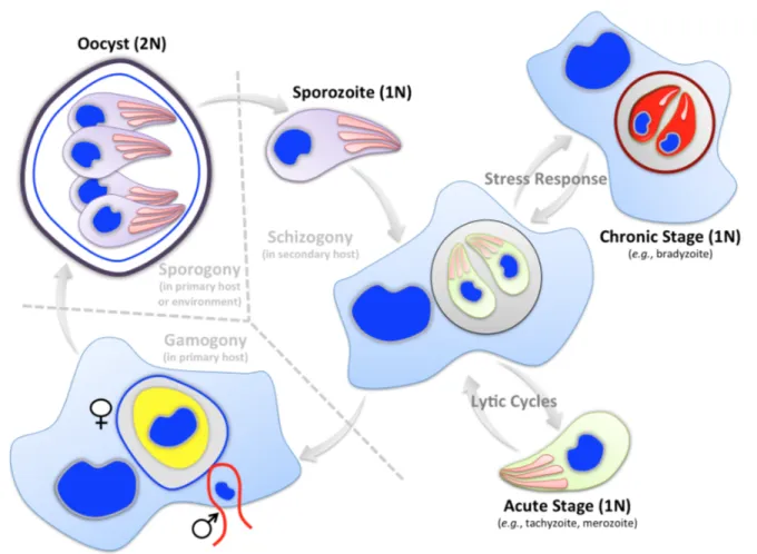
The natural lifecycle of apicomplexan parasites comprises both asexual and sexual reproduction gyrating often between the primary (i.e. definitive or sexual) and secondary (i.e. intermediate or asexual) hosts [3,14] (Fig 3). Toxoplasma and Plasmodium species complete their entire development in two hosts (dixenic), while Eimeria and Cryptosporidium species require only one host (monoxenic) (Fig 2). Plasmodium, Eimeria and Cryptosporidium species are very specific to the host, tissue and cell types they infect; conversely, T. gondii is quite promiscuous with little to no regard to any of these.
Infective stages of the apicomplexan parasites are termed as zoites that are formed after sexual (sporozoite) or after asexual (merozoite, tachyzoite, bradyzoite etc.) reproduction [14,19]. Asexual growth of zoites is followed by sexual development producing male and female gametes that fuse together to form a zygote, which later develops into an oocyst. The oocyst stage undergoes the process of sporogony producing the sporozoite stage and thereby completing the lifecycle.
Fig 3: Simplified lifecycle of apicomplexan parasites. Only the most generic type lifecycle and underlying stages are depicted. Many deviations are seen in nature, particularly with respect to the asexual development. As illustrated, lifecycle is completed either in one or two hosts (dotted line). Most developmental stages of the apicomplexan parasites are haploid except for the oocyst, which is diploid. Inter-host transmission usually requires the entire cycle, which initiates with the formation of sporozoites within oocysts via sporogony (a type of meiosis). Asexual reproduction occurs generally by schizogony (a type of mitosis). Some parasites (e.g., Toxoplasma, Neospora) can also form dormant or chronic stages in response to physicochemical stress.
Asexual reproduction of Plasmodium in its intermediate host begins with the injection of infective sporozoites by the primary host, mosquito [20]. Sporozoites infect and reproduce in hepatocytes, which eventually release thousands of merozoites into bloodstream. Merozoites replicate in red blood cells by multiple rounds of erythrocytic schizogony. A fraction of merozoites differentiates into gametes, which undergo sexual commitment to ultimately yield the sporulated oocysts in the mosquito host. Lifecycle of Eimeria species initiates with the ingestion of sporulated oocysts from the environment, ensued by infection of intestinal epithelium with freed sporozoites, schizogony, gamogony and oocyst development, all occurring in one host [14]. In the case of Toxoplasma, the secondary hosts get infected either by ingesting oocysts containing sporozoites from environment, or by tissue cysts harboring bradyzoites (present in infested meat), both of which differentiate into tachyzoites in intestinal epithelium [21]. Tachyzoites undertake lytic cycles causing tissue necrosis and then spread to immunoprivileged sites (brain, eyes, muscles etc.) before forming bradyzoites.
Bradyzoites can start either schizogony producing merozoites and then sexual reproduction upon predation of a chronically-infected secondary host by a primary host, or alternatively can resume asexual growth as tachyzoites when ingested by another secondary host [21]. Hence, Toxoplasma is unique in its ability to bypass the sexual phase and transmit from asexual to asexual hosts.
1.4 Structure and morphology of an apicomplexan zoite
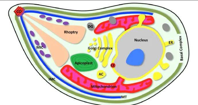
A plethora of intracellular pathogens have evolved to invade and develop in host cells; however, only few would contest the subcellular complexity observed in apicomplexan zoites. The zoite is a highly polarized cell delimited by the pellicle, a tri-bilayer alveolate-specific structure comprising plasma membrane and inner membrane complex (Fig 4). Underneath the pellicle are the apical and basal complexes, located at the anterior and posterior ends, respectively [22]. The pellicle is associated to the subpellicular microtubule network, which acts as the parasite skeleton [22]. The inner membrane complex extends from the apical to the basal complex, and harbors the gliding machinery to drive the actin-myosin-dependent parasite motility. The parasite body contains a multitude of dedicated organelles, which enable it to perform the sequential tasks of invasion and subsequent development [23,24]. In short, a mature zoite is designed to identify and invade the target host cell, proliferate intracellularly, and often differentiate into the next lifecycle stage [23].
The apical complex is an emblematic feature of the phylum, though its constituents vary among members. It consists of the parasite-specific secretory organelles, microneme and rhoptry, and a polar ring (Fig 4). In coccidians, the apical complex also comprises the conoid – a cylinder-like structure of spirally-arranged tubulin – that is extended during gliding motility and invasion. The parasite contains many exclusive organelles (rhoptries, micronemes, dense granules, apicoplast, plant-like vacuole, acidocalcisomes, exonemes, refractile bodies) as well as the typical eukaryotic organelles (nucleus, mitochondrion, centriole, Golgi body, endoplasmic reticulum) [14,19]. Some of these are parasite or even stage-specific, e.g., plant-like vacuole in Toxoplasma [25], food vacuole and exonemes in Plasmodium [26,27] and refractile bodies in Eimeria [28]. Differences in the organelle makeup of each parasite or stage generally correlate with the specialized behavior and adaptation to respective intracellular niche [29,30]. The apicoplast (enclosed by four membranes)
is in fact a vestigial chloroplast of algal origin acquired by secondary endosymbiosis [31,32]. It is present in most members of the phylum except for gregarines (Fig 2). The apicoplast has lost the ability to perform photosynthesis, but retained certain important metabolic pathways [33,34].
Fig 4: Schematic illustration of a zoite. Only the organelles conserved across most parasites are depicted. Not every structure shown here occurs in all apicomplexan parasites. The morphology of individual zoites may differ from the depiction, which signifies the tachyzoite stage of T. gondii (sporozoite, slender elongated;
merozoite, oval to slender elongated; tachyzoite, crescent; bradyzoite, crescent to elongated; ookinete, oval-elongated). To see a merozoite of P. falciparum (within a red blood cell), please refer to Figure 7. AC, acidocalcisome; CE, centrosome; CO, conoid; DG, dense granule; ER, endoplasmic reticulum; IMC, inner membrane complex; MIC, microneme; MT, microtubule
1.5 Asexual reproduction in apicomplexan parasites
The pathology caused by individual parasites is due to multiple rounds of asexual reproduction, often leading to host-cell lysis, tissue necrosis and inflammation. Acute infection occurs by serial lytic cycles that involve invasion, replication, parasite egress and subsequent infection of nearby cells (Fig 5). Once attached and apically oriented onto the host cell surface, the parasite invades within a minute, enclosed in a special membranous structure termed the parasitophorous vacuole (PV) [35,36]. Events of invasion and intracellular establishment require an orchestrated secretion of micronemes, rhoptries and dense granules. Secreted proteins enable several functions that include motility, invasion, maturation of the PV, egress and defense against the host-cell insults [30]. Mechanisms of invasion and formation of nascent PV are mostly conserved since these are regulated by a fairly conserved set of micronemal (MIC) and rhoptry-neck (RON) proteins [37–
40]. However, the way each parasite manipulates its host differs considerably due to divergence in rhoptry-bulb (ROP) and dense granule (GRA) proteins. The ROP and GRA proteins contrast significantly with respect to their presence, number, localization and function within the phylum.
Fig 5: Asexual reproduction in apicomplexan parasites as exemplified for the tachyzoite stage of T. gondii. The zoite stage invades the host cell, proliferates to form progeny, which egress to begin a new lytic cycle (acute infection).
Sporozoites and bradyzoites differentiate into the next stage during the asexual growth, while tachyzoites and merozoites do not always undergo stage switching. Most zoites divide in a parasitophorous vacuole except for Theileria, Babesia and Sarcocystis species, which escape the nascent vacuole to eventually reside in the cytosol. Physicochemical and immune stresses cause certain parasites, notably Toxoplasma and Neospora, to assume dormancy as tissue cyst (chronic infection), which may reactivate upon declined stress to initiate the lytic cycle (recrudescence). Calcium and cyclic nucleotides are the two core regulators of the lytic cycle and stage differentiation. Some parasites (Plasmodium, Babesia) infect non-nucleated host cells.
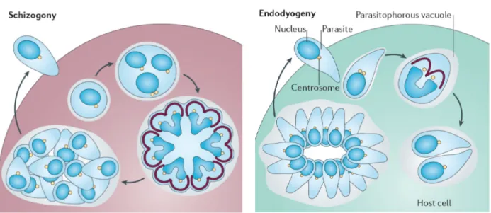
Successive phases of intracellular replication ultimately cause parasites to egress by lysing the host cell, which occurs after variable number of cell cycles depending on the parasite and host cell. In most apicomplexans, egress is a signaling-mediated active process that involves timely sensing of the host environment by the parasite [41–43]. Perturbation of ion homeostasis in the dying host cell and activation of calcium- and cGMP-dependent signaling in the parasite are among many other factors governing the gliding motility and subsequent egress and invasion events. Immune response and physicochemical stress can trigger encystation of certain parasites into tissue cysts [21] (Fig 5), which remain latent until reactivated under favorable condition, such as weakened immunity. The apicomplexans have evolved distinct strategies to execute their cell cycles, which are designed to ensure that progeny are mature enough to initiate a new lytic cycle or further development [44] (Fig 6). Merozoites of Plasmodium, Eimeria and Toxoplasma assume schizogony; in which cytokinesis does not instantly follow karyokinesis. Instead, many cycles of DNA replication and nuclear divisions produce a schizont stage prior to the assembly of several zoites. Toxoplasma tachyzoites divide by endodyogeny, a variant of schizogony, where karyokinesis and cytokinesis proceed soon after DNA replication. Unlike schizogony, endodyogeny yields only two cells per cycle [44]. Cell division is orchestrated by waves of transcript expression encoding for cell cycle
constituents and regulators. It appears to be regulated globally as well as locally, especially in the schizont stage. Global regulation is exerted by diffusible proteins, such as transcription factors, mitosis-specific kinases and cyclins. Local regulation acts on each nucleus and daughter cells [44].
Fig 6: Cell division in apicomplexan parasites. Only two usual modes of asexual growth (Plasmodium, Eimeria and Toxoplasma) are shown. Schizogony: many rounds of DNA replication and nuclear divisions precede budding of daughter cells at the plasma membrane. Endodyogeny: one cycle of DNA replication and nuclear division are immediately followed by budding of two progeny within the mother-cell cytoplasm. Image is adapted from the reference [44]. Centrosome, yellow circles; inner membrane complex, brown; nucleus, blue 1.6 Parasitophorous vacuole and remodeling of infected host cell
Nearly all zoites except for some hematozoan blood stages infect biosynthetically active host cells, which often include even the immune cells (dendritic cells, macrophages, lymphocytes) [45]. The parasites’ foremost tasks are therefore to escape lysosomal fusion and cell-intrinsic immunity, and intercept nutrients. To accomplish all these, they extensively manipulate their respective host cell.
Individual members differ in relationship with intracellular niches, which is mainly dictated by rhoptry-bulb and dense granule proteins [39,40]. Most parasites develop within a PV, which is initially derived from the host plasma membrane during the process of invasion but significantly modified to safeguard the parasite development [46–49]. The mature PV does not intersect with the endocytic and exocytic traffic and avoids lysosomal acidification. As known in T. gondii [47], the PVM forms deep invaginations into the intravacuolar space and long extensions in the host cytosol (Fig 7). The PV of tachyzoites physically tethers with the host endoplasmic reticulum and mitochondria, and recruits cytoskeleton [50]. Interaction of the PVM with the host ER (but not mitochondria) has also been reported in hepatic cells infected with Plasmodium sporozoites [51].
Likewise, the merozoite stage induces the formation of Maurer’s clefts (flat membrane structures), PVM extensions and cytoskeleton rearrangements in the erythrocyte cytosol along with knob-like structures and new permeation pathway on the surface [52–54] (Fig 7). Many other variations in the PV and host-cell manipulation exist in other apicomplexan parasites [39,45].
Fig 7: Remodeling of host cells induced by Toxoplasma and Plasmodium. Images exemplify only selected aspects.
Top panel: Toxoplasma tachyzoites in a nucleated mammalian cell. Intravacuolar space is decorated with the membranous nanotubular network (MNN). Extensions of PV into host cytosol (tubulovesicular network or TVN) and intravacuolar invaginations of the PVM (i.e. Host Organelle Sequestering Tubules; HOST) are also shown. The host-cell ER, mitochondria and cytoskeleton (tubulin, vimentin) become rearranged onto the PVM. Bottom panel: A Plasmodium merozoite in an erythrocyte. The parasite induces the formation of Maurer’s cleft, membrane loops, TVN, knob-like structures (as depicted), as well as cytoskeletal alterations and new permeation pathway (not shown) in the infected cell. Unlike T. gondii tachyzoites, where the PV is the main destination for most rhoptry-bulb and dense granule proteins, the PV of Plasmodium merozoites constitutes a mandatory transit point for the protein cargo en route to host erythrocytes. DG, dense granule;
ER, endoplasmic reticulum; Ex, exoneme; MIC, microneme; RBC, red blood cell; ROP, rhoptry
In addition to specified morphological alterations, infection of host cells with individual parasites also results into transcriptomic and proteomic modulation of distinct pathways, such as protein synthesis, metabolism, immune signaling, membrane trafficking, intrinsic immunity, apoptosis, antigen presentation and microRNA [55–64]. It appears as though a personalized manipulation of the host environment by each parasite is imperative to ensure its survival and reproduction.
1.7 Nutritional basis of intracellular parasitism
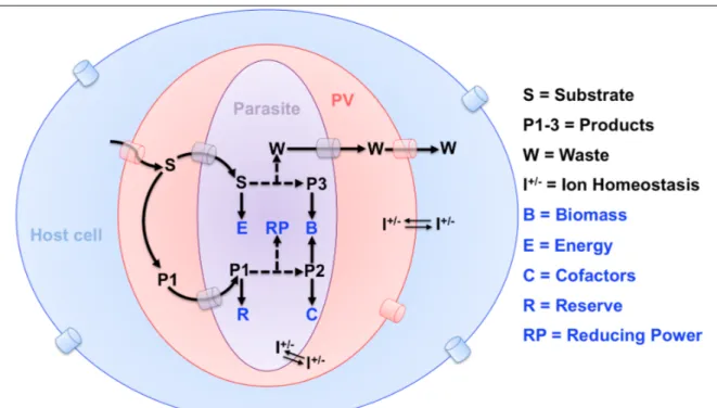
Most apicomplexans during their entire intracellular phase are sheltered within a non-fusogenic vacuole, which shields from host defense, but also restricts them from retrieving metabolites (Fig 8). Parasites have therefore invented ingenious strategies; for example, Toxoplasma, Plasmodium and Eimeria remodel their PVM as such to make it permeable to small molecules [65–67]. The PVM of T. gondii tachyzoite harbors Gra17 and Gra23 proteins, which enable diffusion of molecules up to 1.3-kDa [65,68]. The PVMs of erythrocytic and hepatic stages of Plasmodium also function as molecular sieves allowing the passage of metabolites below 1.4-kDa and 0.85-kDa, respectively [51,66]. Plasmodium merozoites also modify the permeability of erythrocyte plasma membrane to obtain a wide range of nutrients from the blood milieu [69,70]. These membrane sieves are very vital for salvaging those nutrients that parasites are unable to produce themselves, as well as for importing the precursors for de novo synthesis of complex macromolecules within the parasite.
Fig 8: Abridged depiction of metabolic interactions in a parasite-infected cell. Most parasites are separated from exogenous nutrients by at least three physical barriers including the plasma membranes of the host cell and parasite, as well as the parasitophorous vacuole membrane. Exemplary flow of metabolic substrates (S) and waste (W) across these membranes and putative transporters are shown. Just as any other cell, parasites require biomass, energy, cofactors, reducing equivalents, ion homeostasis and nutrient stores for reproduction; however, how they fulfill these metabolic necessities differs considerably. Depending on the nutrient, they can either make use of the endogenous pathways and/or salvage from the host cell.
In free-living organisms, the linked pathways of central carbon metabolism (glycolysis, pentose phosphate shunt, TCA cycle) constitute a metabolic hub to ensure the biomass, energy and redox demands during cell proliferation and differentiation [71,72]. While intracellular parasitism has resulted in a net loss of metabolic pathways in apicomplexans, these three core pathways remain conserved in most intracellular parasites [9]. Evenly, parasites have also often preserved synthesis of fatty acids except for few (e.g., Theileria), which appear to be auxotrophic [73]. Toxoplasma and Eimeria genomes also encode pathways to produce most amino acids [74]. By contrast, Plasmodium merozoites cannot produce them and satisfy their demands for amino acids largely by degrading erythrocyte-derived hemoglobin in food vacuole [75]. Except for the cytostome-mediated uptake of hemoglobin [76,77], there is no tangible evidence for a typical endocytosis-mediated import in apicomplexans [78]. Hence, once across the PV sieve, metabolites translocate to the parasite via substrate-specific transporters located in the plasma membrane. Indeed, these parasites encode a large repertory of transporters (www.membranetransport.org) (Table 1).
Metabolism is also among one of the most affected functional categories in transcriptomics and proteomics studies of host cells infected with Eimeria, Toxoplasma and Plasmodium species, as well as in intracellularly-residing parasites [55,57–59,64,79–81]. These data suggest a robust importance of metabolic interactions between the parasite and host cell. The types of metabolism-associated proteins modulated in parasitized host cells include carbohydrate, lipid and nucleotide pathways, which match across infections. It is also postulated that host mitochondria and ER may donate multiple nutrients to the developing parasites [82], such as lipids, sugar-phosphates, amino acids, fatty acids, glycan intermediates and lipoic acid. Although pending a direct proof, tachyzoites of Toxoplasma may also salvage a range of metabolites (metals, sulfate, lipids, amino acids, sugars) by selectively intercepting the host vesicles via HOST structures [83] (Fig 7). Not much is known about Eimeria species, but given the phylogenetic proximity, their metabolic crosstalk with respective host cells is likely to resemble T. gondii, excluding niche-specific variations. Theileria and Babesia escape the PV immediately after invasion and divide in the cytoplasm of lymphocytes and erythrocytes, respectively. These parasites have more direct access to host-derived nutrients that resonates with their much lower metabolic potential than peers [45].
1.8 Overarching theme and underlying paradigms
Infection, pathogenesis and transmission of apicomplexan parasites depend on distinct abilities of individual pathogens to switch lifecycle stages and exploit host-cell resources including nutrients.
By examining how parasites interact with and respond to the nutritional milieus encountered in their host cells, one can learn how to selectively inhibit them. Apicomplexan parasites have much smaller genomes (Table 1), which signifies that they are under different selective pressures when compared to free-living counterparts. In context of this work, our genome-wide comparisons are indicative of a strong evolutionary influence of metabolism in shaping the world of apicomplexan parasites. They have gained or lost selected metabolic pathways, while optimizing their lifecycles with individual host through the long path of co-evolution. Toxoplasma, Plasmodium and Eimeria for example harbor just about 1300-1700 enzymes, which is much less than the typical mammalian
hosts, human and mouse, encoding for approximately 5000 enzymes (Table 1). An equally strong variance with similar pattern is also observed when comparing the number of unique proteins and transporters. In essence, it reflects multiple metabolic dependencies of designated parasites, which may underlie their ultimate adaptation to a strictly intracellular parasitic lifestyle.
Table 1: Genome-wide comparisons of selected apicomplexan parasites and mammalian hosts
Organisma Genome
size (Mb) Total genes
(Unique proteins) Enzymes
(with EC #) Transporters (# with GO term)
Toxoplasma gondii (ME49) 64 8920 b 1700 b 304 b
Plasmodium falciparum (3D7) 23 5777 b 1311 b 406 b
Eimeria tenella (Houghton) 60 8634 b 1556 b 216 b
Homo sapiens (Human) 3300 23287 (87222) c 4974 d 2725 d
Mus musculus (mouse) 2800 22796 (45557) c 4852 d 2361 d
a Only one representative species of each organism is shown.
b Deduced from the parasite genome databases, www.ToxoDB.org and www.PlasmoDB.org.
c Extracted from The Global Proteome Machine Database (www.thegpm.org).
d As annotated in UniProt Database (www.uniprot.org).
Asexual reproduction of parasites within host cells is achieved by consecutive lytic cycles. A single parasite can usually produce 16-64 daughter cells within a nonfusogenic vacuole, which serves as a safe residence for reproduction. Such an efficient replication and concomitant expansion of the PV requires significant nutritional import and synthesis of macromolecules within the parasite. A number of metabolic precursors are likely transported and utilized to produce biomass. However, the mechanisms of import and subsequent metabolic usage of many such nutrients by parasites (metabolic potential) are very much underappreciated. In addition, it is not well understood how the parasite metabolism is rewired and regulated during the lytic cycle and stage differentiation.
In specifics, this work aimed to investigate:
Ø Central carbon metabolism of Toxoplasma and Plasmodium Ø Mechanisms of membrane biogenesis in Toxoplasma and Eimeria
Ø Host metabolism as a determinant of the parasite infection (Eimeria and Toxoplasma) Ø Regulation of asexual reproduction in Toxoplasma
Ø Potential anti-parasitic drug targets in the parasite metabolism
The following sections describe and discuss the results generated using T. gondii, P. berghei and E.
falciformis as the representative intracellular parasites. Collectively, these parasites have enabled a complementary study on discrete infectious stages of the apicomplexan lifecycle, e.g., tachyzoite, bradyzoite, sporozoite and merozoite (Fig 3). While T. gondii has been pivotal in this work owing to relative ease of culture and genetic tractability, P. berghei and E. falciformis have been imperative to compare and complete the specific findings.
2 RESULTS AND DISCUSSION
2.1 Central carbon metabolism of Toxoplasma gondii and Plasmodium berghei Underlying publications/manuscript (for abstracts, please refer to Section 5 on page 53-57):
Host-derived glucose and its transporter in the obligate intracellular pathogen Toxoplasma gondii are dispensable by glutaminolysis. Blume M, Contreras DR, Landfear S, Fleige T, Soldati DF, Lucius R, Gupta N; Proceedings of National Academy of Sciences USA, 2009, 106(31): 12998-3003 Appendix A [84]
A constitutive pan-hexose permease in Plasmodium and models for high-throughput screening of anti- malarial sugar analogs. Blume M, Hliscs M, Contreras DR, Sanchez M, Landfear S, Lucius R, Matuschewski K, Gupta N; FASEB J, 2011, 25(4): 1218-29 Appendix B [85]
Metabolic cooperation of glucose and glutamine is essential for the lytic cycle of obligate intracellular parasite Toxoplasma gondii. Nitzsche R, Zagoriy V, Lucius R, Gupta N; Journal of Biological Chemistry, 2016,
291(1): 126-41 Appendix C [86]
A Toxoplasma gondii gluconeogenic enzyme contributes to robust central carbon metabolism and is essential for replication and virulence. Blume M, Nitzsche R, Sternberg U, Gerlic M, Masters SL, Gupta N, McConville MJ; Cell Host & Microbe, 2015, 18(2): 210-20 Appendix D [87]
A mitochondrial phosphoenolpyruvate carboxykinase ensures glucose-independent survival of the protozoan parasite Toxoplasma gondii. Nitzsche R, Tischer M, Zagoriy V, Gupta N Appendix E [submitted]
Toxoplasma and Plasmodium are long known to be voracious consumers of glucose [88,89]. Sugar catabolism through glycolysis does not yield significant CO2 by feeding of sugar-derived pyruvate in the parasite mitochondrion; instead, it is reduced to lactate, which is excreted in cultures. This results in a lower energy yield (4 moles of ATP per mole of glucose as opposed to 36 moles when completely oxidized in the mitochondria) and loss of 3 carbons as lactate [90]. The phenomenon has been attributed to hypoxic intracellular environment these parasites are regularly faced with, although the quintessence of such an inefficient metabolism is quite intriguing. In other words, the scope and extent to which glucose satisfies the bioenergetic obligations in these parasites has not been adequately understood. We studied how sugar is imported and utilized for biosynthetic purpose, and whether parasites can deploy alternative sources of carbon.
We identified closely related orthologs of facilitative sugar transporter in the genomes of T. gondii (TgGT1), P. berghei (PbHT1) and P. falciparum (PfHT1) (Appendix A-B). All three transporters exhibit similar substrate specificities. They can transport glucose, fructose, mannose as well as galactose, albeit with somewhat varying affinities. Surprisingly, genetic ablation of TgGT1 inflicted only a minor 30% defect in the tachyzoite growth, even though sugar import by the parasite was nearly abolished. Virulence of the Δtggt1 mutant in mice remained unaffected, indicating dispensability of glucose transport for in vivo reproduction of tachyzoites. The ATP-dependent gliding motility and de novo synthesis of RNA and proteins in the Δtggt1 mutant in standard culture medium were remarkably normal, which implied flexible usage of alternative nutrients. Indeed, we identified glutamine as a second major carbon source used by tachyzoites. Notably, parasites can assimilate
glutamine through the TCA cycle in a constitutive manner regardless of glycolytic flux. The Δtggt1 mutant shows induction of glutaminolysis along with activation of gluconeogenesis, which together support the bioenergetic demands in the absence of glucose import (Fig 9) (Appendix C).
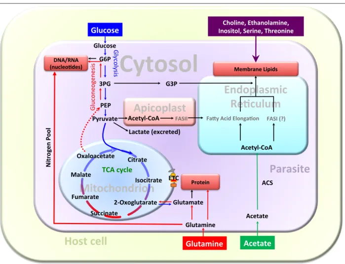
Fig 9: Central carbon metabolism in the tachyzoite stage of T. gondii. The model is constructed based on our work, literature and annotations of selected enzymes expressed in the tachyzoite stage (www.ToxoDB.org). Only those metabolites relevant to this work are shown for simplicity. Glucose and glutamine are co-utilized to satisfy the carbon demands for biomass (protein, nucleotides, lipids), energy and reducing equivalents (not depicted). Parasites can also deploy acetate as a carbon source for lipid synthesis when available in culture.
Lipid synthesis utilizes acetyl-CoA and 3PG (3-phosphoglycerate), which are mostly derived from glucose.
Biogenesis of nucleotides requires ribose 5-phosphate produced by diversion of G6P (glucose-6 phosphate) to the pentose phosphate shunt. Likewise, protein synthesis uses glucose-derived nonessential amino acids, such as serine and glycine (not shown). Glutamine catabolism enables effective biosynthetic utilization of sugar-derived carbon by replenishing the TCA cycle (drained by macromolecule biogenesis in replicating parasites). Glutamine also confers the much-needed nitrogen pool for nucleotides and protein syntheses.
Glutamine-derived carbon flux (TCA cycle and gluconeogenesis) can sustain the parasite survival without a severe growth defect, when glycolysis is compromised in tachyzoites. Other nutrients utilized by the parasite for membrane biogenesis include choline, ethanolamine, serine, inositol and threonine. Carbon metabolism is rewired to meet proliferating (intracellular) and non-proliferating (extracellular) demands as well as in response to the available nutrients. ACS, acetyl-CoA synthetase; ETC, electron transport chain;
FASI/FASII, fatty acid synthase I or II; G3P, glycerol-3-phosphate; PEP, phosphoenolpyruvate
! !
!
Cytosol!
Mitochondrion! !
Citrate!
DNA/RNA!
(nucleo8des)!
Protein!
Membrane!Lipids!
Glucose!
Glucose!
3PG!
Pyruvate!
AcetylBCoA!
Glutamine!
Glutamate!
Apicoplast!
FASII!
Glycolysis!
Gluconeogenesis!
TCA!cycle!
2BOxoglutarate!
Oxaloacetate!
Parasite!
Acetate!
Glutamine! Acetate!
FASI!(?)!
FaLy!Acid!Elonga8on!
G3P!
Malate!
Fumarate!
PEP!
Lactate!(excreted)!
Nitrogen!Pool!
Succinate!
Endoplasmic!
Re8culum!
ACS!
Choline,!Ethanolamine,!
Inositol,!Serine,!Threonine!
G6P!
Host!cell!
Isocitrate! ETC!
AcetylBCoA!
Extracellular tachyzoites depend on either glucose or glutamine to invade host cells, because the exogenous milieu lacks any substitutive nutrients that can generate adequate energy to facilitate the gliding motility and host-cell invasion. Indeed, a pharmacological inhibition of glutaminolysis or oxidative phosphorylation in the glycolysis-deficient mutant arrests the lytic cycle. Intracellular parasites on the other hand show a much greater resilience, possibly by exploiting intermediates of the host-cell glycolysis and other amino acids. Nonetheless, glucose and glutamine are the two key physiologically important nutrients utilized for the synthesis of macromolecules (ATP, nucleic acid, proteins, lipids). They together furnish a major fraction of total biomass carbon and energy in a co-regulated manner, and either of them is sufficient to support the replication of tachyzoites (Appendix C). Ironically, whereas glutamine is capable of driving nucleotide and protein biogenesis when glycolysis is impaired, it falls short in ensuring an optimal fatty acid synthesis, which results in a wide-spectrum deficit of lipids leading to a modest growth defect in the Δtggt1 mutant. Lipid synthesis as well as the lytic cycle of the glycolytic mutant can be restored by acetate supplement, which can recompense for the glucose-derived acetyl-CoA for fatty acid synthesis and elongation via FASII and FAE pathways in the apicoplast and ER (Fig 9). Such a cooperative metabolism of glucose, glutamine and acetate in tachyzoites resembles the physiology of tumor cells [72,90–92].
Our ensuing work on gluconeogenesis has identified two isoforms of the gluconeogenic enzyme fructose 1,6-bisphosphatase (TgFBP1, TgFBP2) in tachyzoites, which are constitutively expressed (Appendix D). It is quite unusual given that expression of gluconeogenic enzymes is repressed when glucose is available to avert a futile cycling of glycolytic metabolites. We found that whereas TgFBP1 is dispensable irrespective of glucose catabolism, TgFBP2 is essential for the parasite survival even when glycolysis is intact. Conditional knockdown of TgFBP2 results in a complete cessation of the parasite growth and virulence in a mouse model. TgFBP2 deficiency translates into altered glycolytic and TCA cycle flux, as well as dysregulation of glycolipid, amylopectin and fatty acid syntheses. We therefore postulate a futile cycle between fructose 1,6-bisphosphatase (gluconeogenic) and phosphofructokinase (glycolytic) enzymes as a novel regulatory mechanism that may allow tachyzoites to rapidly adapt to nutrients available in different host cells. Our work also found two isoforms of phosphoenolpyruvate carboxykinase (PEPCK) protein, only one of which is expressed in the tachyzoite stage (PEPCKmt localized in the mitochondrion; Appendix E).
Parasites tolerated the deletion of either isoforms, indicating a nonessential role of PEPCK enzymes in normal glucose-replete cultures. They could not survive collective ablations of both PEPCKmt and TgGT1 however, because the mitochondrial isoform is required for glutamine- fueled gluconeogenesis, which in turn sustains the biomass production in proliferating parasites upon glycolytic dysfunction. Once again, these results on PEPCKmt-dependent gluconeogenesis converge with cancer cells, which rely on glutamine-derived carbon flux to withstand glucose- independent growth [93].
We also observed that the physiological significance of glucose for Plasmodium contrasts with T.
gondii, even though the former can also assimilate glutamine as a major carbon source [94]. It was not feasible to ablate the PbHT1 gene without parallel complementation with a functional copy,
which confirms an essential role of glucose for erythrocytic stages of P. berghei (Appendix B). These results on genetic essentiality of glycolysis have also been independently confirmed by a similar study on P. falciparum and P. berghei [95]. Our in silico searches show that both Plasmodium species lack gluconeogenesis, which renders glycolysis essential. Unlike glycolysis, most of the enzymes of TCA cycle have been shown to be nonessential for the asexual growth of P. falciparum [96]. These mitochondrial mutants exhibited a normal erythrocytic cycle; however, their development was severely interrupted in the mosquito host. Along the line, we determined that a glucose analog (C3361) potently inhibits the sexual and hepatic development of P. berghei. Therefore, it can be concluded that glucose is indispensable for the entire lifecycle of Plasmodium species. To pursue a translational application of these findings, we generated transgenic strains of Saccharomyces cerevisiae and P. berghei, which express hexose transporter of P. falciparum in lieu of endogenous permease(s), and as a consequence depend on PfHT1 for their survival. These two models together permit a streamlined in vitro prescreening and subsequent in vivo assessment of antimalarial sugar analogs.
Such a platform should facilitate the early drug development against human malaria.
2.2 Phospholipid biogenesis in tachyzoites of Toxoplasma gondii
Intracellular proliferation of tachyzoites and expansion of the PV require substantial synthesis of organelle membranes, which are composed of predominantly neutral and polar lipids. This work explored several primary features of phospholipid metabolism in T. gondii. We demonstrate that tachyzoites harbor standard eukaryotic as well as atypical lipids. Phosphatidylcholine (PtdCho) is the most common lipid, followed by phosphatidylethanolamine (PtdEtn), phosphatidylthreonine (PtdThr), phosphatidylinositol (PtdIns), phosphatidylserine (PtdSer), ethanolamine- phosphorylceramide (EPC), phosphatidylglycerol (PtdGro) and phosphatidate (PtdOH) (Fig 10).
Intracellular tachyzoites express a nearly complete set of enzymes, which catalyze the synthesis of major phospholipids using host-derived carbon sources, namely glucose, glutamine and acetate.
Fig 10: Pathways of phospholipid syntheses in the tachyzoite stages of T. gondii. The model is constructed primarily based on our results. Lipid precursors are shown in blue; the intermediates of lipid synthesis are in black;
and phospholipids are in red. Phospholipid synthesis begins with the synthesis of precursor lipid (PtdOH), which is converted to diacylglycerol (DAG) for making PtdCho and PtdEtn in the ER. PtdEtn can also be generated in the mitochondrion and PV by decarboxylation of PtdSer. Unlike mammalian cells, PtdEtn is not methylated to make PtdCho (red cross). PtdThr and PtdSer are produced exclusively in the parasite ER. PtdOH in the ER and apicoplast also yields CDP-DAG, which serves as a substrate for the synthesis of PtdIns in the Golgi bodies and PtdGro in the mitochondrion. Glycerol backbone and fatty acids are originally derived from glycolysis (Fig 9). Phospholipid synthesis is significantly induced or repressed when tachyzoites switch between intracellular and extracellular phases, respectively, during the lytic cycle.