The illusion of (cell cycle) control. (Adapted from Watterson, 1995)
Functional analysis of CDKA;1, the Arabidopsis thaliana homologue of the p34cdc2 protein kinase
Inaugural-Dissertation
zur
Erlangung des Doktorgrades
der Mathematisch-Naturwissenschaftlichen Fakultät der Universität zu Köln
vorgelegt von
Moritz K. Nowack
aus Marburg
2007
Berichterstatter: Prof. Dr. Martin Hülskamp PD Dr. Frank Sprenger
Prüfungsvorsitzender: Prof. Dr. Siegfried Roth
Tag der mündlichen Prüfung: 20. April 2007
DANKE
Mein besonderer Dank gilt Dr. Arp Schnittger für die Überlassung des Themas und für eine inspirierende wissenschaftliche Betreuung, die weit über das übliche Maß hinausging.
Bei Prof. Dr. Martin Hülskamp möchte ich mich für die freundliche Aufnahme in seinen Lehrstuhl und den Kreis der Pflanzenentwicklungsbiologen bedanken. Für die Übernahme des Zweitgutachtens danke ich PD Dr. Frank Sprenger.
Des weiteren möchte dich denjenigen danke sagen, ohne deren Mitarbeit diese Arbeit nicht zustande gekommen wäre.
Damit ist vor allen anderen Paul E. Grini gemeint, dessen Fachwissen und Arbeitseinsatz für viele Aspekte meiner Arbeit unverzichtbar war. Unsere Zusammenarbeit während meiner Aufenthalte an der Universität Oslo und unser Zusammenkommen auf eigentlich jeder Konferenz haben mir viel gegeben und unheimlich viel Spaß gemacht! In diesem Zusammenhang geht mein Dank auch an Prof. Reidunn Aalen und ihr Team am Department of Molecular Biosciences der Universität Oslo für meine Aufnahme als Gastwissenschaftler, und besonders an Reza Shirzadi für ein tolles Teamwork!
Auch die Arbeiten am Institute of Molecular Medicine and Experimental Immunology des Bonner Universitätsklinikums waren eine gegenseitige Bereicherung an Erfahrungen. Ein dickes Danke an Elmar Endl, Andreas Dolf und Peter Wurst für zahlreiche LSR- und FlowJo- Sitzungen! Keine Experimente!
Ebenso dankbar bin ich Moola, Uli, Elena, Daniel und allen anderen von der Botanik III in Köln für die gute Zusammenarbeit und für so manches Kaffee-Pad.
Äußerst konstruktiv fand ich auch für die Arbeitsatmosphäre am MPI – mein besonderer Dank geht an
Marcel Lafos für die Hilfe am Mutanten-Wühltisch der Sammlung von Czaba Koncz;
Sandra Noir und ihren Pollenstaubsauger;
Hughes Barbier und Mattieu Reymond für all die SNK-Tests;
Enrico für die Westernhilfen;
Elmon Schmelzer für die professionelle Unterstützung an allen Mikroskopen;
Ralf Petri und seinen Einsatz für die IMPRS;
Andreas Lautscham und Frank Eikelmann und ihr Gärtnerteam;
die Kollegen von SUSAN für ihren hervorragenden user-support;
und an Georges Mancel und seine Kollegen aus der Werkstatt, die keine Idee für prinzipiell verrückt hielten.
Zum Schluss kommt immer das Beste: Allen Leuten aus dem Schnittger-lab bin ich sehr dankbar für Jahre guter Zusammenarbeit im Labor sowie ununtertroffenen Gesprächen am Mittagstisch! Dankeschön, Christina, Farshad, Stefan, Marc, Nico, Doris, Suzanne, Alex, Björn und Karo! Danken möchte ich auch den PraktikantInnen und StudentInnen, die uns tatkräftig unterstützt haben, Anni, Janina, Peter, Rafael, Katharina, Markus und vielen mehr.
Ein ganz dickes Dankeschön geht außerdem an Aurélie und Nico für den großartigen Tag- und-Nacht-Einsatz ihrer Adleraugen!
Contents I
CONTENTS
Contents………...…I Zusammenfassung……….……….…………..IV Abstract……….…………. ..VI Publications……….………VIII Abbreviations and gene names………...IX Figure and table index ...XI
1 INTRODUCTION
1.1 Cell cycle control ... 1
1.1.1 CDKs: microprocessors at the heart of cell cycle control... 1
1.1.2 CDKs and cell cycle control in plants... 3
1.2 Plant development ... 4
1.2.1 General features of plant cell division and development ... 4
1.2.2 The plant life cycle ... 5
1.2.3 Double fertilization ... 6
1.2.4 The endosperm: an integrator of seed development... 7
1.3 Imprinting and he role of FIS-class genes during seed development ... 9
1.3.1 The FIS-PRC2 ... 9
1.3.2 Imprinting in angiosperm seed development ... 11
1.4 Aim of the study... 13
2 RESULTS 2.1 Isolation and molecular characterization of a cdka;1 mutant... 14
2.1.1 Isolation of two independent cdka;1 mutant lines ... 14
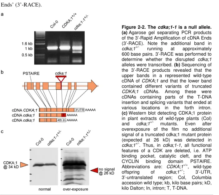
2.1.2 cdka;1-1 is a null allele ... 16
2.2 Analysis of the cdka;1 mutant phenotype ... 17
2.2.1 There are no homozygous cdka;1 mutants... 17
2.2.2 cdka;1 represents a paternal effect mutant ... 17
2.2.3 The pollen phenotype of cdka;1+/- mutants... 19
2.2.4 The homozygous cdka;1-/- mutants ... 21
2.3 Complementation assays... 23
2.3.1 Expression of the CDKA;1 cDNA from a 2 kb CDKA;1 promoter fragment can rescue the cdka;1 mutant... 23
2.3.2 Complementation by ProCDKA;1:CDKA;1:YFP... 23
2.3.3 CDKA;1:YFP dynamics in pollen development... 24
2.4 The secondary phenotype of cdka;1 mutants... 26
2.4.1 cdka;1 pollen is viable and able to germinate in vitro ... 26
2.4.2 cdka;1 pollen causes a single fertilization and exclusively fertilizes the egg cell ... 27
2.4.3 Development of unfertilized endosperm in wt x cdka;1 seeds ... 29
2.5 Combination of the cdka;1 mutant with the medea mutant ... 30
2.5.1 wild-type x cdka;1 seeds abort... 30
2.5.2 mea-/- x cdka;1+/-: a new class of developing seeds... 31
Contents II
2.5.3 mea x cdka;1 seeds can undergo complete development and
develop into normal F1 plants... 33
2.5.4 Post-embryo development of mea-/- x cdka,1+/- F1 plants... 37
2.5.5 mea-/- x cdka;1:yfp+/- results in a mutual rescue... 37
2.5.6 Flow cytometry ... 40
2.5.7 SSLP based paternity test... 40
2.6 Diploid, unfertilized endosperm possesses wild-type characteristics ... 43
2.6.1 Marker lines for endosperm differentiation and development ... 43
2.6.2 Endosperm cellularization... 45
2.6.3. Gene expression is balanced in mea-/- x cdka;1+/- seeds ... 45
3 DISCUSSION 3.1 Cell cycle arrest - the primary cdka;1 phenotype... 48
3.1.1 CDKA;1 function is essential for Arabidopsis development ... 48
3.1.2 CDKA;1 function is indispensable for sporophyte development... 48
3.1.3 Male, but not female cdka;1 mutant gametophytes arrest development ... 49
3.1.4 cdka;1 mutant pollen arrests in G2 phase ... 50
3.2 Single fertilization - the secondary cdka;1 phenotype... 51
3.2.1 cdka;1 pollen produces a fertile gamete... 51
3.2.2 Preferential fertilization of the egg cell... 52
3.2.3 A positive signal from the zygote starts endosperm proliferation . 53 3.2.4 The nature of the proliferation signal ... 55
3.3 The mutual rescue of fis-class mutants with cdka;1 pollen ... 56
3.3.1 wt x cdka;1 seeds abort with an underdeveloped endosperm ... 56
3.3.2 mea x cdka;1 seeds are viable but smaller than wild type seeds.... 56
3.3.3 wt x cdka;1 and mea x cdka;1 copy a maternal excess phenotype 57 3.3.4 Imprinting and the kinship theory of seed development ... 58
3.3.5 mea x cdka;1: paternalization meets maternalization ... 59
3.3.6 PHERES1 – the final dosage is decisive ... 60
3.3.7 mea x cdka;1 – two perspectives on the rescue... 61
3.4 Reflections on the evolutionary origin of the endosperm in angiosperms 65 3.5 Outlook: The cdka;1 mutant as a tool investigate plant development ... 67
4 MATERIALS & METHODS 4.1 Materials ... 69
4.1.1 Chemicals and antibiotics... 69
4.1.2 Enzymes, primers and kits ... 69
4.1.3 Cloning vectors and constructs ... 69
4.1.4 Plant material ... 69
4.1.5 Bacterial strains ... 70
4.2 Methods ... 70
4.2.1 Plant work ... 70
Contents III
Plant growth conditions ... 70
Crossing of plants... 70
Plant transformation ... 70
Seed surface sterilization... 71
Selection of transformants... 71
4.2.2 Microscopy and cytological methods... 71
Microscopy ... 71
LR-White embedding and semi-thin sectioning of seeds... 72
Whole-Mount preparation of seeds ... 72
GUS staining ... 72
Pollen preparation for fluorescence analysis... 72
Pollen DNA measurements ... 73
Pollen viability assay... 73
Pollen in vitro germination assay ... 73
Flow cytometry for seed tissue ploidy analysis ... 73
4.2.3 Molecular-biological methods... 74
Genomic DNA preparation from plant tissue I ... 74
Genomic DNA preparation from plant tissue II... 74
Plasmid DNA preparation from bacteria... 75
DNA-manipulation... 75
Cloning of complementation and reporter constructs ... 75
Identification of cdka;1 mutants by PCR... 75
3’ Rapid Amplification of cDNA Ends (3’ RACE) ... 75
Quantitative PCR... 76
Paternity test of embryo and endosperm ... 76
4.2.4 Biochemical methods ... 77
Protein extraction and Western blotting... 77
5 REFERENCES... 80
APPENDIX Erklärung ... 89
Lebenslauf ... 90
Zusammenfassung IV
ZUSAMMENFASSUNG
CYCLIN-DEPENDENT KINASEs (Cyclin-abhängige Kinasen, CDKs) sind zentrale Steuerungselemente der Zellzykluskontrolle und homologe CDK-Proteine sind in allen Eukaryonten konserviert. Die vorliegende Promotionsarbeit beschreibt die funktionelle Analyse von CDKA;1, einer bedeutenden CDK in Arabidopsis thaliana. CDKA;1 ist im Arabidopsis-Genom mit nur einer Kopie vertreten und nur CDKA;1 ist ein funktionelles Äquivalent der cdc2/CDC28-Kinasen in Hefen.
Ein Screening von zwei T-DNA Insertionsmutanten-Sammlungen ergab die Isolierung von zwei unabhängig entstandenen cdka;1-mutanten Pflanzenlinien. Beide Linien erwiesen sich als Nullmutanten und zeigten den gleichen Phänotyp. Eine nähere Untersuchung ergab, dass CDKA;1 für die Zellzykluskontrolle sowohl in der sporophytischen als auch in der gametophytischen Generation von Arabidopsis benötigt wird. Während heterozygote Sporophyten keinerlei Abweichungen in ihrer Entwicklung aufwiesen, waren homozygote Mutanten nicht lebensfähig und starben während der frühen Embryonalentwicklung.
Außerdem führte das Fehlen der CDKA;1-Funktion im männlichen Gametophyten (Pollen) zu einer Unterbrechung des Zellzyklus-Programms in der G2-Phase vor der letzten pollenspezifischen Mitose. Durch diesen Zellzyklusdefekt bildete sich reifer cdka;1-Pollen mit nur einem statt der üblichen zwei Spermazellen.
Trotz dieses Defekts war cdka;1-Pollen lebensfähig und in der Lage, den weiblichen Gametophyten (Embryosack) zu erreichen und zu befruchten. Dadurch, dass cdka;1-Pollen nur eine Spermazelle zur Befruchtung beisteuern konnte, erfolgte eine einfache Befruchtung anstelle der für Blütenpflanzen typischen Doppelbefruchtung. Interessanterweise wurde bei dieser einfachen Befruchtung ausschließlich die Eizelle befruchtet, während die Zentralzelle, die sich normalerweise nach der Befruchtung zum Endosperm entwickelt, unbefruchtet blieb.
Nichtsdestotrotz begann nach der Befruchtung der Eizelle nicht nur die Embryonalentwicklung, sondern auch der unbefruchtete Zentralzellkern begann sich zu teilen.
Diese Tatsache ließ auf einen Signalmechanismus schließen, der von der befruchteten Eizelle in Gang gesetzt wird und den Zentralzellkern zur Proliferation anregt. Diese autonome Proliferation umfasste allerdings nur maximal fünf mitotische Teilungen, bevor das unbefruchtete Endosperm seine Entwicklung stoppte und anschließend abstarb. Im Folgenden brach auch der Embryo seine Entwicklung ab und der gesamte Samen abortierte in einer frühen Entwicklungsphase. Durch diesen Samenabort kann man dem cdka;1-Pollen einen sogenannten paternalen Effekt zuschreiben, da er unabhängig von der genetischen Situation im weiblichen Kreuzungspartner zu einem Absterben des Samens nach der Befruchtung führt.
Zusammenfassung V
Um die Endosperm-Entwicklung zu verstärken, wurde cdka;1-Pollen auf verschiede fis- Mutanten gekreuzt. Diese Mutanten zeichnen sich durch einen Defekt im FIS-Proteinkomplex aus, der über die weibliche Seite vererbt wird und als Polycomb-group-Komplex die genomische Prägung (Imprinting) im Endosperm kontrolliert. In fis-Mutanten kommt es ohne Befruchtung zu autonomer Endosperm-Proliferation. Befruchtete fis-Mutanten weisen eine starke Überproliferation des Endosperms auf und aufgrund eines maternalen Effekts abortieren ihre Samen zu einem späteren Zeitpunkt der Samenentwicklung.
Wenn cdka;1-Pollen zur Bestäubung von fis-Mutanten verwendet wurde, entwickelte sich das Endosperm deutlich stärker als im cdka;1-bestäubten Wildtyp und viele Samen entwickelten sich über das Stadium des cdka;1-bedingten Aborts hinaus. Überraschenderweise wurde aber auch der durch die maternalen fis-Allele hervorgerufene Samenabort zum Teil aufgehoben und einige Samenanlagen entwickelten sich zu reifen, lebensfähigen Samen.
Diese Rettung der Samenentwicklung ging mit einer deutlichen Verringerung des Expressionsniveaus des MADS-box Transkriptionsfaktors PHERES1 im Endosperm einher.
PHERES1 ist ein direktes Ziel der transkriptionellen Repression durch den FIS-Komplex und ist daher in fis-Mutanten stark überexprimiert. Die Abschwächung des PHERES1 Expressionsniveaus im Endosperm der Kreuzung fis x cdka;1 lässt vermuten, dass die Abwesenheit der paternalen Expression, kombiniert mit der maternalen Überexpression, zu einem Normalisierung des Expressionsniveaus von PHERES1 führte. Möglicherweise sind von dieser Normalisierung auch andere, bisher unbekannte Gene betroffen, deren Expressionsniveau für die Endosperm-Entwicklung von Bedeutung ist.
Zusammengenommen deuten die Ergebnisse der vorliegenden Arbeit deuten darauf hin, dass der FIS-Komplex für die Endosperm-Entwicklung nicht essentiell ist. Vielmehr scheint die Funktion der FIS-Proteine darin zu liegen, die Genexpression von maternal und paternal vererbten Genen aufeinander abzustimmen. Darüber hinaus verdeutlichen die hier gewonnenen Erkenntnisse, dass das paternale Genom für die Entwicklung eines funktionellen Endosperms in Arabidopsis nicht benötigt wird, wenn das Imprinting im maternalen Genom durch einen Defekt im FIS-Komplex umgangen wird.
Die Tatsache, dass ein rein maternal vererbtes Endosperm für eine funktionelle Samenentwicklung ausreicht, unterstützt eine Hypothese von Eduard Strasburger aus dem Jahr 1900. Strasburger mutmaßte bereits damals, dass der evolutive Ursprung des Endosperms im weiblichen Gametophyten zu suchen sei und sich die Doppelbefruchtung als Auslöser für die Endosperm-Proliferation entwickelt habe.
Abstract VI
ABSTRACT
CYCLIN-DEPENDENT KINASEs (CDKs) are the central gatekeepers of cell cycle progression and conserved in all eukaryotes. In this study, the Arabidopsis thaliana master cell cycle regulator CDKA;1 was functionally analyzed. CDKA;1 is a single gene in Arabidopsis and homologous to the human Cdk1 and the yeast cdc2/CDC28. Screening of two T-DNA insertion mutant collections resulted in the isolation of two independent cdka;1 null mutant alleles, which displayed the same phenotype. CDKA;1 was found to be required for both the sporophytic and the male gametophytic generations of the flowering plant Arabidopsis. While during sporophyte development, heterozygous mutant plants were unaffected, homozygous cdka;1 mutants were not viable and died as young embryos. During male gametophyte (pollen) development, the lack of CDKA;1 function caused a cell cycle arrest in the G2 phase prior to the last mitotic division. This cell cycle defect led to cdka;1 mutant pollen with only one instead of the usual two sperm cells.
Nevertheless, the mutant cdka;1 pollen was viable and could fertilize the female gametophyte (embryo sac). Because cdka;1 pollen grains had only one instead of two sperm cells, they only performed single fertilization and thus, disrupted the double fertilization event characteristic of flowering plants. Interestingly, the cdka;1 mutant single fertilization exclusively targeted the egg cell, leaving the progenitor of the endosperm, the central cell, unfertilized. However, upon cdka;1 fertilization of the egg cell, not only the embryo started to develop, but the unfertilized central cell nucleus also began to divide. This onset of endosperm development without fertilization revealed a hitherto unrecognized endosperm proliferation signal emitted from the fertilization of the egg cell.
The autonomous endosperm in cdka;1-fertilized seeds only underwent up to five nuclear division cycles before it stopped proliferating, followed by an early abortion of the whole seed. Thus, the cdka;1 mutant belongs to a rare class of paternal effect mutants that cause seed abortion irrespective of the genetic constitution of the female partner.
In order to enhance endosperm proliferation in cdka;1-fertilized seeds, cdka;1 pollen was crossed to various fis-class mutants. These mutants are defective in the maternally inherited FIS-complex, a Polycomb-group repressive complex controlling genomic imprinting in the endosperm. In fis-class mutants, autonomous endosperm develops in the absence of fertilization. When fertilized, the fis-class mutant endosperm over-proliferates and due to a maternal effect these seeds abort later during development.
Abstract VII
The endosperm development in cdka;1-fertilized fis-mutant seeds was substantially enhanced and led to a partial rescue of the cdka;1-mediated seed abortion. Unexpectedly, the maternally conferred seed abortion caused by fis-class mutants was also partially reversed, producing viable seeds among the fis-class x cdka;1 offspring. This rescue was characterized by a down- regulated expression of the MADS-box transcription factor PHERES1, a downstream target of FIS-complex repression which is highly over-expressed in fertilized fis-class mutants.
The down-regulation of PHERES1 in fis-class x cdka;1 endosperm suggests that the lack of paternal expression in combination with the defective gene repression of fis-class mutants results in a more balanced gene dosage of PHERES1 and potentially other genes of which the dosage is pivotal for regular seed development.
These results indicate that the FIS-complex is not essential for endosperm development, but is important to harmonize maternal and paternal gene expression by the control of imprinting in the female genome. Furthermore, these data demonstrate that the paternal genome is not required for functional endosperm development if maternally derived genomic imprinting is bypassed due to mutations in the FIS-complex.
The finding that a solely maternally derived endosperm can sustain seed development supports a hypothesis raised by Eduard Strasburger, who proposed in 1900 that the endosperm of flowering plants is of female gametophytic origin and that central cell fertilization might have evolved as a trigger to start endosperm proliferation.
Publications VIII
PUBLICATIONS
Novel Functions of Plant Cyclin-Dependent Kinase Inhibitors, ICK1/KRP1, Can Act Non-Cell-Autonomously and Inhibit Entry into Mitosis
Weinl, C., S. Marquardt, S. J. Kuijt, M. K. Nowack, M. J. Jakoby, M. Hulskamp and A.
Schnittger. Plant Cell 17(6): 1704-22 (2005).
• For this paper, I cloned the fusion construct ProGL2:GUS:YFP:KRP1109 and generated the corresponding transgenic plant lines
A positive signal from the fertilization of the egg cell sets off endosperm proliferation in angiosperm embryogenesis
Nowack, M. K., P. E. Grini, M. J. Jakoby, M. Lafos, C. Koncz and A. Schnittger. Nat Genet 38(1): 63-7 (2006).
• Apart from the analysis of the mea/fis1 x cdka;1 crosses and the ultra-structural pollen analysis, which were done by P.E.G. and the cloning of the ProCDKA;1:CDKA;1 rescue construct, which was done by M.J.J., I did all the work for this paper
T-Loop Phosphorylation of Arabidopsis CDKA;1 Is Required for Its Function and Can Be Partially Substituted by an Aspartate Residue
Dissmeyer, N., M. K. Nowack, S. Pusch, H. Stals, D. Inzé, P. E. Grini and A. Schnittger.
Plant Cell (in print)
• For this paper, I performed parts of the mutant analyses of the non-phosphorylatable CDKA;1T161V and the phospho-mimicry CDKA;1T161D version, including analyses of pollen and embryo development as well as ploidy-analyses by flow cytometry.
Bypassing genomic imprinting allows seed development
Nowack, M. K., R. Shirzadi, N. Dissmeyer, A. Dolf, E. Endl, P. E. Grini and A. Schnittger.
(Manuscript under review)
• Apart from major parts of the quantitative PCR and the in situ hybridisations, which were done by R.S. and P.E.G., and the cloning of the ProCDKA;1:CDKA;1:YFP rescue construct, which was done by N.D., I did all the work for this paper.
Abbreviations and gene names IX
Abbreviations and gene names
% Percent
°C degree Celsius
3' three prime end of a DNA fragment
35S 35S promotor from the Cauliflower Mosaic virus 5' five prime end of a DNA fragment
ANOVA Analysis of variance, statistical method ATP Adenosinetriphosphate
bp base pair
C DNA content of a haploid genome CAK CDK ACTIVATING KINASE CDK CYCLIN DEPENDENT KINASE CDKA;1 CYCLIN-DEPENDENT KINASE A1 cDNA complementary DNA
CDS coding sequence
CKI CYCLIN DEPENDENT KINASE INHIBITOR CKS1 CDC KINASE SUBUNIT 1
CLF CURLY LEAF
Col Arabidopsis thaliana Columbia accession CYC CYCLIN
CZE chalazal endosperm d.a.g. days after germination d.a.p. days after pollination
DAPI 4',6'-diamidino-2-phenylindole DME DEMETER
DMSO Dimethylsulfoxide DNA desoxyribonucleic acid DP DIMERIZATION PARTNER
E(z) Enhancer of zeste (Drosophila melanogaster) e.g. exempli gratia [Lat.] for example
E2F ADENOVIRUS E2 PROMOTOR BINDING FACTOR EDTA ethylenediaminetetraacetic acid
Esc Extra sex combs (Drosophila melanogaster) et al. et alii / et aliae [Lat.] and others
F1, F2, F3 first, second, third... filial generation after a cross FDA fluorescein diacetate
FIE FERTILIZATION-INDEPENDENT ENDOSPERM Fig. Figure
FIS2 FERTILIZATION-INDEPENDENT SEED 2 FIS-class proteins forming the core of the FIS-PRC2
FIS-PRC2 Arabidopsis PRC2 containing MEA, FIS2, FIE, and MSI1 G1 Gap phase between M phase and S phase
G2 Gap phase between S phase and M phase gene-/- homozygous mutant of a gene
gene+/- heterozygous mutant of a gene GFP green fluorescent protein GUS beta-glucuronidase h.a.p. hours after pollination
H3K27 Lysine residue 27 of the histone H3 i.e. id est [Lat.] that is
Abbreviations and gene names X
ICK/KRP INHIBITOR OF CYCLIN-DEPENDENT KINASE / KIP-RELATED PROTEIN (plant CKI) Ilgf2 mammalian Insulin-like growth factor 2
kb 1000 base pairs kD kilo Dalton LB T-DNA left border
Ler Arabidopsis thaliana Landsberg erecta accession m maternally inherited genome
M phase mitotic phase of the cell cycle MCE micropylar endosperm
MEA MEDEA
MET1 METHYLTRANSFERASE 1 mRNA messenger RNA
MSI1 MULTICOPY SUPPRESSOR OF IRA 1
n Number
p paternally inherited genome PBS phosphate bufferd saline buffer PcG Polycomb-group
PCR polymerase chain reaction PEN peripheral endosperm PHE1 PHERES1
PMI pollen-specific mitosis one PMII pollen-specific mitosis two PRC2 Polycomb Repressive Complex 2 ProGENE promoter sequence of a GENE QPCR quantitative Real-time PCR RB T-DNA right border
RBR1 RETINOBLASTOMA RELATED 1 Arabidopsis homologue of the Retinoblastoma gene RNA ribonucleic acid
rpm rotations per minute
RT PCR reverse transcription followed by a polymerase chain reaction S phase synthetic phase of the cell cycle
SDS PAGE sodium dodecylsulfate polyacrylamide gel electrophoresis
SET domain common to cytosine methyltransferases, derived from Su(var), E(z) and Trithorax SSLP Single Sequence Length Polymorphism
Su(var) Suppressor of variegation (Drosophila melanogaster) Su(z) Suppressor of zeste (Drosophila melanogaster) SWN SWINGER
T1, T2, T3 first, second, third transgenic generation after stable plant transformation T-DNA transferred DNA
Tris/HCl buffer containing 2-amino-e-hydroxymethyl-1,3-propanediol and HCl UTR untranslated region
UV ultra-violet light WEE WEE kinase
wt wild type
x crossed to (crosses are always indicated in the order: female x male) YFP yellow fluorescent protein
The nomenclature for plant genes follows the Arabidopsis standard: GENES are written in upper case italics, while mutant genes are indicated in lower case italics. PROTEINS appear in upper case regular letters, mutant proteins in lower case regular letters.
Figure and table index XI
Figure and table index
Figures
Figure 1-1. CDKs are the core of eukaryotic cell cycle control... 2
Figure 1-2. The plant life cycle ... 6
Figure 1-3. Seed development in Arabidopsis thaliana... 8
Figure 1-4. The Arabidopsis FIS-PRC2... 10
Figure 2-1. T-DNA insertion mutants of the Arabidopsis CDKA;1... 15
Figure 2-2. The cdka;1-1 is a null allele ... 16
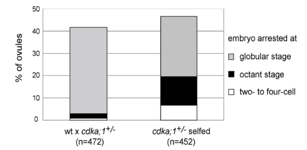
Figure 2-3. Seed abortion in cdka;1+/- mutants ... 18
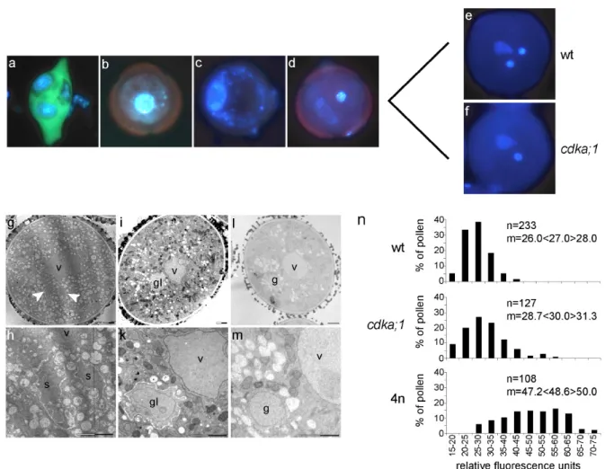
Figure 2-4. Phenotype of cdka;1 mutant pollen... 20
Figure 2-5. Homozygous cdka;1 mutants abort early in embryo development... 22
Figure 2-6. CDKA;1:YFP dynamics during pollen development... 25
Figure 2-7. Viability and in vitro germination ability of cdka;1 mutant pollen ... 27
Figure 2-8. Seeds expressing the fertilization reporter ProCDKA;1:GUS... 28
Figure 2-9. Seed development in wild-type plants fertilized with cdka;1+/- mutant pollen... 30
Figure 2-10. Seed development in mea-/- mutant plants fertilized with cdka;1+/- mutant pollen... 32
Figure 2-11. Expression of the ProCDKA;1:CDKA;1:YFP transgene after fertilization to the mea-/- mutant... 34
Figure 2-12. Rescue of mea-conferred embryo abortion after pollination with cdka;1 mutant pollen... 35
Figure 2-13. Post-embryo development of mea-/- x cdka;1:yfp+/- F1 plants ... 38
Figure 2-14. Flow cytometry of mea-/- x cdka;1+/- seeds ... 41
Figure 2-15. Laser microdissection of small mea-/- x cdka;1+/- rescue seeds at 6 d.a.p... 42
Figure 2-16. Endosperm differentiation of a uniparental, diploid endosperm in mea-/- x cdka;1+/- seeds proceeds as in wild type... 44
Figure 2-17. Quantitative real-time PCR monitoring PHE1 expression in mea-/- x cdka;1+/- seeds ... 46
Figure 3-1. The crosstalk between embryo and endosperm during early seed development ... 54
Figure 3-2. The endosperm perspective ... 62
Figure 3-3. The embryo perspective ... 65
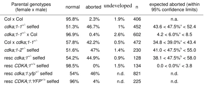
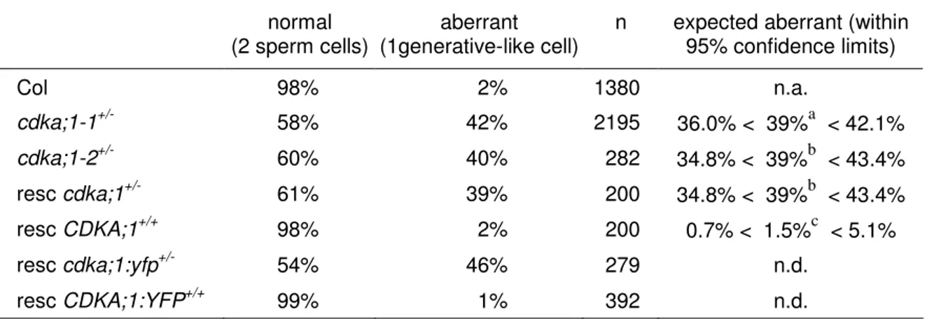
Tables Table 2-1. Percentage of aborted seeds in cdka;1 mutant lines... 18
Table 2-2. Transmission of the cdka;1-1 allele ... 19
Table 2-3. Phenotype of cdka;1 pollen at anther dehiscence ... 21
Table 2-4. Seed development in mea-/- x cdka;1-1+/- plants ... 31
Table 2-5. Endosperm development in mea-/- x cdka;1+/- plants... 33
Table 2-6. Viable seeds in mea-/- x cdka;1+/-... 36
Table 2-7. Flowering time in mea-/- x cdka;1:yfp+/- F1-plants ... 36
Table 2-8. Transmission frequencies of the cdka;1 mutant allele ... 39
Table 2-9. Results of the laser-microdissection PCR ... 42
Table 4-1. Primers and probes ... 78
Introduction 1
1 INTRODUCTION
“Omnis cellula e cellula”: The fundamental biological dogma that every cell is created by division of a pre-existing cell was formulated some 150 years ago and opposed the idea of spontaneous generation of life (Virchow, 1855). To date, extensive research has provided a detailed molecular understanding of how one cell is derived from another cell and it is now known that all living organisms depend on the duplication of their genetic material and subsequent cell division to reproduce, grow, and develop. These events are tightly coordinated in a highly conserved cellular programme known as the cell cycle.
1.1 Cell cycle control
1.1.1 CDKs: microprocessors at the heart of cell cycle control
The basic mitotic cell cycle is divided in four phases: During the synthetic (S) phase, the DNA is replicated, while during the mitotic (M) phase, the sister chromatids are segregated to the newly forming daughter cells that are afterwards separated by cytokinesis. Between M phase and S phase, there are two gap phases (G1 and G2), in which cells proceed with important physiological functions and eventually prepare for the entry into the next cell cycle phase. There are several modifications of the basic cell cycle theme and one widespread cell cycle mode is an endocycle, in which the M phase is skipped while the DNA continues to be replicated leading to polyploid cells.
Progression through the mitotic cell cycle is controlled at two major checkpoints, the G1/S transition and the G2/M transition. The molecular machinery that controls progression through these checkpoints is highly conserved in all eukaryotes investigated so far (Inze and De Veylder, 2006). Its core consists of CYCLIN-DEPENDENT KINASEs (CDKs).
CDKs serve as information processors that integrate intracellular and extracellular signals to ensure the appropriate progress of the cell cycle (Morgan, 1997). Upon favourable conditions, e.g. the presence of nutrients or mitogens, cells advance in the cell cycle, while in response to negative cues such as DNA damage, cells arrest at a checkpoint (Fig. 1-1 a).
Information is conferred to the CDKs by a complex molecular machinery that tightly controls the patterns of CDK catalytic activity throughout the cell cycle. CDK activity depends on the association with subunits; most importantly with CYCLINs (CYC) the oscillating concentrations of which create the basic cell-cycle dependent activity patterns
Introduction 2
Figure 1-1. CDKs are the core of eukaryotic cell cycle control. (a), CDK/CYC complexes trigger the progression through the cell cycle at two major checkpoints, the transition from G1 to S phase and the transition from G2 to M phase. CDKs act as processors of multiple signalling pathways conferring intrinsic and extrinsic cues to the cell cycle machinery. (b), CDK activity is controlled by multiple mechanisms, including association with activating (CYC) or inhibiting (CKI) subunits. CDK activity is further controlled by activating or inactivating phosphorylations effected by CDK- ACTIVATING KINASES (CAK) or WEE kinases, respectively. The action of CDK activators (green) eventually causes cell cycle progression, while CDK inhibitors (red) can effect a cell cycle arrest at certaincheckpoints.
of CDKs. CYCLINs enhance the CDK/CYC substrate specificity and specific CYCLINs bind to CDKs in different cell cycle phases (Morgan, 1997).
Next to CYCLINs, other interactors modify the activity of the CDK/CYC complex:
CYCLIN-DEPENDENT KINASE INHIBITORS (CKIs) are able to block CDK/CYC kinase function when cell cycle progression needs to be stopped or modified (Fig. 1-1 b) (Sherr and Roberts, 1999).
Additionally, CDK/CYC activity is controlled by a regulatory network of protein kinases and antagonistic protein phosphatases causing CDK/CYC activation or inactivation by phosphorylation or dephosphorylation (Morgan, 1997). These regulatory mechanisms help to fine-tune the intrinsic activity patterns of CDK/CYC complexes (Pomerening et al., 2003) and represent an additional pathway to feed external signals into the cell cycle control (Fig. 1-1 b) (De Schutter et al., 2007).
Introduction 3
Once fully activated, CDK/CYC complexes phosphorylate a vast number of target proteins that directly or indirectly prepare the cell for the entry into a new cell cycle phase (Ubersax et al., 2003).
1.1.2 CDKs and cell cycle control in plants
In plants, as in other eukaryotes, the conserved CDK/CYC core cell cycle machinery controls progression through the cell cycle. Many features of CDK/CYC activity control are conserved in plants although some of the regulation is realized in plant-specific ways (Boudolf et al., 2006; Inze and De Veylder, 2006).
In contrast to unicellular eukaryotes such as yeasts, in which a single CDK controls the progression through all cell cycle phases, in multicellular organisms like animals and plants, small families of CDKs have evolved. The CDK family of the model plant Arabidopsis thaliana consists of twelve members including one A-type, four B-type, two C-type, three D-type, one E-type and one F-type CDK (Vandepoele et al., 2002). Only the single A-type CDK in Arabidopsis, CDKA;1, contains the conserved PSTAIRE motif and is able to complement the cdc2 mutant in fission yeast (Schizosaccharomyces pombe) and the cdc28 mutant in budding yeast (Saccharomyces cerevisiae) (Ferreira et al., 1991;
Hirayama et al., 1991; Porceddu et al., 1999). Therefore CDKA;1 is likely to be an important plant cell cycle regulator. The exact function of the other CDKs is not entirely understood. CDKBs become active at the G2-M transition and thus, might be involved in the control of mitosis (Boudolf et al., 2004a; Boudolf et al., 2004b; Inze and De Veylder, 2006)
The observation that CDKA;1 protein levels remain constant throughout the cell cycle (Magyar et al., 1997; Porceddu et al., 2001; Sorrell et al., 2001) and that CDKA;1 activity can be detected at both checkpoints (Hemerly et al., 1995; Porceddu et al., 2001; Joubes et al., 2004) lead to the assumption that CDKA;1 is participating in all cell cycle transitions.
Consistently, CDKA;1 is expressed in all tissues that show cell division or else are competent to divide. This expression pattern suggested a general role of CDKA;1 in the establishment of proliferative competence (Martinez et al., 1992; Hemerly et al., 1995).
While over-expression of native CDKA;1 did not alter the cell cycle nor plant development, misexpression of a dominant negative CDKA;1DN version caused lethality, suggesting an essential role of CDKA;1 in Arabidopsis cell cycle control (Hemerly et al., 1995).
Introduction 4
In tobacco (Nicotiana tabaccum), the same dominant-negative construct had no effects on plant viability, but strongly decreased cell division rates. However, G1/G2 ratios were unaltered, indicating the participation of CDKA;1 at both cell cycle checkpoints (Hemerly et al., 1995). In maize (Zea mays) endosperm, expression of a CDKA;1DN version reduced endoreplication, arguing for a role of CDKA;1 in the control of G1/S transition (Leiva- Neto et al., 2004).
Other experimental approaches to study the function of CDKA;1 were made by misexpression of CDKA;1 inhibitors called INHIBITORS OF CDK/KIP-RELATED PROTEINS (ICK/KRPs). ICK/KRPs bind to CDKA;1 and block its kinase activity (De Veylder et al., 2001). In short, these experiments demonstrated that lowered CDKA;1 kinase activity leads to an overall reduction of cell division resulting in smaller plants (Wang et al., 2000; De Veylder et al., 2001). Furthermore, while high levels of ICKs/KRPs block CDKA;1 activity at both G1/S and G2/M transition, moderate levels specifically target the G2/M-specific CDKA;1/CYC complexes. Taken together, CDKA;1 appears to function as a master cell cycle regulator essential for both the G1/S and the G2/M transition in plants.
1.2 Plant development
1.2.1 General features of plant cell division and development
In both plants and animals, the control of cell division is crucial to the correct realization of the genetically programmed body plan. However, in contrast to animals, the plant body develops mainly post-embryonically, producing its biomass out of small clusters of pluripotent stem cells called meristems. While the shoot meristem typically produces the above-ground organs of a plant, such as shoots, leaves, and the reproductive organs, the root meristems located in the root tips builds up the root system. The iterative development of life-long sustained populations of stems cells leads to an enormous potential of morphological plasticity in plant development.
However, fully differentiated cells also show a remarkable pluripotency: In root pericycle cells, for instance, re-activation of the cell cycle leads to the formation of a new meristem that subsequently forms a new side root (Casimiro et al., 2003).
Introduction 5
Furthermore, plant cells are surrounded by a rigid cell wall and tightly connected to neighbouring cells in the tissue, which means that cell migration is largely impossible and other means to build up the body structure have been implemented. Therefore, cell division ratios and the orientation of cell division planes are of great importance for the plant body architecture (Hemerly et al., 2000).
1.2.2 The plant life cycle
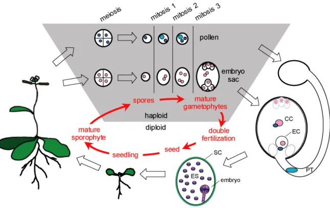
A major feature characteristic for plants is their two-phase life cycle of alternating generations of sporophytes and gametophytes (Fig. 1-2). In flowering plants (angiosperms), the predominant generation is the typically diploid sporophyte. This generation builds up the main plant body with roots, shoots, leaves and flowers, but it does not undergo sexual reproduction. Instead, meiotic divisions in specialized floral tissues lead to the formation of haploid microspores and megaspores. These spores are the starting point for the second, haploid generation, called gametophyte. The purpose of the gametophytes is to produce the male and the female gametes and to bring them together during sexual reproduction. In Arabidopsis, a typical angiosperm, the gametophytic generation is reduced to minute few-celled organisms which are embedded in the maternal tissue and completely dependent from the mother sporophyte.
Microspores are produced in the anthers of a flower and undergo two cell cycle rounds to complete their development into mature male gametophytes or pollen grains. During pollen-mitosis one (PMI), the microspore undergoes an unequal division to form the large vegetative cell and the small generative cell. Subsequently, the pollen-mitosis two (PMII) divides the generative cell in two sperm cells (McCormick, 2004).
Megaspores usually undergo three cell cycle rounds to produce the female gametophyte or embryo sac containing eight nuclei. Subsequent cellularization forms the mature seven- celled embryo sac, which includes the two gametes, the egg cell and the homodiploid central cell, as well as the accessory synergids and the antipodal cells. The mature embryo sac is surrounded by several layers of maternally derived integuments. The entity of embryo sac and the integuments is called the ovule, and this structure will develop into a seed after successful fertilization (Drews and Yadegari, 2002; Yadegari and Drews, 2004).
Introduction 6
Figure 1-2. The plant life cycle. Plants have a two-phase life cycle of alternating generations. The typically diploid sporophyte produces haploid spores through meiosis. The spores develop into the haploid gametophytes. Microspores undergo two mitotic cycles to form the mature pollen that contains two sperm cells (dark blue). Megaspores produce a seven-celled gametophyte by three mitotic divisions. The female gametes are the egg cell (EC) and the central cell (CC), respectively.
After the pollen tube (PT) has transported the two sperm cells to the embryo sac, double fertilization occurs: One of the sperm cells fuses with the egg cell to form the diploid embryo, while the other sperm cell fuses with the homodiploid central cell to give rise to the triploid endosperm (ES). Embryo and endosperm are surrounded by the maternally derived seed coat (SC). After completion of embryo development, the seedling is released from the seed and develops into the new sporophyte. (Adapted from Berger et al. 2006).
1.2.3 Double fertilization
For the fertilization process in flowering plants, pollen is released and has to be transported to the stigma, a specialized receptive tissue formed by the maternal sporophyte. In contact with the stigma cells, the pollen germinates and forms a pollen tube which penetrates the maternal sporophytic tissue and grows towards an ovule. After penetration of the ovule, the pollen tube releases the two sperm cells. In a process called double fertilization, one of the sperm cells fertilizes the haploid egg cell to form the embryo while the second sperm cell fertilizes the homodiploid central cell to give raise to the triploid endosperm (Faure et al., 2002).
Introduction 7
Successful double fertilization requires a sequence of signalling events, starting from stigma-pollen interaction followed by guidance of the pollen tube to the ovules (Higashiyama et al., 2003), and terminating with signalling that accompanies the actual fertilization process (Berger et al., 2006).
1.2.4 The endosperm: an integrator of seed development
Successful double fertilization leads to seed and fruit development. The seed containing the embryo and a fertilized endosperm has exclusively evolved in the angiosperms. Seeds are located within the carpel tissue which after pollination will develop into the fruit.
The seed itself is made up of three basic units representing three different organisms: the embryo is the new sporophyte generated by the fusion of the egg cell and the sperm cell;
the endosperm is the fertilization product of a second sperm cell and the homodiploid central cell; and the seed coat which is produced by the mother sporophyte.
In order to form one functionally integrated whole, these three organisms have to tightly coordinate their growth and development. Proliferation of the endosperm and the embryo has to be balanced and the seed integuments have to grow accordingly. One has to postulate the existence of repeated signalling events coordinating the development of these components during seed development (Berger et al., 2006).
During seed development the embryo grows and develops to the mature seedling undergoing specific developmental stages of morphogenesis (Fig. 1-3 a-f): At first the zygote develops into a globular embryo with a filiform suspensor. With the subsequent onset of embryonic leaf formation, the embryo acquires a heart shape. The heart stage embryo gains in length during the torpedo stage and finally bends in the mature seed (Mansfield, 1994; Jurgens et al., 1995). The mature embryo consists of an embryonic root, shoot and a pair of embryonic leaves as well as two embryonic meristems. These meristems will establish the postembyonic plant body, later supported by secondary meristems.
Introduction 8
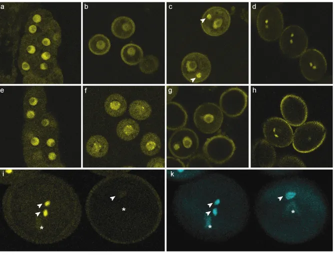
Figure 1-3. Seed development in Arabidopsis thaliana. (a-f), Seed development from before fertilization to seed maturation. (a), Mature female gametophyte prior to fertilization. Visible are the egg cell (white arrowhead), the central cell nucleus (black arrowhead) and the two synergid cells (S). (b), 2 days after pollination (d.a.p.), the two-celled proembryo (arrowhead) is accompanied by the early endosperm syncytium in the central cell (CC). (c), 4 d.a.p., globular-stage embryo and syncytial endosperm. (d), 6 d.a.p. heart-stage embryo, at this point the endosperm starts to cellularize around the embryo. (e), 8 d.a.p., torpedo-stage embryo, with completely cellularized endosperm. (f), 12 d.a.p., embryo with bent embryonic leaves near maturity, filling the major part of the seed. (g), Seed with a globular stage embryo (arrowhead) and the endosperm differentiated in the three domains central peripheral endosperm (PEN), chalazal endosperm (CZE), and miroyplar endosperm (MCE). Scale bars are 20 µm in a, and 100 µm in b-f.
Growing alongside the embryo in the seed, the endosperm also develops according to a well defined though fundamentally different programme (Fig. 1-3 a-f) (Mansfield, 1994;
Boisnard-Lorig et al., 2001). Endosperm development is characterized by four phases:
syncytium, cellularization, differentiation and death (Berger, 1999). First, mitotic cycles of the fertilized central cell in the absence of cytokinesis form a syncytial endosperm containing several hundreds nuclei that fill the central cell. The endosperm nuclei are
Introduction 9
organized in mitotic domains that display different rates of proliferation. Morphologically, three domains are recognized: The micropylar endosperm (MCE) surrounds the embryo at the anterior pole of the seed, the peripheral endosperm (PEN) lines the inside of the seed integuments and the chalazal endosperm (CZE) has dense cytoplasm and occupies the posterior pole of the seed (Brown et al., 1999).
In the seed stage characterized by a heart-shaped embryo, the syncytial phase of the endosperm ends and cellularization sets in (Sorensen et al., 2002). In Arabidopsis, the cellularized endosperm gets mostly consumed by the growing embryo and dies after seed germination (Berger, 1999).
The endosperm has been connected with nutrient acquisition from the mother plant and is thought to be a nurse tissue for the developing embryo (Hirner et al., 1998; Berger, 2003).
Furthermore, the endosperm has been described as a central integrator of signals during seed development (Berger et al., 2006). There is evidence of reciprocal signalling between the seed integuments and the endosperm, taking influence on the final size of the seed. On the one hand, the seed integuments can influence endosperm proliferation as shown in sporophytic mutations leading to reduced or enhanced growth of endosperm and embryo (Ray et al., 1996; Garcia et al., 2005; Schruff et al., 2006). Conversely, enhanced or reduced endosperm growth promotes or inhibits growth of the seed integuments (Garcia et al., 2005; Luo et al., 2005).
Data so far suggest both a maternal sporophytic and a zygotic control of seed development, the latter apparently mediated via the endosperm. In comparison to the endosperm, the embryo seems to play less of a role during communication in seed development (Berger et al., 2006).
1.3 Imprinting and he role of FIS-class genes during seed development
1.3.1 The FIS-PRC2
One important class of genes controlling the development of endosperm in Arabidopsis are the FIS-class genes. FIS-class proteins form a Polycomb-group (PcG) complex homologous to the Polycomb Repressive Complexes (PRCs) of animals (Chanvivattana et al., 2004). In mammals, the PRC2 is a complex involved, for instance, in the inactivation
Introduction 10
of the X-chromosome (Wang et al., 2001). It contains subunits conferring a H2K27- specific histone methyltransferase activity targeting lysine 27 in the basic tail domain of histone H3 (hereafter referred to as H3K27) (Cao et al., 2002).
In plants, there are several PRC2s built up by members of small gene families that show homologies to their animal PRC2 counterparts. Of importance for seed development and imprinting is the FIS-PRC2, composed of at least four core components: MEDEA (MEA), FERTILIZATION INDEPENDENT SEED 2 (FIS2), FERTILIZATION INDEPENDENT ENDOSPERM (FIE) and MULTICOPY SUPPRESSOR OF IRA1 (MSI1) (Fig. 1-4) (Guitton and Berger, 2005a). MEA is a SET-domain protein homologous to the Drosophila Enhancer of zeste E(z), which has been shown to confer histone lysine methyltransferase activity to the PRC2 (Cao et al., 2002; Muller et al., 2002). In the Arabidopsis FIS-PRC2, MEA interacts with FIE, the homologue of the Drosophila Extra sex combs (Esc) in vitro and in vivo (Luo et al., 2000; Spillane et al., 2000; Yadegari et al., 2000; Bracha-Drori et al., 2004). FIS2 is another member of the FIS-PRC2, and is the homologue of Su(z)12, a zinc finger protein which is important for the association of PRC2 with a selected set of target genes (Guitton and Berger, 2005a). Finally, MSI1 has also been identified as part of the FIS-PRC2 (Kohler et al., 2003a). MSI1 is a histone-binding protein homologous to the Drosophila p55.
Mutants of the Arabidopsis FIS-complex components have first been identified a decade ago. Their loss-of-function phenotypes are quite similar and display two effects:
Figure 1-4. The Arabidopsis FIS-PRC2.
The FIS-PRC2 consists at least of four core proteins, MEA, FIS2, FIE, and MSI1. The FIS-PRC2 is exclusively active in the female gametophyte and in the endosperm to control the expression status of imprinted genes via maintenance of H3K27 methylation. The repressive influence of the FIS-PRC2 on the expression of endosperm genes is thought to restrict endosperm proliferation.
(Adapted from Kinoshita et al. 2001).
Introduction 11
First, lack of FIS-class proteins confers a failure of cell cycle arrest in the mature female gametophyte, leading to autonomous endosperm proliferation without fertilization inside embryo-less seed-like structures (Ohad et al., 1996; Chaudhury et al., 1997; Grossniklaus et al., 1998; Kohler et al., 2003a). An exception is the msi1 mutant, which shows occasional development of non-viable parthenogenetic embryos (Guitton and Berger, 2005b).
Second, after fertilization, maternally inherited mutant fis-class alleles lead to heterochronically altered endosperm development and subsequent seed abortion (Kiyosue et al., 1999; Guitton et al., 2004; Ingouff et al., 2005). In fis-mutant endosperms, mitotic domains are missing or ill-defined. The fis-class endosperms are delayed in differentiation, leading to over-proliferation and lack of cellularization and differentiation (Grossniklaus et al., 1998; Kiyosue et al., 1999). The molecular nature of this pleiotropic phenotype is not understood to date (Guitton and Berger, 2005a).
1.3.2 Imprinting in angiosperm seed development
Investigations of gene expression patterns suggested that some FIS-class genes (MEA, eventually also FIS2 and FIE) are exclusively expressed from their maternal alleles in the endosperm, while their paternal alleles are silenced (Kinoshita et al., 1999; Luo et al., 2000; Yadegari et al., 2000).
Asymmetric paternal and maternal expression patterns are characteristic for the fertilization products of mammals and plants; this phenomenon has been described as genomic imprinting.
Up to now, almost 100 imprinted genes have been identified in mammals (Morison et al., 2005). As a consequence of imprinting, gene functions derived from both the maternal and the paternal genome are required for normal embryo development in mammals. Embryos with either only maternal or only paternal genomes show aberrant development and eventually die (Barton et al., 1984; McGrath and Solter, 1984; Surani et al., 1984).
Like mammals, flowering plants have been found to imprint certain genes resulting in a parent-of-origin dependent expression during seed development (Gehring et al., 2004;
Autran et al., 2005; Guitton and Berger, 2005a; Kohler and Grossniklaus, 2005; Scott and Spielman, 2006). The experimental evidence so far restricts imprinting in plants to the endosperm and has first been observed as a functional non-equivalency of the parental