INTRODUCTION
Bone formation on bioceramics, like calcium phosphate ceramics (CPCs), is well documented in animal models1–4
and in humans.5 These ceramics showed to be appropriate carriers for bone morphogenetic proteins (BMPs)6,7 and cells.9,10 The main limitation of CPCs is to find the appro- priate balance between degradability and mechanical sta- bility.8,9 An ideal bone substitute should be degradable and should be replaced by newly formed bone in a short time10 adapting to a dynamic processes as growth thought remodel- ing.11 The degradation pattern of those ceramics is a critical issue especially in areas of highly aesthetic demands as the craniofacial region, because the grafted site should remain unaltered in shape after the ceramic is replaced by newly formed bone. Due to the need of long-lasting shape stability in craniofacial surgery, hydroxyapatite (HA)–based ceram- ics have been traditionally used as nonresorptive materials for augmentation of the facial skeleton12 and cranial vault reconstruction.16,17 Beside the limited resorptive capacity of HA, bone formation has also been observed as limited.13,14
Disclosure: Supported by the German Ministry of Education and Research (Bundesministerium für Bildung und Forschung, BMBF), grant No. 0315019, a joint project of the Friedrich-Baur-Research Institute (R.D.), Bayreuth, and the Department of Cranio-Maxillofacial Plastic Surgery, University of Regensburg (J.C.R.). The Article Processing Charge was paid for by the authors.
Bone Morphogenetic Protein-7 Enhances
Degradation of Osteoinductive Bioceramic Implants in an Ectopic Model
From the *Division of Pediatric Facial Plastic Surgery and Craniofacial Anomalies, Catholic Children’s Hospital Wilhelmstift, Hamburg, Germany; †Teaching Hospital of the University of Lübeck, Germany;
‡Department of Cranio-Maxillofacial Plastic Surgery, University of Regensburg, Regensburg, Germany; §Friedrich-Baur-Research- Institute for Biomaterials, University of Bayreuth, Bayreuth, Germany;
¶Department of Cardiothoracic Surgery, University of Regensburg, Regensburg, Germany; ║Department of Orthodontics, University of Regensburg, Regensburg, Germany; **Department of Biomaterials, Cell Biology and Tissue Engineering, University of Erlangen, Germany; and
††Bio-Cer Entwicklungs-GmbH, Bayreuth, Germany.
Received for publication February 2, 2017; accepted April 21, 2017.
Preliminary results were presented at the 20th International Conference on Oral and Maxillofacial Surgery, November 1–4, 2011, Santiago de Chile, Chile.
Copyright © 2017 The Authors. Published by Wolters Kluwer Health, Inc. on behalf of The American Society of Plastic Surgeons. This is an open-access article distributed under the terms of the Creative Commons Attribution-Non Commercial-No Derivatives License 4.0 (CCBY-NC-ND), where it is permissible to download and share the work provided it is properly cited. The work cannot be changed in any way or used commercially without permission from the journal.
DOI: 10.1097/GOX.0000000000001375 J. Camilo Roldán, MD, DMD,
PhD*†‡
Tim Klünter, DMD‡
Peter Schulz, DMD‡
Ulrike Deisinger, PhD§
Claudius Diez, PhD¶
Waltraud Waiss, MD, DMD‡
Christian Kirschneck, DMD║
Torsten E. Reichert, MD, DMD, PhD‡
Rainer Detsch, PhD**††
Roldán et al.
Background: The aim of the present study was to evaluate the degradation pattern of highly porous bioceramics as well as the bone formation in presence of bone morphogenetic protein 7 (BMP-7) in an ectopic site.
Methods: Novel calcium phosphate ceramic cylinders sintered at 1,300ºC with a total porosity of 92–94 vol%, 45 pores per inch, and sized 15 mm (Ø) × 5 mm were grafted on the musculus latissimus dorsi bilaterally in 10 Göttingen minipigs: group I (n = 5):
hydroxyapatite (HA) versus biphasic calcium phosphate (BCP), a mixture of HA and tricalcium phosphate (TCP) in a ratio of 60/40 wt%; group II (n = 5): TCP versus BCP. A test side was supplied in situ with 250 μg BMP-7. Fluorochrome bone labeling and computed tomography were performed in vivo. Specimens were evaluated 14 weeks after surgery by environmental scanning electron microscopy, fluorescence microscopy, tartrate-resistant acid phosphatase, and pentachrome staining.
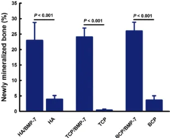
Results: Bone formation was enhanced in the presence of BMP-7 in all ceramics (P = 0.001).
Small spots of newly formed bone were observed in all implants in the absence of BMP-7.
Degradation of HA and BCP was enhanced in the presence of BMP-7 (P = 0.001). In those ceramics, osteoclasts were observed. TCP ceramics were almost completely degraded inde- pendently of the effect of BMP-7 after 14 weeks (P = 0.76), osteoclasts were not observed.
Conclusions: BMP-7 enhanced bone formation and degradation of HA and BCP ce- ramics via osteoclast resorption. TCP degraded via dissolution. All ceramics were os- teoinductive. Novel degradable HA and BCP ceramics in the presence of BMP-7 are promising bone substitutes in the growing individual. (Plast Reconstr Surg Glob Open 2017;5:e1375; doi: 10.1097/GOX.0000000000001375; Published online 22 June 2017.)
2017
2017
PRS Global Open • 2017
Long-term studies on HA cements for cranial reconstruction inform about a high rate of complications, particularly when using in proximity to the paranasal sinuses or in extended cranioplasties.16,20,21 Cranial full-thickness reconstruction re- mains the major limitation for CPC.21 A highly degradable tricalcium phosphate (TCP) ceramic was proposed recently for cranial reconstruction. To maintain the implant shape a titan mesh was incorporated into the scaffold.5 A more ratio- nal approach is to manage ceramic degradation by material composition. Biphasic calcium phosphate (BCP), a mixture of HA and TCP, combines the benefit of stability (HA) and degradability (TCP).10 The authors recently informed about enhanced degradability of BCP in the presence of BMP-7 in a cranioplasty minipig model showing integrity of the grafted site after ceramic degradation occurred. Implants not loaded with BMP-7 collapsed, producing an irregular and depressed grafted site.15 There is limited knowledge about the mechanism of bioceramic degradation, whether through cellular digestion (osteoclast activity) or chemical dissolution.10 Understanding degradation mechanisms of ceramics plays a critical role in the design of specific im- plants, considering individual mechanical and aesthetic demands.8 The ectopic animal implant model, also known as extra-skeletal or heterotopic model, is of value to evalu- ate biomaterial degradation by dissolution (without cellular skeletal components) as well as for evaluation of bone for- mation in the presence of BMPs16 or seeded cells.17
The hypotheses were (1) BCP implants will have a su- perior degradation profile and improved bone formation compared with HA or TCP alone and (2) the addition of BMP-7 will significantly improve bone formation and bone degradation when compared with carrier alone.
MATERIALS AND METHODS
Ceramics used in this study were prepared from com- mercially available HA and TCP powders (Merck, Darm- stadt, Germany). For fabricating three-dimensional (3D) scaffolds, the polyurethane (PU) replica technique (Schwartzwalder-technique) was chosen. Therefore, com- mercially available polyurethane-foams (pore size, 45 ppi) were coated with a calciumphosphate-slurry and sintered at 1,300°C for 1 hour. This procedure was repeated twice.
The porous structure of the PU-foam was exactly repli- cated, resulting in highly porous CaP ceramic scaffolds.18
Ten adult Göttingen minipigs aged 36 months in aver- age and weighing 33–41 kg in average (Ellengard Göttingen Minipigs ApS, Dalmose, Denmark) were operated on. The animals were fed with 2 × 250 g standard soft diet (Altromin 9023 Atronium International GmbH, Lange, Germany) and water ad libitum. The study was approved by the Dis- trict of Oberpfalz, Bavaria (No. 54-2531.1-02/07) according to the animal protection law (TierSchG- § 8 Abs. 1) and the local Ethic Committee at the University of Regensburg. All experiments were performed at the Experimental Animal Facility at the University of Regensburg, Germany.
Experimental Procedure
The infrascapular region was cleaned with a povidone–
iodine solution and draped with sterile towels. Two paral-
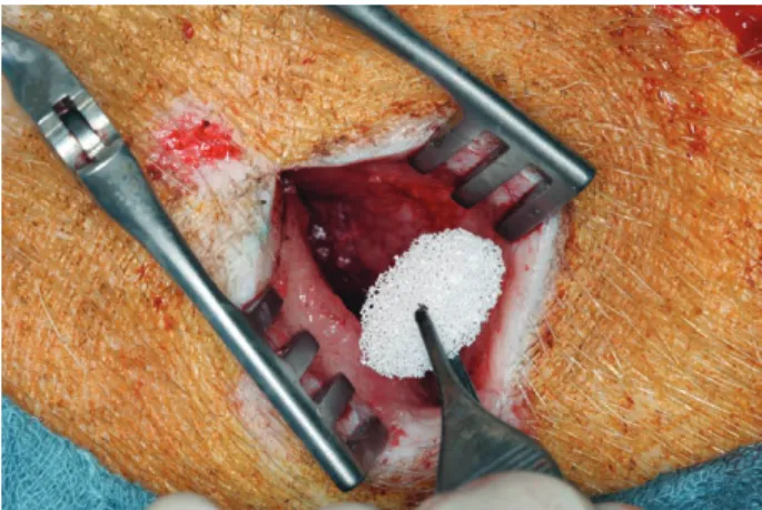
lel incisions were marked and shaved below the scapular border laterally at a distance of 4 cm from each other, bilaterally. The skin incision was performed through the subcutaneous tissue until the latissimus dorsi muscle in the depth; gentle preparation of the skin flap above the muscle was performed. Highly porous CPC implants sized 15 × 5 mm developed at the Friedrich-Baur-Research- Institute for Biomaterials at University of Bayreuth, Ger- many, were implanted (Fig. 1). Ceramics used were HA, TCP, and a BCP in a weight ratio of 60/40% of HA/TCP.
CPCs were sintered at 1,300º C.18 The CPCs have intercon- necting macropores with a bimodal pore size distribution (360–440 μm and 900–1150 μm). The total porosity was 92–94 vol%.2,18 Previous studies on the CPCs in vitro19 and in vivo in the mouse and minipig2,15 proved to be suitable for bone regeneration.
Two animal groups were evaluated (each n = 5); group I: HA versus BCP with and without BMP-7 (4 implants in each animal); group II: TCP versus BCP with or without BMP-7 (4 implants in each animal). In total, 40 implants were evaluated and compared for statistical purposes: HA (n = 5), TCP (n = 5), BCP (n = 10), HA + BMP-7 (n = 10), TCP + BMP-7 (n = 5), BCP + BMP-7 (n = 10). The test side was supplied in situ with 250 μg BMP-7 diluted in 20 mM acetate buffer with 5% mannitol (pH = 4.5) applied with a pipette (Eppendorf, Hamburg, Germany) providing full embedment of the implant (generous gift from Professor S. Vukicevic, Laboratory for Mineralized Tissues Center for Translational and Clinical Research, University of Za- greb, School of Medicine and Genera, Krapinske Toplice, Croatia). The chosen dose seems to be appropriate, as no bone overgrowth behind the scaffold was observed when applying 250 μg BMP-7 in a cranioplasty model.15
In vivo computed tomography (CT) was performed at weeks 1, 10, and 14 after surgery in intravenous anes- thesia as reported previously.15 Multiple in vivo CTs were performed to prove the presence of the ceramic after surgery and the progression of degradation before ter- mination of the study at week 14. CT does not differenti- ate between the ceramic and newly formed bone under the selected conditions (material composition).15 Histo-
Fig. 1. Ceramic implantation in the lateral subscapular region. Skin flap raised on the musculus latissimus dorsi. two parallel incisions were placed in both sides as implant recipient. Ceramics in the test side were supplied in situ with 250 μg Bmp-7.
morphometry was performed by using environmental scanning electron microscope (ESEM).2,20 Core volume was evaluated according to Parfitt21 (core volume evalu- ated area in mm2). 3D-CT reconstructions were per- formed for visualization.
An in vivo polychrome sequential labeling of miner- alizing tissues was performed as previously described.22 Intraperitoneal injection of fluorochrome started 1 week after the surgical procedure and continued sequentially in weeks 6, 8, and 12 after surgery as follows: xylenol orange, calcein green, alizarin complexone, and doxycycline as re- ported previously.15,23
Animals were killed 14 weeks after surgery by an intravi- tal and intracardial perfusion of a saline and a Sörensen’s fixation solution24; a lateral thoracotomy was preferred.15 Histology
Each sample was harvested en bloc and sectioned in the middle after performing an x-ray. The surrounding soft tissues were preserved. One half of the specimens were prepared for methyl methacrylate (MMA) embedment ac- cording to Donath,23,25,26 and the other half were prepared for paraffin embedment. Histomorphometry of newly formed bone and ceramic degradation was evaluated on en bloc MMA specimens analyzed by ESEM as previously described.2,15 The following parameters were evaluated21 by using an imaging software analySIS Pro3.2 (Soft Imaging Systems GmbH, Muenster, Germany): area of newly min- eralized bone and area of ceramic (ratio evaluated in per- centage). Subsequently, the MMA blocks were cut, ground, and polished into sections with a thickness of 90 μm. Those sections were evaluated for fluorescence labeling of newly formed bone.26
Paraffin-embedded specimens were treated with tar- trate-resistant acid phosphatase (TRAP) for osteoclast identification. Movat’s pentachrome staining was per- formed to visualize newly formed bone by self-induction (in the absence of BMP-7).27
Statistical Analysis
The statistical analysis was done with Stata 10.1 SE for Windows (StataCorp., Inc. College Station, Tex.) and Sig- maPlot 11 for Windows (SYSTST Inc., Chicago, Ill.). Graphs were created with SigmaPlot 11. Data were graphically tested for normality with Q–Q plots, followed by a formal analysis with the Shapiro-Wilk test. Continuous, normally distributed data from more than 2 groups were compared with analysis of variance followed by pairwise comparison (Holm-Sidak). Comparison of 2 groups was done by Stu- dent’s t test. Continuous, nonnormally distributed data from more than 2 groups were analyzed by means of the Kruskal-Wallis test, followed by a pairwise analysis with the Mann-Whitney test. Data are presented as mean value ± SD.
A P value < 0.05 was considered as statistically significant.
RESULTS
Animals gained in average 5% weight during the ex- perimental period of 14 weeks. The evolution was un- eventful; no infections were observed.
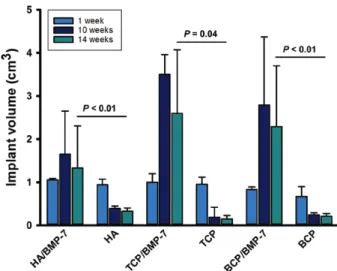
Implant Volume on In-Vivo CT
Baseline in vivo CT data were acquired at week 1 after surgery (day 7) for all 3 ceramics (HA, TCP, and BCP). CT showed a progressive volume reduction in all implants not loaded with BMP-7 at weeks 10 and 14. The extension of the volume reduction was less on HA implants. TCP was almost not detectable at weeks 10 and 14. BMP-7 expand- ed the volume of all ceramics (HA and BCP, P = <0.01;
TCP, P = <0.4), showing a volume reduction at week 14 (Fig. 2). Newly formed bone was not distinguished from the ceramic on CT.
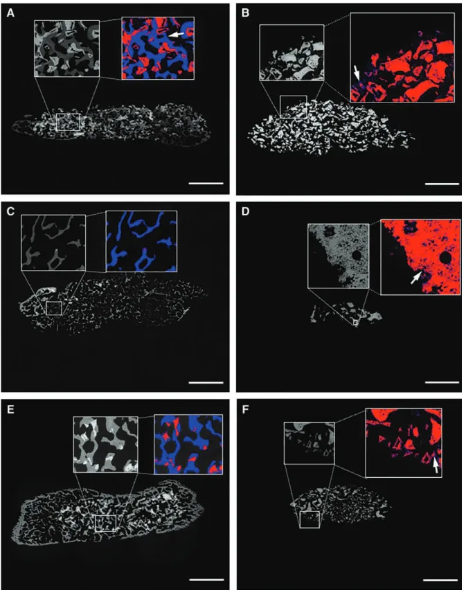
ESEM with Correlation to TRAP and Movat’s Staining Bone formation and ceramic degradation were eval- uated by ESEM (Fig. 3). Bone formation was equally enhanced in the presence of BMP-7 in all evaluated ce- ramics (HA, TCP, or BCP; P = 0.001; Fig. 3A; Fig. 4).
Small spots of newly formed bone were observed in the absence of BMP-7 in all ceramics (Fig. 3B). The histo- morphometry showed less bone formation in the almost collapsed TCP (P = 0.01; Fig. 3D; Fig. 4). Ceramic deg- radation was enhanced in the presence of BMP-7 in the HA and in the BCP groups (P = 0.001; Fig. 3A; Fig. 5).
In those ceramics, osteoclasts were observed via TRAP staining (Fig. 6). BMP-7 did not show a significant statis- tical effect on TCP degradation after 14 weeks (P = 0.74;
Fig. 5); osteoclasts were not observed via TRAP staining (Fig. 6). TCP without BMP-7 was almost completely de- graded at the termination of the study; no osteoclasts were observed. Bone formation without BMP-7 was con- firmed in all 3 ceramics in the pentachrome staining ac- cording to Movat, showing dispersed immature primary bundle bone in front of the muscle; in the presence of
Fig. 2. implant volume assessed in vivo by Ct at weeks 1, 10, and 14. Ceramics not loaded with Bmp-7 drastically lost volume at week 10; some volume recovered as observed at week 14 is explained by late bone formation, evident in fluorochrome labeling at week 10 (see also Fig. 7). Bmp-7–stimulated ceramics showed a statistically significant volume increment. Volume reduction under Bmp-7 was observed from week 10 to week 14, explained by enhanced ceramic degradation evident in the ESEm (see Fig. 3; Student’s t test after testing for normality [tCp ceramics] or mann-Whitney test for non- normally distributed data [Ha and BCp ceramics]).
PRS Global Open • 2017
Fig. 3. ESEm images of explanted grafted CpC (total core) from the dorsal area. Small rectangle demarks the area of magnification. Colored areas show newly formed bone in blue and ceramic in red: newly formed bone is observed on and inside Ha ceramic in the presence of Bmp-7. Ha ceramic volume decreases in the presence of Bmp-7 (a). Small spots of newly formed bone (blue areas), marked with a white arrow, are observed on Ha ceramic without Bmp-7, confirming the self-osteoinductive property of this material (B). tCp ceramic in the presence of Bmp-7 has almost disappeared while bone formation was enhanced (C). tCp ceramic without Bmp-7 is almost completely degraded; small spots of newly formed bone (blue areas), marked with a white arrow, are observed (D). BCp ceramics in the presence of Bmp are fully integrated in newly formed bone (E), Bmp-7 enhanced ceramic degradation (see also Fig. 6). BpC ceramic without Bmp-7 is partially collapsed. Small spots of newly formed bone (blue areas, white arrow) confirm again the self-osteoinductive property of this material (F), (undecalcified in mma embedded specimens en bloc analyzed, composite images, scale bar: 3.5 mm).
BMP-7, extensive lamellar bone was observed in the to- tal core (images not shown).
Temporal Bone Formation Assessed by Intravital Fluorochrome Labeling
Fluorochrome staining revealed bone formation in the presence of BMP-7 at week 1 {xylenol orange = yellow;
Fig. 7A, C, E [HA (A), TCP (C), BCP (E)]}. Fluorochrome histomorphometry showed a higher bone formation at week 6 (calcein green = green) compared with weeks 8 and 12 (P = 0.005). No significant difference was observed in terms of bone formation in the presence of BMP-7 in
all ceramics at week 6 (P = 0.32), week 8 (P = 0.70), and week 12 (P = 0.18). Bone formation without BMP-7 was confirmed through fluorochrome labeling at week 8, de- tected by small red spots (alizarin complexone = red) in a partially collapsed scaffold (Fig. 7).
DISCUSSION
In the present study, the degradability of CPC could be demonstrated by a cellular (osteoclast) and by chemical dissolution in an ectopic site. HA and BCP were signifi- cantly degraded in the presence of BMP-7 (P = 0.001). In those ceramics, osteoclasts were observed on TRAP stain- ing. Osteoclasts were also observed on BCP without BMP- 7, but in this case, no statistically significant enhanced degradability was observed.
Degradation of CPC in the Experimental Setting
In previous in vitro studies on the present CPCs, osteo- clastogenesis was evaluated by using RAW 264.7 murine monocytes stimulated with receptor activator of NF-κB ligand and macrophage colony-stimulating factor for 21 days. Those cells differentiated in 14 days in osteoclast- like cells. Resorptive lacunae on BCP were measured with a depth of 10–12 μg and diameters of 30–40 μm on ESEM. Osteoclast lacunae were also observed on HA but not in ß-TCP.28 In the present study, osteoclasts were not observed on TCP; at termination of the study at week 14, almost no ceramic was observed and the effect of BMP-7 did not change the degradability of this material. Control TCP (without BMP-7) was almost completely degraded, only small spots of newly formed bone (osteoinductive bone) were observed. The degradability of TCP is attrib- uted mainly to chemical dissolution.10 Furthermore, the chemical dissolution of TCP increases the Ca2+ ion con- centration, which inhibits osteoblast formation in vitro.29 Yamada et al. showed enhanced osteoclastogenesis in vi- tro on BCP (in a ratio of HA:ß-TCP of 100:0 and 80:20).
Lacunae pits were not observed in TCP.30 Mayr et al. pro- posed an HA:ß-TCP ratio of 80:20 to 60:40 based on a osteoclastogenesis in vitro model.31 In the present study on BCP, an HA/TCP ratio of 60:40 was chosen. It is well known that osteoclastogenesis is influenced by different material properties, such as chemical composition and crystallinity of the ceramic, topography, and micro- as well as macroporosity of the scaffolds. All these aspects influ- ence cell adhesion and thus osteoclastogenesis.10 Because the geometry, composition, and sintering process of CPC directly influence bone formation and also biodegrad- ability,32 each particular biomaterial should be character- ized, defining the biodegradable properties (dissolution versus resorption) for a specific anatomical area.29
Whether the degradability is an important feature of a scaffold, replacement by newly formed bone is also a criti- cal issue because the grafted site should remain unaltered in shape. Recently, the authors informed about enhanced deg- radation of cranial implants in the presence of BMP-7, where- as the grafted site remained unchanged in shape. Ceramics nonloaded with BMP-7 showed a delayed bone formation in a collapsed scaffold changing the shape of the grafted site.15 Fig. 4. area of newly mineralized bone assessed by ESEm. Enhanced
bone formation was statistically observed in all ceramics (Ha, tCp, and BCp) in the presence of Bmp-7 (P = 0.001), no statistical differ- ences were observed between the ceramics (normality test Shapiro- Wilk, failed P < 0.05).
Fig. 5. area of calcium phosphate ceramics assessed by ESEm. Bmp- 7 enhanced ceramic degradation on Ha and BCp (P = 0.001). Osteo- clasts were observed in those ceramics (see Fig. 8). Degradability of tCp was not affected by Bmp-7 (P = 0.76). tCp was degraded via chemical dissolution as no cellular component (osteoclasts) was ob- served in the trap staining (see Fig. 8;, normality test Shapiro-Wilk, failed P < 0.05).
PRS Global Open • 2017
Effects of BMP on Calcium Phosphate Ceramics and Degradation
The effects of BMP on ceramics are widely document- ed for bone formation, but the effects on ceramic degra- dation are scarce. To the best of our knowledge, this study is the first one evaluating biodegradability of CPC, pro- viding evidence of osteoclast activity or chemical dissolu- tion in the presence of BMP-7 in vivo. We also previously observed an enhanced ceramic degradation in the pres- ence of BMP-7 in an ectopic mouse model.2 Osteoclasts have been observed in the present ceramic for the first time by TRAP staining in vivo. Ripamonti et al.33 evalu- ated sintered HA ceramics implanted ectopically (rectus abdominis muscle) in the adult primate in the presence of 5, 25, and 45 μg BMP-7. After 30 and 90 days, bone formation was enhanced, but the ceramic did not show a statistically significant degradation. Draenert et al.34 stated an enhanced bone remodeling and ß-TCP “resorption”
when adding BMP-7, but osteoclasts were not evaluated.
The mechanism of degradation of CPC in vivo is often not considered; the term “resorption” is generally used for degradation whether or not osteoclastic resorption or chemical dissolution has been observed. Abe et al.35 evalu- ated the effect of BMP-2/4 on osteoblasts and osteoclasts in vitro: Noggin, a BMP antagonist, blocked osteoblastic as well as osteoclastic differentiation, thus explaining the
effect of BMP on bone remodeling. Thus, ceramic degra- dation and ceramic replacement by newly formed bone should resemble bone remodeling as an orchestrated pro- cess of bone formation and bone/ceramic resorption; this process is supported by BMP. In the present study, HA as well as BCP combined with BMP-7 showed no statistical difference in terms of bone formation and ceramic deg- radation. The benefit of BCP combined with BMP-7 lies in the adjustable degradability of the implant by changing the HA/TCP ratio matching the implant to the mechani- cal demands in a particular anatomical area while BMP- 7 strengthens the highly degradable TCP component through bone formation.
Bone Formation on Calcium Phosphate Ceramics:
Osteoinduction by BMP and Self-Osteoinduction
In the present study, BMP-7 enhanced bone formation in all ceramics equally. This result underlines that the limi- tation of BMP is the scaffold and the critical issue is the scaffold degradability. The temporal apposition of bone assessed by in vivo fluorochrome labeling disclosed early bone formation as a critical issue for further bone apposi- tion before the ceramic collapse, as also observed when the ceramic is implanted at a skeletal site.15 Bone forma- tion was observed at week 1 in BMP-7 stimulated implants, supporting further bone apposition. Nonstimulated BMP- Fig. 6. trap staining on CpC explanted from the dorsal area. Osteoclasts are identified on Ha and BCp
ceramics in the presence of Bmp-7 (a and B) and on BCp ceramics in the absence of Bmp-7 (C; black ar- row). no osteoclasts were seen on tCp whether or not stimulated with Bmp-7 (D) (magnification 10x).
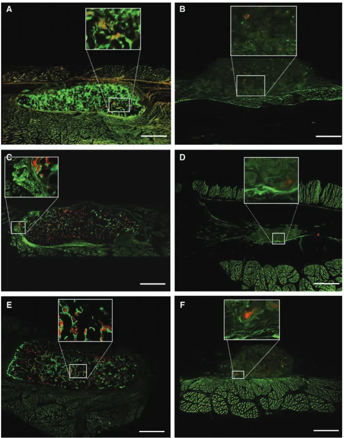
Fig. 7. Fluorochrome labeling of explanted grafted CpC from the dorsal area (total core), (xylenol orange/orange yellow marker at week 1; calcein green/green marker at week 6; alizarin complexone/red marker at week 8; doxycycline/ brown marker at week 12). rectangle marks the area of magnification. Ceramics stimulated with Bmp-7 (a, C, E) show newly formed bone starting at week 1 (yellow marker), successive bone formation (weeks 6, 8, and 12) takes place throughout the implant site. new bone formation in the absence of Bmp-7 is detected as small red spots (week 8) on Ha (B), tCp (D), and BCp (F), confirming osteoinductive property of the evaluated ceramics (unde- calcified in mma-embedded specimens en block analyzed, composite images, scale bar 3.5 mm).
PRS Global Open • 2017
7 implants collapsed. TCP needs a strong BMP stimulus, before the ceramic disappears, as observed in the control site (no BMP), which was evident at week 10 on in vivo CT.
All studied ceramics showed osteoinductive properties.
This effect is not only influenced by the material composi- tion36 but also by the geometry.19,37,38 Deproteinized bovine bone (Bio-Oss) does not display osteoinductive properties in an ectopic site, even if the scaffold architecture is similar to human bone.26 Ceramic osteoinduction (without BMP) was detected in the present study as small spots of newly formed bone in ESEM and fluorochrome labeling starting at week 8. This effect was confirmed in the pentachrome staining directly in front of the muscle, where the vascu- larization is higher. This observation supports the critical role of angiogenesis in bone formation and also the ecto- pic site as an ideal implant site for scaffold prefabrication in a clinical setting. Nevertheless, the scarce amount of newly formed bone in absence of BMP-7 was not relevant for a clinical purpose under the selected conditions.
The osteoinductive properties of BMPs, described for the first time by Urist39 in 1965, have not been observed in the presence of any other cytokine signal or material; BMP remains the most effective therapeutic tool in bone regen- eration.32 Attempts for enhancing effect of BMP with an appropriate functionalized osteoinductive scaffold40 aims to reduce dose and cost. Furthermore, dose should be matched to the defect size and localization.41
Clinical Relevance: Perspective
According to the mentioned characteristics of CPC under the influence of BMP, the following clinical ben- efits can be mentioned: CPC is degradable via resorption (HA-based CPC) and dissolution (TCP). This effect is en- hanced by BMP. CPC under BMP undertakes a remodel- ing process based on bone formation and bone/ceramic resorption. This remodeling process is a prerequisite to fit those implants to architectural changes in the growing infant. CPC is unlimited in source and can be tailored as a 3D implant with a defined inter- and intraconnective pore system and tailored as a patient-specific implant via com- puter-aided design. Implant straightness can be achieved changing the HA:TCP ratio, being higher augmenting the HA content. In those implants, a higher dose of BMP could be required to enhance degradation and remod- eling. All mentioned aspects indicate that the future in bone regeneration lies in the development of patient-spe- cific implants, not only in terms of surface shape but also in terms of internal architecture and composition. CPC seams to meet those requirements.
CONCLUSIONS
The evaluated CPCs are resorbable (HA), dissolv- able (TCP), and osteoinductive. BMP-7 enhanced HA and BCP degradation via osteoclastic resorption and en- hanced bone formation, integrating the ceramic in the bone remodeling process. In terms of bone formation and ceramic degradation, BCP was not superior to HA in the presence of BMP-7 as hypothesized. The benefit of BCP combined with BMP-7 lays in the adjustable degradabil-
ity of the implant by changing the HA/TCP ratio, while BMP-7 strengthens the highly degradable TCP compo- nent through bone formation. Those features make this biomaterial promising for the treatment of bone defects, especially in the growing infant, where implant remodel- ing is of major importance.
J. Camilo Roldán, MD, DMD, PhD Department of Cranio-Maxillofacial Plastic Surgery University of Regensburg Franz-Josef-Strauß-Allee 11 93053 Regensburg E-mail: jcamilo.roldan@yahoo.com
ACKNOWLEDGMENTS
The authors greatly thank Professor Slobodan Vukicevic, Laboratory for Mineralized Tissues Center for Translational and Clinical Research, University of Zagreb School of Medicine, for providing us with BMP-7 and his critical evaluation of the study design; and Antje Böttinger for her excellent technical support at the Bone Research Laboratory at the Department of Cranio-Max- illofacial Plastic Surgery, University of Regensburg.
REFERENCES
1. Le Nihouannen D, Daculsi G, Saffarzadeh A, et al. Ectopic bone formation by microporous calcium phosphate ceramic particles in sheep muscles. Bone. 2005;36:1086–1093.
2. Roldán JC, Detsch R, Schaefer S, et al. Bone formation and deg- radation of a highly porous biphasic calcium phosphate ceramic in presence of BMP-7, VEGF and mesenchymal stem cells in an ectopic mouse model. J Craniomaxillofac Surg. 2010;38:423–430.
3. Fellah BH, Gauthier O, Weiss P, et al. Osteogenicity of biphasic calcium phosphate ceramics and bone autograft in a goat model.
Biomaterials. 2008;29:1177–1188.
4. Yuan H, van Blitterswijk CA, de Groot K, et al. Cross-species compari- son of ectopic bone formation in biphasic calcium phosphate (BCP) and hydroxyapatite (HA) scaffolds. Tissue Eng. 2006;12:1607–1615.
5. Engstrand T, Kihlström L, Lundgren K, et al. Bioceramic implant induces bone healing of cranial defects. Plast Reconstr Surg Glob Open. 2015;3:e491.
6. Urist MR, Lietze A, Dawson E. [beta]-tricalcium Phosphate deliv- ery system for bone morphogenetic protein. Clin Orthop Relat Res.
1984;187:277.
7. Yuan H, Zou P, Yang Z, et al. Bone morphogenetic protein and ce- ramic-induced osteogenesis. J Mater Sci Mater Med. 1998;9:717–721.
8. Hollister SJ, Lin CY, Saito E, et al. Engineering craniofacial scaf- folds. Orthod Craniofac Res. 2005;8:162–173.
9. Habraken W, Habibovic P, Epple M, et al. Calcium phosphates in biomedical applications: materials for the future? Materials Today. 2016;19:69–87.
10. Detsch R, Boccaccini AR. The role of osteoclasts in bone tissue engineering. J Tissue Eng Regen Med. 2015;9:1133–1149.
11. Feroze AH, Walmsley GG, Choudhri O, et al. Evolution of cranio- plasty techniques in neurosurgery: historical review, pediatric con- siderations, and current trends. J Neurosurg. 2015;123:1098–1107.
12. Gosain AK, Riordan PA, Song L, et al. A 1-year study of hy- droxyapatite-derived biomaterials in an adult sheep model: III.
Comparison with autogenous bone graft for facial augmenta- tion. Plast Reconstr Surg. 2005;116:1044–1052.
13. Gosain AK, Riordan PA, Song L, et al. A 1-year study of osteo- induction in hydroxyapatite-derived biomaterials in an adult sheep model: part II. Bioengineering implants to optimize bone replacement in reconstruction of cranial defects. Plast Reconstr Surg. 2004;114:1155–1163; discussion 1164.
14. Gosain AK; Plastic Surgery Eductional Foundation DATA Committee. Biomaterials for reconstruction of the cranial vault.
Plast Reconstr Surg. 2005;116:663–666.
15. Roldán JC, Schulz P, Klünter T, et al. BMP-7 preserves surface integrity of degradable-ceramic cranioplasty in a Göttingen mini- pig model. Plast Reconstr Surg Glob Open. 2017;5:e1255.
16. Ripamonti U. Bone induction by recombinant human osteogen- ic protein-1 (hOP-1, BMP-7) in the primate Papio ursinus with ex- pression of mRNA of gene products of the TGF-beta superfamily.
J Cell Mol Med. 2005;9:911–928.
17. Nakano K, Murata K, Omokawa S, et al. Promotion of osteo- genesis and angiogenesis in vascularized tissue-engineered bone using osteogenic matrix cell sheets. Plast Reconstr Surg.
2016;137:1476–1484.
18. Deisinger U. Synthetisches Knochenersatzmaterial mit spongiosa-ähnli- cher Struktur. Berlin: Mensch & Buch Verlag; 2009.
19. Roldán JC, Chang E, Kelantan M, et al. Quantifying migration and polarization of murine mesenchymal stem cells on differ- ent bone substitutes by confocal laser scanning microscopy.
J Craniomaxillofac Surg. 2010;38:580–588.
20. Detsch R, Hausherr J, Schlüfter S, et al. Charakterisierung von Knochenwachstum auf einer Calcium- phosphat-Mischkeramik:
Beurteilung einer zerstörungsfreien und 3- dimensionalen Charakterisierungsmethode. In Krenkel W. ed. Verbundwerkstoffe und Werkstoffverbunde. Bayreuth. 2008:707–712.
21. Parfitt AM. Bone histomorphometry: standardization of nomen- clature, symbols and units (summary of proposed system). Bone.
1988;9:67–69.
22. Rahn BA, Perren SM. Xylenol orange, a fluorochrome useful in polychrome sequential labeling of calcifying tissues. Stain Technol. 1971;46:125–129.
23. Roldán JC, Jepsen S, Schmidt C, et al. Sinus floor augmentation with simultaneous placement of dental implants in the presence of platelet-rich plasma or recombinant human bone morphoge- netic protein-7. Clin Oral Implants Res. 2004;15:716–723.
24. Roldán JC, Knueppel H, Schmidt C, et al. Single-stage sinus aug- mentation with cancellous iliac bone and anorganic bovine bone in the presence of platelet-rich plasma in the miniature pig. Clin Oral Implants Res. 2008;19:373–378.
25. Donath K, Breuner G. A method for the study of undecalcified bones and teeth with attached soft tissues. The Säge-Schliff (sawing and grinding) technique. J Oral Pathol. 1982;11:
318–326.
26. Roldán JC, Jepsen S, Miller J, et al. Bone formation in the pres- ence of platelet-rich plasma vs. bone morphogenetic protein-7.
Bone. 2004;34:80–90.
27. MOVAT HZ. Demonstration of all connective tissue elements in a single section; pentachrome stains. AMA Arch Pathol.
1955;60:289–295.
28. Detsch R, Schaefer S, Deisinger U, et al. In vitro: osteoclastic ac- tivity studies on surfaces of 3D printed calcium phosphate scaf- folds. J Biomater Appl. 2011;26:359–380.
29. Detsch R, Mayr H, Ziegler G. Formation of osteoclast-like cells on HA and TCP ceramics. Acta Biomater. 2008;4:139–148.
30. Yamada S, Heymann D, Bouler JM, Daculsi G. Osteoclastic resorp- tion of calcium phosphate ceramics with different hydroxyapatite/
beta-tricalcium phosphate ratios. Biomaterials. 1997;18:1037–1041.
31. Mayr H, Schlüfter S, Detsch R, Ziegler G. Influence of phase composition on degradation and resorption of biphasic calcium phosphate ceramics. Key Eng Mater. 2008;361–363:1043–1046.
32. García-Gareta E, Coathup MJ, Blunn GW. Osteoinduction of bone grafting materials for bone repair and regeneration. Bone.
2015;81:112–121.
33. Ripamonti U, Crooks J, Rueger DC. Induction of bone forma- tion by recombinant human osteogenic protein-1 and sintered porous hydroxyapatite in adult primates. Plast Reconstr Surg.
2001;107:977–988.
34. Draenert ME, Kunzelmann KH, Forriol F, et al. Primary cancel- lous bone formation with BMP and micro-chambered beads: ex- perimental study on sheep. Bone. 2013;52:465–473.
35. Abe E, Yamamoto M, Taguchi Y, et al. Essential requirement of BMPs-2/4 for both osteoblast and osteoclast formation in mu- rine bone marrow cultures from adult mice: antagonism by nog- gin. J Bone Miner Res. 2000;15:663–673.
36. Tang Z, Wang Z, Qing F, et al. Bone morphogenetic protein Smads signaling in mesenchymal stem cells affected by osteo- inductive calcium phosphate ceramics. J Biomed Mater Res A.
2015;103:1001–1010.
37. Ripamonti U, Richter PW, Thomas ME. Self-inducing shape mem- ory geometric cues embedded within smart hydroxyapatite-based biomimetic matrices. Plast Reconstr Surg. 2007;120:1796–1807.
38. Yuan H, Fernandes H, Habibovic P, et al. Osteoinductive ceram- ics as a synthetic alternative to autologous bone grafting. Proc Natl Acad Sci U S A. 2010;107:13614–13619.
39. Urist MR. Bone: formation by autoinduction. Science.
1965;150:893–899.
40. Schuessele A, Mayr H, Tessmar J, et al. Enhanced bone morpho- genetic protein-2 performance on hydroxyapatite ceramic sur- faces. J Biomed Mater Res A. 2009;90:959–971.
41. Choi JW, Jeong WS, Yang SJ, Park EJ, Oh TS, Koh KS. Appropriate and effective dosage of BMP-2 for the ideal regeneration of calvar- ial bone defects in beagles. Plast Reconstr Surg. 2016;138:64e–72e.