AUS DEM LEHRSTUHL FÜR ANÄSTHESIOLOGIE
PROF. DR. BERNHARD M. GRAF, MSc.
DER FAKULTÄT FÜR MEDIZIN DER UNIVERSITÄT REGENSBURG
FILTER ADSORPTION OF ANIDULAFUNGIN TO A POLYSULFONE-BASED HEMOFILTER DURING CVVHD IN VITRO
Inaugural – Dissertation zur Erlangung des Doktorgrades
der Medizin
der
Fakultät für Medizin der Universität Regensburg
vorgelegt von Peter Kolbinger
2018
Dekan: Prof. Dr. Dr. Torsten E. Reichert
1. Berichterstatter: Prof. Dr. Karl-Peter Ittner
2. Berichterstatter: Prof. Dr. Bernhard Banas
Tag der mündlichen Prüfung: 18.09.2018
Inhalt
Einleitung ... 6
Material und Methoden ... 8
In vitro CVVHD System ... 8
Experimente ... 9
Hochleistungsflüssigkeitschromatographie (HPLC) ... 10
Berechnungen ... 12
Ergebnisse ... 12
Dialyse von Kochsalzlösung ... 13
Albuminhaltige Kochsalzlösung ... 13
Blut ... 14
Diskussion ... 15
Lieraturverzeichnis ... 20
Originalversion des Artikels ... 24
Danksagung ... 33
Lebenslauf ... 34
Filteradsorption von Anidulafungin an einem polysulfon-basierten Hämofilter während kontinuierlicher venovenöser Hämodialyse in vitro
(Filter Adsorption of Anidulafungin to a Polysulfone-Based Hemofilter During CVVHD In Vitro;
Kolbinger, P., Gruber, M., Roth, G., Graf, B. M. and Ittner, K.-P. (2017).Artificial Organs. doi:10.1111/aor.12982)
Einleitung
In der Behandlung von Intensivpatienten werden sowohl Antimykotika als auch kontinuierliche Nierenersatzverfahren in erheblichem Umfang eingesetzt. Die Angaben in der Literatur bezüg- lich der Häufigkeit eines akuten Nierenversagens (ANV) schwanken zwischen etwa einem Drittel [1] und bis zu zwei Drittel [2] aller Intensivpatienten. In jedem Fall ist ein ANV eine häufige Komplikation bei intensivpflichtigen Patienten, wobei die Inzidenz in den letzten Jahren deutlich zugenommen hat [1, 2]. Dies belegen Zahlen aus den USA: Hier war ein ANV 1980 in 1,8/10.000 Fällen der Grund für eine Krankenhauseinweisung, im Jahr 2005 dagegen in
36,5/10.000 Fällen [3].
Weitere häufige Komplikationen bei Intensivpatienten sind Candidämien. Daten aus chirurgi- schen Intensivstationen in den Vereinigten Staaten ergaben 9,82 Candida Blutbahninfektionen pro 1.000 Patienten [4]. Insbesondere hiervon betroffen sind Erkrankte mit einem begleitenden ANV, welche ein um das Vierfache erhöhte Risiko besitzen, an einer Candidämie zu erkranken [4].
Zur Behandlung von Candidämien (mit Ausnahme von Candida parapsilosis Infektionen) werden Echinocandine empfohlen, insbesondere auch in der Behandlung schwer kranker Patien- ten [5, 6]. In einer aktuellen Studie ergaben sich keine Hinweise auf eine zunehmende Resistenz von invasiven Candida-Isolaten gegen Echinocandine [7]. Der neueste Vertreter in der Gruppe der Echinocandine ist Anidulafungin [8, 9]. Es ist ein großes Lipopeptidmolekül mit einer
Molekülmasse von 1140,3 Da [10]. Wie auch bei den übrigen Echinocandinen handelt es sich um ein amphiphiles Hexapeptid, in diesem Falle kombiniert mit einer lipophilien
Alkoxytriphenyl (Terphenyl) Seitenkette [9]. Wie alle Echinocandine ist es fungizid gegen die meisten Candida spp. [7, 9, 11, 12] und fungistatisch gegen eine Vielzahl Aspergillus spp. [9, 12, 13]. Nach parenteraler Applikation besitzt Anidulafungin ein vorhersehbares
pharmakokinetisches Profil, einschließlich dosisproportionaler Plasmakonzentrationen [14].
Bereits nach einer Initialdosis (doppelte Erhaltungsdosis) und einer Tagesdosis wird der Steady State erreicht; das Verteilungsvolumen beträgt etwa 33 Liter [14]. Anidulafungin ist zu mehr als 99% an Plasmaproteine gebunden [10]. Es unterliegt einem chemischen Abbau und wird an- schließend über die Galle eliminiert; es wird nicht hepatisch metabolisiert oder renal ausge- schieden [15]. Unabhängig von Leber- oder Nierenfunktionsstörungen beträgt die Clearance von Anidulafungin daher etwa 1 Liter/Stunde [16].
Im Gegensatz zu bekannten pharmakokinetischen Eigenschaften eines Medikaments, wie den oben genannten, kann die Dosierung während einer Nierenersatztherapie schwierig sein, da hierdurch viele Faktoren beeinflusst werden können [17, 18]. Beispielsweise können unter- schiedliche Mengen einer Substanz an der Oberfläche des Hämofilters adsorbieren [19-23].
Da insbesondere schwer kranke Patienten in Intensivstationen Antimykotika und Nierenersatz- therapien benötigen, ist die Kenntnis der Pharmakokinetik der verwendeten Medikamente von großer Bedeutung. Ebenso wichtig ist die Kenntnis über eine mögliche Beeinflussung, z.B.
durch eine Nierenersatztherapie.
Die genannten Punkte machen deutlich, dass die Untersuchung des Einflusses von Nierener- satzverfahren auf Anidulafungin auch von klinischem Interesse ist.
So gibt es mehrere Studien, die sich mit der Auswirkung verschiedener Nierenersatzverfahren auf die Blutkonzentration von Anidulafungin beschäftigt haben: Dowell et al. [16] untersuchten die Anidulafunginblutkonzentration bei Patienten mit terminaler Niereninsuffizienz und konn- ten zeigen, dass sie durch die Hämodialyse nicht beeinflusst wird. Die Studie von Leitner et al.
(CVVHF), Aguilar et al. [25] mit kontinuierlicher venovenöser Hämodiafiltration (CVVHDF) und Burkhardt et al. [26] mit Patienten, welche mit „extended daily dialysis“ (EDD) therapiert wurden. Eine Notwendigkeit zur Dosisanpassung ergab sich in den genannten Studien nicht.
Das Ziel der vorliegenden Studie war es, die Adsorption von Anidulafungin an der Oberfläche eines Hämofilters während eines kontinuierlichem Nierenersatzverfahrens (kontinuierliche venovenöse Hämodialyse, CVVHD) und deren Einfluss auf die Anidulafunginkonzentration zu untersuchen. Zu diesem Zweck wurden verschiedene dialysierte Lösungen untersucht.
Material und Methoden
In vitro CVVHD System
Die Dialyse wurde mit einem Edwards bm11a/bm14 Dialysesystem durchgeführt (Edwards Lifesciences Germany GmbH, Unterschleißheim, Deutschland). Bei den Hämofiltern handelte es sich um Fresenius Ultraflux AV 600S (Fresenius Polysulfon, Filteroberfläche 1,4 m2, Frese- nius Medical Care, Bad Homburg, Deutschland). Als Dialysat wurde SH 27 Lösung (B. Braun, Melsungen, Deutschland) verwendet. Das Reservoir für die in vitro Dialyseversuche war eine 2 Liter Glasflasche. Dieses Flüssigkeitsreservoir wurde während der Dialyse kontinuierlich mit einem Magnetrührer gerührt, um eine homogene Durchmischung der Lösungen sicherzustellen.
Alle Experimente wurden doppelt ausgeführt. Zur Bestimmung der nach dem Spülen im Schlauchsystem und Hämofilter vorhandenen Kochsalzlösung, wurde das System vor und nach dem Spülen gewogen. Es ergab sich ein Volumen von 350 ml. Dieses wurde in den durchge- führten Berechnungen berücksichtigt.
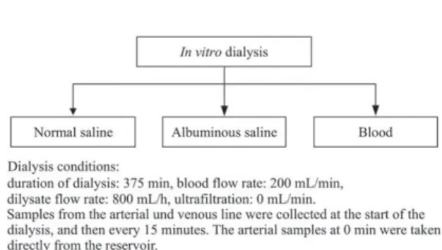
Die Dialyseexperimente wurden mit drei verschiedene Lösungen durchgeführt: Zunächst mit Kochsalzlösung, anschließend mit albuminhaltiger Kochsalzlösung und schließlich mit einer Ersatzmischung für Frischblut. In den ersten Versuchen wurden 1.650 ml Kochsalzlösung eingesetzt. Die eiweißhaltige Kochsalzlösung bestand aus 1.250 ml NaCl und 400 ml Human Albumin 20% (Baxter, Wien, Österreich). Die Albuminkonzentration betrug bei den Experi-
menten mit albuminhaltiger Kochsalzlösung 40 g/l. Als Ersatz für Frischblut wurde eine Mi- schung aus 600 ml humanen Erythrozyten, 400 ml gefrorenem Frischplasma (GFP), 100 ml 8,4% NaHCO3 und 10.000 IE Heparin (2 ml) hergestellt, von welcher 1.000 ml eingesetzt wurden (pH 7,13 ± 0,03 [Standardabweichung], Hämatokrit 34,5 ± 2,12). Die Gesamteiweiß- konzentration in diesen Versuchen betrug ca. 21,5 g/l, basierend auf einer typischen GFP- Eiweißkonzentration von 80 g/l [27].
Das Gesamtvolumen, einschließlich der im Schlauchsystem verbliebenen Kochsalzlösung, betrug in den Experimenten mit Kochsalzlösung und albuminhaltiger Kochsalzlösung 2.000 ml, in den Versuchen mit Blut 1.350 ml.
Experimente
Die Parameter der Dialyse waren: Dauer der Dialyse: 375 min; Blutflussrate: 200 ml/min und Dialysatflussrate: 800 ml/h. Die Ultrafiltration war auf Null eingestellt. Zu Beginn der Experi- mente herrschten Anidulafunginkonzentrationen entsprechend typischen Plasmaspitzenkonzent- rationen [16]. Im weiteren Verlauf
der Versuche wurde Anidulafun- gin nach jeweils 60 Minuten zugegeben, um potentielle Verlus- te an den Hämofilter oder das Dialysat auszugleichen. Die hie- raus resultierenden eingesetzten Konzentrationen sind in Abbil- dung 1 dargestellt.
Zu Beginn und anschließend in Abständen von 15 Minuten wurden bis zum Ende der Versuche Proben (ca. 2 ml) aus dem arteriellen und venösen Blutschlauch entnommen. Um bereits Daten vor der ersten Passage des Hämofilters zu erhalten, wurden die arteriellen Proben zum Zeitpunkt 0 min direkt aus dem Reservoir entnommen. Aus diesem Grund wurde bei diesen Proben eine Abbildung 1. Während der Dialyse zugegebenes Anidulafungin
etwas höhere Konzentration in Kauf genommen, da noch keine Durchmischung mit der Koch- salzlösung im Schlauchsystem stattgefunden hatte. Die Blutproben wurden unmittelbar nach Entnahme zentrifugiert (Heraeus Biofuge pico, 5 Minuten bei 10.000 g, Heraeus, Osterode, Deutschland).
Dialysatproben wurden nach 60, 120, 180, 240, 300, 330 und 360 Minuten asserviert. Alle Proben wurden nach Entnahme bei -20°C gelagert.
Nach dem Ende der Dialyse wurde der Hämofilter geöffnet, indem die Enden mit Vergussmasse abgesägt wurden, wie in Abbildung 2 darge- stellt. Im Filter verbliebene Flüssig-
keiten wurden verworfen, die Filterkapillaren aus dem Gehäuse entfernt und gewogen. Die durchschnittliche Masse der Filterkapillaren betrug 127,1 ± 13,7 g. Diese wurden anschließend auf einer Fläche ausgebreitet und fünf 1 Gramm-Proben entnommen. Die Proben bestanden aus kompletten Kapillaren (von Filterein- bis zum -auslass), um einer Verfälschung aufgrund von bevorzugter Adsorption, wie sie an bestimmten Teilen des Filters vorkommen könnte, vorzu- beugen. Auch wurden die Proben gleichmäßig aus dem Bündel von Kapillaren entnommen, um den kompletten Filter zu repräsentieren. Jede Probe wurde insgesamt dreimal in frischer Spüllö- sung für 10 Minuten in ein Ultraschallbad gegeben, um an den Kapillaren angelagertes Anidula- fungin auszuspülen: Zunächst zweimal in je 10 ml 10 mM KH2PO4 (Fluka, Buchs, Schweiz) Puffer (pH 4,5) und abschließend in 10 ml Acetonitril (HPLC Gradient Grade, Roth, Karlsruhe, Deutschland)/10 mM KH2PO4 Puffer (50/50, v/v).
Hochleistungsflüssigkeitschromatographie (HPLC)
Als HPLC diente ein Agilent Serie 1200 HPLC System mit einem Agilent Serie 1200 Fluores- zenz-Detektor. Zur Bestimmung von Anidulafungin in Plasma, Kochsalzlösung und Dialysat kam eine modifizierte Methode nach Dowell et al. [16] zum Einsatz. Für jede Probe wurden
Abbbildung 2. Geöffneter Hämofilter
100 µl Kochsalzlösung, Plasma oder Dialysat mit 5 µl (0,069 µg) des internen Standards Lau- danosin (Aldrich, Milwaukee, Wisconsin, USA) versetzt und 250 µl Acetonitril/10 mM KH2PO4 Puffer (10/90, v/v) zugegeben. Diese Mischung wurde auf eine mit 1 ml Acetonitril/10 mM KH2PO4 Puffer (10/90, v/v) vorbehandelte 130-mg Festphasenextraktionssäule (Bond Elut-Certify, 130 mg, 1 ml, Teilenummer 12102083, Varian, Darmstadt, Deutschland) aufgetra- gen. Die Festphasenextraktionssäule wurde anschließend mit 500 µl Acetonitril/10 mM KH2PO4 Puffer (10/90, v/v) sowie mit 500 µl Chloroform (Fluka, Buchs, Schweiz) gewaschen.
Anidulafungin und Laudanosin wurden mit 2 ml Acetonitril/10 mM KH2PO4 Puffer (70/30, v/v) eluiert und in konischen Glasröhren gesammelt. Nach Verdampfung bei 40°C wurde der Rückstand in 50 µl Acetonitril/10 mM KH2PO4 Puffer (50/50, v/v) gelöst und in 300 µl Mikrovialeinsätze (Klaus Trott Chromatographie-Zubehör, Kriftel, Deutschland) überführt. Es wurden jeweils 5 µl in die HPLC injiziert. Im automatischen Probengeber wurden die Proben bis zur Analyse auf 8°C gekühlt. Der Fluoreszenz-Detektor arbeitete mit einer Anregungswel- lenlänge von 225 nm und Emissionswellenlängen von 431 nm für Anidulafungin und 300 nm für den internen Standard Laudanosin. Die Retentionszeiten betrugen 9,9 Minuten für Anidula- fungin und 6,8 Minuten für Laudanosin. Zur Trennung der Substanzen kam ein binärer Gradient und eine mit einer 4 x 2 mm C18 Vorsäule (Phenomenex, Aschaffenburg, Deutschland) ausge- stattete Silice Upti-prep Strategy 100A 2.2 µm PRO Trennsäule (Interchim, Montluçon, Frank- reich) zum Einsatz. Der Gradient [Lösungsmittel A: 10 mM KH2PO4 Puffer; Lösungsmittel B:
Acetonitril] startete mit einer Flussrate von 0,4 ml/min mit 5% Lösungsmittel B mit linearem Verlauf zu 90% B bei 10 Minuten und dann zurück auf 5% B bei 12 Minuten. Die Gesamtlauf- zeit betrug 15 Minuten. Die matrixfreie Nachweisgrenze lag bei 8 ng/ml, die matrixfreie Quanti- fizierungsgrenze bei 32 ng/ml und der Korrelationskoeffizient >0,999. Die Inter-day-Variabilität war 23,7% und die Intra-day-Variabilität 14,6%.
Abbildung 3. Anidulafunginkonzentrationen (Durchschnitt ± Standardabweichung) während der Dialyseversuche
Berechnungen
Die Berechnung der Dialyse Clearance (CLP, recovery clearance; CLP = CD · VolD/P · t; CD = Konzentration im Dialysat, VolD = Volumen des Dialysat, P = Konzentration am Eingang des Dialysators, t = Dauer der Dialyse) [18], Prozentsatz des an die Hämofiltermembran gebunde- nen Anidulafungins (PAF; PAF = CAF · WF/AT · 100%; CAF = Durchschnittskonzentration des an Filterkapillaren gebundenem Anidulafungins, WF = durchschnittliche Masse der Filterkapilla- ren, AT = Gesamtmenge des in den Experimenten eingesetzten Anidulafungins, 20 mg) sowie deskriptive Statistik (Durschnitt und Standardabweichung), wurden mit Excel 2007 durchge- führt.
Ergebnisse
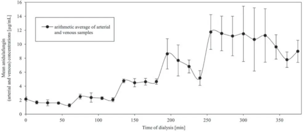
In keinem der durchgeführten Versuche war Anidulafungin im Dialysat nachweisbar. Dies ist übereinstimmend mit den Ergebnissen weiterer Studien zu anderen Nierenersatzverfahren [16, 24-26]. Die Dialyse Clearance beträgt somit 0 ml/min. Abbildung 3 zeigt den Verlauf der ge- messenen Anidulafunginkonzentrationen der untersuchen Lösungen (Durchschnitt der arteriel- len und venösen Proben ± Standardabweichung).
Nach dem Ende eines Versuches wurden die Kapillaren wie oben angegeben aus den Hämofil- tern entfernt. Nach Behandlung der Kapillaren mit 10 mM KH2PO4 Puffer war in den Proben der Waschlösung jeweils kein Anidulafungin nachweisbar.
Dialyse von Kochsalzlösung
Sowohl nach dem Beginn der Dialyse, als auch nach jeder Zugabe fiel die Anidulafunginkon- zentration deutlich ab. Nach einer Stunde betrug die gemessene Konzentration 0,02 µg/ml (Durchschnittliche Konzentration aller arteriellen und venösen Proben 60 Minuten nach Beginn der Dialyse; theoretische Konzentration von Anidulafungin zu diesem Zeitpunkt: 3 µg/ml;
Abbildung 1). Nach 120 Minuten war die Konzentration auf 0,04 µg/ml (theoretische Konzent- ration 5 µg/ml), nach 180 Minuten auf 0,08 µg/ml (theoretische Konzentration 7,5 µg/mL) und nach 240 Minuten auf 0,16 µg/ml (theoretische Konzentration 10 µg/ml) gefallen. Danach erfolgten keine weiteren Zugaben von Anidulafungin. Bis zum Ende des Experiments fiel die gemessene Anidulafunginkonzentration auf 0,04 µg/ml. Im untersuchten Konzentrationsbereich von bis zu 10 µg/ml war nach jeweils einer Stunde Dialyse kaum mehr Anidulafungin in der dialysierten Lösung vorhanden.
Nach dem Waschen mit Acetonitril/10 mM KH2PO4 Puffer (50/50, v/v) fanden sich Anidula- funginkonzentrationen von 164,5 ± 11,7 µg/g in den Filterkapillaren. Unter Einbeziehung der Masse der Kapillaren ergibt sich, dass in diesen Experimenten etwa 98,5 ± 7% der eingesetzten 20 mg Anidulafungin an den Hämofilter gebunden wurden.
Albuminhaltige Kochsalzlösung
In den Experimenten mit albuminhaltiger Kochsalzlösung waren höhere Anidulafunginkonzent- rationen in der dialysierten Lösung nachweisbar. Innerhalb einer Stunde fiel die Konzentration auf 1,52 µg/ml (theoretische Konzentration 3 µg/ml). Nach 120 Minuten betrug die Konzentra- tion 1,48 µg/ml (theoretische Konzentration 5 µg/ml), nach 180 Minuten 2,78 µg/ml (theoreti- sche Konzentration 7,5 µg/ml) und nach 240 Minuten 3,73 µg/ml (theoretische Konzentration
10 µg/ml). Bis zum Ende des Experiments nach 375 Minuten fiel die Konzentration auf 1,75 µg/ml.
Die Proben der Waschlösung der Hämofilterkapillaren mit Acetonitril/10 mM KH2PO4 Puffer (50/50, v/v) ergaben Anidulafunginkonzentrationen von 103,1 ± 44,1 µg/g in den Filterkapilla- ren. Bezogen auf den gesamten Hämofilter ergibt sich eine Anidulafunginbindungsrate von 60,3
± 25,8% der eingesetzten 20 mg.
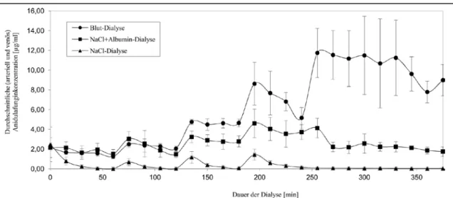
Blut
In den Experimenten mit Blut bestand die geringste Adsorption von Anidulafungin an den Hämofilter. Nach 60 Minuten Dialyse betrug die Anidulafunginkonzentration 1,26 µg/ml (theo- retische Konzentration 2,22 µg/ml), nach 120 Minuten Dialyse 2,06 µg/ml (theoretische Kon- zentration 3,70 µg/mL) und nach 180 Minuten 4,66 µg/ml (theoretische Konzentration 7,41 µg/ml), nach 240 Minuten 5,16 µg/ml (theoretische Konzentration 11,11 µg/ml) und nach 300 Minuten (theoretische Konzentration 14,81 µg/ml) betrug die gemessene Anidulafunginkon- zentration 11,49 µg/ml. Bis zum Ende des Experiments, nach 375 Minuten, nahm sie auf 8,99 µg/ml ab. In diesem Experiment war die Streuung der gemessenen Anidulafunginkonzentratio- nen nach der letzten Zugabe (nach 240 Minuten) im Vergleich zu den Experimenten mit gerin- geren Konzentrationen (Dialyse von Kochsalzlösung und albuminhaltiger Kochsalzlösung) höher. Eine Ku-
mulation der Substanz in den Experimenten mit höheren Konzent- rationen könnte diese Beobachtung erklären. Die Proben der
Zeit [min]
Anidulafunginkonzentration [µg/ml]
NaCl-Dialyse NaCl+Albumin-
Dialyse.
Blut-Dialyse Eingesetzt Gemessen Eingesetzt Gemessen Eingesetzt Gemessen
0 3 2,48 3 2,16 2,22 2,15
60 3 0,02 3 1,52 2,22 1,26
120 5 0,04 5 1,48 3,7 2,06
180 7,5 0,08 7,5 2,78 7,41 4,66
240 10 0,16 10 3,73 11,11 5,16
300 10 0,05 10 2,57 14,81 11,49
375 10 0,04 10 1,75 14,81 8,99
Tabelle 1. Übersicht über eingesetzte und nachgewieseneAnidulafunginkon- zentrationen (Durchschnitt ± Standardabweichung)
Waschlösung nach dem Waschen der Kapillaren mit Acetonitril/10 mM KH2PO4 Puffer (50/50, v/v) zeigten Anidulafunginkonzentrationen von 48,7 ± 13,3 µg/g in den Filterkapillaren. Dies deutet darauf hin, dass 35,3 ± 9,6% der eingesetzten 20 mg Anidulafungin an der Filteroberflä- che adsorbierten. Tabelle 1 gibt einen Überblick über die deutliche Differenz der eingesetzten zu den nachgewiesenen Anidulafunginkonzentrationen im Verlauf der Versuche.
Diskussion
Das Hauptergebnis dieser Studie war, dass bei der Dialyse von Blut etwa 35% des eingesetzten Anidulafungins an der Oberfläche des Hämofilters adsorbieren. Es war das Ziel, diesen Sach- verhalt in vitro unter CVVHD zu untersuchen, da bekannt ist, dass Adsorption an der Oberflä- che eines Hämofilters die Blutkonzentration eines verabreichten Medikaments beeinflussen kann [19-23].
Die empfohlene Dosierung von Anidulafungin für Patienten, die an einer Candidämie oder Candida Infektionen wie z. B. einem intraabdominellen Abszess oder einer Peritonitis leiden, beträgt 100 mg/Tag nach einer Initialdosis von 200 mg am ersten Tag [10]. Bei diesem Dosisregime wird im Steady State bei gesunden Probanden eine Maximalkonzentration (Cmax) von 8,6 µg/ml und eine AUC (Area under the curve, Fläche unter der Konzentration/Zeit- Kurve) von 111,8 mg·h/l erreicht [10]. Ähnliche Werte finden sich bei Patienten mit Pilzinfek- tionen [10]. Patienten, die unter einer ösophagealen Candidiasis leiden, sollen nach einer Initaldosis von 100 mg eine Erhaltungsdosis von 50 mg/Tag erhalten [10]. Mit dieser Dosierung werden bei Patienten Plasmaspiegel zwischen 1,6 µg/ml und 4,2 µg/ml erreicht [10]. Die mini- male Hemmkonzentration (minimum inhibitory concentration, MIC) von Anidulafungin, die benötigt wird, um mindestens 90% der Proben zu hemmen (MIC90), war für 5.346 getestete Proben von Candida spp. 2 mg/l und die MIC50 0,06 mg/l [7]. Die mittlere minimale effektive
Konzentration (minimum effective concentration, MEC) beträgt für Aspergillus fumigatus 0,12 mg/l und für Aspergillus terreus und Aspergillus flavus jeweils 0,25 mg/l [28].
In der vorliegenden Studie betrug der adsorbierte Anteil des Anidulafungins bei der Dialyse von Blut 35%. Bei Patienten, die mit dem Dosisregime von 100 mg täglich behandelt werden, würde das bedeuten, dass die Plasmaspiegel ausreichend sind, um die meisten Candida Infektionen zu behandeln. Bei Patienten, die 50 mg Anidulafungin pro Tag erhalten, könnte eine Reduktion der Plasmakonzentration um 35% jedoch zu Konzentrationen unterhalb des benötigten Niveaus führen. Dies gilt insbesondere bei Infektionen mit Candida spp., welche höhere Anidulafungin- konzentrationen erfordern, wie C. parapsilosis (MIC90: 2 mg/l [7, 29]), obwohl Echinocandine nicht Mittel der ersten Wahl zur Therapie von C. parapsilosis Infektionen sind [6].
In unserer Studie zeigte sich in der Gegenwart von menschlichem Plasma eine geringere Ad- sorption von Anidulafungin an den Hämofilter, als in den Experimenten mit albuminhaltiger Kochsalzlösung. Dies kann durch einen größeren proteingebundenen Anteil des Anidulafungins in den Experimenten mit menschlichem Plasma erklärt werden. Die Ursache dafür könnte sein, dass Anidulafungin neben Albumin auch an andere Serumbestandteile gebunden ist, oder dass die Gegenwart von Serum die Bindungsfähigkeit von Albumin verändert. Beide Theorien wurden von Zeitlinger et al. [30] bei der Untersuchung der Auswirkung von Proteinbindung auf Fluorchinolone eingeführt.
Die Studie von Dowell et al. [16] ergab, dass bei Patienten die Hämodialyse erhalten, keine Anpassung der Anidulafugindosierung nötig ist. Leitner et al. [24] diskutierten in ihrer Studie eine Verminderung der Anidulafunginkonzentration unter CVVHF aufgrund von Adsorption an der Oberfläche von Polyethersulfon Hämofiltern. Aguilar et al. [25] untersuchten CVVHDF mit Polysulphon Hämofiltern und fanden keine Adsorption an der Filteroberfläche. In dieser Studie wurde daher gefolgert, dass die Beobachtung von Leitner et al. [24] mit einem Verdünnungsef- fekt durch die Substitutionslösung erklärt werden könnte [25]. In der Studie von Burkhardt et al.
[26] wurden Polysulphon Hämofilter für EDD verwendet und es wurde keine Adsorption an die
Abbildung 4. Strukturformeln von Anidulafungin und Polysulfon Filtermembran beschrieben. Die genannten Studien [24-26] kamen übereinstimmend mit Dowell et al. [16] zu dem Ergebnis, dass keine Dosisanpassung nötig ist.
Im Gegensatz dazu zeigten sich die Anidulafunginkonzentrationen in den in vitro Dialyseexpe- rimenten der vorliegenden Studie deutlich beeinflusst. Das kann ein Hinweis darauf sein, dass der Effekt in vivo möglicherweise weniger relevant ist. Allerdings besteht die Möglichkeit, dass ein Nierenersatzverfahren eine parallele Anidulafungintherapie beeinflusst.
In diesem Kontext sollte auch beachtet werden, dass die meisten pharmakokinetischen Daten lediglich die Gesamtkonzentration eines Medikaments betrachten. In Anbetracht der Adsorption von 99% bei der Dialyse von Kochsalzlösung gegenüber 35% bei der Dialyse von Blut, scheint ein Hämofilter laut unseren Daten jedoch vorwiegend die freie Anidulafunginkonzentration zu beeinflussen. Die chemische Struktur von Anidulafungin mit drei linear angeordneten aromati- schen Ringen deutet darauf
hin, dass eine starke Inter- aktion mit einer syntheti- schen linearen poly- aromatischen Oberfläche, wie sie bei einem
Polysulfon Hämofilter vorliegt, sehr wahrscheinlich ist (Abbildung 4). Dass die verminderte Adsorption in Gegenwart von Albumin oder Blut infolge einer „Blockade“ der Filteroberfläche durch Proteine bewirkt wird, ist aufgrund der geringen Adsorption von Albumin an der Polysulfonoberfläche [31] weniger wahrscheinlich.
Anidulafungin ist entgegen der anfänglichen Annahme von 84% [9] in großem Maß (>99%) an Plasmaproteine gebunden [10]. Die weiteren Echinocandine Micafungin (99,8%) und Caspo- fungin (96%) weisen ebenso eine sehr hohe Proteinbindung auf [9]. Die Proteinbindung scheint auch großen Einfluss auf die Wirksamkeit dieser Substanzklasse zu haben. So weist Anidula- fungin in der Gegenwart von Serum deutlich erhöhte MIC Werte auf [32-34]. Auch die in vitro
spp. nähern sich in Anwesenheit von Serum oder in vivo an, sodass unter diesen Bedingungen teils vergleichbare MIC und MEC Werte vorliegen [34]. Ob dieser Effekt darauf beruht, dass proteingebundenes Anidulafungin inaktiv ist und nur ein kleiner freier Anteil antimikrobiell aktiv ist, oder ob Anidulafungin auch im proteingebundenen Zustand aktiv ist, ist bisher unklar [34].
Da laut unseren Versuchen durch einen Hämofilter insbesondere das freie Anidulafungin ver- mindert wird, könnten die Auswirkungen auf Patienten stärker sein, als durch die alleinige Reduktion der Gesamtkonzentration zu erwarten wäre. So gibt es bisher keine Studien zur Gewebeverteilung von Anidulafungin im Menschen. In Tierversuchen wurde Anidulafungin schnell in die am häufigsten von Mykosen betroffenen Gewebe verteilt [35]. Eine gleichzeitige Nierenersatztherapie könnte also besonderen Einfluss auf die Konzentration in diesen Organen haben, wenn das nicht proteingebundene Anidulafungin entzogen wird. Diesbezüglich wären weitere Studien zur Untersuchung der Auswirkung von Nierenersatzverfahren auf die Wirksam- keit von Anidulafungin in vivo aufschlussreich. Zum Beispiel könnten Mikrodialyseexperimente bei Intensivpatienten Aufschluss über den Einfluss von Nierenersatztherapien auf die Gewebe- spiegel von Anidulafungin geben [36]. Derartige Erkenntnisse würden auch größere klinische Relevanz besitzen, als reine Plasmakonzentrationen.
Ein weiterer Aspekt, der in diesem Kontext untersucht werden sollte, ist die Tatsache, dass schwer kranke Patienten oft mangelernährt sind. So könnten verminderte Eiweißspiegel bei diesen Patienten zu einer vermehrten Menge an freiem Medikament führen [37]. -Allerdings könnte eine gleichzeitige Nierenersatztherapie auch zu einer stärkeren Medikamentenadsorption an den Hämofilter führen.
In der vorliegenden in vitro Studie wurde ein Hämofiltertyp untersucht. Da die Membran erheb- lichen Einfluss auf die Adsorption eines Medikamentes haben kann [21], sind weitere Studien zur Untersuchung anderer Hämofiltertypen nötig. In unserer Studie wurde lediglich die Anidulafunginadsorption unter CVVHD untersucht. In Studien anderer Autoren zu weiteren Nierenersatzverfahren [24-26] ergab sich kein Hinweis auf die Notwendigkeit einer
Dosisanpassung. Dennoch scheinen weitere Studien sinnvoll, um auch den Effekt dieser Nie- renersatzverfahren auf die Adsorption von Anidulafungin an die Hämofiltermembran zu unter- suchen.
Zusammenfassend zeigt die vorliegende in vitro Studie, dass unter CVVHD etwa 35% des verabreichten Anidulafungins an der synthetischen Oberfläche des Hämofilters adsorbieren können. Dennoch scheinen die dadurch zu erwartenden Blutkonzentrationen ausreichend zur Behandlung der meisten Candida und Aspergillus Infektionen [7, 28, 29].
Die überwiegende Menge der verfügbaren pharmakokinetischen Daten bezieht sich auf die Gesamtkonzentrationen einer Substanz. Dagegen sind der biologisch aktive Anteil von Anidula- fungin in vivo sowie die Gewebeverteilung im Menschen weitgehend unerforscht. Aus diesem Grund ist es unklar ob und in welchem Ausmaß ein Hämofilter die aktive Anidulafunginkon- zentration und die Gewebespiegel am Ort der Infektion beeinflusst.
Lieraturverzeichnis
1. Brady HR, Singer GG. Acute renal failure. Lancet. 1995;346:1533-40.
2. Hoste EAJ, Schurgers M. Epidemiology of acute kidney injury: How big is the prob- lem? Crit Care Med. 2008;36:S146-S51.
3. Centers for Disease Control and Prevention (CDC). Hospitalization discharge diagnoses for kidney disease—United States, 1980-2005. Morbidity and Mortality Weekly Report.
2008;57:309-12.
4. Blumberg HM, Jarvis WR, Soucie JM, Edwards JE, Patterson JE, Pfaller MA, et al.
Risk factors for candidal bloodstream infections in surgical intensive care unit patients: the NEMIS prospective multicenter study. The National Epidemiology of Mycosis Survey. Clin Infect Dis. 2001;33:177-86.
5. Pappas P, Kauffman C, Andes D, Benjamin D, Calandra T, Edwards J, et al. Clinical practice guidelines for the management of candidiasis: 2009 Update by the Infectious Diseases Society of America. Clin Infect Dis. 2009;48:503-35.
6. Cornely OA, Bassetti M, Calandra T, Garbino J, Kullberg BJ, Lortholary O, et al.
ESCMID*-guideline for the diagnosis and management of Candida diseases 2012: non- neutropenic adult patients. Clin Microbiol Infect. 2012;18:19-37.
7. Pfaller MA, Boyken L, Hollis RJ, Kroeger J, Messer SA, Tendolkar S, et al. In vitro susceptibility of invasive isolates of Candida spp. to anidulafungin, caspofungin, and mica- fungin: Six years of global surveillance. J Clin Microbiol. 2008;46:150-6.
8. Murdoch D, Plosker GL. Anidulafungin. Drugs. 2004;64:2249-58.
9. Denning DW. Echinocandin antifungal drugs. Lancet. 2003;362:1142-51.
10. Anidulafungin (Eraxis). [Prescribing Information]. Pfizer Inc, New York, NY. 2013.
11. Pfaller MA, Diekema DJ, Ostrosky-Zeichner L, Rex JH, Alexander BD, Andes D, et al.
Correlation of MIC with outcome for Candida species tested against caspofungin, anidula-
fungin, and micafungin: Analysis and proposal for interpretive MIC breakpoints. J Clin Microbiol. 2008;46:2620-9.
12. Zhanel GG, Karlowsky JA, Harding GAJ, Balko TV, Zelenitsky SA, Friesen M, et al. In vitro activity of a new semisynthetic echinocandin, LY-303366, against systemic isolates of Candida species, Cryptococcus neoformans, Blastomyces dermatitidis, and Aspergillus species.
Antimicrob Agents Chemother. 1997;41:863-5.
13. Serrano MdC, Valverde-Conde A, Chávez M, Bernal S, Claro RM, Pemán J, et al. In vitro activity of voriconazole, itraconazole, caspofungin, anidulafungin (VER002, LY303366) and amphotericin B against Aspergillus spp. Diagn Microbiol Infect Dis. 2003;45:131-5.
14. Dowell JA, Knebel W, Ludden T, Stogniew M, Krause D, Henkel T. Population phar- macokinetic analysis of anidulafungin, an echinocandin antifungal. J Clin Pharmacol.
2004;44:590-8.
15. Damle BD, Dowell JA, Walsky RL, Weber GL, Stogniew M, Inskeep PB. In vitro and in vivo studies to characterize the clearance mechanism and potential cytochrome P450 interac- tions of anidulafungin. Antimicrob Agents Chemother. 2009;53:1149-56.
16. Dowell JA, Stogniew M, Krause D, Damle B. Anidulafungin does not require dosage adjustment in subjects with varying degrees of hepatic or renal impairment. J Clin Pharmacol.
2007;47:461-70.
17. Atkinson AJ, Jr, Lertora JJL. Chapter 5 - Effect of renal disease on pharmacokinetics.
In: Atkinson AJ, Jr, Huang S-M, Lertora JJL, Markey SP, eds. Principles of Clinical Pharma- cology (Third Edition). San Diego, CA: Academic Press 2012:55-64.
18. Atkinson AJ, Jr, Susla GM. Chapter 6 - Pharmacokinetics in patients requiring renal replacement therapy. In: Atkinson AJ, Jr, Huang S-M, Lertora JJL, Markey SP, eds. Principles of Clinical Pharmacology (Third Edition). San Diego, CA: Academic Press 2012:65-79.
19. Meyer B, Ahmed el Gendy S, Delle Karth G, Locker GJ, Heinz G, Jaeger W, et al. How to calculate clearance of highly protein-bound drugs during continuous venovenous hemofiltra-
20. Tian Q, Gomersall CD, Wong A, Leung P, Choi G, Joynt GM, et al. Effect of drug concentration on adsorption of levofloxacin by polyacrylonitrile haemofilters. Int J Antimicrob Agents. 2006;28:147-50.
21. Choi G, Gomersall CD, Lipman J, Wong A, Joynt GM, Leung P, et al. The effect of adsorption, filter material and point of dilution on antibiotic elimination by haemofiltration: An in vitro study of levofloxacin. Int J Antimicrob Agents. 2004;24:468-72.
22. Ittner KP, Roth G, Gruber M, Pawlik M, Taeger K. Clearance of moxifloxacin during continuous haemofiltration (CVVHF) in vitro. J Antimicrob Chemother. 2005;56:360-4.
23. Meyer B, Traunmuller F, Hamwi A, Hollenstein UM, Locker GJ, Sperr WR, et al.
Pharmacokinetics of teicoplanin during continuous hemofiltration with a new and a 24-h used highly permeable membrane: rationale for therapeutic drug monitoring-guided dosage. Int J Clin Pharmacol Ther. 2004;42:556-60.
24. Leitner JM, Meyer B, Fuhrmann V, Saria K, Zuba C, Jager W, et al. Multiple-dose pharmacokinetics of anidulafungin during continuous venovenous haemofiltration. J Antimicrob Chemother. 2011;66:880-4.
25. Aguilar G, Azanza JR, Carbonell JA, Ferrando C, Badenes R, Parra MA, et al. Anidula- fungin dosing in critically ill patients with continuous venovenous haemodiafiltration. J
Antimicrob Chemother. 2014;69:1620-3.
26. Burkhardt O, Kaever V, Burhenne H, Kielstein JT. Extended daily dialysis does not affect the pharmacokinetics of anidulafungin. Int J Antimicrob Agents. 2009;34:282-3.
27. Jelkmann W. Blut. In: Schmidt RF, Lang F, Heckmann M, eds. Physiologie des Men- schen: mit Pathophysiologie. Berlin, Heidelberg: Springer Berlin Heidelberg 2011:477-502.
28. Antachopoulos C, Meletiadis J, Sein T, Roilides E, Walsh TJ. Comparative in vitro pharmacodynamics of caspofungin, micafungin, and anidulafungin against germinated and nongerminated Aspergillus conidia. Antimicrob Agents Chemother. 2008;52:321-8.
29. Ostrosky-Zeichner L, Rex JH, Pappas PG, Hamill RJ, Larsen RA, Horowitz HW, et al.
Antifungal susceptibility survey of 2,000 bloodstream Candida isolates in the United States.
Antimicrob Agents Chemother. 2003;47:3149-54.
30. Zeitlinger M, Sauermann R, Fille M, Hausdorfer J, Leitner I, Muller M. Plasma protein binding of fluoroquinolones affects antimicrobial activity. J Antimicrob Chemother.
2008;61:561-7.
31. Mares J, Thongboonkerd V, Tuma Z, Moravec J, Matejovic M. Specific adsorption of some complement activation proteins to polysulfone dialysis membranes during hemodialysis.
Kidney Int. 2009;76:404-13.
32. Spreghini E, Orlando F, Sanguinetti M, Posteraro B, Giannini D, Manso E, et al. Com- parative effects of micafungin, caspofungin, and anidulafungin against a difficult-to-treat fungal opportunistic pathogen, Candida glabrata. Antimicrob Agents Chemother. 2012;56:1215-22.
33. Odabasi Z, Paetznick V, Rex JH, Ostrosky-Zeichner L. Effects of serum on in vitro susceptibility testing of echinocandins. Antimicrob Agents Chemother. 2007;51:4214-6.
34. Paderu P, Garcia-Effron G, Balashov S, Delmas G, Park S, Perlin DS. Serum differen- tially alters the antifungal properties of echinocandin drugs. Antimicrob Agents Chemother.
2007;51:2253-6.
35. Damle B, Stogniew M, Dowell J. Pharmacokinetics and tissue distribution of anidula- fungin in rats. Antimicrob Agents Chemother. 2008;52:2673-6.
36. Hope WW, Drusano GL. Antifungal pharmacokinetics and pharmacodynamics: bridg- ing from the bench to bedside. Clin Microbiol Infect. 2009;15:602-12.
37. Ashley ESD, Lewis R, Lewis JS, Martin C, Andes D. Pharmacology of systemic anti- fungal agents. Clin Infect Dis. 2006;43:28-39.
Originalversion des Artikels
Filter Adsorption of Anidulafungin to a Polysulfone-Based Hemofilter During CVVHD In Vitro
Kolbinger, P., Gruber, M., Roth, G., Graf, B. M. and Ittner, K.-P. (2017). Artificial Organs.
doi:10.1111/aor.12982
Filter Adsorption of Anidulafungin to a Polysulfone-Based Hemofilter During CVVHD In Vitro
*Peter Kolbinger , †Michael Gruber, †Gabriel Roth, †Bernhard M. Graf, and †‡Karl-Peter Ittner
*Clinic of Radiology, Minimally Invasive Therapies and Nuclear Medicine, SLK-Kliniken Heilbronn GmbH, Heilbronn;
†Department of Anaesthesiology, University Hospital Regensburg; and‡Didactic Department of Pharmacology, University Hospital Regensburg, Regensburg, Germany
Abstract: Candidemia is frequent in critically ill patients, especially in combination with an acute kidney injury (AKI). Echinocandins generally are recommended for therapy of such infections. Recent studies found no need for dosage adjustment in patients with end-stage renal dis- ease receiving hemodialysis, or patients with AKI receiv- ing continuous venovenous hemofiltration. The aim of this in vitro study was to examine the adsorption of anidulafun- gin to the surface of the hemofilter during continuous venovenous hemodialysis (CVVHD) and its effect on ani- dulafungin concentrations. The concentration of anidula- fungin in the dialyzed fluid, and the dialysate during CVVHD in vitro was examined using three different dia- lyzed fluids (saline; saline with 40 g/L human albumin; and a mixture of human erythrocytes and fresh frozen plasma).
After the end of dialysis, the hemofilter was opened and portions of the filter capillaries were also analyzed to deter- mine the amount of anidulafungin adsorbed. When dialyz- ing saline, about 99% of the anidulafungin used adsorbed to the hemofilter capillaries; in the experiments with saline with 40 g/L albumin, about 60% adsorbed to the hemofil- ter’s surface, and when blood was dialyzed, 35% was found adsorbed after analyzing the filter capillaries. Anidulafun- gin was not detectable in the dialysate of any of the experi- ments, consequently the dialysis clearance was 0 mL/min.
In conclusion, during CVVHD in vitro we found remark- able adsorption of anidulafungin to the hemofilter’s sur- face, yet the effect on the tissue concentration needs further examination. Key Words: Renal replacement therapy—Echinocandins—Artificial membranes.
Acute kidney injury (AKI) is a frequent compli- cation in critically ill patients, which occurs in nearly every third admission to an intensive care unit (ICU), and the incidence has greatly increased in recent years (1). The rate of hospitalization for AKI in US hospitals in 1980 was 1.8/10 000 and increased to 36.5/10 000 in 2005 (2). Candidemia is a frequent reason for bloodstream infections, par- ticularly in ICUs (3). In surgical ICUs in the United States, 9.82 Candida bloodstream infections per
1000 admissions were reported, whereas patients with AKI had a fourfold increased risk in compari- son to patients without this diagnosis (3). These facts show that both antifungal drugs and continu- ous renal replacement therapy are widely used in ICUs. Consequently, the pharmacokinetic charac- teristics of an antifungal drug during renal replace- ment therapy are of great importance for numerous patients.
Especially in critically ill patients, an echinocan- din is generally recommended for therapy of Candi- demia, except for Candida parapsilosis infections (4,5). This recommendation is emphasized by the fact that a recent study found no evidence for emerging resistance against echinocandins among invasiveCandida isolates (6). Anidulafungin is the newest agent in the group of echinocandins (7,8). It is, as all echinocandins, fungicidal against most
doi: 10.1111/aor.12982
Received February 2017; revised April 2017; accepted May 2017.
Address correspondence and reprint requests to Peter Kolbinger, Clinic of Radiology, Minimally Invasive Therapies and Nuclear Medicine, SLK-Kliniken Heilbronn GmbH, Am Gesundbrunnen 20-26, 74078 Heilbronn, Germany.
E-mail: peter.kolbinger@slk-kliniken.de
bs_bs_banner
CopyrightVC 2017 International Center for Artificial Organs and Transplantation and Wiley Periodicals, Inc.
Candida spp. (6,8–10) and also shows high fungi- static efficacy against many Aspergillus species (8,10,11).
Due to poor oral bioavailability, which is typical for echinocandins (8), anidulafungin has to be administered intravenously. After intravenous administration, anidulafungin has a predictable phar- macokinetic profile, including dose-proportional plasma concentrations (12). It has a volume of distri- bution of about 33 L at steady state, which is achieved after a loading dose (double the daily dose) and one normal dose (12). The protein bind- ing of anidulafungin is>99% (13). Anidulafungin is a large lipopeptide molecule with a molecular weight of 1140.3 Da (13). As all echinocandins it is an amphiphilic hexapeptide with an N-linked acyl lipid side-chain, in the case of anidulafungin consist- ing of a lipophilic alkoxytriphenyl (terphenyl) side- chain (8).
Anidulafungin does not undergo hepatic metabo- lization or renal excretion, but is degraded nonen- zymatically and eliminated in the feces (14).
Therefore, the clearance of anidulafungin is approximately 1 L/h, regardless of hepatic or renal insufficiency (15).
In contrast, drug dosing under renal replacement therapy is quite difficult since it is subject to many factors (16,17). For example, variable amounts of the drugs might adsorb to a hemofilter’s surface (18–22).
Dowell et al. (15) showed that the anidulafungin blood concentration is not affected by hemodialysis in patients with end-stage renal disease, and Leit- ner et al. (23) confirmed that no dosage adjustment is necessary in patients receiving continuous veno- venous hemofiltration (CVVHF). In addition, the studies of Aguilar et al. (24) and Burkhardt et al.
(25) found no need for dosage adjustment in patients receiving continuous venovenous hemodia- filtration (CVVHDF) and extended daily dialysis (EDD).
The aim of this study was to analyse the effect of continuous renal replacement therapy (continuous venovenous hemodialysis, CVVHD) on anidulafun- gin concentrations due to adsorption to the surface of the hemofilter. For this investigation, the results of different compositions of dialyzed solutions were examined.
MATERIALS AND METHODS In vitro CVVHD system
A 2 L glass bottle was used as a reservoir for the in vitro dialysis. Three different solutions were
used (Fig. 1): First, 1650 mL of normal saline. Sec- ond, albuminous saline, consisting of 1250 mL of normal saline and 400 mL of 20% human albumin (80 g of albumin in total, Baxter, Vienna, Austria) to give a total volume of 1650 mL. Third, a mixture of 600 mL of human erythrocytes, 400 mL of fresh frozen plasma (FFP), 100 mL of 8.4% NaHCO3
and 10 000 IU heparin (2 mL), of which 1000 mL were used (pH 7.1360.03 [SD], hematocrit 34.562.12, about 29 g of plasma proteins in total, based on a typical concentration of 80 g/L of the FFP (26)) in substitution for 1 L of fresh human blood. To calculate the dilution volume of the tube system and hemofilter, we weighed the system before and after flushing it with saline, which showed a total volume of 350 mL. We considered this volume in our calculations, as we included it in the total volume of 1350 mL in the experiments with human blood and 2000 mL in the experiments with pure saline/albuminous saline, respectively.
An overview of the experiments is shown in Table 1. The total albumin concentration in the experiments with albuminous saline was 40 g/L. In the experiments with blood the total protein con- centration was approx. 21.5 g/L. During all experi- ments, a magnetic stirrer was used to continuously mix the solutions. Dialysis was performed with an Edwards bm11a/bm14 dialysis system (Edwards Lifesciences Germany GmbH, Unterschleissheim, Germany). SH 27 Solution (B. Braun, Melsungen, Germany) was used as dialysate and the hemofil- ters were Fresenius Ultraflux AV 600S (Fresenius Polysulfone, surface area 1.4 m2, Fresenius Medical Care, Bad Homburg, Germany). All experiments were done in duplicate.
High-performance liquid chromatography
A modified method for detecting anidulafungin in plasma, saline and dialysate was used according to Dowell et al. (15). The high-performance liquid chromatography (HPLC) was an Agilent 1200 Series HPLC system with an Agilent 1200 Series
FIG. 1. Experiments performed.
FILTER ADSORPTION OF ANIDULAFUNGIN 201
fluorescence detector working at 225 nm excitation, and 431 and 300 nm emission wavelengths for ani- dulafungin and internal standard laudanosine (Aldrich, Milwaukee, WI, USA), respectively. A binary gradient (solvent A: 10 mM KH2PO4[Fluka, Buchs, Switzerland], pH 4.5; solvent B: acetonitrile [HPLC Gradient Grade, Roth, Karlsruhe, Ger- many]) was used to separate the target substances on a Silice Upti-prep Strategy 100A 2.2 mm PRO column (Interchim, Montluc¸on, France) equipped with a 4 3 2 mm C18 pre-column (Phenomenex, Aschaffenburg, Germany). The gradient started at a flow rate of 0.4 mL/min with 5% solvent B. It changed linearly to 90% B at 10 min and then back to 5% B at 12 min. The total run time was 15 min.
The retention time for anidulafungin and laudano- sine was 9.9 and 6.8 min, respectively.
For every sample, 100 mL of saline, plasma or dialysate was treated with 5mL (0.069 mg) of inter- nal standard laudanosine, then 250 mL acetonitrile/
10 mM KH2PO4buffer (10/90, vol/vol) was added.
This mixture was applied to a 130-mg SPE column (Bond Elut-Certify, 130 mg, 1 mL, part number 12102083, Varian, Darmstadt, Germany), which was preconditioned with 1 mL of acetonitrile/
10 mM KH2PO4buffer (10/90, vol/vol). Afterwards, the SPE column was washed with 500mL of aceto- nitrile/10 mM KH2PO4 buffer (10/90, vol/vol), fol- lowed by 500 mL of chloroform (Fluka).
Anidulafungin and laudanosine were eluted with 2 mL of acetonitrile/10 mM KH2PO4buffer (70/30, vol/vol) and collected in conical glass tubes. After evaporation at 408C, the residue was resolved in 50 mL of acetonitrile/10 mM KH2PO4 buffer (50/50, vol/vol) and transferred into 300 mL micro vial inserts (Klaus Trott Chromatographie-Zubeh€or, Kriftel, Germany). Five microliters were injected into the HPLC. In the autosampler the extracts
8
limit of detection was 8 ng/mL, the matrix-free limit of quantification was 32 ng/mL and the corre- lation coefficient was >0.999. Inter- and intra-day variability were 23.7 and 14.6%, respectively.
Experiments
The experiments were started with anidulafungin concentrations similar to typical peak plasma con- centrations (15). After the start of dialysis, anidula- fungin was added as shown in Table 2, to substitute for any potential losses to the hemofilter or the dialysate. The resulting anidulafungin concentra- tions are shown in Fig. 2.
Dialysis conditions were as follows: duration of dialysis: 375 min, blood flow rate: 200 mL/min and dialysate flow rate: 800 mL/h. Ultrafiltration was set to zero. Samples from the arterial und venous TABLE 1. Experimental conditions
Experiment CVVHD conditions
1–3 CVVHD for 375 min, blood flow 200 mL/min, dialysate flow 800 mL/h
Experiment Reservoir conditions
1–3 350 mL of normal saline residual in the tube system and hemofilter
1 1650 mL of normal saline, total volume of 2000 mL
2 1250 mL of normal saline, 400 mL of 20%
human albumin, total volume of 2000 mL 3 600 mL of human erythrocytes, 400 mL of fresh
frozen plasma, 100 mL of 8.4% NaHCO3, 2 mL of heparin, of which 1000 mL were used, total volume of 1350 mL
FIG. 2.Administered anidulafungin concentrations during TABLE 2. Administered anidulafungin
Experiments with normal saline and albuminous saline, total volume of 2000 mL
Time
Anidulafungin amount (mg)
Anidulafungin concentration
(mg/mL)
At the beginning of dialysis 6 3
After 60 min 10 5
After 120 min 15 7.5
After 180 min 20 10
Experiments with human erythrocytes, fresh frozen plasma, NaHCO3, total volume of 1350 mL
Time
Anidulafungin amount (mg)
Anidulafungin concentration
(mg/mL)
At the beginning of dialysis 3 2.22
After 60 min 5 3.70
After 120 min 10 7.41
After 180 min 15 11.11
After 240 min 20 14.81
P. KOLBINGER ET AL.
202
line (approx. 2 mL) were collected at the start of dialysis, and then every 15 min until the end of the experiment at 375 min. The arterial samples at 0 min were taken directly from the reservoir to establish baseline data before the first pass of the hemofilter, accepting a slightly higher concentration in these samples since the reservoir had not yet been mixed with the normal saline in the tube system. Dialysate samples were collected at 60, 120, 180, 240, 300, 330, and 360 min. Blood samples were centrifuged (Her- aeus Biofuge pico, 5 min at 10 000 g, Heraeus, Oster- ode, Germany) and then stored at 2208C; all other samples were also stored at 2208C after collection.
After dialysis, the hemofilter was opened by cutting away the ends with potting compound. Remaining fluids inside the filter were discarded. The capillaries were then removed from the housing and weighed.
The arithmetic mean weight of the capillaries of all the filters examined was 127.1613.7 g. The bundle of capillaries was spread on a flat surface and five 1 g samples consisting of complete capillaries (i.e., from the inlet to the outlet) were collected from evenly distributed portions to represent the whole fil- ter and to prevent distortion due to preferential adsorption that might occur at certain parts of the fil- ter. Each of these samples was put into 10 mL of 10 mM KH2PO4 buffer solution, stored in an ultra- sound bath for 10 min, and samples from the buffer solution were analyzed. Then the procedure was repeated with fresh buffer solution and finally 10 mL of acetonitrile/10 mM KH2PO4 buffer (50/50, vol/
vol) were used to extract the remaining anidulafun- gin from the hemofilter capillaries.
Calculations
Calculation of dialysis clearance (CLP, recovery clearance; CLP5CD VolD/P t; CD5dialysate concentration, VolD5volume of dialysate, P5arterial plasma concentration, t5dialysis time), (17) percentage of anidulafungin bound to the hemofilter membrane (PAF; PAF5CAF WF/AT
100%; CAF5arithmetic mean concentration of ani- dulafungin bound to filter capillaries, WF5arith- metic mean weight of the filter capillaries, AT5total amount of anidulafungin used in the experiment, 20 mg), as well as descriptive statistics (arithmetic mean and standard deviation), were performed with Excel 2007.
RESULTS Normal saline
After the beginning of dialysis, the anidulafungin concentration of the dialyzed saline dropped to 0.02 mg/mL within 1 h (arithmetic mean of all arte- rial and venous samples, taken after 60 min of dial- ysis; Fig. 3; theoretical concentration of anidulafungin at that time: 3 mg/mL; Fig. 2). Also after each administration of anidulafungin, the measured concentrations dropped within 1 h to 0.04 mg/mL (theoretical concentration 5 mg/mL) and 0.08 mg/mL (theoretical concentration 7.5 mg/
mL) after 120 and 180 min, respectively. After the last administration of anidulafungin (after 3 h of dialysis, theoretical concentration 10 mg/mL), the measured concentration dropped to 0.16 mg/mL within 1 h and decreased further until the end of the experiment (after 375 min of dialysis), at which the concentration was 0.04 mg/mL. Thus, in the examined range of concentrations of up to 10 mg/
mL anidulafungin (Fig. 2), there was nearly no ani- dulafungin left in the dialyzed fluid after 1 h. Due to the absence of anidulafungin in the dialysate, the hemofilters were opened after each experiment as described earlier. In the analysis of the hemofilters, we found no detectable anidulafungin in the wash- ing fluid after washing the filter capillaries in 10 mM KH2PO4 buffer. But after edulcorating them with acetonitrile/10 mM KH2PO4 buffer (50/
50, vol/vol), anidulafungin concentrations of 164.5611.7 mg/g could be found in the filter capillaries. Taking into account the weight of the
FIG. 3. Anidulafungin concentrations (mean6SD) during dialysis of saline.
FILTER ADSORPTION OF ANIDULAFUNGIN 203
hemofilter capillaries and the fact that 20 mg of anidulafungin were used during each experiment, about 98.567% of the anidulafungin was bound to the hemofilter in these experiments.
Albuminous saline
In the experiments with albuminous saline, higher concentrations of anidulafungin were achieved in the dialyzed fluid. After the beginning of dialysis, the anidulafungin concentration dropped continuously to 1.52 mg/mL after 1 h (Fig.
4; theoretical concentration 3 mg/mL). After 120 and 180 min of dialysis, the measured values for anidulafungin in the dialyzed fluid were 1.48mg/mL (theoretical concentration 5 mg/mL) and 2.78 mg/
mL (theoretical concentration 7.5 mg/mL), respec- tively. After the last administration of anidulafun- gin after 3 h of dialysis (theoretical concentration 10 mg/mL), the anidulafungin concentration in the dialyzed fluid dropped to 3.73 mg/mL and 1.75 mg/
mL at 240 and 375 min, respectively.
After washing the hemofilter capillaries with ace- tonitrile/10 mM KH2PO4 buffer (50/50, vol/vol), anidulafungin concentrations of 103.1644.1 mg/g in the filter capillaries could be found. Extrapolating this value to the weight of the examined hemofilter capillaries results in an anidulafungin binding rate of 60.3625.8% of the 20 mg used, explaining the
decline in the measured anidulafungin concentra- tions in the dialyzed fluids.
Blood
In the experiments with blood, the measured ani- dulafungin concentrations in the dialyzed fluids were closest to the expected concentrations. The adsorbed amount of anidulafungin was the lowest of all performed experiments. After 1 h of dialysis, the anidulafungin concentration in the dialyzed fluid was 1.26mg/mL (Fig. 5; theoretical concentra- tion 2.22mg/mL). After 120 and 180 min of dialysis, the measured values for anidulafungin in the dia- lyzed fluid were 2.06mg/mL (theoretical concentra- tion 3.70 mg/mL) and 4.66 mg/mL (theoretical concentration 7.41 mg/mL), respectively. After 240 min of dialysis (theoretical concentration 11.11mg/
mL), the measured anidulafungin concentration was 5.16 mg/mL and 60 min after the last adminis- tration of anidulafungin (after 300 min of dialysis, theoretical concentration 14.81 mg/mL), the mea- sured anidulafungin concentration was 11.49mg/mL and dropped to 8.99mg/mL at the end of the exper- iment after 375 min of dialysis. In particular in this experiment the dispersion of the measured anidula- fungin concentrations after the last administration of anidulafungin (after 240 min) was higher com- pared to the experiments with lower concentrations
FIG. 4. Anidulafungin concentrations (mean6SD) during dialysis of albu- minous saline.
FIG. 5. Anidulafungin concentrations (mean6SD) during dialysis of blood.
P. KOLBINGER ET AL.
204
(saline and albuminous saline). A drug accumula- tion in the system in the experiments with higher concentrations, might explain this finding. The sam- ples from the washing fluid that were taken after washing the filter capillaries with acetonitrile/
10 mM KH2PO4buffer (50/50, vol/vol) showed ani- dulafungin concentrations of 48.7613.3 mg/g in the filter capillaries. This finding indicates that 35.369.6% of the total 20 mg anidulafungin administered adsorbed to the filter surface.
Consistent with the findings of other authors for different renal replacement methods (15,23–25), no anidulafungin was detectable in the dialysate of any of the experiments performed, thus the dialysis clearance was 0 mL/min.
DISCUSSION
The main finding in our study was that about 35% of anidulafungin adsorbs to the hemofilter sur- face when dialyzing blood. Consistent with the find- ings of other authors (15,23–25) anidulafungin was not dialyzable.
Correct drug dosing is crucial, especially in critically ill patients, and this is sometimes difficult in patients receiving renal replacement therapy since it is often influenced by many factors (16,17). For example, the concentration of a drug may be influenced by the hemofilter due to adsorption to the filter surface (18–22). Thus, the aim of our in vitro study was to assess the amount of anidulafungin adsorbing to the synthetic surface of a hemofilter during CVVHD.
The proposed dosage regimen for anidulafungin is 100 mg/day, after a 200 mg loading dose on Day 1 for patients with candidemia and other Candida infections, such as intra-abdominal abscess or peri- tonitis (13). Administered this way, a Cmax of 8.6 mg/mL and an AUC of 111.8 mgh/L is achieved in healthy adults, and similar values are seen in patients with fungal infections (13). Patients suffer- ing from esophageal candidiasis should be treated with 100 mg of anidulafungin on Day 1, followed by a maintenance dose of 50 mg/day (13). Follow- ing this dosage regimen, anidulafungin concentra- tions between 1.6 and 4.2 mg/mL are achieved in patients with fungal infections (13). The minimum inhibitory concentration of anidulafungin required to inhibit the growth of at least 90% of isolates (MIC90) was 2 mg/L and the MIC50 was 0.06 mg/L in 5346 tested isolates of Candida spp. (6). For Aspergillus fumigatus, the median minimum effec- tive concentration (MEC) is reported to be 0.12 mg/L, for Aspergillus terreus and Aspergillus flavus, the MEC is 0.25 mg/L (27).
Anidulafungin is extensively bound to plasma proteins (>99%) (13) and shows considerably increased MICs in the presence of serum (28–30).
The effect of the presence of serum on anidulafun- gin, or rather all echinocandins, is also confirmed by the fact that the echinocandins show consider- ably different MICs and MECs for Candida spp.
and Aspergillusspp., respectively, in vitro, whereas they adapt to each other in the presence of serum and in vivo, and show comparable MICs and MECs under these conditions (30). It is not clear whether this effect derives from protein-bound anidulafun- gin being inactive with only a small amount of free drug left over for antimicrobial activity, or if anidu- lafungin is still active when bound to serum pro- teins (30).
Dowell et al. (15) found that no dosage adjust- ment is necessary in patients undergoing hemodialy- sis. In contrast, we found the anidulafungin concentration to be considerably altered in our in vitro dialysis experiments. On the one hand, this shows that the effect in vivo may be less relevant, which is also supported by the findings of other authors (23–25). On the other hand, however, it could also be that there is a possibility that renal replacement therapy affects a parallel anidulafungin therapy, consistent with the findings of Leitner et al.
(23) who concluded a loss of anidulafungin due to adherence to synthetic surfaces when using a polye- thersulfone hemofilter for CVVHF. Aguilar et al.
(24) used a polysulphone hemofilter for CVVHDF and found no adherence to synthetic surfaces and assumed that the findings of Leitner et al. (23) might be explained by different plasma collection methods (24). In the study of Burkhardt et al. (25), a polysul- phone hemofilter was used for EDD and no adher- ence to the filter membrane was reported.
We found the adsorbed amount of administered anidulafungin to be 35% when dialyzing blood. If about 35% of the anidulafungin adsorbed to a hemofilter, this would indicate that the resulting plasma concentration in patients who are treated with 100 mg anidulafungin per day would still be above the required concentration to treat mostCan- dida infections. However, in patients who receive 50 mg anidulafungin per day, a reduction of 35% in anidulafungin plasma concentration could result in concentrations below the desired level, especially when infections with Candida spp., which require higher anidulafungin concentrations, likeC. parapsi- losis (MIC90: 2 mg/L (6,31)), are present. Although echinocandins are not recommended as the first-line therapy forC. parapsilosisinfections (5).
FILTER ADSORPTION OF ANIDULAFUNGIN 205
Most pharmacokinetic data focus on the total con- centration of a drug. According to our data, the hemofilter seems to particularly influence the free anidulafungin concentration with the adsorbed amount of anidulafungin being 35 and 99% when dialyzing blood and saline, respectively. Anidulafun- gin’s chemical structure with three linear arranged aromatic rings indicates that a strong interaction with a linear polyaromatic synthetic surface, such as a polysulfone hemofilter, without sterical hindrance is most likely. A reduction in the adsorption of ani- dulafungin to the filter surface in the presence of albumin or blood because of a blocking of the filter surface by proteins is less probable due to low albu- min adhesion to the polysulfone surface (32).
The fact that, in our experiments, the presence of human plasma—in contrast to saline and albumin—
resulted in a lower adsorption to the hemofilter might be explained by a larger amount of anidula- fungin being protein-bound in these experiments.
The reason for this might either be the fact that anidulafungin is not only bound to albumin, but also to other serum components, or a different binding potency of albumin in the presence of serum. Both theories were introduced by Zeitlinger et al. (33) when examining the effect of protein binding on fluoroquinolones.
Consequently, the effect on patients receiving both renal replacement therapy and anidulafungin might exceed the expected effect from the sole reduction of the total anidulafungin concentration due to the pri- mary decrease of free anidulafungin. There are no specific studies on the tissue distribution of anidula- fungin in humans, but in animal studies anidulafungin was quickly distributed into tissues most commonly affected by fungal infections (34). Consequently, a parallel renal replacement therapy might have a greater effect on the anidulafungin concentration in these key organs. Based on this, it might be reason- able to perform further studies to investigate the effect of renal replacement therapies on the antifun- gal efficacy of anidulafungin in vivo, as this may have more clinical relevance than total plasma concentra- tions. Microdialysis experiments in intensive care patients could investigate the influence of CVVHD on anidulafungin penetration in tissues (35).
Another aspect that should be further evaluated is the fact that critically ill patients are often mal- nourished. In these patients, the effect of reduced serum proteins could yield higher concentrations of free drug (36), but simultaneous renal replacement therapy could lead to a higher amount of drug
The fact that this was an in vitro study, and only one type of hemofilter was examined are potential limitations of the present study. Further studies to evaluate other types of hemofilters are necessary, since the type of membrane can have a major influ- ence on the adsorption of various drugs (20). In addition, the length of time a hemofilter has been exposed to a particular substance may also influence its effect on the substance’s concentration (22).
Another critical point is that we only examined ani- dulafungin adsorption during CVVHD. Since the findings of other authors indicated no need for dos- age adjustment during other renal replacement methods (23–25), we did not investigate these in the present study. However, further studies to examine their effect on the adsorption of anidulafungin to the hemofilter membrane seem warranted.
CONCLUSION
In conclusion, this in vitro study showed that about 35% of the anidulafungin administered can adsorb to a hemofilter, but still the blood concen- tration seems to remain above the level required to fight most Candida and Aspergillus infections (6,27,31). However, most data focus on total drug concentrations and there is only little information available on the biologically active concentration of anidulafungin and the tissue distribution in humans.
Therefore, it is not clear to what extent a hemofil- ter influences the active amount of anidulafungin, and whether the necessary drug concentration can be reached at the site of infection.
Acknowledgments: We thank the staff of the research laboratory of the Department of Anaesthe- siology of the University Hospital Regensburg, Regensburg, Germany.
Author Contributions: Peter Kolbinger: perform- ing of the experiments, data analysis/interpretation, writing of article. Michael Gruber: laboratory analysis, data analysis/interpretation. Gabriel Roth: concept/
design, data analysis/interpretation. Bernhard M. Graf:
concept/design. Karl-Peter Ittner: concept/design, data analysis/interpretation, literature research, critical revi- sion of article.
Conflict of Interest: The authors declare no con- flict of interest.
REFERENCES
1. Brady HR, Singer GG. Acute renal failure. Lancet 1995;
P. KOLBINGER ET AL.
206