Pals1 functions as a tumor suppressor regulating cell polarity, Hippo signaling and
cancer progression
DISSERTATION ZUR ERLANGUNG DES
DOKTORGRADES DER NATURWISSENSCHAFTEN (DR. RER. NAT.) DER FAKULTÄT FÜR BIOLOGIE UND
VORKLINISCHE MEDIZIN DER UNIVERSITÄT REGENSBURG
vorgelegt von Panichkina Olga
aus Kiew, Ukraine
im Jahr
2017
Das Promotionsgesuch wurde eingereicht am:
Die Arbeit wurde angeleitet von:
Prof. Dr. Dr. Michael Krahn
Unterschrift:
Table of contents
I. Abbreviations ... 1
II. Introduction ... 6
2.1 Epithelial tissue and cell polarity ... 6
2.1.1 The Par complex ...9
2.1.2 The Crb complex ... 11
2.1.3 The Scribble complex ... 13
2.2 Disruption of cell polarity, epithelial-to-mesenchymal transition and cancer ... 14
2.3 Hippo signaling pathway and its link to cell polarity ... 19
III. Aim of the study ... 23
IV. Materials and Methods ... 24
4.1 Equipment ... 24
4.2 Materials ... 25
4.2.1 Consumables ... 25
4.2.2 Chemicals ... 27
4.2.3 Antibodies ... 29
4.2.4 Enzymes ... 30
4.2.5 Oligonucleotides... 31
4.2.6 Primers for quantitative RT–PCR ... 33
4.2.7 Sequencing primers and cloning vectors ... 35
4.2.8 Kits ... 36
4.2.9 Cell culture media and additives ... 37
4.2.10 Solutions and reagents ... 37
4.2.11 Data bases and software ... 42
IV.III Methods ... 43
4.3 Cloning ... 43
4.3.1 Oligonucleotide hybridization for shRNA (gRNA) ... 43
4.3.2 Polymerase chain reaction (PCR) ... 44
4.3.3 Vector preparation ... 45
4.3.4 Plasmid digestion ... 45
4.3.5 Agarose gel electrophoresis ... 45
4.3.6 Purification of vector/PCR product ... 46
4.3.7 Vector dephosphorylation/PCR product digestion ... 47
4.3.8 Ligation ... 47
4.3.9 CRISPR/Cas9 cloning ... 48
4.3.10 Transformation of chemically competent E. coli cells – DH5 alpha ... 49
4.3.11 DNA-preparation ... 49
4.3.12 Mini-preparation ... 50
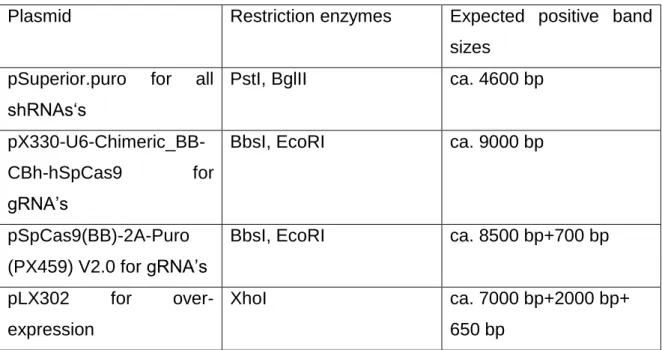
4.3.13 Restriction digestion ... 50
4.3.14 Midi-preparation and sequencing ... 52
4.4 Cell culture... 52
4.4.1 Cell line propagation ... 52
4.4.2 Transfection ... 55
4.4.3 Conventional liposome/lipid-mediated transfection ... 56
4.4.4 Lentivirus-mediated gene delivery (transduction) ... 56
4. 5 Cell proliferation and invasion assays ... 58
4.5.1 MTT cell proliferation assay ... 58
4.5.2 Transwell cell migration assay... 59
4.5.3 Scratch/wound healing assay ... 60
4.5.4 Soft Agar Colony Formation Assay ... 60
4.6 Western Blot ... 61
4.6.1 Western Blot and SDS-PAGE sample preparation ... 61
4.6.2 Western Blot ... 64
4.7 Immunofluorescence ... 65
4.8 Real-time PCR ... 66
4.8.1 RNA-isolation ... 66
4.8.2 Reverse transcription ... 67
4.8.3 Real-time PCR... 68
4.9 Animal treatment and xenotransplantation ... 70
4.10 Statistical analysis... 71
V. Results ... 72
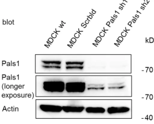
5.1 Pals1 knockdown affects TJ formation ... 72
5.2 Loss of Pals1 results in altered expression of polarity proteins ... 76
5.3 Lack of Pals1 expression leads to enhanced cell motility ... 78
5.4 Pals1 downregulation leads to overexpression of EMT markers and affects Hippo signaling pathway ... 79
5.5 Pals1 knockdown affects cell polarity in HCT116 colon cancer cell line ... 82
5.6 Pals1-deficient cells show augmented cell proliferation ... 87
5.7 Lack of Pals1 expression leads to enhanced cell motility in epithelial and colon cancer cell lines ... 89
5.8 Deregulation of Pals1 expression leads to cellular transformation and augmented tumorigenicity ... 91
5.9 Pals1 knockout in HCT116 cell line enhances cell motility and metastases formation in mice ... 93
5.10 TGF-β pathway is implicated in Pals1-dependend cell motility ... 95
VI. Discussion ... 97
VII. Conclusion and Outlook ... 105
VIII. Supplements ... 106
8.1. List of figures ... 106
8.2. List of tables ... 106
IX. References ... 109
Acknowledgments
First and foremost, I would like to thank Prof. Dr. Dr. Michael Krahn for giving me the opportunity to do my Ph.D. within his research group. Thank you for continuous support and for being my first referee.
I owe my deepest gratitude to PD Dr. Anja K. Wege, who provided me an opportunity to join their team and who was supporting me during all animal studies. Without this collaboration, it would not be possible to conduct this research.
I would like to thank Prof. Dr. Klein and Prof. Dr. Sprenger for being my Ph.D. mentors, for your support, patience and the intense discussions which gave me exciting insights into the project. Thank you for your encouragement and the extra time spent. Besides my advisors and mentors, I would like to thank the rest of my thesis committee: Prof. Dr. Witzgall and Prof. Dr. Längst.
A heartfelt thank you to all my colleagues from the Institute of Molecular and Cellular Anatomy at the University of Regensburg, for your great support, especially V.Menath, C. Maassen, L.Osten and O.Maier. Sincere thanks to all the members of Pathology Department at Uniklinik Regensburg, especially to Rudolf Jung, for helping with IHC studies. Many thanks to all the Ph.D. students within the research group not only for providing direct back-up and advice but also for a very friendly atmosphere. In particular, I would like to acknowledge S.
Feicht, R. Sun, B. Schwertner, L.Kullman for their friendship, excellent insights into the work and for sharing their comprehensive knowledge. I would also like to thank you to all undergraduate students I was supervising during my Ph.D., for your hard work and for fun activities we’ve done together.
I wish to express my sincere thanks to Dr. O. Stelmashenko and to R.Sun for comments and criticisms from reading various drafts and parts of my thesis.
Dear Alex, mom, dad and all my family members, thank you for the opportunity to complete my Ph.D., your unceasing encouragement and support.
Thank you to all my friends I got to know during my Ph.D., for helping me focus
on the research while having a great time! Thank you to every mentioned person
as well as anyone I might have forgotten.
Abstract
Epithelial cell polarity is of vital importance for the organization and function of epithelial tissues and is primarily maintained by three protein complexes, the Crumbs complex, the Par complex and the Scribble complex.
Most of the polarity proteins within these complexes are highly conserved and
play pivotal roles in embryonic development, cell-cell adhesion and cell
migration. Recent studies have demonstrated that deregulation of epithelial
polarity is a hallmark of tumor progression, in particular during the metastatic
process. This study was conducted to examine the role of the tight junction
protein Pals1 in the maintenance of cell polarity, and cancer. RNA interference
and CRISPR/Cas9 gene deletion approaches were used to downregulate or
deplete Pals1 in MDCKII, HCT116 and DLD1 cell lines. Reduction in Pals1
resulted in atypical expression levels of polarity proteins and defects in Hippo
pathway regulation. Moreover, Pals1 loss caused E-cadherin reduction and
enhanced cell migration. Pals1 deficient cells exhibited typical markers, inferring
epithelial-to-mesenchymal transition. Further in vivo xenograft experiments
revealed a function of Pals1 in cancer progression, as tumors derived from
Pals1-deficient cells showed increased growth and more extensive liver and lung
metastases. Taken together, these findings support a close link between
epithelial cell polarity and tumorigenesis, and suggest the existence of a novel
Pals1-mediated mechanism of tumor suppression. Thus, the pathophysiological
consequences of Pals1 alteration must be investigated further, and used to
develop new therapeutic strategies against this devastating disease.
I. Abbreviations
Abbreviation Name
AJ Adherens junctions
AJC Apical junctional complex
AMOT Angiomotin
ANOVA Analysis of variances
AP Alkaline phosphatase
APC Adenomatous polyposis coli aPKC Atypical protein kinase C
APS Ammonium persulfate
ATP Adenosine triphosphate
Birc2 Baculoviral inhibitors of apoptosis repeat-containing 2
BSA Bovine serum albumin
C.elegans Caenorhabditis elegans
Cas9 CRISPR-associated protein 9 Cdc42 Cell division control protein 42
Crb Crumbs
CRIB Cdc42/Rac interactive binding
CRISPR Clustered regularly interspaced short palindromic repeats CTGF Connective tissue growth factor
Cyr61 Cysteine-rich angiogenic inducer 61 DAPI 4',6-diamidino-2-phenylindole
dd
H
2O Double distilled water
DLD1 Colorectal adenocarcinoma cell line, isolated by D.L.Dexter
DLG Disks large
DMEM Dulbecco’s Modified of Eagle’s Medium DMSO Dimethyl sulfoxide
DNA Deoxyribonucleic acid
dNTP Deoxyribonucleotide triphosphate
DTT Dithiothreitol
E.coli Escherichia coli
ECM Extracellular matrix
EDTA Ethylenediaminetetraacetic acid EMT Epithelial-to-mesenchymal transition
EtBr Ethidium bromide
FCS Fetal calf serum
FERM 4.1, Ezrin, Radixin, Moesin
gRNA Guide RNA
GSK Glycogen synthase kinase GTP Guanosine-5'-triphosphate
Guk Guanylate kinase
HCT116 Human colon carcinoma tissue 116 HEK Human embryonic kidney cell line
HRP Horseradish peroxidase
IF Immunofluorescence
IHC Immunohistochemistry
IKK Inhibitors of κB kinase
kb Kilobases
KD Knockdown
kDa Kilodalton, 1000 unified atomic mass units
KO Knockout
LATS Large tumor suppressor
LB Lysogeny broth
LD Loading dye
Lgl Lethal giant larvae
LSM Laser scanning microscope MAPK Mitogen-activated protein kinase
MDCK Madin-Darby Canine Kidney Epithelial Cells MET Mesenchymal-to-epithelial transition
MTT 3-(4,5-dimethylthiazol-2-yl)-2,5-diphenyltetrazolium bromide NF-κB Nuclear factor kappa-light-chain-enhancer of activated B cells
NSG Nod Scid gamma
OD Optical density
PAGE Polyacrylamide gel electrophoresis Pai1 Plasminogen activator inhibitor 1 Pals1 Protein associated with Lin seven 1 par Partitioning defective
Patj Pals1-associated tight junction protein PBS Phosphate buffered saline
PBTw Phosphate buffered saline with tween
PDZ Postsynaptic density protein 95, Dlg, ZO-1
PEI Polyethyleneimine
PFA Paraformaldehyde
PMSF Phenylmethylsulfonylfluoride
PNK Polynucleotide kinase
PTEN Phosphatase and tensin homolog deleted on chromosome 10
puro Puromycine
RNA Ribonucleic acid
rpm Revolutions per minute
RPMI Roswell Park Memorial Institute medium
SAV1 Salvador family WW domain-containing protein 1
Scrbld Scrambled
SD Standard deviation
SDS Sodium dodecyl sulfate SEM Standard error of the mean
SH3 Src Homolgy3
shRNA Short hairpin RNA
SMA Smooth muscle actin
Smad Sma and Mad related proteins
TAO Thousand-and-one amino acid protein kinases TAZ Transcriptional coactivator with PDZ-binding motif TBS Tris-buffered saline
TEAD TEA domain
TEMED Tetramethylethylenediamine
TGF Transforming growth factor
TIAM1 T-cell lymphoma invasion and metastasis 1
TJ Tight junctions
TNF Tumor necrosis factor
TRAF TNF receptor-associated factor
V Volt
WB Western Blot
wt Wild type
YAP Yes-associated protein YTA Yeast extract tryptone agar
ZEB1 Zinc finger E-box-binding homeobox 1
ZO Zonula occludens
II. Introduction
2.1 Epithelial tissue and cell polarity
Mammalian anatomy and physiology are one of the most sophisticated areas of biology and medicine. Remarkably, in humans, there are only four tissue types identified: epithelial, connective, muscular, and nervous tissues. Cell similarity and their related function give tissue identity, although different types of tissues can be found in distinct organs (Mescher, Junqueira 2010). Additionally, each basic tissue has a unique extracellular matrix (ECM) - a highly dynamic structure, consisting of components as water, proteins and polysaccharides.
ECM continuously undergoes structural remodeling, provides a crucial scaffold for all the cellular constituents and participates in cell-cell interactions, tissue homeostasis, morphogenesis and biochemical signaling (Frantz et al. 2010).
Whereas nervous tissue is composed of "finger-like" long cells as axons and dendrites, epithelial tissue is mostly made up of polyhedral cells. They form different layers with strong cell-cell contacts, and line most internal body cavities.
Depending on the number of layers and cell shape in the upper layers, epithelial tissues are divided into several types. Simple epithelium is presented by one layer of cells, while stratified epithelia are considered to have more than two cell lining layers. Pseudostratified epithelium belongs to the simple epithelia, as the cell nuclei are lying at different levels and only giving an appearance of stratification. Further separation of epithelia into subcategories is based on differences in cell shape: flat, cubic or elongated morphology, which dictates the wide variety of epithelia functions. As the epithelial tissue forms boundaries between environments, its main function is a physical and semipermeable barrier, regulating absorption and secretion of substances such as water, nutrients and ions (Betts 2013; Mescher, Junqueira 2010).
All cells in epithelial tissues exhibit some differences in shape and structure due to differences in their function, with two types of cell polarization:
apicobasal polarity and planar cell polarity. This refers to morphological and
functional asymmetry within an epithelial cell or in the plane of tissue and is
coordinates cell distribution and behavior across the tissue plane, correlates to collective cell movements during body axes elongation, embryonic development, hair and bristle growth in Drosophila and mammals. A continuously changing environment and polarity information from neighboring cells requires stringent coordination and dynamic changes including cell rearrangement, cell division and cell-shape changes (Devenport 2014; Zallen 2007). Apico-basal cell polarity is established by two different cell-cell junctions - tight junctions (TJ) and adherens junctions (AJ), dividing the cell into two biochemically and structurally distinct domains. Whereas an apical membrane faces the outside surface of the body or the lumen of internal cavities, the basolateral membrane is oriented towards the basal lamina and adjacent cells (Khursheed, Bashyam 2014).
Despite differences in the distribution of cell-cell junctions, many studies in Drosophila, C.elegans and mammals illustrate evolutionary conservation of core mechanisms determining cell polarity (Gibson, Perrimon 2003). Three major polarity complexes have been identified to date and they serve as the core proteins maintaining apicobasal identity in epithelial cells. First, the Crumbs (Crb) complex, consisting of the transmembrane protein Crb, the adaptor protein associated with Lin seven 1 (Pals1) and Pals1-associated tight junction protein (Patj). Together with the partitioning defective (Par) - atypical protein kinase C (aPKC) complex, with the adaptor proteins Par3 and Par6 they activate the kinase aPKC. Crb and Par-aPKC complexes are required to establish the apical plasma membrane. In contrast, the Scribble homolog (Scrib)–Lethal giant larvae homolog (Lgl)–Discs-large homolog (Dlg) complex defines the basolateral plasma domain (Martin-Belmonte, Perez-Moreno 2012).
Par-aPKC complex serves as a core complex maintaining cell polarity and
together with Crb complex it is implicated in TJ formation. TJ help to keep
epithelial cell polarity by forming a paracellular diffusion barrier between the
apical and basolateral cell domains and hence prevent the movement of solutes
and lipids across the cell layer (Zihni et al. 2014; Hurd et al. 2003). To date, TJ
represent the greatest number of known transmembrane (occludin and claudin
families) and adaptor zonula occludens (ZO) proteins (ZO-1, ZO-2 and ZO-3) as
well as signaling proteins, including protein kinases, phosphatases, guanosine-
5'-triphosphate (GTP)-binding proteins, transcriptional and post-transcriptional
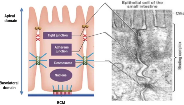
regulators. Together with more basally formed adherens junctions and desmosomes, they establish the apical junctional complex (AJC) and maintain polarity and cell-cell adhesion. Figure 2.1 is a simplified representation of the organization of epithelial tissue.
Figure 2.1: Epithelial cell-cell contacts and adhesion
Organization and maintenance of cell-cell adhesion in epithelial tissues. (Retrieved and adapted from comprehensive system for life science education, University of Tokyo, http://csls-text3.c.u- tokyo.ac.jp).
The AJ regulate epithelial paracellular permeability and are linked to the
actin and microtubule cytoskeletons. The main transmembrane protein of AJ in
epithelial cells is E-cadherin. It belongs to the family of Ca
2+-dependent
glycoproteins and promotes tight cell-cell contacts by homophilic interaction with
the cadherin molecules of the neighboring cells. E-cadherin consists of five
extracellular cadherin repeat domains, one transmembrane domain and a highly
conserved intracellular tail. Several phosphorylation sites in the intracellular
domain promote binding affinity to p120-catenin (Reynolds, Roczniak-Ferguson
2004) and β-catenin (Mehta et al. 2015), further interaction with α-catenin and
thus actin cytoskeleton.
2.1.1 The Par complex
The par-titioning-defective or Par proteins were first described in Caenorhabditis elegans and afterwards were found in almost all living organisms, all the way up to mammals. Interestingly, not only the components of Par complex are conserved - they also show a remarkable conservation of their localization in epithelial cells from worms to flies and humans. Par proteins comprise a complex cell polarity network and are involved in development, asymmetric cell division, spatial restriction of the cytoskeleton, formation of cells’
polarity axes as well as asymmetric cell division (Aranda et al. 2008). Two proteins of the Par family, Par3 and Par6, together with aPKC and small GTPase cell division control protein 42 (Cdc42) assemble in the so-called Par complex.
Par6 serves as a cornerstone protein and is likely to promote an interplay and coordination of all polarity complexes (Pieczynski, Margolis 2011a). There are three different Par6 isoforms, identified to date in mammals: Par6α, Par6β and Par6γ. They are all around 40 kDa in size and share structural organization;
however, they have different patterns of tissue and subcellular localization. Par6 is a scaffolding protein and contains a single postsynaptic-density protein 95-Dlg- ZO-1 (PDZ) domain, responsible for protein-protein interactions usually with other PDZ domains or with specific carboxy-terminal motifs, and contributes to direct interaction with Par3. Adjacent to PDZ, the CRIB (Cdc42/Rac interactive binding) domain binds the Rho GTPase Cdc42 in a GTP-dependent manner, causing conformational changes in Par6 and increasing the affinity of the Par6 PDZ for its carboxy-terminal ligand by ∼13-fold (Peterson et al. 2004).
Two Par3 genes have been identified so far in mammals – Par3A and Par3B. Par3A encodes three protein isoforms (180 kDa, 150 kDa, 100 kDa), whereas Par3B encodes only one around 140 kDa. Par3A and Par3B are both shown to localize apically to TJ at cell-cell contact regions in mammalian cells.
All three Par3A proteins are characterized by three PDZ domains and a C-
terminal region that interacts with the kinase domain of aPKC. Par3B is not fully
described to date, however, in contrast to Par3A, it does not seem to bind to
aPKC. Par3A is broadly expressed in various tissues and can dimerize through
its N-terminal tail. In the early stages of polarization and junction development,
Par3A is recruited to the junctional adhesion molecule, thus further serving as an
anchor. It also initiates Par6 and aPKC transition to TJ at later developmental stages (Assemat et al. 2008). The functionality of Par3 is highly dependent on its phosphorylation state, regulated by aPKC.
Figure 2.2: Protein complexes maintaining cell polarity
Although many proteins participate in apicobasal polarity, three major polarity complexes serve as the core proteins maintaining apicobasal cell polarity. These complexes are the apical Crb (includes Crb, Pals1 and Patj proteins), Par (includes Par6, Par3 and aPKC proteins) and the basolateral Scribble (includes Scribble, Dlg and Lgl proteins) complexes. All of these proteins are in a tight interrelation with each other and are highly conserved through evolution (Pieczynski, Margolis 2011b).
Par3 also shows Par6/aPKC-independent activity, as Par3 can interact with some of the adhesion molecules through its PDZ domain, including p75 and
Crb Pals1
1
Patj
Par6
Par3 aPKC
ar3
Lgl
Dlg
Scribble
Crumbs Complex “Apical”
Par Complex “Mobile”
Scribble Complex “Lateral”
Direct
Phosphorylation Genetic
Interactions
deleted on chromosome 10 (PTEN) might participate in phosphatidylinositide 3 kinase (PI3K)-Akt-mTOR signaling (Chen, Zhang 2013).
Two different aPKC isoforms (aPKCλ/ι and aPKCζ) both play a pivotal role in Par complex function. They share a similarity in their unique protein structure, having a PB1 domain in the N-terminal, which is required for aPKC-Par6 interaction. The C-terminal catalytic domain is known to promote phosphorylation of Par3 by aPKC (Plant et al. 2003; Chen, Zhang 2013). aPKC-depended phosphorylation of other polarity regulators outside the Par complex, like Crb3 or Lgl proteins, provides the cross talk among all polarity determinants and is crucial in apical domain establishment (Aranda et al. 2008). Figure 2.2 represents the interaction and signaling redundancy of all polarity complexes.
Apart from controlling different aspects of cell polarity, Par proteins are also implicated in migratory processes (Crespo et al. 2014), asymmetric cell division (Betschinger et al. 2003) and microtubule organization, particularly in the positioning and orientation of centrosomes and spindle formation. In neurons, Par6-aPKC was shown to co-localize together with dynein to the centrosome and thus might play a role in dynein-based transport and glial-guided migration (Solecki et al. 2004). In addition, Par complex coordinates actomyosin fiber activity, thus is involved in polarized cell migration (Aranda et al. 2008).
Despite recent progress in the field, the biochemical relationship and crosstalk between Par6, Par3, aPKC and Cdc42 is still not fully understood and the functional redundancy of different isoforms makes this network even more complicated.
2.1.2 The Crb complex
The Crb complex is highly conserved in both invertebrates and
vertebrates, consisting of four scaffolding proteins (Crb, Pals1, Patj and Lin-7)
with numerous protein-protein interaction domains. Decades of research have
showed Crb to be the key regulator of epithelial integrity. Fly embryos lacking
Crb were characterized by disruption of apicobasal polarity and zonula
adherence defects (Tepass, Knust 1993), whereas the overexpression of Crb
leads to an enormous expansion of the apical domain with simultaneous
contraction of the basolateral plasma domain (Wodarz et al. 1995). Crb is a
transmembrane protein with a highly conserved intracellular part, containing PDZ- and 4.1/ezrin/radixin/moesin (FERM) binding domains (Figure 2.3). In flies, through its PDZ-binding motif, the Crb protein can recruit other members of the Crb complex, like Stardust (Pals1 homolog in Drosophila), Par6 and aPKC to the plasma membrane. The FERM domain is required to recruit Expanded protein to the apical membrane, linking Crb complex to Hippo pathway (Pocha, Knust 2013). In mammals, Crb is encoded by three Crb genes (Crb1, Crb2 and Crb3).
Crb1 and Crb2 are found to be highly expressed in human retina and brain. Crb3 is the primary form of Crb and is highly expressed in mammalian epithelial tissues (Slavotinek 2016). Crb3 maintains cell polarity and is crucial for correct positioning of the Crb complex at the apical membrane (Makarova et al. 2003).
Figure 2.3: Structure of the Crb complex in Drosophila (Bulgakova, Knust 2009)
Proposed model, depicting the structure and organization of Drosophila Crb complex, comprising of Crb, Sdt (Pals1 homolog in flies), PATJ and Lin-7 proteins.
Pals1 (also known as membrane-associated palmitoylated protein 5
(MPP5) is a member of a guanylate kinase family protein consisting of two Lin-2
and Lin-7 (L27) domains (L27N and L27C), a PDZ domain, an SH3 (Src
Homolgy3) domain, and a Guk (Guanylate kinase) domain. The PDZ domain of
Pals1 binds Crb3, meanwhile Pals1 via its L27 domain interacts with Pals1-
associated tight junction protein Patj - another member of Crb complex. Hence,
Pals1 serves as a membrane-associated protein scaffold, linking and mediating
the indirect interaction of Crb3 and Patj. For instance, Perrimon et al. identified a
mechanism in vertebrates by which Crb might act through Pals1 to recruit the Par3-Baz complex to the apical cell domain (Hurd et al. 2003). Furthermore, mammalian Pals1 is shown to maintain the delivery of E-cadherin to the cell surface and is not only sufficient for tight junction development but also is shown to be necessary for adherens junction maintenance (Wang et al. 2007).
Interestingly, Pals1 was also shown to be expressed in T lymphocytes and plays a role in nuclear factor kappa-light-chain-enhancer of activated B cells (NFκB) signaling (Carvalho et al. 2011a).
In contrast to Drosophila (Figure 2.3), mammalian Patj contains ten PDZ domains and localizes to TJ. According to Shin et al., MDCK cells lacking Patj had delayed tight junction formation and cell polarization defects, suggesting that Patj plays and important role in Crb complex assembly (Shin et al. 2005).
Moreover, Patj has been demonstrated to regulate Pals1 accumulation at TJ, and Crb3 localization and trafficking (Michel et al. 2005). Furthermore, Patj regulates the positioning of aPKC and Par3 to the leading edge during directional cell migration (Shin et al. 2007). In flies, Patj regulates AJ stability, by direct binding to the myosin-binding subunit, subsequent dephosphorylation and inactivation of myosin (Sen et al. 2012). In addition, Patj has a wide range of interaction partners, including claudin-1, ZO-1 (Knust, Bossinger 2002), ZO-3 (Roh et al. 2002a), junctional adhesion molecule 1, nectins (Adachi et al. 2009) and angiomotin (AMOT) proteins (Whiteman et al. 2014). Thus, Patj is functionally significant for epithelial polarization and plays a key role in establishing the apical plasma domain.
2.1.3 The Scribble complex
The Scribble protein was first identified in flies as a crucial regulator of
morphogenesis and septate junction formation. In combination with the lethal
giant larvae Lgl and disc large Dlg proteins, it forms the Scribble complex and
creates the basolateral determinant of a polarized cell. These three proteins are
known to be tumor suppressors and show multiple interactions via their PDZ-
domain with polarity determinants in Par and Crb complexes. Scribble is shown
to regulate epithelial cell adhesion and migration, whereas Lgl is likely more
involved in cell-cell cross-talk and promotes antagonistic interactions with the
aPKC-Par3-Par6 complex. Phosphorylation of Lgl by aPKC depends on its binding to Par6 and aPKC and leads to a release of Lgl from the plasma membrane, and thus helps to control cell shape formation during development.
Interestingly, besides Dlg function in junctional development which is necessary for its targeting to the cytoskeleton, Dlg takes part in proliferation control. Taken together, the antagonistic crosstalk between the basal Scribble and apical Par and Crb polarity complexes is critical for the establishment of correct apicobasal polarity (Su et al. 2012; Woods et al. 1996; Humbert et al. 2008)
2.2 Disruption of cell polarity, epithelial-to-mesenchymal transition and cancer
Epithelial cell polarity is fundamental for tissue organization and allows cells to respond appropriately to signals from the surrounding microenvironment.
Hence, a loss of cell polarity might delay or even abort cell responses to inhibitory signals and consequently the cell may evade differentiation, senescence or apoptosis. Decades of research have demonstrated that a wide number of polarity proteins are either tumor suppressors or proto-oncoproteins.
For instance, Crb3 may act as a tumor suppressor, as downregulation of Crb3 promotes disruption of cell-cell junctions and increased metastatic potential. Loss of Par3, as well as overexpression of Par6α and Par6β proteins, was shown to cause hyperproliferation and enhanced invasiveness in breast cancer cell lines and primary tumor tissues (McCaffrey et al. 2012). In addition, Par6γ acts as a tumor-suppressor by repressing Akt pathway (Marques et al. 2016). Another member of Par complex, aPKCζ, is crucial in pancreatic and hepatocellular tissues, as its overexpression correlates with invasiveness and enhanced cell proliferation. In addition, all three members of the Scribble complex are known to be tumor suppressors, maintaining cell polarity and inhibiting uncontrolled cell proliferation (Martin-Belmonte, Perez-Moreno 2012).
Polarity proteins might play a crucial role in the metastatic progression of
cancer, as they are known to mediate front-rear polarization in epithelial cell
migration during development and tissue repair. Cdc42 is an important regulator
of directed cell migration. Cdc42 activation of the Par-aPKC complex is shown to
be important for actin and microtubule network repolarization along the migration
axis during spatial migration process (Etienne-Manneville et al. 2005). In addition, Par6 and aPKC promote E3 ubiquitin ligase-mediated degradation of RhoA and thus control disorganization of the actin cytoskeleton (Wang et al.
2003). Meanwhile, Par3 is known to control the leading edge of migration by activating Rac exchange factor, TIAM1 (T-cell lymphoma invasion and metastasis 1) (Pegtel et al. 2007). In intestinal epithelial cells, TIAM1 suppresses tumor growth and invasion by regulating Wnt and Hippo signaling (Diamantopoulou et al. 2017). Furthermore, numerous studies have underlined the physiological importance of proper E-cadherin regulation not only in cell-cell adhesion but predominantly in metastatic progression. E-cadherin deregulation and depletion cause cellular dedifferentiation and promotes invasiveness in human cancers (Rodriguez et al. 2012; Onder et al. 2008; Schneider et al. 2014;
Singhai et al. 2011).
During metastasis, cancer spreads from its original place through the
lymph or blood stream to other organs, where it gives rise to secondary tumors
at the distant site. Epithelial-to-mesenchymal transition (EMT) –
transdifferentiation of epithelial cells into motile fibroblast-like mesenchymal cells
has been shown to be decisive for tumor cell dissemination in epithelium-derived
carcinogenesis. EMT is first and foremost essential in embryonic development,
as during EMT cell loses junctional and apicobasal polarity, undergoes a change
in the cytoskeleton and signaling programs which enables plasticity and distinct
cell development during embryogenesis (Lamouille et al. 2014). Moreover, in
adults, EMT also plays a crucial role, promoting wound healing and tissue
regeneration and is constituted to the second type of EMT. EMT type III is
involved in malignant cancer progression (Kalluri 2009). Once the migratory
cancer cell in type III EMT anchors at the distant epithelial tissue, it undergoes a
mesenchymal-to-epithelial transition (MET), the reverse process to EMT, and is
able to form secondary tumors, characterized by an epithelial phenotype
(Figure 2.4).
Figure 2.4: Epithelial-to-mesenchymal transition and reverse process of mesenchymal- to-epithelial transition (Kolbl et al. 2016)
EMT is a process by which the cell loses its epithelial phenotype, adopts migratory mesenchymal characteristics, enters the blood stream and might migrate to distinct body parts, where through reverse metamorphosis MET is able to give rise to remote metastases.
EMT and epithelial polarity are closely connected, as during EMT initiation epithelial cell-cell junctions are reorganized and the expression pattern of polarity proteins is drastically changed. Upon EMT, RhoA degradation by ubiquitin ligase Smurf1 leads to disassembly of tight junctions in epithelial cells, and is characterized by subsequently reduced claudin-1 and claudin-7 expression (Tabaries, Siegel 2016; Moustakas, Heldin 2007), ZO-1 downregulation and further internalization. Furthermore, disruption of adherens junctions, desmosomes and cadherin switch comprise an integral part of the EMT (Knights et al. 2012). E-cadherin is cleaved at the plasma membrane by metalloproteases (i.e. MMP7 and ADAM10) and replaced by N-cadherin. The shredded C-terminal of E-cadherin translocates into the nucleus resulting in dissociation of E-cadherin – β-catenin – p120 catenin complex and AJ disruption (Grabowska, Day 2012).
Rho GTPases also counterpart to EMT, as RhoA promotes increased cell
contractility and actin stress fiber formation, whereas Cdc42 plays a role in lamellipodia and filopodia organization (Lamouille et al. 2014).
Recent evidence suggests that the transforming growth factor (TGF)-β pathway can trigger an EMT initiation (Katsuno et al. 2013). Indeed, TGF-β was shown to initiate loss of cell polarity due to Par complex disassembly during EMT (Ozdamar et al. 2005). Although TGF-β is mostly known as a proinflammatory cytokine, its excrescent secretion leads to EMT activation via two interrelated pathways: canonical and non-canonical TGF-β pathways (Figure 2.5).
Figure 2.5: Smad-dependent (canonical) and -independent (non-canonical) TGF-β pathways (Papageorgis, Stylianopoulos 2015)
TGF-β acts through the downstream mediators to exert a vast range of biological activities during inflammation, EMT and cancer progression.
TGF-β binding to its receptors leads to phosphorylation of Smad2/3, activating the canonical Smad-dependent pathway. It causes gene reprogramming by directly activating the key EMT transcription factors including Snail, Twist and zinc-finger E-box-binding (ZEB) proteins.
Snail1, the mostly studied member of Snail family, is characterized by the SNAG box in its N-terminus, required for transcriptional repression of CDH1 cadherin gene by recruitment of histone deacetylases or C-terminal binding proteins during cancer progression. At the C-terminus Snail1 contains four zinc finger domains and a nuclear localization signal motif, required for deoxyribonucleic acid (DNA) binding and nuclear translocation, respectively.
ZEB1 shares structural similarity with Snail1, however preliminary Snail induction is required to trigger ZEB1 expression, as Snail1 also contributes to NFκB nuclear import. Twist belongs to the basic helix-loop-helix (bHLH) family of transcription factors and is also able to repress the CDH1 gene, although indirectly via chromatin remodeling complex recruitment (Diaz et al. 2014).
Dynamic co-interaction of Snail, Zeb and Twist proteins promote the downregulation of genes responsible for tight junction and adherens junction polarity protein processing and simultaneously enhance fibronectin, vimentin, N- cadherin and matrix metalloproteinases expression, considered to be the hallmarks of a mesenchymal phenotype (Peinado et al. 2007).
Additionally, TGF-β induces non-Smad signaling pathways, leading to activation of Rho GTPases, MAP kinase (MAPK) pathways and the PI3-Akt- mTOR pathway (Saitoh 2015; Yilmaz, Christofori 2009; Nistico et al. 2012;
Lamouille et al. 2014; Derynck et al. 2014).
In the non-canonical TGF-β signaling pathway, TGF-β has been
demonstrated to activate tumor necrosis factor (TNF)-receptor-associated factor
6 (TRAF6). In turn, TRAF6 activates mitogen-activated protein kinases, including
extracellular signal-related kinase (ERK) 1/2, p38 mitogen-activated kinase (p38)
and c-Jun N-terminal kinase pathways. TRAF6 also activates the inhibitors of the
κB (IκB) kinase (IKK) complex. IκBs phosphorylation results in their
polyubiquitination and subsequent degradation, allowing NF-κB to translocate
into the nucleus and activate the transcription of many κB-dependent genes
implicated in the regulation of apoptosis and EMT (Mu et al. 2012; Wu, Zhou 2010).
Changes and deregulation in protein translation process are also considered to participate in cancer progression. The PI3K-Akt-mTOR pathway is known to regulate protein biosynthesis and cell size in mammals. TGF-β induced PI3K and mTOR signaling activity are demonstrated to be highly enhanced in a broad number of cancers, including breast and colorectal cancer. Activated Akt can phosphorylate many target proteins, most notably glycogen synthase kinase 3β (GSK-3β). In turn, GSK-3β stimulates the transcription of Snail and thus acts indirectly as a repressor of E-cadherin expression and EMT induction (Yu, Cui 2016; Bachelder et al. 2005). Taken together, cell polarity is essential for the proper functioning of eukaryotic cells, relies on a strong interplay between polarity proteins and a wide variety of signaling pathways and is crucial to regulate tumor progression and metastatic processes.
2.3 Hippo signaling pathway and its link to cell polarity
The Hippo pathway (Figure 2.6) was first described in studies of Drosophila that examined genes involved in tissue growth (Justice et al. 1995;
Xu et al. 1995; Tapon et al. 2002). For some time, the Hippo pathway was thought to be evolutionarily conserved from flies to mammals, however, in mammals the Hippo signaling network seems to be more extensive and complicated. To date, this signaling pathway is known to control organ size by inhibiting cell proliferation and promoting apoptosis, and to play an important role in tissue repair and regeneration. Deregulation of the Hippo pathway is closely connected with cancer progression and metastasis, as mutations in its core proteins have been shown to result in overgrowth phenotype (Yu et al. 2015).
The core of the Hippo pathway includes several kinase cascades that
work redundantly to regulate the activity and localization of the downstream
effectors Yes-associated protein (YAP) and transcriptional coactivator with a
PDZ-binding motif (TAZ) (Figure 2.6). Initially, the cascade begins with MST
protein activation via its phosphorylation by thousand-and-one amino acid (TAO)
protein kinases.
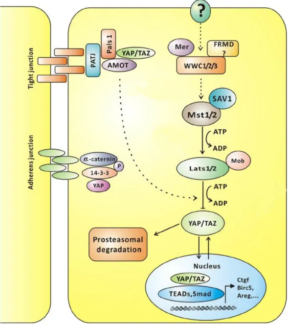
Figure 2.6: Hippo pathway signaling in mammals (adapted from Hao et al. 2014)
The Hippo pathway is known to regulate cell size and tissue growth via several kinase cascades, the common goal of which is the regulation of YAP/TAZ localization via physical interaction with AMOT and other proteins. These kinases promote YAP/TAZ phosphorylation and nuclear sequestration, thus controlling and regulating gene transcription.
Phosphorylated MSTs together with the adaptor protein Salvador family WW domain-containing protein (SAV)1/WW45 are able to phosphorylate and activate large tumor suppressor (LATS) proteins, especially LATS1 and LATS2, promoting their autophosphorylation (Meng et al. 2016; Ehmer, Sage 2016).
Afterwards, the LATS protein subset is believed to phosphorylate and inactivate
YAP and TAZ. Phosphorylation of YAP and TAZ leads to their binding with 14-3-
ubiquitin-depended degradation. When the Hippo pathway is off, non- phosphorylated YAP and TAZ translocate into the nucleus, bind the TEA domain (TEAD) transcription factor family and prompt expression of a wide range of genes that are involved in cell proliferation and migration (Yu et al. 2015; Meng et al. 2016). Connective tissue growth factor (CTGF), Cysteine-rich angiogenic inducer 61 (Cyr61) and baculoviral inhibitors of apoptosis repeat-containing 2 (Birc2) have been shown to be direct targets induced by YAP-dependent TEAD activation, and to play a role in proliferation and anchorage-independent growth (Liu et al. 2015; Zhao et al. 2010b). Moreover, YAP and TAZ are known to interact with Smad2 and Smad3, connecting another transcriptional target of YAP plasminogen activator inhibitor 1 (Pai1) with activation of TGF-β signaling during tissue fibrosis and cancer (Zhao et al. 2010a).
Additionally, Hippo pathway activity is regulated by upstream signals. In Drosophila, the Mer/Ex/Kibra complex acts as an apical tumor suppressor complex, recruits Hpo (MST1/2 flies homolog) to the plasma membrane and enhances its phosphorylation, which could also influence YAP localization (Mo et al. 2014). In mammals, Kibra was shown to interact with aPKC and angiomotin family proteins (Zhang et al. 2014). AMOT was first described as a protein promoting endothelial cell migration and angiogenesis. However, two different isoforms of AMOT exhibit different functions, as only p80 was shown to promote cell migration and is not able to bind to YAP (Yi et al. 2013). Binding p130 AMOT to YAP family proteins is crucial for YAP and TAZ phosphorylation and their right localization to the tight junction. Furthermore, AMOT inhibits YAP-induced transformation and loss of cell contact inhibition in the epithelial cells.
Remarkably, AMOT is shown to interact through its C-terminal PDZ
binding motif with Pals1 and Patj, maintain their proper localization and thus is
important for the long-term TJ integrity in epithelial cells (Wells et al. 2006). The
phosphorylation state of AMOT is also highly important and drives the
association with Pals1/Patj at the plasma membrane, whereas the non-
phosphorylated form concomitantly induces YAP and TEAD association,
enhances its transcriptional activation and promotes uncontrolled cell
proliferation (Moleirinho et al. 2017). Interestingly, in MCF10A human breast
epithelial cell line Pals1 and Patj were determined to co-precipitate with YAP.
This might involve AMOT proteins as well, as they were also present in pull- downs, verified by Western Blot (Zhao et al. 2011; Varelas et al. 2010). In addition, AMOT is able to form a complex with Patj and by controlling GTPase activity regulates migration capacity of endothelial cells (Ernkvist et al. 2009).
Recent data also identified AMOT as a novel binding partner of cadherin -11, a marker of EMT in prostate cancer cells. Together with cadherin-11, AMOT could influence mediated migration of these cells (Ortiz et al. 2014)
In addition, Crb3 is considered to interact with AMOT and Merlin to regulate YAP- and TAZ-dependent transcription and MAP kinase signaling, hence Crb3 is linked to cell-density-dependent regulation of proliferation and cancer (Varelas et al. 2010; Zihni et al. 2014).
Taken together, precise control of cell-cell adhesion in combination with
decisive physiological function and molecular interplay of polarity determinants
with Hippo and other signaling pathways is crucial in development as well as in
cancer progression.
III. Aim of the study
Every year the number of cancer suffering patients increases drastically, reflecting the biggest medical worldwide problem to date. By 2020, there will be approximately 15 million new cancer patients diagnosed (Bray, Moller 2006). For this reason, the mechanisms triggering cancer, and the basic differences between a 'normal' cell and cancer cells must be intensely investigated.
Proper regulation and maintenance of cell polarity are required for development and homeostasis. There is strong evidence that the disruption of cell polarity and loss of cell-cell adhesion are implicated in cancer growth. Two fundamental roles of epithelial cells contribute to tumor development: the regulation of asymmetric cell division and the maintenance of the apical junction complex (Royer, Lu 2011).
Several cell polarity genes have been identified as tumor suppressors in flies and mammals, simultaneously playing a fundamental role in cell fate decisions and epithelial cell polarity. Mutations of Dlg, Lgl, Scribble, deregulation of Par3 and Crb3 cause excessive non-controlled epithelial growth, loss of apical-basal polarity, enhance EMT and metastatic progression (Wodarz 2000;
McCaffrey et al. 2012; Karp et al. 2008; Elsum et al. 2013).
However, little is known about the direct Crb3 interaction partner protein Pals1, its implication in establishment and maintenance of cell polarity as well as cell proliferation and migration. Hence, the present study was designed to assess the variety of Pals1 functions in epithelial cell polarity, with the goal of describing its possible implication in tumor progression. The role of Pals1 in both apicobasal polarity maintenance and junction formation was investigated.
Moreover, the function of Pals1 in the Hippo pathway and TGF-β signaling
remains perplexing. Since Pals1 is involved in E-cadherin trafficking, the next
objective of the study was to explore the possible influence of Pals1 on cell
migration and EMT. In addition, it was of interest to analyze the potential role of
Pals1 in tumor growth and metastasis development in vivo.
IV. Materials and Methods
4.1 Equipment
Table 1 Equipment
Equipment Label Manufacturer
Autoclave 5050 ELV Tuttnauer
Cell culture incubator CB 160 Binder
Centrifuge Multifuge 3 Heraeus
Centrifuge Microstar 12 VWR
Chemical balance BL 1500 S Sartorius
Chemiluminescence- system
Fusion FX 7 Vilber
Drying chamber Heraeus T 6200 Thermo Scientific Drying chamber Heraeus UT 6120 Thermo Scientific Electric pipetting aid Pipetboy Integra
Electrophoresis Module Eco-Mini Biometra Floor centrifuge Varifuge 3.0 RS Heraeus
Freezer -20 °C GNP 3376 Liebherr
Freezer -80 °C GFL Chest Freezer 6485 Novolab Heating block Thermomixer comfort Labnet Heating/shaking block Thermomixer R Eppendorf
Imager GelDoc XR+ Biorad
Incubator shaker Unitron Infors HT
Laminar Airflow Bench LaminAir HBB2472S Heraeus Laminar Flow Hood Herasafe HS18 Heraeus
Light microscope Eclipse TS100 Nikon
Laser scanning microscope
LSM 510 Meta Zeiss
Light/fluorescence microscop
Axiovert 200 Zeiss
LightCycler 480 System Roche
4.2 Materials 4.2.1 Consumables
Table 2 Consumables
Disposables Manufacturer
96 Biosphere
®Filter Tips, 0.1 – 10 µl Sarstedt 96 Biosphere
®Filter Tips, 2 – 20 µl Sarstedt
Autoclave tape, 50 m Brand
Cell Scraper 25 cm Sarstedt
Cuvettes, 10 x 4 x 45 mm Sarstedt
Delicate Task Wipes – White Kimberly-Clark
Folded Filters (Qual.), Ø 240 mm Sartorius
Micro tube, 1.5 ml Sarstedt
Microscope Cover Glasses, Ø 22 mm Thermo Scientific Microscope Slides, 25 x 75 x 1.0 mm Thermo Scientific
Multiply -Pro cup, 0.2 ml Sarstedt
Magnet stirring bar retriever
MR3001 Heidolph
Microwave 8018 E Privileg
pH Meter CG842 Schott
Platform shaker Duomex 1030 Heidolph
Refrigerator Medline Liebherr
Sonicator Sonopuls HD2070 Bandelin
Spectrometer Nanodrop 2000 Thermo Scientific
Table centrifuge CT 15 RE Hitachi
Thermocycler Mastercycler nexus Eppendorf
transilluminator UV slider Intas
Vortex mixer Vortex Genie 2 Intas
Waterbath 3042 Koetterman
Nitril Extra-Sensitive Gloves Nitrisense
Parafilm “M” Bemis
Pipette tip, 1000 µl Sarstedt
Pipette tip, 200 µl Sarstedt
Pipette tip, 20 µl Sarstedt
Pipette tip, 10 µl Sarstedt
SafeSeal micro tube, 2 ml Sarstedt
Serological pipette, 10 ml Sarstedt
Serological pipette, 5 ml Sarstedt
Serological pipette, 2 ml Sarstedt
TC-Dish 150, Standard Sarstedt
TC-Flask T25, Stand., Vent. Cap Sarstedt TC-Flask T75, Stand., Vent. Cap Sarstedt
TC-Plate 6 Well, Standard, F Sarstedt
TC-Plate 12 Well, Standard, F Sarstedt
TC-Plate 24 Well, Standard, F Sarstedt
Transfer pipette, 3.5 ml Sarstedt
Tube, 15 ml Sarstedt
Tube, 50 ml Sarstedt
Gel blotting paper Roth
Nitrocellulose membrane GE Healthcare
96 wells light cycler plates Sarstedt
96 wells light cycler plates sealing foils Sarstedt
Culture-Inserts 2 Well for self-insertion Ibidi
Cell culture insert 24 well 8.0 μm pore size Falcon
4.2.2 Chemicals
Table 3 Chemicals
Name Source
6x Loading Dye (LD) Thermo Scientific
Acrylamide Roth
Agarose Bioenzym scientific
Ammonium per sulfate (APS) Thermo scientific
Ampicillin Roth
Bovine serum albumin (BSA) Roth
Bromphenol blue Pharmacia Biotech
Chloroform Merck
Crystal violet Roth
Diamidino-2-phenylindol Dihydrochlorid (DAPI) Roth
Dimethyl sulfoxide (DMSO) Sigma
Dithiothreitol (DTT) Roth
Dntps (datp, dgtp, dctp, dttp) Thermo scientific
Ethanol Roth
Ethidium bromide (EtBr) Sigma
Ethylenediaminetetraacetic acid (EDTA) Roth
Fugene Promega
GeneRule 1 kilo bases (kb) DNA ladder Thermo Scientific
Glycerol Merck
Glycine Merck
Hydrochloric acid Fluka
Isopropanol Merck
Kanamycin Roth
Lysogeny broth (LB)-agar Roth
LB-medium Roth
Mercaptoethanol Merck
Methanol Merck
Methylene blue Sigma
Monopotassium phosphate KH
3PO
4Merck
Mowiol Calbiochem N,n,n’,n’-tetramethylethylenediamine (TEMED) Sigma
PageRuler Prestained protein ladder Thermo Scientific
Paraformaldehyde (PFA) Polysciences
Polybrene Sigma
Polyethylenimine (PEI) Polysciences
Penicillin/streptomycin Sigma
Ponceau S solution Sigma
Potassium acetate Merck
Potassium chloride Roth
RNase Roth
SB431542 Sigma
Skimmed milk powder Sucofin
Sodium chloride Thermo scientific
Sodium dodecyl sulfate (SDS) Merck
Sodium hydroxide Merck
Sodium phosphate dibasic Na
2HPO
4Merck
Triethylamine Sigma
Trizma (TRIS base) Sigma
Trypsin Sigma
Trypton Becton-Dickinson
Tween 20 Roth
Verteporfin Sigma
Yeast extract Becton-Dickinson
Zeocine Invitrogen
Thiazolyl Blue Tetrazolium Bromide (MTT) reagent Sigma
Bradford Roti-Quant solution Roth
4.2.3 Antibodies
Table 4 Antibodies used for Western Blot (WB) and Immunofluorescence (IF)
Antigen Organism Utilization (Dilution) Source
Actin Mouse WB (1:1000) Santa Cruz sc-47778
aPKC (PKCζ) Rabbit WB (1:500) Santa Cruz, sc-216
Cdc42 Rabbit WB (1:200) Santa Cruz, sc-87
dogE-cadherin Mouse WB/IF (1:10) DSHB #rr1
dogZO-1 Rat IF (1:10) DSHB #R26.4C
E-Cadherin Mouse IF (1:200) Santa Cruz, sc-21791
E-Cadherin Mouse WB (1:1000) BD #610181
Pals1 Rabbit WB/IF (1:1000) Thermo Scientific PA5- 30966
Pals1 Mouse WB (1:100) /IF (1:200) Santa Cruz sc-365411
Par3 Rabbit WB (1:1000) Millipore 07-330
Par6α Rabbit WB (1:100) Santa Cruz, sc-25525
Par6β Rabbit WB (1:500) Santa Cruz, sc-67392
Par6γ Rabbit WB (1:500) Santa Cruz, sc-85097
Patj Rabbit WB (1:1000) Millipore 07-330
Patj Rabbit WB (1:1000) Abcam ab151257
pYAP Rabbit WB (1:1000) Cell signaling 4911
Smooth Muscle Actin (SMA)
Mouse WB (1:100) Abcam ab7817
Vimentin Mouse WB (1:200) Santa Cruz sc-32322
YAP Rabbit WB (1:1000)/ IF (1:400) Cell signaling 4074
Table 5 Secondary antibodies for Western Blot
Sec. Antigen Organism Dilution Source Anti-rabbit horseradish
peroxidase (HRP)
Donkey 1:10000 Invitrogen
Anti-mouse HRP Donkey 1:10000 Invitrogen
Table 6 Secondary antibodies for Immunofluorescence
Sec. Antigen Organism Dilution Source
Alexa Fluor 488-anti Rat Goat 1:200 Life Technologies Alexa Fluor 488-anti Rabbit Donkey 1:200 Life Technologies Alexa Fluor 488-anti Mouse Donkey 1:200 Life Technologies Alexa Fluor 488-anti Goat Donkey 1:200 Life Technologies Alex Fluor a 568-anti Rabbit Donkey 1:200 Life Technologies Alexa Fluor 568-anti Mouse Donkey 1:200 Life Technologies Alexa Fluor 647-anti Rabbit Donkey 1:200 Life Technologies Alexa Fluor 647-anti Rat Goat 1:200 Life Technologies Alexa Fluor 647-anti Goat Donkey 1:200 Life Technologies
4.2.4 Enzymes
Table 7 Restriction enzymes
Name Restriction site Buffer Source
AscI GG/CGCGCC yellow buffer Fermentas/Thermoscientific
BamHI G/GATCC unique buffer Fermentas/Thermoscientific
BbsI GAAGAC/GAAGAC green buffer Fermentas/Thermoscientific
BglI GCCNNNN/NGGC orange buffer Fermentas/Thermoscientific
BglII A/GATCT orange buffer Fermentas/Thermoscientific
EcoRI G/AATTC unique buffer Fermentas/Thermoscientific
EcoRV GAT/ATC red buffer Fermentas/Thermoscientific
HindIII A/GCTT red buffer Fermentas/Thermoscientific
NotI GC/GGCCGC orange buffer Fermentas/Thermoscientific
PstI CTGCA/G orange buffer Fermentas/Thermoscientific
SacII CCGC/GG blue buffer Fermentas/Thermoscientific
StuI AAG/CCT blue buffer Fermentas/Thermoscientific
XhoI C/TCGAG red buffer Fermentas/Thermoscientific
Table 8 Enzymes
Name Source
Accuzyme Polymerase New England BioLabs
FAST- alkaline phosphatase (AP) Thermo Scientific
Polynucleotide kinase (PNK) Thermo Scientific
T4 DNA Ligase Fermentas
Taq Polymerase homemade
4.2.5 Oligonucleotides
Table 9 Short hairpin RNA (shRNA) oligonucleotides
Name Sequence Source
dogPals1-shRNA-1-F GATCCCGCATGGTACGCTGACATT TGTTTCAAGAGAACAAATGTCAGC GTACCATGC TTTTTA
Invitrogen designer shRNA, Microsytnth dogPals1-shRNA-1-R AGCTTAAAAAGCATGGTACGCTGA
CATTTGTTCTCTTGAAACAAATGTC AGCGTACCATGC GG
Invitrogen designer shRNA, Microsytnth dogPals1-shRNA-2-F GATCCCAGAGGATAGTAGACAAG
TTCTTTCAAGAGAAGAACTTGTCT ACTATCCTCT TTTTTA
designed by DSIR shRNA tool,
Microsynth dogPals1-shRNA-2-R AGCTTAAAAAAGAGGATAGTAGAC
AAGTTCTTCTCTTGAAAGAACTTG TCTACTATCCTCT GG
designed by DSIR shRNA tool,
Microsynth hPals1-shRNA-1-F GATCCCGCCAGTTCATCATAAGGA
AGGTTCAAGAGACCTTCCTTATGA TGAACTGGCTTTTTA
designed by DSIR shRNA tool,
Microsynth
hPals1-shRNA-1-R AGCTTAAAAAGCCAGTTCATCATA AGGAAGGTCTCTTGAACCTTCCTT ATGATGAACTGGCGG
designed by DSIR shRNA tool,
Microsynth hPals1-shRNA-2-F GATCCCGCTACAGTTCGTAATGAA
ATGTTCAAGAGACATTTCATTACG AACTGTAGCTTTTTA
designed by DSIR shRNA tool,
Microsynth hPals1-shRNA-2-R AGCTTAAAAAGCTACAGTTCGTAA
TGAAATGTCTCTTGAACATTTCATT ACGAACTGTAGCGG
designed by DSIR shRNA tool,
Microsynth
Table 10 Guide RNA (gRNA) sequences for CRISPR/Cas9 gene deletion
Name Sequence Source
dogPals1-CRISPR1-F CACC
GGCGTAGGCGAGAGGAAG AA
CRISPR
design tool, Microsynth dogPals1-CRISPR1-R AAAC TTCTTCCTCTCGCCTACGCC CRISPR
design tool, Microsynth hPals1-CRISPR1-F CACC
GCCCTGGAGATTTGGGCACC
CRISPR
design tool, Microsynth hPals1-CRISPR1-R AAAC
GGTGCCCAAATCTCCAGGGC
CRISPR
design tool,
Microsynth
4.2.6 Primers for quantitative RT–PCR
Table 11 Primers for quantitative RT–PCR
Gene name Primer name Sequence Source
dogSnail1 (231 bp)
dogSnail1-F AAGATGCACATCCGA
AGCCA
Primer- BLAST
NCBI, Microsynth
dogSnail1-R GGAGAAGGTTCGGG
AACAGG dogZEB1
(295 bp)
dogZEB1-F CAGTCCGGGGGTAAT
CGTAA
dogZEB1-R TTTGCCGTATCTGTG
GTCGT dogCyr61
(116 bp)
dCyr61-F CAATGACAACCTTGA
ATGCC
dCyr61-R TCTTGGTCTTGCTGC
ATTTC dogCTGF
(126 bp)
dCtgf-F ATGTGCCTCCTCTTT
GGAGT
dCtgf-R AGAACTTGACCCAGC
CTCAT dogBirc2
(124 bp)
dBirc2-F CTATTACGTGGGTCG
CAATG
dBirc2-R CAAGAACTCACACCT
GGGAA dogPai-1
(134 bp)
dPai1-F GTGGAGAGAGCCAG
GTTCAT
dPai1-R CCGTTGAAGTAGAGG
GCATT dogGAPDH
(125 bp)
dGapdh-F AGTGGATATTGTCGC
CATCA
dGapdh-R TGACAAGTTTCCCGT
TCTCA
hSnail1 (237 bp)
hSnail1-F AAGATGCACATCCGA
AGCCA
hSnail1-R CATTCGGGAGAAGGT
CCGAG hZEB1
(242 bp)
hZEB1-F AGATCAAAGACATGT
GACGCAG
hZEB1-R TGAGTCCTGTTCTTG
GTCGC hCyr61
(265 bp)
hCyr61-F GAAGCGGCTCCCTGT
TTTTG
hCyr61-R TGGTTCGGGGGATTT
CTTGG hCTGF
(285 bp)
hCtgf-F GTGTGCACCGCCAAA
GATG
hCtgf-R AAACGTGTCTTCCAG
TCGGT hBirc2
(297 bp)
hBirc2-F CTGGCCATCTAGTGT
TCCAGT
hBirc2-R TGAATAATTGGTGGG
TCAGCAT hPai-1
(257 bp)
hPai1-F GACCTCAGGAAGCC
CCTAGA
hPai1-R ACTGTTCCTGTGGGG
TTGTG hTwist1
(244 bp)
hTwist-F TCTCGGTCTGGAGGA
TGGAG
hTwist-R TTTTAAAAGTGCGCC
CCACG hLamine A/C
(507 bp)
hLam A/C-F CTCGTCGTCCTCAAC
CACAGT
hLam A/C-R TGCGTACGGCTCTCA
TCAACT
4.2.7 Sequencing primers and cloning vectors
Table 12 Sequencing primers for cloning vectors
Vector name Primer Sequence Source
pSuperior.puro pSuperior- copy-seq-F
CTCCCCTACCCGGTAGAA Microsynth pX330-U6-
Chimeric_BB- CBh-hSpCas9
px-seq-R CGTCAATGGAAAGTCCCTATT
GGCGT
Microsynth
pSpCas9(BB)- 2A-Puro (PX459) V2.0
px-seq-R CGTCAATGGAAAGTCCCTATT
GGCGT
Microsynth
CWG CMV-seq-F GGCGTGTACGGTGGGAGG Microsynth
pLX302 CMV-seq-F GGCGTGTACGGTGGGAGG Microsynth
pENTR M13-seq-F CGTTGTAAAACGACGGCCAG Microsynth
pENTR M13-seq-R CAGGAAACAGCTATGAC Microsynth
Table 13 Cloning vectors
Vector name
Backbone size (bp)
Vector type
Bacterial resistance in E.coli
Selectable markers
Source
pSuperior.puro 4354 Mammalian Expression, RNAi
Ampicillin Puromycin Oligoengine
pX330-U6- Chimeric_BB- CBh-hSpCas9
8854 Mammalian Expression, CRISPR
Ampicillin Zeocine Addgene, Feng Zhang lab pSpCas9(BB)-
2A-Puro (PX459) V2.0
9200 Mammalian Expression, CRISPR
Ampicillin Puromycin Addgene, Feng Zhang lab
CWG 6194 Mammalian
Expression
Ampicillin Neomycin (G418)
Homemade
pLX302 9573 Mammalian Expression, Lentiviral;
Gateway Destination vector
Ampicillin Puromycin Addgene
pENTR 2351 Gateway
entry vector
Kanamycin - Thermo
Fisher Scientific
4.2.8 Kits
The kits shown in the following table were always performed according to their instructions.
Table 14 Commercially available kits