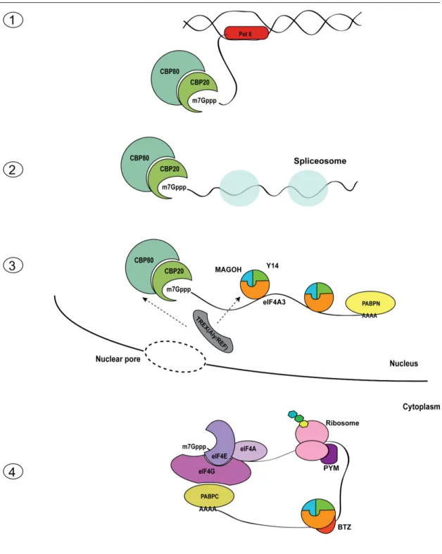
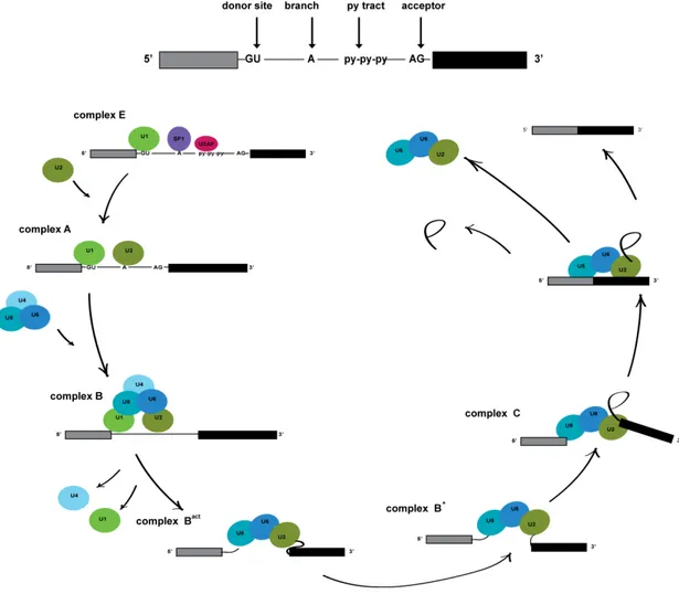
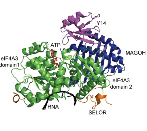
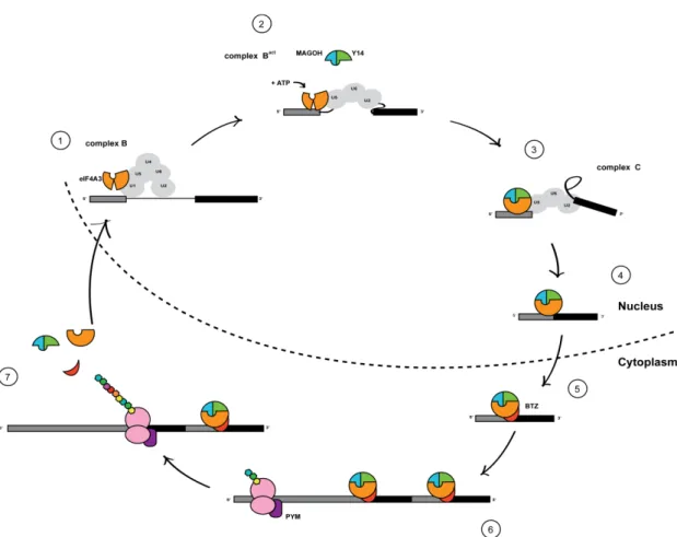
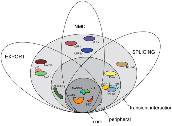
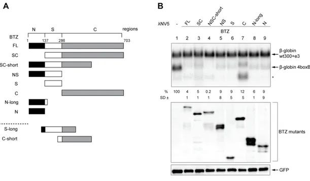
• Keine Ergebnisse gefunden

Separate  functions  of  BTZ  during  post-­transcriptional  gene  regulation

N/A
N/A
Protected

Academic year: 2021

Aktie "Separate  functions  of  BTZ  during  post-­transcriptional  gene  regulation"

Copied!
101
0
0

Wird geladen.... (Jetzt Volltext ansehen)

Volltext

(1)

Separate  functions  of  BTZ   during  post-­transcriptional  

gene  regulation  


Inaugural-­‐Dissertation   zur  

Erlangung  des  Doktorgrades  

der  Mathematisch-­‐Naturwissenschaftlichen  Fakultät   der    Universität  zu  Köln  


vorgelegt  von   Simona  Ciriello   aus  Neapel,  Italien    


Köln,  01.10.14    


(2)


Berichterstatter  :   PD  Dr.  Niels  H.  Gehring    

      Prof.  Dr.  Jürgen  R.  Dohmen    


Tag  der  mündlichen  Prüfung  :   28.11.14  


(3)

Table  of  Contents  

List  of  Abbreviations...III  

1.  Abstract...1  

1.1  Deutsche  Zusammenfassung... 2  

2.  Introduction...3  

2.1  mRNA  life:  from  transcription  to  translation ... 3  

2.2  The  Exon  Junction  Complex ... 9  

2.2.1  EJC  structure... 9  

2.2.2  EJC  assembly  and  disassembly... 11  

2.2.3  Peripheral  EJC  components ... 12  

2.2.4  EJC  functions... 13  

2.3  Nonsense-­mediated  mRNA  decay ... 16  

2.4  Barentsz... 19  

2.4.1  BTZ  structure ... 19  

2.4.2  BTZ  functions ... 20  

2.5  EJC  and  NMD  in  D.melanogaster  and  C.elegans ... 23  

3.  Aims  of  the  Project ... 25  

4.  Results ... 26  

4.1  Functions  of  BTZ  domains... 26  

4.1.1  Reduction  of  mRNA  levels  upon  BTZ  mutants  tethering ... 26  

4.1.2  BTZ  C-­‐terminus  activates  polyadenylation... 28  

4.1.3  Point  mutations  in  the  SELOR  domain  affect  NMD  function ... 30  

4.1.4  EJC  binding  and  NMD  inducing  domains  influence  RNA  decay... 33  

4.1.5  Conservation  of  the  SELOR  domain  in  different  species... 35  

4.2  BTZ-­induced  NMD ... 36  

4.2.1  BTZ  domains  induce  NMD  via  different  pathways ... 37  

4.2.2  XRN1-­‐dependent  degradation... 38  

4.3  BTZ  binding  to  the  RNA ... 40  

4.3.1  in  vitro  RNA  binding... 41  

4.3.2  in  vivo  RNA  binding ... 44  

4.4  BTZ  protein  interactome ... 46  

4.4.1  SRSF  proteins  contribute  to  NMD ... 48  

4.4.2  The  NTC  complex  induces  mRNA  degradation  in  a  NMD-­‐independent  way .. 50  

5.  Discussion... 52  

5.1  Separate  functions  of  the  three  BTZ  regions ... 52  

5.1.1  BTZ  induced  NMD ... 52  

5.1.2  BTZ  induced  polyadenylation ... 55  

5.2  RNA  binding ... 56  

5.3  SELOR  protein  interactome ... 58  

5.3.1  Functions  of  S-­‐long  binding  partners ... 59  

5.5  Conclusions ... 60  

6.  Materials  and  Methods... 62  

6.1  Materials ... 62  

6.1.1  Cell  lines ... 62  

6.1.2  Bacterial  strains ... 62  

6.1.3  Plasmids ... 62  

6.1.4  Antibodies... 62  

(4)

Table  of  Contents  

6.1.5  siRNAs  and  Primers... 63  

6.1.6  Buffers... 64  

6.2  Methods ... 67  

6.2.1  Cloning ... 67  

6.2.2  Plasmid  transfections ... 68  

6.2.3  siRNA  and  plasmid  transfection ... 68  

6.2.4  RNA  extraction  and  Northern  blot... 69  

6.2.5  Tethering  assay ... 69  

6.2.6  Immunoprecipitation ... 71  

6.2.7  Western  blot ... 71  

6.2.8  Silver  stain... 71  

6.2.9  Crosslinking  and  immunoprecipitation  (CLIP) ... 71  

6.2.10  Mass  spectrometry... 72  

6.2.11  EMSA  assay... 72  

6.2.12  in  vitro  protein  purification... 73  

6.2.13  Immunofluorescence ... 73  

7.  Supplemental  material ... 75  

8.  Bibliography... 78  

9.  Acknowledgement... 94  


(5)

List  of  Abbreviations  


Aa     Amino  acid   Bp       Base  pair  

EJC     Exon  Junction  Complex  

EMSA     Electrophoretic  mobility  shift  assay   IP     Immunoprecipitation  

KD     knockdown  

mRNA     Messenger  RNA  

mRNP     Messenger  ribonuceoprotein     NES     Nuclear  export  signal  

NLS     Nuclear  localization  signal  

NMD     Nonsense-­‐mediated  mRNA  decay   Nt     Nucleotide  

o/n     Over  night  

ORF     Open  reading  frame   PCR     Polymerase  chair  reaction   Pol  II     RNA  Polymerase  II  

PTC     Premature  termination  codon   RNP     Ribonucleoprotein  

RRM     RNA  recognition  motif   RT     Room  temperature  

snRNP     Small  nuclear  ribonucleic  particles   UTR     Untranslated  region  


(6)

Abstract  

1.  Abstract  


In  metazoans,  the  exon  junction  complex  (EJC)  is  a  central  component  of  spliced   messenger   ribonucleoprotein   particles   (mRNPs).   EJCs   are   assembled   by   the   spliceosome   and   deposited   upstream   of   exon-­‐exon   boundaries   in   the   nucleus.  

The   heterotetrameric   core   of   the   EJC   is   composed   of   the   proteins   eIF4A3   (DDX48),   MAGOH,   RBM8   (Y14)   and   CASC3/MLN51/Barentsz   (BTZ).   EJCs   contribute   to   different   steps   of   post-­‐transcriptional   gene   expression   including   splicing,   translation   and   nonsense-­‐mediated   mRNA   decay   (NMD).   BTZ   is   an   important  functional  component  and  is  involved  in  the  stimulation  of  translation   and  nonsense-­‐mediated  mRNA  decay.  Here,  I  show  that  both  the  N-­‐terminal  and   the   SELOR   domain   of   BTZ   elicit   NMD   in   a   tethering   assay.   They   activate   NMD   following   two   different   pathways,   BTZ-­‐dependent   and   UPF2   dependent,   which   get   reunited   once   UPF1   is   activated.   In   contrast,   the   C-­‐terminal   region   of   BTZ   does   not   seem   to   be   involved   in   NMD.   Instead,   this   region   plays   a   role   in   a   different   process   that   leads   to   the   polyadenylation   of   a   reporter   mRNA   at   an   upstream,  non-­‐canonical  polyadenylation  site.  Moreover,  I  show  that  binding  of   the   SELOR   domain   to   mRNA   in   vivo   is   EJC-­‐dependent.   In   addition   the   SELOR   domain  in  vivo  interacts  with  several  SR  proteins  for  a  subset  of  which  an  NMD-­‐

activating  function  is  observed.  These  findings  uncover  novel  EJC-­‐dependent  and  

-­‐independent   functions   of   BTZ   during   post-­‐transcriptional   gene   expression  

regulation.  

(7)

1.1  Deutsche  Zusammenfassung    

In  Metazoen  stellt  der  Exon-­‐Verbindungs  Komplex  (`exon  junction  complex´,  EJC)   eine   zentrale   Komponente   von   gespleißten   Ribonukleoproteinpartikeln   (mRNPs)   dar.   EJCs   werden   mithilfe   von   Spleißosomen   im   Zellkern   zusammengesetzt   und   nahe   Exon-­‐Exon   Grenzen   auf   der   mRNA   platziert.   Der   heterotetramere   Kern   des   EJC   besteht   aus   den   Proteinen   eIF4A3   (DDX48),   MAGOH,   RBM8   (Y14)   und   CASC3/MLN51/Barentz   (BTZ).   EJCs   sind   in   verschiedenste   Schritte   der   posttranskriptionalen   Genexpression   involviert,   unter   anderem   das   Spleißen,   die   Translation   und   den   nonsense-­‐vermittelten   mRNA-­‐Abbau   (NMD).   BTZ   ist   eine   wichtige   funktionale   Komponente   innerhalb   dieser   Prozesse   und   an   der   Stimulation   von   sowohl   Translation   als   auch   NMD   beteiligt.   In   dieser   Arbeit   wird   gezeigt,   dass   die   N-­‐terminale   sowie   die   SELOR   (`Speckle  Localizer  and  RNA-­‐binding´)  Domäne  von  BTZ,  NMD  im  Rahmen  eines   Tethering-­‐Assays   induzieren   können   und   dabei   zwei   verschiedene   Wege   der   NMD-­‐Aktivierung  genutzt  werden.  Darüber  hinaus  wird  dargestellt,  dass  die  C-­‐

terminale  Domäne  eine  nicht-­‐kanonische  Polyadenylierungsstelle  innerhalb  der  

3ʹ   UTR   einer   Reporter-­‐mRNA   aktiviert,   was   darauf   hinweist,   dass   BTZ   an  

alternativer   Polyadenylierung   beteiligt   ist.   Zudem   wird   präsentiert,   dass   die  

SELOR   Domäne   in   virto   präferenziell   bestimmte   mRNAs   bindet   und   in   vivo   die  

mRNA-­   Interaktion   von   SELOR   EJC-­‐abhängig   ist.   die   Zusätzlich   konnten  

verschieden   SR-­‐Proteine   in   SELOR-­‐assoziierten   Proteinkomplexen   identifiziert  

werden,   wobei   für   einige   dieser   SR   Proteine   eine   NMD-­‐aktivierende   Funktion  

nachgewiesen  werden  konnte.  Diese  Ergebnisse  zeigen  eine  neue,  EJC-­‐abhängige  

sowie   –unabhängige,   regulatorische   Funktion   von   BTZ   innerhalb   der  

posttranskriptionalen  Geneexpression.    

(8)

  Introduction  

2.  Introduction  


2.1  mRNA  life:  from  transcription  to  translation  


Eukaryotic  gene  expression  is  a  multistep  process  that  requires  the  coordination   of   several   events   that   are   spatially   and   temporally   separated   (Moore,   2005;  

Moore  and  Proudfoot,  2009;  Reed,  2003).  All  these  steps  are  needed  in  order  to   correctly  decode  the  information  of  the  DNA  into  a  functional  product  (Figure  1).  

The  following  sections  describe  these  processes  with  a  special  emphasis  on  the   steps  most  important  for  this  work.  


Transcription  

Gene  expression  begins  with  transcription  of  DNA  into  messenger  pre-­‐RNA  (pre-­‐

mRNA)   by   the   RNA   Polymerase   II   (Pol   II)   (Kornberg,   1999;   Sims   et   al.,   2004)   (Figure   1).   The   different   steps   of   transcription   (initiation,   elongation   and   termination)  are  marked  by  rearrangement  of  the  components  that  binds  Pol  II   (Cheung  and  Cramer,  2012).  Transcription  initiation  starts  upon  addition  of  two   initiating  nucleoside  triphosphates  (NTPs)  complementary  to  the  DNA  sequence   and  the  formation  of  the  first  phosphodiester  bond.  When  the  pre-­‐mRNA  is  23-­‐

nt-­‐long  the  rearrangement  of  the  transcriptional  factors  around  Pol  II  marks  the   beginning   of   the   elongation   phase   (Pal   and   Luse,   2002;   Roberts   et   al.,   2004).  

Polyadenylation   (see   below)   and   specific   termination   factors   mediate   the   termination   of   transcription   (Kireeva   et   al.,   2000;   Lykke-­‐Andersen   and   Jensen,   2007;  Richard  and  Manley,  2009).    


5ʹcapping  

The  5ʹ  end  capping  is  the  first  modification  that  nascent  pre-­‐mRNA  undergoes,   when   it   is   22-­‐25   nt   long   and   emerging   from   the   RNA   exit   channel   of   Pol   II   (Shatkin,   1976;   Shatkin   and   Manley,   2000).   The   5ʹ   cap   protects   mRNA   against   exonucleases  and  promotes  transcription,  polyadenylation,  splicing  and  nuclear   export  (Gu  and  Lima,  2005;  Lewis  and  Izaurralde,  1997).    


(9)


Figure  1.  Steps  of  gene  expression.  1)  Pol  II  transcribes  the  information  of  DNA  in  pre-­‐mRNA.  

5ʹ  cap  (m7Gppp)  is  added  when  the  pre-­‐mRNA  is  22-­‐25  nt  long  and  is  subsequently  bound  by  the   nuclear  Cap  Binding  Complex  (nCBC:  CBP20  and  CBP80).  2)  Splicing  occurs  cotranscriptionally   and  removes  the  introns.  3)  As  a  consequence  of  splicing,  the  pre-­‐EJC  (MAGOH,  Y14  and  eIF4A3)   is   deposited   on   the   mRNA.   At   the   end   of   transcription,   the   mRNA   is   polyadenylated,   and   the   poly(A)  tail  is  bound  by  the  nuclear  Poly(A)  binding  protein  (PABPN).  Finally,  the  export  complex   TREX  addresses  the  mRNA  to  the  nuclear  pore.  4)  In  the  cytoplasm,  BTZ  joins  the  EJC  complex,   the  eIF4F  complex  (eIF4E,  eIF4A  and  eIF4G)  replaces  the  nCBC,  the  cytoplasmic  PABP  (PABPC)   replaces  PABPN,  the  ribosome  translates  the  mRNA  into  protein  and  PYM  displaces  the  EJC  upon   translation.  


(10)

  Introduction  

mRNA   capping   requires   three   reactions:   first,   the   5ʹ-­‐γ-­‐phosphate   group   of   the   first  transcribed  nucleotide  of  pre-­‐mRNA  is  removed  by  the  RNA  triphosphatase;  

second,   a   guanine   monophosphate   (GMP)   nucleotide   is   transferred   by   a   guanylyltransferase   to   the   RNA   5ʹ-­‐diphosphate   end;   third,   a   RNA   methyl   transferase   adds   a   methyl   group,   yielding   the   7-­‐methylguanosine   cap   (m7GpppN)  (Mao  et  al.,  1995;  Yue  et  al.,  1997).  The  5ʹ  cap  is  bound  to  the  nuclear   cap  binding  complex  (nCBC)  in  the  nucleus,  consisting  of  the  cap-­‐binding  subunit   CBP20  and  the  auxiliary  protein  CBP80  that  stabilizes  the  interaction  of  CBP20   with  the  cap  (Mazza  et  al.,  2001;  Mazza  et  al.,  2002).  After  the  mRNA  is  exported   to   the   cytoplasm,   eIF4E   (eukaryotic   translation   initiation   factor   4E),   a   component  of  the  eIF4F  complex,  binds  the  cap  and  promotes  the  recruitment  of   the  small  ribosomal  subunit  for  translation  initiation.  The  other  two  components   of  the  complex  are  the  DEAD-­‐box  helicase  eIF4A,  responsible  for  RNA  unwinding   (Feoktistova  et  al.,  2013)  and  eIF4G.  eIF4G  binds  eIF4E  and  PABPC  via  its  C-­‐  and   N-­‐terminal   region,   respectively   (see   polyadenylation).   These   interactions   enhance  eIF4F  binding  to  the  cap  (Kahvejian  et  al.,  2005b),  as  well  as  enabling   the  formation  of  the  so-­‐called  closed-­‐loop  which  facilitates  re-­‐initiation  of  mRNA   translation  (Hinnebusch  and  Lorsch,  2012;  Jackson  et  al.,  2010)  (Figure  1).    


Splicing  

The   second   modification   that   most   pre-­‐mRNAs   undergo   is   the   excision   of   the   intervening   sequences   (introns).   Specific   elements   inside   the   gene   sequence   mark  the  position  of  an  intron  (Clancy,  2008;  Konarska  et  al.,  1985)  (Figure  2):    

• donor   site,   at   the   5ʹ   end   of   the   intron,   it   contains   a   GU   sequence,   surrounded  by  a  less  conserved  region;  

• branch  site,  20-­‐50  nt  upstream  of  the  acceptor  site,  it  includes  an  A;  

• acceptor  site,  at  the  3ʹend  of  the  intron,  it  contains  an  AG  sequence  and  is   preceded  by  a  polypyrimidine  tract.  

The   spliceosome   is   a   complex   consisting   of   several   small   nuclear  

ribonucleoproteins   (snRNP),   which   catalyzes   nuclear   pre-­‐mRNA   splicing   (Wahl  

et   al.,   2009).   The   spliceosome   assembly   is   spatially-­‐temporally   organized   and  

proceeds  through  the  assembly  of  the  complexes  E,  A,  B,  B

act

 ,  B*  and  C  (Bessonov  

(11)

et  al.,  2010).  Complex  E  is  comprised  of  U1  snRNP  binding  the  donor  site,  U2AF   the  polyprimidine  tract  and  SF1  the  branch  point  sequence  (BPS).  Subsequently,   U2   snRNP   is   recruited   to   the   BPS,   forming   complex   A.   U4/U6   and   U5   are   then   recruited  as  pre-­‐assembled  tri-­‐snRNP,  leading  to  the  formation  of  complex  B.    


Figure   2.   Splicing   reaction.   Schematic   representation   of   an   mRNA,   with   indicated   donor   site,   branch  site,  polypyrimidine  tract  and  acceptor  site.  The  steps  required  for  the  splicing  reaction   are  indicated  with  the  name  of  the  complex  formed  (E,  A,  B,  B

act

 ,  B*  and  C).  


This   complex   is   still   inactive   and   requires   a   conformational/compositional  

change   to   be   functional:   U1   and   U4   are   released   and   the   complex   becomes  

splicing  competent  (complex  B

act

).  After  a  structural  rearrangement  into  complex  

B

*

,   the  splicing  reaction  takes  place  in  complex  C.  The  first  reaction   consists  of  

cleaving  of  the  pre-­‐mRNA  at  the  5ʹ  end  of  the  intron  and  lariat  formation.  During  

the   second   reaction   the   exons   are   ligated,   the   intron   released   and   the  

(12)

  Introduction  

spliceosome  disassembled  with  the  snRNP  being  recycled  (Bessonov  et  al.,  2010)   (Figure  2).    


Polyadenylation  

Similary   to   5ʹ   capping,   3ʹ   polyadenylation   is   required   for   maturation   of   pre-­‐

mRNA  and  is  necessary  for  nuclear  export,  stability  of  the  mature  transcript  and   efficient   translation   (Sachs,   1990).   The   vast   majority   of   eukaryotic   mRNAs   contain   a   long   stretch   of   untemplated   adenosines   in   their   3′   end   termed   the   poly(A)   tail.   The   3ʹ   end   polyadenylation   is   a   two-­‐steps   process   that   involves   endonucleolytic   cleavage   of   the   transcript   and   the   addition   of  a   poly(A)   tail.   In   human  cells,  the  average  length  of  the  poly(A)  tail  varies  between  250  and  300   adenines,   while   the   length   in   yeast   fluctuates   between   70   and   80   adenines.   In   general,  the  number  of  adenines  is  restricted  and  varies  between  species  (Elkon   et   al.,   2013).   mRNAs   with   a   shortened   poly(A)   tail   are   normally   degraded   or   stored   in   a   translationally   dormant   state   (DʹAmbrogio   et   al.,   2013;   Guhaniyogi   and   Brewer,   2001).   Polyadenylation   requires   several   cis-­‐   and   trans–acting   elements.  The  Poly(A)  signal  (PAS)  dictates  the  selection  of  the  cleavage  site  and   is  commonly  located  15-­‐20  nt  upstream  of  it  (Proudfoot  and  Brownlee,  1976).  In  

~  70%  of  human  mRNAs,  the  PAS  consists  of  the  nucleotide  sequences  AAUAAA  

or   AUUAAA.   The   remaining   ~   30%   of   RNAs   contain   other   sequences,   such   as  

UAUAAA,  AACAAA  or  ACUAAA  (MacDonald  and  Redondo,  2002),  suggesting  the  

possibility  of  alternative  polyadenylation  (APA).  Alternative  polyadenylation  can  

generate   transcript   isoforms   with   alternative   3′   ends   (Elkon   et   al.,   2013).   In  

addition   to   the   PAS,   U-­‐   or   GU-­‐rich   downstream   sequence   elements   (DSEs)   and  

less   well-­‐defined   upstream   sequence   elements   (USEs)   enhance   cleavage  

efficiency.  The  cleavage  and  polyadenylation  specificity  factor  (CPSF)  recognizes  

the   PAS,   the   cleavage   stimulating   factor   (CSTF)   binds   the   DSEs   (Mandel   et   al.,  

2008;  Proudfoot,  2011),  and  cleavage  factors  Im  (CFIm)  and  IIm  (CFIIm)  bind  the  

USEs  (Brown  and  Gilmartin,  2003;  Yang  et  al.,  2011).  The  interaction  of  PABP  (in  

humans   primarily   PABPC1)   with   the   cap-­‐associated   eIF4G   in   the   cytoplasm   is  

thought   to   facilitate   circularization   of   mRNAs,   thereby   supporting   efficient  

translation  termination,  ribosome  recycling  and  translation  initiation  (Amrani  et  

al.,  2004;  Kahvejian  et  al.,  2005a;  Wells  et  al.,  1998)  (Figure  1).  

(13)

Export  

After   the   completion   of   all   processing   steps   (capping,   splicing   and   polyadenylation),   the   mature   mRNA   needs   to   be   exported   to   the   cytoplasm   in   order   to   be   translated   into   protein.   mRNA   export   is   already   initiated   during   splicing   of   the   first   intron   when   the   transcription   export   (TREX)   complex   is   deposited   at   the   5ʹ   end   of   the   mRNA   (Cheng   et   al.,   2006;   Masuda   et   al.,   2005).  

TREX  is  a  multiprotein  complex  that  is  composed  of  the  THO  complex,  the  RNA   helicase   UAP56   and   the   adaptor   molecule   Aly/REF   (Zhou   et   al.,   2000).   The   mechanism   of   recruitment   of   TREX   to   the   mRNA   is   still   unclear.   Different   evidence  suggest  an  EJC-­‐dependent  (Stutz  et  al.,  2000;  Zhou  et  al.,  2000)  or  a  cap-­‐

dependent   (Cheng   et   al.,   2006;   Luna   et   al.,   2012)   recruitment.   Once   recruited,   Aly/REF  interacts  with  the  heterodimeric  mRNA  export  receptor  TAP/p15  (also   known   as   NXF1/NXT1)   (Viphakone   et   al.,   2012),   which   binds   to   mRNA   and   translocates  it  across  the  nuclear  pore  to  the  cytoplasm  (Hurt  et  al.,  2000;  Segref   et  al.,  1997)  (Figure  1).  


Translation  

After   export   to   the   cytoplasm,   the   information   of   the   mRNA   is   translated   into   protein.   This   process   can   be   divided   into   different   steps,   which   are   initiation,   elongation,   termination   and   recycling.   Translation   initiation   is   carried   out   by   a   network   of   factors   and   involves   eukaryotic   initiation   factors   (eIFs),   the   ribosomal  subunits  and  the  mRNA  (Aitken  and  Lorsch,  2012).  These  factors  scan   the   mRNA   until   an   AUG   start   codon   is   encountered   (Aitken   and   Lorsch,   2012;  

Jackson  et  al.,  2010).  Specifically,  eIF3  in  the  initiation  phase  interacts  with  eIF4F   and  contributes  to  the  recognition  of  AUG  (Hinnebusch,  2006).  Elongation  of  the   amino   acid   chain   proceeds   until   a   termination   codon   is   reached   (UAG,   UAA,   UGA).  The  eukaryotic  release  factors  (RFs)  are  responsible  for  the  release  of  the   completed  protein  product  (Jackson  et  al.,  2012).  At  this  step  the  ribosome  is  still   assembled   on   the   mRNA.   The   recycling   process   is   still   unclear,   but   what   is   known  is  that  the  ribosome  subunits  are  separated  from  the  mRNA.  This  enables   the   ribosomal   subunits   to   be   recruited   again   to   the   AUG   of   mRNA   to   start   another  round  of  translation  (Nurenberg  and  Tampe,  2013).  


(14)

  Introduction  

2.2  The  Exon  Junction  Complex  

Messenger   ribonuceoproteins   (mRNPs)   are   dynamic   complexes,   changing   their   composition   during   the   lifetime   of   an   mRNA.   As   an   example,   the   nuclear   cap   binding  protein  (CBP20/80)  binds  mRNA  in  the  nucleus  (Lewis  and  Izaurralde,   1997;   Sonenberg   and   Hinnebusch,   2009),   while   the   eukaryotic   translation   initiation  complex  4F  replaces  CBP20/80  in  the  cytoplasm  (Jackson  et  al.,  2010;  

Sonenberg  and  Hinnebusch,  2009).  Similarly,  the  nuclear  poly(A)  binding  protein   PABPN  (Krause  et  al.,  1994;  Wahle,  1991)  is  replaced  by  PABPC  in  the  cytoplasm   (Gorlach  et  al.,  1994).  The  exon  junction  complex  (EJC)  differs  from  the  above-­‐

mentioned  mRNPs.  It  remains  bound  to  the  mRNA  in  the  cytoplasm  after  being   deposited   in   the   nucleus   during   splicing   (Le   Hir   et   al.,   2000a;   Le   Hir   et   al.,   2000b).  


2.2.1  EJC  structure  

The   core   of   the   EJC   consists   of   four   proteins   known   as   MAGOH,   Y14   (RMB8A),   eIF4A3  (DDX48)  and  Barentsz  (BTZ,  also  known  as  CASC3  and  MLN51)  (Gehring   et  al.,  2009a)  (Figure  3).  In  living  cells,  the  EJC  functions  as  a  binding  platform  for   many   other   proteins   responsible   for   different   processes   during   the   mRNA   life   cycle   (see   paragraphs   2.2.3   and   2.2.4).   eIF4A3   is   a   DEAD-­‐box   helicase   protein.  

The  proteins  of  the  DExD/H  family  share  a  conserved  helicase  core,  consisting  of   two   globular   RecA-­‐like   domains,   the   N-­‐terminal   RecA   domain   1   and   the   C-­‐

terminal   RecA   domain   2   connected   by   a   flexible   linker   (Caruthers   and   McKay,   2002).  These  domains  correspond  to  amino  acids  38-­‐240  and  251-­‐411  in  eIF4A3   respectively.   Their   orientation   is   not   defined   in   the   absence   of   ATP   and   RNA.  

However,   upon   binding   to   ATP   and   RNA,   the   RecA-­‐like   domain   1   and   2   are  

brought   in   close   proximity   forming   a   cleft   where   ATP   (in   red)   binds.   On   the  

opposite   surface   the   two   RecA-­‐like   domains   bind   the   RNA   (in   black).   This  

binding   is   sequence   independent   because   eIF4A3   binds   the   ribose-­‐phosphate  

backbone  of  the  RNA  (Bono  et  al.,  2006).  The  SELOR  domain  of  BTZ  (aa  137-­‐286  

(Degot  et  al.,  2004))(in  orange)  extends  with  two  separate  stretches  over  both  

domains   of   eIF4A3.   The   N-­‐terminal   stretch   (aa   168–196)   binds   to   domain   2   of  

eIF4A3   and   the   C-­‐terminal   stretch   (aa   214–248)   binds   to   domain   1.   These  

(15)

stretches   are   connected   by   a   flexible   linker.   In   addition,   the   SELOR   domain   of   BTZ   contributes   to   the   interaction   of   BTZ   to   the   RNA   and   to   MAGOH   (in   blue)   (Bono  et  al.,  2006).  The  structure  of  the  dimer  MAGOH-­‐Y14  in  the  EJC  is  almost   identical   to   one   of   the   isolated   dimer   (Fribourg   et   al.,   2003;   Lau   et   al.,   2003).  

MAGOH  folds  into  flat  antiparallel  β   sheets  flanked  on  one  side  by  two  parallel  α   helices,  which  bind  the  RNA  binding  domain  (RBD)  of  Y14  (in  violet).  In  the  EJC   the  dimer  MAGOH/Y14  interacts  mainly  with  the  RecA-­‐like  domain  2  of  eIF4A3   (Bono  et  al.,  2006).  Interestingly,  MAGOH/Y14  binds  eIF4A3  only  in  the  ATP-­‐and   RNA-­‐bound   state.   This   explains   why   in   vitro   MAGOH-­‐Y14   inhibits   the   ATPase   activity  of  eIF4A3.  In  contrast,  SELOR  stimulates  ATPase  activity  of  eIF4A3  (Bono   et  al.,  2006).  


Figure   3.   EJC   crystal   structure.   The   crystal   structure   of   the   EJC   complex   was   modeled   with   pyMOL  (PyMOL  Molecular  Graphics  System,  Version  1.5.0.4  Schrödinger,  LLC)  and  the  PDB  file   2J0S  (Bono  et  al.,  2006).    


(16)

  Introduction   2.2.2  EJC  assembly  and  disassembly  

As   a   consequence   of   splicing,   the   EJC   is   deposited   on   the   mRNA   20-­‐24   nt   upstream  of  the  exon-­‐exon  junction  marking  the  position  of  the  former  introns   (Le   Hir   et   al.,   2000a).   The   deposition   of   the   EJC   on   the   mRNA   is   a   multi-­‐step   process.   EJC   assembly   requires   CWC22,   a   component   of   the   spliceosome   and   a   newly   identified   binding   partner   of   eIF4A3   (Steckelberg   et   al.,   2012).   Before   exon-­‐exon   ligation   takes   place,   CWC22   brings   eIF4A3   in   close   proximity   to   the   mRNA  (in  the  spliceosomal  complex  B).  The  subsequent  conformational  change,   in  the  presence  of  ATP,  induces  the  binding  of  eIF4A3  to  the  mRNA.  As  a  result,   the   MAGOH/Y14   heterodimer   can   interact   with   eIF4A3   (spliceosomal   complex   B

act

),  stabilizing  the  trimeric  pre-­‐EJC  on  the  mRNA.  The  trimeric  pre-­‐EJC  remains   stably   associated   to   the   mRNA   while   the   introns   are   spliced   out   (spliceosomal   complex   C).   Next,   the   mRNA   associated   complex   is   exported   to   the   cytoplasm   where,   according   to   the   current   model,   the   protein   BTZ   joins   the   complex   (Gehring  et  al.,  2009a;  Steckelberg  et  al.,  2012)  (Figure  4,  from  1  to  5).  Indeed,   there  is  evidence  that  BTZ  and  the  spliceosome  bind  the  same  eIF4A3  residues,   supporting   the   hypothesis   of   BTZ   joining   to   the   EJC   after   completion   of   the   splicing   reaction.   This   assumption   is   corroborated   by   previous   results,   where   BTZ  was  not  identified  in  purified  spiceosomal  complexes  (Bessonov  et  al.,  2008;  

Gehring   et   al.,   2009a).   EJC   proteins   need   to   be   recycled   quickly   and   efficiently  

because   of   the   limited   cellular   amount   of   EJC   proteins   and   the   high   number   of  

exon   junctions   in   the   steady-­‐state   transcriptome.   EJCs   located   within   the   open  

reading  frame  (ORF)  are  removed  by  the  ribosome  in  the  cytoplasm  during  the  

first  round  of  translation  (Dostie  and  Dreyfuss,  2002;  Lejeune  et  al.,  2002).  The  

protein  PYM  binds  the  dimer  MAGOH-­‐Y14  with  its  N-­‐terminal  domain  (Bono  et  

al.,  2004;  Forler  et  al.,  2003),  but  it  also  interacts  with  the  ribosome  (Diem  et  al.,  

2007).   For   this   reason,   PYM   was   proposed   to   bridge   the   ribosome   and   the   EJC  

and  to  stimulate  the  release  of  the  EJC  proteins  from  the  mRNA.  Binding  of  PYM  

to  the  ribosome  guarantees  that  PYM  removes  the  EJC  only  within  the  ORF  from  

translated   mRNA.   This   type   of   binding   represents   a   safe   mechanism   that  

minimizes  the  amount  of  free  PYM,  in  order  to  avoid  EJC  removal  from  not  yet  

translated  mRNAs  (Gehring  et  al.,  2009b).  According  to  the  proposed  model,  the  

(17)

binding   of   PYM   to   the   dimer   MAGOH-­‐Y14   induces   a   conformational   change   in   MAGOH-­‐Y14  that  impairs  their  binding  with  eIF4A3.  In  this  way,  eIF4A3  is  not   locked   on   the   mRNA   anymore   and   is   released   together   with   the   other   EJC   components  (Nielsen  et  al.,  2009)  (Figure  4,  6-­‐7)  .  


Figure  4.  Model  of  EJC  assembly  and  recycle.  Steps  1  to  5  show  the  assembly  of  the  EJC  in  the   nucleus   and   the   export   to   the   cytoplasm.   PYM   displaces   the   EJC   in   the   cytoplasm   during   translation  (steps  6  and  7).  This  image  was  inspired  by  N.H.  Gehring  drawing.  


2.2.3  Peripheral  EJC  components  

As  explained  in  paragraph  2.2.4,  the  EJC  executes  several  functions.  In  order  to  

mediate   these   functions,   it   needs   to   interact   with   other   proteins   (Degot   et   al.,  

2004;  Diem  et  al.,  2007;  Ferraiuolo  et  al.,  2004;  Gatfield  et  al.,  2001;  Kataoka  et  

al.,  2001;  Kataoka  et  al.,  2000;  Kim  et  al.,  2001;  Le  Hir  et  al.,  2001;  Le  Hir  et  al.,  

2000a;  Le  Hir  et  al.,  2000b;  Li  et  al.,  2003;  Luo  et  al.,  2001;  Lykke-­‐Andersen  et  al.,  

(18)

  Introduction  

2000;   Palacios   et   al.,   2004;   Zhou   et   al.,   2000).   In   Figure   5,   EJC-­‐interacting   proteins  are  shown  and  their  functions  are  described  in  the  next  paragraphs.  


Figure   5.   EJC-­interacting   proteins   and   functions.   The   core   of   the   EJC   (MAGOH,Y14,BTZ   and   eIF4A3)  (dark  gray  circle)  interacts  with  several  peripheral  EJC  components  (middle  gray)  and   with  transient  binding  partners  (light  gray)  to  execute  different  functions,  e.g.  NMD,  export  and   splicing   (The   proteins   involved   in   a   common   function   are   enclosed   in   an   oval   shape,   with   the   function  indicated).  


2.2.4  EJC  functions  

The   EJC   influences   the   life   of   an   mRNA   through   several   processes,   such   as   localization,  splicing,  translation  and  decay.  With  the  exception  of  decay,  which  is   described   in   paragraph   2.3,   this   paragraph   discusses   how   the   EJC   influences   these  processes.  


Localization  

The  EJC  is  required  for  proper  localization  of  the  oskar  mRNA  in  D.melanogaster  

oocytes.  Oskar  mRNA  is  synthesized  in  the  nurse  cell  nuclei  and  then  transported  

along  the  microtubules  into  the  adjacent  oocyte  (St  Johnston,  2005).  Expression  

(19)

of   oskar   occurs   exclusively   at   the   posterior   pole   is   precisely   regulated:   during   transport   along   the   microtubules,   Bruno   represses   the   translation   of   oskar   (Gunkel  et  al.,  1998;  Kim-­‐Ha  et  al.,  1995).  Once  oskar  reaches  the  posterior  pole,   Staufen   activates   its   translation   (Micklem   et   al.,   2000).   Translation   occuring   exclusively   at   the   posterior   pole   is   essential   for   the   formation   of   the   abdomen   and   the   germ   line   of   the   embryo   (Johnstone   and   Lasko,   2001).   Several   trans-­‐

acting  factors  control  repression,  transport  and  translation  of  the   oskar  mRNA.  

Examples   of   these   controlling   factors   include   the   D.melanogaster   EJC   core   components   eIF4A3,   Barentz,   Mago   Nashi   and   Tsunagi   (MAGOH   and   Y14   in   human)  (Palacios,  2002;  Palacios  et  al.,  2004).  In  particular,  BTZ  is  required  for   transport   of   oskar   to   the   posterior   pole.   BTZ   does,   however,   not   affect   other   processes   such   as   transcription,   export,   colocalization   with   Staufen,   or   translation  (van  Eeden  et  al.,  2001).  Similarly,  van  Eeden  and  collegues  observed   that  mutations  in   mago  nashi  and  btz  resulted  in  the  same  phenotype  and  that   the   two   proteins   depend   on   each   other   for   proper   localization   at   the   posterior   pole.  Subsequently,  Y14  and  eIF4A3  were  identified  to  be  responsible  for  proper   oskar  localization  as  well  (Hachet  and  Ephrussi,  2001;  Palacios  et  al.,  2004).  The   oskar   mRNA   localization   is   the   best   studied   example   of   the   involvement   of   the   EJC   in   mRNA   localization.   Another   example   of   the   contribution   of   the   EJC   proteins  in  mRNA  localization  comes  from  dendrites  of  mammalian  hippocampal   neurons.   The   dendrites   are   highly   polarized   cells   and   require   translation   of   specific  mRNAs.  MAGOH,  Y14  and  BTZ  accumulated  in  the  dendrites  where  BTZ   associates  with  the  mRNA  transport  factor  Staufen1.  Staufen1  assembles  in  RNPs   responsible   to   transport   and   localize   RNAs   into   dendrites   of   mature   hippocampal  neurons  (Giorgi  and  Moore,  2007;  Glanzer  et  al.,  2005;  Macchi  et  al.,   2003;  Monshausen  et  al.,  2004).    


Splicing  

The  recognition  of  the  splice  site  occurs  in  two  different  ways  in  long  and  short  

intron  containing  genes:  in  short  introns  (<200  bp),  the  5ʹ  and  3ʹ  splice  sites  are  

recognized  across  the  intron  (intron  definition);  in  long  introns  (>250  bp),  splice  

sites  of  bordering  exons  have  to  be  recognized  before  splicing  can  occur  (exon  

definition)  (Fox-­‐Walsh  et  al.,  2005;  Sterner  et  al.,  1996).  The  influence  of  the  EJC  

(20)

  Introduction  

over   the   splicing   process   is   associated   with   this   second   phenomenon   of   exon   definition.  In  D.melanogaster  the  nuclear  EJC  influences  splicing  of  the  mapk  gene   exclusively.   The   splicing   of   the   other   genes   of   the   RAS1/MAPK   pathway   is   not   influenced  by  the  EJC,  indicating  a  selective  choice  for  a  specific  mRNA  (Ashton-­‐

Beaucage  et  al.,  2010).  EJC  removal  leads  to  exon  skipping  (Ashton-­‐Beaucage  et   al.,  2010)  and,  of  the  EJC  core  components,  only  the  depletion  of  BTZ  does  not   affect   splicing   of   the   mapk   pre-­‐mRNA.   This   is   in   line   with   the   observation   that   BTZ   deposition   on   the   mRNA   occurs   after   completion   of   the   splicing   reaction   (Gehring  et  al.,  2009a).  So  far  it  was  not  possible  to  define  if  the  effect  of  the  EJC   was  direct  or  indirect.  One  possibility  is  that  the  EJC  works  as  a  binding  platform   for  the  splicing  effectors  SRSF  proteins  (Long  and  Caceres,  2009).  Another  option   is  that  the  EJC  stabilizes  the  interaction  between  the  spliceosome  and  the  splice   site  (Ashton-­‐Beaucage  et  al.,  2010).  In  2012,  Michelle  and  collegues  analyzed  the   effect   of   three   categories   of   EJC   components   on   splicing   of   the   apoptotic   regulator   Bcl-­x:  core,  peripheral  and  NMD  associated.  The  depletion  of  the  core   components  Y14  and  eIF4A3  and  the  peripheral  RNPS1,  Acinus  and  SAP18  was   associated   with   an   increase   of   the   proapoptotic   splicing   variant   Bcl-­x

s

.   On   the   contrary,  no  effect  was  observed  when  the  export  factors  UAP56,  ALY/REF  and   the  NMD  proteins  UPF1,  UPF2  and  UPF3b  were  depleted.  Similarly,  no  effect  was   observed  when  BTZ  was  depleted.  Interestingly,  the  function  and  the  deposition   of the   EJC   components   in   Bcl-­x   modulation   depend   on   cis-­‐acting   elements,   suggesting  a  different  type  of  EJC  assembly  and  regulation  (Michelle  et  al.,  2012).    


Translation    

It  was  first  observed  that  intron-­‐containing  transcripts  had  a  higher  translation  

rate  compared  to  the  intron-­‐less  upon  injection  in  Xenopus  oocytes  (Matsumoto  

et  al.,  1998).   Subsequently,  in  mammalian  cells  an  increased  mRNA  translation  

was   observed   upon   the   deposition   of   the   EJC   (Lu   and   Cullen,   2003;   Nott   et   al.,  

2003).   No   detectable   positive   effect   on   the   expression   of   the   mRNA   was  

observed  for  the  human  β-­‐globin  gene  where  the  intron  was  positioned  so  close  

to  the  5ʹ  end  of  the  mRNA  that  the  EJC  could  not  assemble,  even  though  splicing  

of  this  mRNA  still  occurred.  In  contrast,  tethering  of  the  EJC  proteins  RNPS1  and  

SRm160   increases   expression   of   intronless   β-­‐globin   (Wiegand   et   al.,   2003).  

(21)

Similarly,   Nott   and   colleagues   observed   that   tethering   of   the   EJC   components   Y14,   MAGOH   and   the   more   peripheral   component   RNPS1   led   to   translational   stimulation  of  the  reporter  transcript.  The  EJC  components  might  contribute  to   the   increased   mRNA   translation   in   different   ways.   One   explanation   is   the   interaction  of  PYM  with  the  dimer  MAGOH/Y14.  The  binding  of  the  N-­‐terminus   of  PYM  to  MAGOH/Y14  and  the  C-­‐terminus  to  ribosome  would  recruit  EJC  bound   mRNAs  to  the  translation  machinery  (Diem  et  al.,  2007).  This  model  is  supported   by   previous   observation   that   cytoplasmic   polysome   association   resulted   in   enhanced  spliced  mRNAs  (Nott  et  al.,  2004).  Another  possibility  is  the  interaction   of  BTZ  with  the  translation  initiation  factor  eIF3  (paragraph  2.4.2)  (Chazal  et  al.,   2013)   or   the   recruitment   of   the   TREX   complex.   Export   of   the   mRNA   to   the   cytoplasm  would  then  make  it  available  for  the  translation  machinery  (Le  Hir  et   al.,  2001;  Luo  and  Reed,  1999;  Luo  et  al.,  2001;  Zhou  et  al.,  2000).    


2.3  Nonsense-­mediated  mRNA  decay  

During   gene   expression,   a   frequently   occurring   error   is   the   generation   of   premature   termination   codons   (PTCs).   PTCs   can   arise   at   the   DNA   level   as   a   consequence   of   nonsense   mutations,   deletion,   insertions   or   somatic   rearrangements.  Alternatively,  mutations  in  functional  motifs,  such  as  splice  site   or   splice   regulatory   elements   that   lead   to   alternative   splicing   events,   can   generate  PTCs  (Nicholson  et  al.,  2010).  One-­‐third  of  all  alternative  splicing  events   lead  to  the  formation  of  PTCs  (Lewis  et  al.,  2003).  In  principle,  the  presence  of   PTCs   can   lead   to   synthesis   of   truncated,   non-­‐functional   and   sometimes   deleterious   proteins   (Frischmeyer   and   Dietz,   1999;   Holbrook   et   al.,   2004).  

Eukaryotes   have   acquired   an   evolutionary   conserved   surveillance   mechanism,  

named   nonsense-­‐mediated   mRNA   decay   (NMD),   to   identify   and   degrade   these  

aberrant   mRNAs   (Amrani   et   al.,   2006;   Behm-­‐Ansmant   et   al.,   2007;   Conti   and  

Izaurralde,   2005;   Culbertson,   1999;   Muhlemann   et   al.,   2008).   According   to   the  

current   model,   in   higher   eukaryotes   a   stop   codon   is   recognized   as   premature  

when   situated   more   that   50   nt   upstream   of   an   exon-­‐exon   junction   and,  

consequently,  of  a  deposited  EJC  (Figure  6-­‐1)  (Nagy  and  Maquat,  1998;  Zhang  et  

al.,   1998a;   Zhang   et   al.,   1998b).   Two   main   complexes   are   required   for   NMD  

(22)

  Introduction  

activation:   one   is   the   EJC,   downstream   of   the   stop   codon,   the   other   the   SURF   complex.  When  the  ribosome  stops  either  at  a  normal  stop  codon  or  at  a  PTC,  the   eukaryotic  release  factors  (eRF)  1  and  3  interact  with  the  A-­‐site  of  the  ribosome   (Figure   6-­‐2).   For   efficient   translation   termination,   the   interaction   of   eRF3   with   the   cytoplasmic   poly(A)-­‐binding   protein   (PABPC1)   is   required   (Cosson   et   al.,   2002a;  Cosson  et  al.,  2002b).  However  the  up-­‐frameshift  protein  UPF1  competes   with   PABPC1   for   binding   to   eRF3,   affecting   the   normal   termination   process   (Ivanov  et  al.,  2008).  eRF3-­‐bound  UPF1  in  turn  interacts  with  SMG1,  forming  the   so-­‐called   SURF   complex   (SMG1,   UPF1,   eRF   1   and   3)   (Czaplinski   et   al.,   1998;  

Kashima  et  al.,  2006)  (Figure  6-­‐3).    


Figure   6.   Model   of   NMD   activation.   1-­‐   EJC   is   deposited   20-­‐24   nt   upstream   of   the   exon-­‐exon   junction.  2-­‐  The  ribosome  stalls  at  the  PTC  and  recruits  the  eukaryotic  release  factors  (eFR1-­‐3).  

3-­‐The   SURF   complex   (SMG1,   UPF1,   eRF   1   and   3)   is   assembled.   4-­‐UPF3b   and   UPF2   bridge   the   SURF  complex  to  the  EJC.  5-­‐  SMG5,  SMG  7  and  SMG  6  are  recruited  and  activate  the  degradation   process  6-­‐  Generation  of  the  two  unprotected  RNA  fragments.  


The  up-­‐frameshift  proteins  UPF3b  and  UPF2  bridge  the  EJC  to  the  SURF  complex   through  binding  to  UPF1  (Chamieh  et  al.,  2008;  Kashima  et  al.,  2006)(Figure  6-­‐

4).   This   bridging   activates   the   kinase   activity   of   SMG1,   which   phosphorylates  

UPF1  (Figure  6-­‐4)  (Isken  and  Maquat,  2008;  Kashima  et  al.,  2006;  Ohnishi  et  al.,  

2003;   Yamashita   et   al.,   2001).   Once   UPF1   is   phosphorylated,   it   serves   as   a  

binding  platform  for  the  effector  proteins  SMG5,  SMG6  and  SMG7  (Figure  6-­‐5).  

(23)

Two   possible   pathways   are   activated   to   degrade   the   mRNA:   the   first   is   SMG5-­‐

SMG7   dependent   and   activates   deadenylases   and   decapping   enzymes,   thereby   leading   to   an   exonucleolytic   degradation   of   the   mRNA   (Loh   et   al.,   2013;  

Unterholzner   and   Izaurralde,   2004);   the   second   is   SMG6   dependent.   SMG6   executes   an   endonucleolytic   cleavage   of   the   substrate,   thereby   generating   two   unprotected   RNA   fragments,   which   are   subsequently   degraded   by   the   5ʹ-­‐to-­‐3ʹ   exonuclease  XRN1  or  by  the  3ʹ-­‐to-­‐5ʹ  exosome  complex  (Figure  6-­‐6)  (Eberle  et  al.,   2009;  Huntzinger  et  al.,  2008).  In  addition  to  the  EJC-­‐dependent  NMD,  another   way   has   been   described   to   activate   NMD.   In   presence   of   a   long   3ʹ   UTR,   the   ribosome   might   fail   to   terminate   properly   due   to   the   long   distance   between   PABPC1  and  the  eRFs  (“faux”  3ʹ UTR  model)  (Amrani  et  al.,  2004;  Behm-­‐Ansmant   et   al.,   2007;   Eberle   et   al.,   2008;   Singh   et   al.,   2008).   In   this   case,   as   well   as   described   above,   UPF1   binds   the   eRFs,   activating   the   downstream   cascade   of   events  that  leads  to  the  mRNA  degradation  (Kashima  et  al.,  2006).  

The  medical  impact  of  NMD  was  first  observed  in  β-­‐thalassemia,  where  a  single  

nucleotide  deletion  produces  a  premature  stop  codon,  leading  to  a  reduced  half  

life  of  the  corresponding  mRNA  (Kinniburgh  et  al.,  1982).  β-­‐thalassemia  is  a  valid  

example   of   the   correlation   between   NMD   and   pathologies.   In   case   of   a  

homozygous  mutation,  the  β-­‐globin  chain  is  not  produced  due  to  degradation  of  

its   mRNA   via   NMD.   In   heterozygous   mutations,   the   normal   allele   can   produce  

enough   β-­‐globin   subunits   to   support   near   normal   hemoglobin   levels   (Hall   and  

Thein,  1994).  A  third  situation  is  the  NMD-­‐insensitive  last-­‐exon  PTC  mutations,  

where  truncated  nonfunctional  β-­‐globin  is  produced  (Thein  et  al.,  1990).  NMD  is  

not   only   responsible   for   the   degradation   of   mutated   mRNA   that   would   lead   to  

the   translation   of   truncated   and   deleterious   proteins,   it   also   targets   5-­‐10%   of  

naturally  occurring  transcript  (Mendell  et  al.,  2004).  One  example  are  members  

of   the   SR-­‐family   of   splicing   factor:   SRSF2   and   SRSF3   promote   the   alternative  

splicing   of   their   own   mRNA,   leading   to   NMD   sensitive   variants   (Sureau   et   al.,  

2001).  Another  example  is  the  regulation  of  the  mammalian  selenium-­‐dependent  

glutathione   peroxidase   1   (Se-­‐GPx1)   mRNA.   This   mRNA   contains   a   UGA   codon  

that   is   recognized   as   a   codon   for   the   nonstandard   amino   acid   selenocysteine  

(Sec).  But  in  presence  of  reduced  concentrations  of  selenium,  the  UGA  codon  is  

(24)

  Introduction  

decoded  as  a  stop  codon,  leading  to  the  subsequent  degradation  of  the  transcript   (Moriarty  et  al.,  1998).  


2.4  Barentsz  

Human  BTZ  was  originally  identified  in  a  screening  of  breast  cancer  metastatic   lymph  node  cDNAs,  hence  also  being  known  as  MLN51  (Metastatic  Lymph  Node   51).   It   is   located   on   the   long   arm   of   chromosome   17   in   the   q11-­‐q21.3   region   (Tomasetto  et  al.,  1995).  The  name  CASC3  (Cancer  Susceptibility  Candidate  Gene   3)  derives  from  a  different  screening  of  genes  of  which  expression  was  increased   in   cancer   (Arriola   et   al.,   2008),   while   the   name   BTZ   derives   from   the   D.melanogaster   ortholog.   The   protein   is   highly   conserved   in   mammals   (90%  

homology   with   cow,   mouse   rat   and   pig)   and   in   invertebrates   (41%   and   48%  

similarity  respectively  with   C.elegans  and   D.melanogaster).  The  region  168-­‐256   of  human  BTZ  was  found  to  be  more  conserved  than  the  rest  of  the  protein,  with   100%  identity  among  human,  rat  and  mouse.  Because  this  region  is  responsible   for  RNA  binding  and  localization  to  the  nuclear  speckles,  it  was  named  SELOR,   for  speckle  localizer  and  RNA  binding  module  (Degot  et  al.,  2004).    


2.4.1  BTZ  structure  

The  ORF  of  BTZ  is  4119  bp  long  and  encodes  a  703  aa  protein  with  a  predicted  

molecular   weight   of   76   kDa   (Degot   et   al.,   2002).   Figure   7   displays   a   schematic  

representation   of   BTZ.   The   full-­‐lenght   protein   localizes   to   the   cytoplasm,   with  

the   nuclei   weakly   stained,   despite   two   nuclear   localization   signals   (NLSs)  

(PKGRQRK,  204  –  210;  PRRIRKP,  255  –261)  being  present  in  the  SELOR  domain  

(Degot   et   al.,   2002).   However,   these   NLSs   are   functional:   expressing   only   the  

sequence  containing  the  NLS  (203-­‐261  and  1-­‐351)  of  BTZ  in  cell  culture  lead  to  

localization  of  these  proteins  to  the  nucleus  (Degot  et  al.,  2002).  On  the  contrary,  

the  C-­‐terminal  domain,  which  contains  a  nuclear  export  signal  (NES)  (462-­‐472),  

localizes  to  the  cytoplasm  (Degot  et  al.,  2004).  According  to  the  running  behavior  

during  SDS  PAGE,  the  apparent  molecular  weight  of  BTZ  is  110kD,  although  the  

calculated  weight  is  just  76  kD.  Several  factors  contribute  to  the  different  protein  

migration.  A  putative  coiled-­‐coil  domain  is  present  in  the  N-­‐terminal  part  of  the  

(25)

protein  (92  –  130)  contributing  to  protein  oligomerization  (Degot  et  al.,  2002).  In   addition,   BTZ   has   a   high   proline   content   (14.4%   of   the   total   amino-­‐acid   composition),  especially  in  the  C-­‐terminal  domain  (22.7%  from  amino  acids  352   –  703)  and  a  conserved  glutamine-­‐rich  region  (608-­‐675).  The  proline-­‐rich  region   forms   relatively   rigid   structures   that   confer   a   retarded   migration   on   conventional   SDS-­‐containing   gel   (Hansen   et   al.,   1998;   Schreiber   et   al.,   1998).  

Moreover,  the  proline-­‐rich  region  contributes  to  the  protein  oligomerization  by   binding   the   Src   homology   region   3   (SH3)   domain   (Degot   et   al.,   2002).   Two   classes  of  SH3  binding  motifs  have  been  described  according  to  the  position  of  a   positively  charged  residue  in  the  environment  of  the  PXXP  binding  motif.  Class  I   and  class  II  SH3  binding  motifs  correspond  to  the  following  consensus  sequences   +XXPXXP  and  PXXPX+  (where  +  refers  to  a  positively  charged  amino  acid)  (Kay   et   al.,   2000).   One   class   I   motif   (RPVPEPP,   528   –   534)   and   three   class   II   motifs   (PPPPDR,  392  –  398;  PTPPTK,  442  –  447;  PSPPRR  678  –  683)  are  present  in  BTZ.  

Another   factor   contributing   to   the   protein   oligomerization   is   one   putative   tyrosine  phosphorylation  site  (Y404)  (Degot  et  al.,  2002).  This  site  can  be  bound   by   Src   homology   region   2   (SH2)   domains,   which   bind   to   phospho-­‐tyrosine   (Mayer  et  al.,  1988;  Songyang  et  al.,  1993).    


Figure   7.   Schematic   representation   of   BTZ.   The   figure   is   drawn   to   scale   with   the   coiled-­‐coil   domain  in  red,  the  NLSs  in  yellow,  the  SH3  domain  binding  sites  in  black,  SH2  in  green  and  the   NES  in  turquoise.  The  aa  position  is  indicated  above.  


2.4.2  BTZ  functions  

Functionally,   BTZ   can   be   divided   in   three   regions:   N-­‐terminus,   SELOR   and   C-­‐

terminus  (Figure  8).  The  role  of  the  SELOR  domain,  as  a  mediator  of  EJC  and  RNA  

binding,   was   described   in   paragraph   2.2.   Concerning   the   N-­‐terminus,   the   only  

function  reported  so  far  was  its  role  in  NMD.  Gehring  and  colleagues  showed  in  

(26)

  Introduction  

2009   that   the   removal   of   the   N-­‐terminal   domain   of   BTZ   significantly   impaired   BTZ   efficiency   in   tethering   assays.   Several   other   BTZ   functions   have   been   reported  and  are  described  in  this  paragraph  separately.  


Figure   8.   BTZ   domain   structure.   Three   functional   domains   compose   the   protein   BTZ.   N-­‐

terminus  (aa  1-­‐137)  involved  in  mRNA  degradation;  SELOR  (aa  137-­‐286),  responsible  for  eIF4A3   and  mRNA  binding;  C-­‐terminus  (aa  286-­‐703),  involved  in  stress-­‐granule  assembly.    


Stress  granule  assembly  

The   C-­‐terminus   of   BTZ   was   proposed   to   be   the   region   responsible   for   the   assembly   of   BTZ   in   stress   granules   (SGs)   (Baguet   et   al.,   2007).   SGs   are   cytoplasmic   aggregates   composed   of   proteins   and   RNAs   and   are   formed   under   unfavorable   conditions.   They   represent   an   adaptive   cellular   response   to   environmental  stress  and  are  mainly  known  as  dynamic  cytoplasmic  foci  where   stalled  48S  preinitiation  complexes  accumulate  (Kedersha  et  al.,  2002).  Many  SG-­‐

resident   proteins   are   RNA-­‐binding   proteins   involved   in   different   aspects   of   mRNA  function,  such  as  translation  (TIA1,  TIAR  and  PABP),  stability  (HuR,  TTP),   degradation   (G3BP   and   PMR1)   and   localization   (Staufen,   Smaug   and   FMRP)   (Anderson  and  Kedersha,  2006).  These  proteins  shuttle  rapidly  in  and  out  of  SGs,   supporting   the   idea   that   SG   are   not   static   storage   centers,   but   rather   dynamic   structures  (Anderson  and  Kedersha,  2006;  Kedersha  and  Anderson,  2002).  BTZ   also  shuttles  rapidly  in  and  out  of  the  SGs,  with  the  isolated  C-­‐terminus  having  a   higher   mobility   compared   to   the   full-­‐length   protein.   Specifically,   the   220   C-­‐

terminal  aa  are  responsible  for  BTZ  recruitment  to  the  SGs  (Baguet  et  al.,  2007).  

The  same  region  is  dispensable  for  EJC  assembly  (Degot  et  al.,  2004).  TIA1  and   Pumilio   2   have   been   shown   to   be   targeted   to   SGs   via   a   glutamine-­‐rich   prion-­‐

related  domain  (PRD),  which  is  responsible  for  their  self  aggregation  (Gilks  et  al.,  

(27)

2004;   Vessey   et   al.,   2006).   The   C-­‐terminal   domain   of   BTZ   contains   several   phosphorylation   sites   at   serine   residues,   SH2   and   SH3   binding   sites   and   a   conserved   glutamine-­‐rich   region   (aa   608-­‐675)   (Degot   et   al.,   2004;   Degot   et   al.,   2002).  Both  phosphorylation  sites  and  the  conserved  glutamine-­‐rich  region  may   govern  BTZ  association  with  SGs  (Baguet  et  al.,  2007).  

  NMD  

In   order   to   activate   NMD,   two   pathways   were   suggested:   BTZ-­‐dependent   and   UPF2-­‐dependent.   The   BTZ-­‐dependent   pathway   requires   a   fully   assembled   EJC,   where  UPF3b  binds  UPF1,  without  bridging  by  UPF2.  In  this  pathway,  the  EJC  is   fundamental   and   any   mutation   in   the   four   core   components   that   affect   their   binding  disrupts  the  NMD  function  (Gehring  et  al.,  2009a).  The  existence  of  this   UPF2-­‐independent  NMD-­‐activating  mRNP  can  also  explain  some  earlier  findings.  

UPF3   proteins   can   coimmunoprecipitate   UPF1   independently   of   the   binding   to   UPF2  (Ohnishi   et   al.,   2003).  In  addition,  UPF3a  (short   isoform)   and   UPF1   have   both  been  detected  in  high  molecular  weight  complexes  lacking  UPF2  (Schell  et   al.,   2003).   Finally,   the   interaction   of   UPF2   with   tethered   UPF3b   seems   to   be   dispensable  for  NMD,  a  finding  that  was  difficult  to  reconcile  with  a  linear  UPF3-­‐

UPF2-­‐UPF1  pathway  (Gehring  et  al.,  2003).  The  second  proposed  pathway  is  EJC-­‐

independent  and  UPF2-­‐dependent.  In  this  pathway  RNPS1  binds  the  mRNA  via   its  RRM  (RNA  recognition  motif)  domain  and  activates  UPF1  through  UPF3b  and   UPF2  (Gehring  et  al.,  2009a).  


Translation  initiation  

BTZ  has  been  identified  as  binding  partner  of  eIF3  and  was  proposed  to  stabilize   the   initiating   complex,   promoting   translation   initiation   (Chazal   et   al.,   2013).  

Chazal   and   colleagues   showed   that   BTZ   overexpression   is   associated   with  

increased  translation  of  the  Firefly  luciferase  reporter.  Similarly,  BTZ  depletion  

reduced   mRNA   translation,   whereas   complementation   of   the   depleted   extracts  

by  de  novo-­‐synthesized  BTZ  restored  translation  efficiency,  showing  that  BTZ  is  

a  bona  fide  regulator  of  translation.  Although  BTZ  is  able  to  stimulate  translation  

of  mRNAs  that  have  not  undergone  splicing,  its  effect  is  much  more  pronounced  

on  spliced  mRNA.  This  function  of  BTZ  is  EJC-­‐dependent,  because  BTZ  mutants  

Abbildung

Figure   1.   Steps   of   gene   expression.   1)   Pol   II   transcribes   the   information   of   DNA   in   pre-­‐mRNA
Figure    2.    Splicing    reaction.    Schematic    representation    of    an    mRNA,    with    indicated    donor    site,    branch   site,   polypyrimidine   tract   and   acceptor   site
Figure    3.    EJC    crystal    structure.    The    crystal    structure    of    the    EJC    complex    was    modeled    with    pyMOL   (PyMOL   Molecular   Graphics   System,   Version   1.5.0.4   Schrödinger,   LLC)   and   the   PDB   file    2J
Figure   4.   Model   of   EJC   assembly   and   recycle.   Steps   1   to   5   show   the   assembly   of   the   EJC   in   the    nucleus    and    the    export    to    the    cytoplasm
+7

Referenzen

ÄHNLICHE DOKUMENTE

Die Bezugshöhe (BZH) ist die Straßenhöhe, die sich in der Mitte der Gebäudefas- sade des jeweiligen Vorhabens ergibt, wenn die im Bebauungsplan eingetrage- nen Straßenhöhen als

* Die Gruppenkennung sowie die Kassamarkt-ID werden von der Eurex Deutschland entsprechend der nachfolgenden Tabelle vergeben und dienen unter anderem der Festlegung

Es hat sich gezeigt, dass eine frühzeitige Einbindung der Personalvertretung für beide Seiten gewinnbringend ist und so ein Verständnis geschaffen wird, warum und wie der HPR

survival to adulthood and the prd mutant adult segmentation phenotype (Table 1; Fig. 3D), which is undistinguishable from wild-type (Fig. These results suggest that the prdRes

Der verunglückte Motorradfahrer blutete so stark, dass er im Krankenhaus weiter behandelt werden musste. Der Sohn erbte nach dem Tode seines Vaters so viel Geld, ohne dass er

Beeinträchtigungen von Natura 2000-Gebieten sowie die Gefahr schwerer Unfälle sind durch das Vorhaben nicht zu erwarten und es werden keine Vorhaben zuge- lassen, die einer Pflicht

c, The luciferase expression of HEK293 cells after transfection with the indicated luciferase reporter plasmid variants (Luc, Mot2 or M4) and mCherry-PAL under the control of

On top of the transcriptional regulation by the clock, circadian rhythms in mRNAs rely in large parts on post-transcriptional regulation, including alternative pre-mRNA splicing,